Article

Determination of the Antioxidant Capacity of Germinated and Yeast-Fermented Sweet and Bitter Lupin Seeds and Sprouts via Cyclic Voltammetry Compared to the Spectrophotometric and Photochemiluminescence Methods

by
Danuta Zielińska
1,
Piotr Gulewicz
2,
Małgorzata Kasprowicz-Potocka
3 and
Henryk Zieliński
4,*
1
Department of Chemistry, University of Warmia and Mazury in Olsztyn, Plac Łódzki 4, 10-727 Olsztyn, Poland
2
Faculty of Social Sciences, King Stanisław Leszczyński Higher School of Humanities, Królowej Jadwigi 10, 64-100 Leszno, Poland
3
Department of Animal Nutrition, Poznan University of Life Sciences, Wolynska 33, 60-637 Poznan, Poland
4
Institute of Animal Reproduction and Food Research, Department of Chemistry and Biodynamics of Food, Polish Academy of Science, 10-748 Olsztyn, Poland
*
Author to whom correspondence should be addressed.
Appl. Sci. 2025, 15(2), 729; https://doi.org/10.3390/app15020729
Submission received: 26 October 2024 / Revised: 31 December 2024 / Accepted: 3 January 2025 / Published: 13 January 2025
(This article belongs to the Special Issue Food Fermentation: New Advances and Applications)

Abstract

:
This paper describes the total antioxidant capacity (TAC) of sweet lupin (Lupinus luteus cv. Lord—LLL) and narrow-leaved bitter lupin (Lupinus angustifolius cv. Mirela—LAM) sprouts fermented by yeast, determined by deploying the updated analytical strategy based on three assays. The procedures covered electrochemical, spectrophotometric, and photochemiluminescence methods. Cyclic voltammetry (CV), the scavenging of 2,2-diphenyl-1-picrylhydrazyl radicals (DPPH⦁), and photochemiluminescence (PCL) assays against superoxide anion radicals were applied to hydrophilic (ACW) and lipophilic (ACL) fractions, and the data obtained were used to calculate the TAC after sequence extraction of the samples with 80% methanol followed by methanol/hexane (4:1; v/v). The total polyphenol content (TPC) in the hydrophilic fractions was measured using Folin–Ciocalteu reagent. The fermentation of the LLL and LAM seeds had no impact on the antioxidant capacity of their H fractions, whereas it increased the content of their L fractions up to 56%. The germinated lupin seeds of both cultivars showed slightly increased TAC than the fermented ones. The TPC in the yeast-fermented sprouts was slightly higher compared to the LLL sprouts, about twofold higher than in the fermented LLL seeds, and finally almost fivefold higher compared to the LLL seeds. A beneficial effect of fermentation was found with respect to LAM materials. The TAC of the fermented LLL and LAM sprouts measured via CV and PCL assays was almost twofold higher and eight and six times higher, respectively, compared to the seeds. These findings clearly suggest that the seeds of yellow sweet lupin (Lupinus luteus cv. Lord) are the most suitable for producing fermented sprouts with a high content of electroactive polar compounds able to scavenge multiple free radicals of biological and non-biological origin. The updated analytical strategy for the determination of the total antioxidant capacity proved to be a viable tool for screening processed lupin seeds.

1. Introduction

Legumes are a rich source of plant protein and are a viable alternative for crop rotation and biodiversity improvement. In particular, they are desirable in sustainable and ecological agriculture as they allow for limiting the use of fertilizers and pesticides [1]. Legumes are also a source of many non-nutritional compounds that exhibit biological activity. Their nutritional and functional properties can be improved by means of various processes, like dehulling and its combined treatment, enzyme and extraction treatments, heat treatment, fermentation, and germination. Lupin has been demonstrated to be an outstanding raw material source, superior to most crops and suitable for manufacturing foods with good antioxidant and nutritional properties [2]. Its sweet and bitter cultivars foster huge potential as alternative sources of protein and valuable non-nutritional compounds, such as the raffinose family of oligosaccharides. Lupin seeds may be a potential source of alimentary cellulose for the production of dietetic foods. Their high-protein fraction (25–40%) could be used as a substance for enriching different kinds of products, such as pastries, breads, chips, and milk substitutes, and also be the main food component when animal proteins are eliminated. Multiple processes have been proposed so far to improve the nutritional value of lupin seeds. In lupin seeds, the raffinose family of oligosaccharides and alkaloids, in the case of bitter cultivars, are the main non-nutritive compounds [3,4]. Germination is an easy and useful process to increase the nutritional value of legumes and cereals; additionally, it causes a reduction in the content of alkaloids, phytynians, and oligosaccharides [5]. Both fermentation and germination are effective processes for enhancing free radical scavenging ability. Thus far, many studies have been published on the germination of legume seeds and their fermentation [6,7]; however, the combination of these two processes has not been tested so far. It was reported that germination modified the quantitative and qualitative polyphenolic composition of lupin (Lupinus angustifolius L.) seeds during different days of the process, leading to a significant increase in the antioxidant capacity [6].
A variety of analytical methodologies have been deployed to study antioxidant capacity [8,9,10], including, among others, DPPH radical scavenging activity (DPPH assay), Trolox-equivalent antioxidant capacity (TEAC, known as the ABTS assay), ferric-reducing antioxidant power (FRAP assay), and oxygen radical absorption capacity (ORAC) as the most often used ones. In recent years, electrochemical methods and sensitive photochemiluminescence approaches have gained prominence for their rapid estimation of the antioxidant capacity of food extracts [11,12,13,14]. Voltammetry, a widely used electrochemical technique, includes several methods such as cyclic voltammetry (CV), differential pulse voltammetry (DPV), and square-wave voltammetry (SWV). The photochemiluminescence assay (PCL) is suitable for measuring the radical scavenging properties of single antioxidants as well as more complex food systems in the nanomolar range [15].
The aim of this study was to (1) evaluate the effects of germination and fermentation on the antioxidant capacity of sweet and bitter lupin seeds and sprouts fermented by yeast and (2) test different methods for assessing antioxidant capacity by means of the updated analytical strategy based on the recently recommended voltammetric, photochemiluminescence, and spectrophotometric methods.

2. Materials and Methods

2.1. Chemicals

PCL ACW (antioxidant capacity of water-soluble substances) and PCL ACL (antioxidant capacity of lipid-soluble substances) kits for PCL assay were acquired from Analytik Jena AG (Jena, Germany). Methanol (HPLC-grade), n-hexane, Trolox (6-hydroxy-2,5,7,8-tetramethylchroman-2-carboxylic acid), and DPPH (2,2-diphenyl-1-picrylhydrazyl) were provided by Merck (Darmstadt, Germany). All other reagents of reagent-grade quality were from POCh, Gliwice, Poland. Water was purified with a Mili-Q system (Milipore, Bedford, MA, USA).

2.2. Plant Material, Germination, and Fermentation

Seeds of the yellow, sweet lupin—Lupinus luteus cv. Lord (LLL) and seeds of the blue, also called narrowleaf, bitter lupin—Lupinus angustifolius cv. Mirela (LAM)—were kindly supplied by Dr. Stanislaw Stawiński from the Breeding Station of the Plant Breeding and Acclimatization Institute in Przebędowo, near Poznań (Poland). The seeds were first washed in running tap water for 1 min. Then, 100 g of lupin seeds were soaked for 20 min in 350 mL of 0.07% sodium hypochlorite for disinfection and then rinsed in distilled water until the pH was 7.0. Clean seeds were poured with 500 mL of distilled water and soaked for 8 h while also being shaken every 30 min. The seeds were also fermented by yeasts, and the obtained material was labeled as fermented LLL and fermented LAM seeds. The soaked seeds were placed on wet laboratory paper and covered with wet laboratory paper in previously disinfected cuvettes. The germination process was carried out in the dark at a temperature of 24 °C for 72 h. The material (LLL sprouts and LAM sprouts) was freeze-dried, milled, and stored in plastic bags in a refrigerator at 4 °C for further analysis as previously described [16].
Fresh sprouts were used for fermentation. They were first gently crushed in a previously disinfected mortar, and 10% of a 12-h culture of Saccharomyces cerevisiae was added to the pulp prepared in this way. Fermentation was conducted under aerobic conditions in Dreschler’s bottles at 25 °C for 24 h. The obtained material was labeled as fermented LLL and LAM sprouts. After fermentation, the samples were freeze-dried, milled, and stored at 4 °C in plastic bags for further analysis.

2.3. Preparation of Hydrophilic (H) and Lipophilic (L) Extracts

Hydrophilic extract. About 100 mg of dried and pulverized sample was extracted with 1 mL of 80% methanol by 90 s sonication. Next, the mixture was vortexed for 60 s, again sonicated, and centrifuged for 5 min (5000× g, 4 °C). That step was repeated five times on the residue with the next volume of 1.0 mL of the solvent. Supernatants were collected into 5 mL flasks. Finally, the extracts were kept at −20 °C prior to further determination of the antioxidant capacity by ABTS, DPPH, and PCL ACW assays and total polyphenol content (TPC). The final concentration of the extracts was 20 mg/mL. Additional extraction was performed for the CV assay with the final concentration of 100 mg/mL.
Lipophilic extracts. The residue after extraction with 80% methanol was further extracted with a mixture of 200 µL of n-hexane and 800 µL of methanol by 90 s sonication. Next, the mixture was vortexed for 60 s, again sonicated, and centrifuged for 5 min (5000× g, 4 °C). This step was repeated five times on the residue with the next volume of 1.0 mL of the solvent. Supernatants were collected into 5 mL flasks. Finally, the extracts were stored at −20 °C prior to further determination of the antioxidant capacity by PCL ACL assays. The final concentration of the extracts was 20 mg/mL. Additional extraction was performed for the CV assay with the final concentration of 100 mg/mL.

2.4. Determination of the Total Phenolic Compound (TPC) Content

The TPC of the hydrophilic extracts (20 mg/mL of the stock solution) was determined according to Shahidi and Naczk [17]. Briefly, 0.25 mL of the extract was mixed with 0.25 mL of the Folin–Ciocalteu reagent previously diluted with distilled water (1:1 v/v), 0.5 mL of saturated sodium carbonate (Na2CO3), and 4 mL of water. The mixture was incubated at room temperature for 25 min and centrifuged at 2000× g for 10 min. Supernatant absorbance was measured at 725 nm using a spectrophotometer (UV-160 1PC, Shimadzu, Kyoto, Japan). TPC was standardized against gallic acid (GA) and expressed as milligrams of GA equivalents (GAE) per gram of the sample. The linearity range for this assay was determined as 0.1–1.5 mg GAE/mL (R2 = 0.99), giving an absorbance range of 0.06–0.85 AU.

2.5. Cyclic Voltammetric (CV) Experiments

The cyclic voltammetry measurements were performed at room temperature in hydrophilic and lipophilic extracts mixed with 0.2 M sodium acetate–acetic buffer (pH 4.5) at the ratio of 1:1 (v/v) as described recently [14]. The applied electrochemical system was based on (1) a 3 mm diameter glassy carbon working electrode (BAS MF-2012), (2) an Ag/AgCl electrode as a reference one, and (3) a platinum electrode as a counter electrode. Exactly 100 µL of the extract (100 mg/mL) and 100 µL of buffer were mixed in the voltammetric apparatus cell. The cyclic voltammograms were recorded by scanning the potential from −100 to +1200 mV at a scanning rate of 100 mV s−1. A potentiostat/galvanostat G 750 (Gamry Ins., Warminster, PA, USA) was used for voltammetric experiments. The total charge below the anodic wave curve of the CV measurements. voltammogram reflecting the area under the curve (AUC) was used to calculate the antioxidant capacity expressed as µmol Trolox/g. To this end, the cyclic voltammograms of the 80% methanol and hexane/methanol solutions of Trolox within the concentration range of 0.10–2.5 mM were recorded. The total antioxidant/reducing capacity was calculated as total values provided for hydrophilic and lipophilic fractions.

2.6. Photochemiluminescence Assay

The total antioxidant capacity of the extracts was measured using both ACW and ACL analytical kits provided by Analytik Jena (Leipzig, Germany) designed to measure the antioxidant capacity formed by hydrophilic and lipophilic compounds, respectively [18]. The hydrophilic extracts for ACW and lipophilic extracts for ACL measurements were centrifuged (5 min at 16,000× g) prior to analysis. Measurements were performed with a Photochem® apparatus (Analytik Jena, Leipzig, Germany). The total antioxidant capacity was calculated as a sum of the ACW and ACL values as described previously [19].

2.7. DPPH Assay

The DPPH⦁ radical scavenging capacity of the hydrophilic fraction of the samples was expressed in terms of Trolox equivalents (the absorbance at 515 nm; UV-160 1PC spectrophotometer with CPS-Controller, Shimadzu, Japan) as described previously in detail [20].

2.8. Statistical Analysis

Data are presented as a mean (three replications) with the standard deviation. One-way analysis of variation (ANOVA) supported by the Duncan multiple range test for a uniform group at the p ≤ 0.05 level was performed. The Pearson correlation coefficient was also calculated. The Statistica ver. 5.0 software (General Convention and Statistica, StatSoft, Tulsa, OK, USA, 1995) was used for all analyses.

3. Results and Discussion

3.1. Total Phenolic Compound (TPC) Content

The TPC of lupin seeds is provided in Table 1. TPC analysis of the hydrophilic fractions shows that LLL seeds (0.76 mg/g GEA) are a richer source of TPC than LAM seeds (6.99 mg/g GEA). The germination process caused a ninefold increase in TPC of the LLL sprouts as compared to the seeds, but in contrast no statistically significant changes were found in TPC of the LAM sprouts. After yeast fermentation of the LLL seeds, a twofold increase in their TPC was noted; however, no impact of the yeast fermentation was observed on the LAM seeds. The TPC in sprouts fermented by yeast was 6% higher when compared to the LLL sprouts, about twofold higher than in the fermented LLL seeds, and finally almost fivefold higher when compared to the LLL seeds.
Beneficial findings were found in respect of the LAM materials. The TPC in the yeast-fermented sprouts decreased by 16% compared to the LAM sprouts, and about 20% lower TPC was noted compared to both the fermented LAM and LAM seeds. These impacts of the technological processes clearly suggest that seeds of the yellow, sweet lupin—Lupinus luteus cv. Lord are the most suitable to produce fermented sprouts with a high content of total phenolic compounds. On the other hand, the data indicate the highest content of TPC in fermented LAM sprouts; however, the effect of processing was less tangible in this case.
The total phenolic content varies among the genera of the lupins. Almost all the data available in the literature refer to the soluble free phenolic fraction, which is more abundant and features better bioaccessibility. The literature available on the phenolic composition and antioxidant capacity of lupins deals mainly with raw seeds (either bitter or sweet) [2]. The processes to which the seeds are subjected also modify the TPC. Previously, Siger et al. [21] showed that yellow lupin seeds (LAM) had the highest content, followed by narrow-leaved lupin seeds and finally white lupin with the lowest TPC. On the other hand, Król et al. [22] showed that the content of TPC was higher in the bitter cultivars than in the sweet ones of narrow-leaved lupin; however, this was not corroborated in our study.

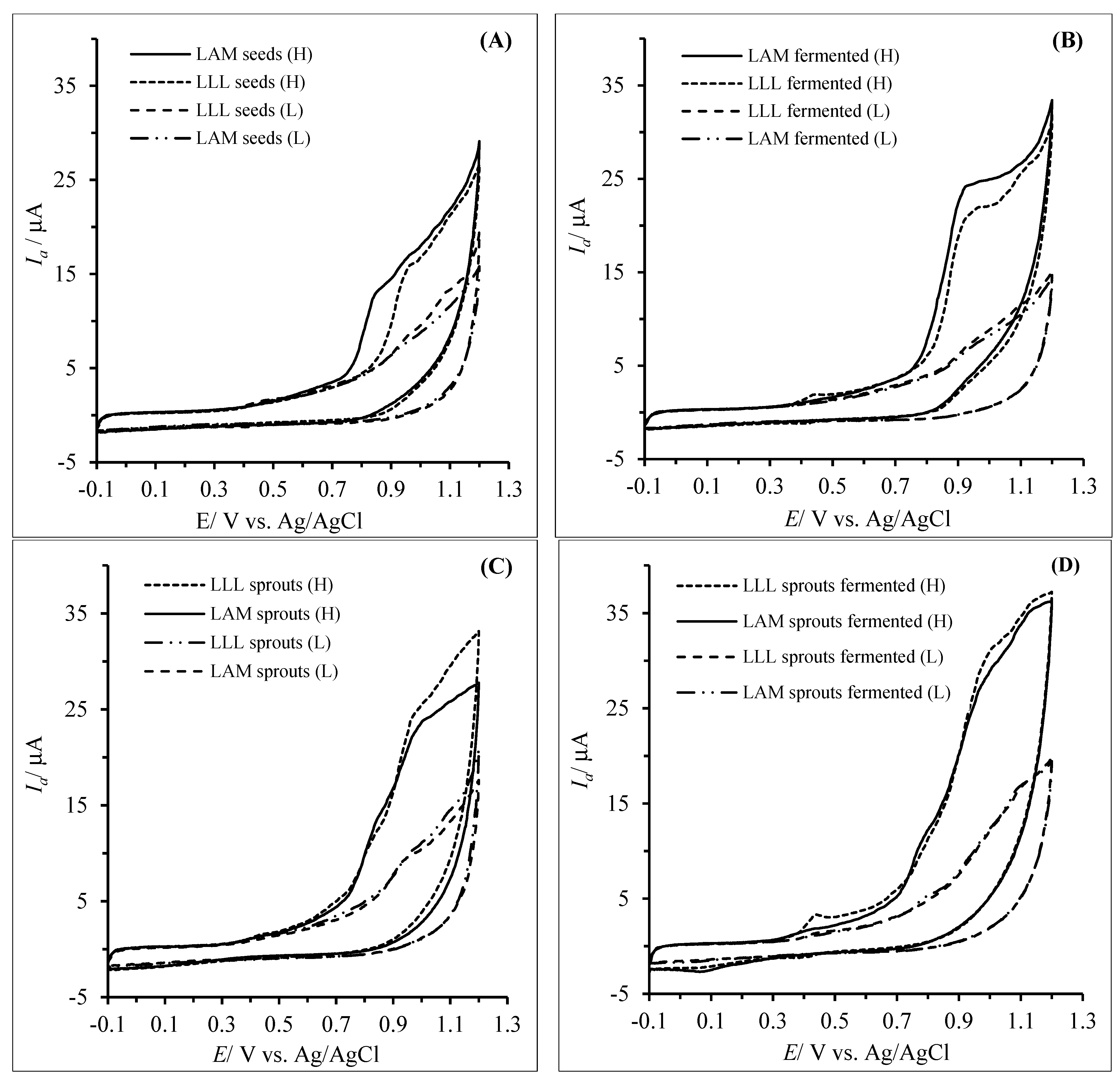
3.2. Antioxidant/Reducing Capacity of the Hydrophilic and Lipophilic Fractions Determined via the Cyclic Voltammetry Method

Fermentation and germination of lupin seeds enable not only improving their nutritional value and reducing their content of non-nutrients (such as alkaloids and raffinose family oligosaccharides) but also boosting their antioxidant capacity [4,6,23]. The content of antioxidant compounds might be modified during both processes.
In this study, the cyclic voltammetry method was applied to determine the antioxidant capacity of lupin seeds after germination and fermentation with yeast, and the results were compared with those provided by the DPPH and FRAP assays. The area under the anodic current waveform (AUC) was taken to reflect the reducing capacity of the samples compared to a set of Trolox solutions, as suggested by other authors [12,13,24]. Greater AUC area indicates a higher reducing capacity of the investigated extract.
The voltammograms of the extracts (50 mg/mL) recorded from −0.1 to 1.3 mV at a scan rate of 100 mV/s showed that lupin seeds of the cv. Lord (LLL seeds) and cv. Mirela (LAM) exhibited the oxidation and reduction voltammetric peaks with a broad anodic peak between 0.7 and 1.1 V (Figure 1A). This peak was related to the presence of hydrophilic and lipophilic antioxidants with different oxidation potentials. The results provide a marked advantage in some cases, particularly when the AUC wave represents more than a single component.
As presented in Figure 1A, the hydrophilic (H) and lipophilic (L) fractions of lupin seeds of cv. Mirela (LAM seeds) showed higher antioxidant capacity than the H and L fractions of the lupin seeds cv. Lord (LLL seeds). Moreover, the antioxidant capacity of the H fraction of LAM seeds was 37% higher compared to the H fraction of LLL; however, the antioxidant capacity of the L fraction of LAM seeds was 19% lower than that of the L fraction of LLL seeds. As a result, the total antioxidant capacity of bitter lupin seeds of cv. Mirela was higher by 19% compared to the TAC of the sweet lupin seeds of cv. Lord (Table 2).
The H fractions of the LLL and LAM lupin seeds of both cultivars fermented by yeast showed a higher antioxidant capacity by 65 and 46%, respectively, whereas the antioxidant capacity of the L fractions was lower by 28% and 36% (Figure 1B). Well-defined oxidative peaks at 0.95 V were clearly visible on the obtained voltammograms for the H fractions. As a result, the total antioxidant capacity of the fermented bitter lupin seeds of cv. Mirela was higher by 11% compared to the TAC of the sweet lupin seeds of cv. Lord (Table 2). After germination of the lupin seeds, the hydrophilic fractions of LLM and LAM showed twofold to threefold higher antioxidant capacity than the lipophilic fractions (Figure 1C), and the well-defined oxidative peaks at 0.95 V were still seen on the obtained voltammograms, thus indicating the presence of the same reductive substances as those found in the H fractions after LLL and LAM fermentation. These fractions were richer in the reductive substances as compared to the H and L fractions of the seeds. Among them, polyphenolic compounds, vitamins E and A, and others may be the most important contributors to the formation of the antioxidant capacity of both fractions due to their different solubility [11]. As a result, the total antioxidant capacity of the germinated bitter lupin seeds of cv. Mirela was higher by 19% compared to the TAC of the sweet lupin seeds of cv. Lord (Table 2). It should be mentioned that germinated lupin seeds of both cultivars showed increased TAC than the fermented ones.
The antioxidant properties of lupin sprouts may be improved after their fermentation, as was achieved in this study for the first time ever. It was noted that the fermented LLL and LAM sprouts had the highest antioxidant capacity (Figure 1D) when compared to the sprouts (Figure 1C) and seeds (Figure 1A). Moreover, the antioxidant capacity of the H fractions was significantly higher than that of the respective L fractions. The TAC of the fermented LLL and LAM sprouts measured by the cyclic voltammetric method calculated as the sum of antioxidant capacity of H and L fractions was almost twofold higher compared to the TAC of the seeds (Table 2), which corroborated earlier findings reported by Cho et al. [25] and Zielinska et al. [19]. In general, after processing the lupin seeds, about 80% of the electroactive substances were present in their hydrophilic fractions.

3.3. Antioxidant Capacity of the Hydrophilic and Lipophilic Fractions Determined by the PCL Method

The total antioxidant capacity of the processed lupin seeds, calculated as a sum of the antioxidant capacity of water-soluble compounds (PCL ACW) present in the hydrophilic fraction and the antioxidant capacity of lipid-soluble compounds (PCL ACL) present in the lipophilic fraction, is provided in Table 3.
The hydrophilic (H) and lipophilic (L) fractions of lupin seeds of cv. Lord (LLL) and cv. Mirela (LAM seeds) showed comparable antioxidant capacity measured against superoxide anion radicals. Therefore, the TAC of both cultivars did not differ significantly. The H fractions of the LLL and LAM lupin seeds of both cultivars fermented by yeast showed about threefold higher antioxidant capacity, whereas the antioxidant capacity of the L fractions was lowered by 37 and 47%, respectively, thus confirming the findings provided by the cyclic voltammetry technique. Moreover, the antioxidant capacity of the H fractions of the fermented LLL and LAM seeds exceeded six and four times the antioxidant capacity of the L fractions. As a result, the TAC of the fermented lupin seeds of both cultivars was twofold higher when compared to the unfermented seeds. These findings are in agreement with other studies, as a significant increase in both TPC and antioxidant capacity was also noted after the solid-state fermentation of L. angustifolius flour with different bifidobacteria and lactobacillus strains [26,27].
After germination of the lupin seeds, the hydrophilic fractions of LLL and LAM sprouts showed a threefold to fivefold higher antioxidant capacity compared to the H fractions of the seeds. The antioxidant capacity of the H fractions of the germinated LLL and LAM seeds was four times higher than that of the L fractions. As a result, the TAC of germinated lupin seeds of both cultivars was threefold higher compared to the unfermented seeds. Similar findings were previously provided by Fernández-Orozco et al. [6] and Frias et al. [24], who noted an increase in the antioxidant capacity of white lupin (Lupinus albus) sprouts after a nine-day germination due to the increased contents of phenolics and vitamins E and C.
The fermented LLL and LAM sprouts showed the highest antioxidant capacity as compared to the seeds, sprouts, and fermented seeds. The antioxidant capacity of the H fractions were about twelve and nine times higher than those of the respective L fractions of LLL and LAM seeds. Moreover, a threefold increase was also observed in the antioxidant capacity of the L fractions. The TAC of the fermented LLL and LAM sprouts measured by the photochemiluminescence assay was eight and six times higher compared to the seeds. It was also confirmed, similarly to the findings provided by the CV technique, that the compounds able to scavenge the superoxide anion radicals were mainly present in the hydrophilic fractions of the sprouts, fermented sprouts, and fermented seeds but not in seeds. A combination of germination and fermentation of L. angustifolius seeds with a tempeh starter fungus (Rhizopus sp.) showed that the fermentation of the 12-hour germinated seeds improved their antioxidant capacity as well as total phenolic compound content [6].

3.4. Antioxidant Capacity of the Hydrophilic and Lipophilic Fractions Determined Against DPPH⦁ Radicals

The total antioxidant capacity of the processed lupin seeds, calculated as a sum of the antioxidant capacity of water-soluble and lipid-soluble compounds able to scavenge DPPH⦁ radicals, is provided in Table 4.
The antioxidant capacity of the hydrophilic (H) fractions of sweet lupin seeds of cv. Lord (LLL) and bitter seeds of cv. Mirela (LAM) did not differ significantly but was four and five times higher than the antioxidant capacity of the lipophilic fractions, respectively. The TAC of the seeds of both cultivars was comparable without any statistically significant differences (2.75–2.80 µmol TE/g DM). The fermentation of the LLL and LAM lupin seeds had no impact on the antioxidant capacity of the H fractions, while it increased by 56 and 25% in the respective L fractions. As an outcome, the TAC was only increased within the range of 3–10%. After germination of the lupin seeds, the hydrophilic fractions of LLL and LAM showed higher antioxidant capacity by 28% and 12%, whereas no changes were noted in the antioxidant capacity of the L fractions. The TAC of the sprouts was higher within the range of 11–18% when compared to the seeds.
The fermented LLL sprouts showed an increase in the antioxidant capacity of their H fraction by only 13%, whereas no changes were observed in the antioxidant capacity of the H and L fractions from LAM. Finally, the TAC of fermented sprouts of both lupin cultivars was on the same comparable level as it was noted for the fermented seeds and sprouts (2.53–3.31 µmol TE/g DM). In accordance with the results provided by the CV and PCL assays, it was confirmed that the compounds able to scavenge the DPPH radicals were mainly present in the hydrophilic fractions of the seeds, fermented seeds, sprouts, and fermented sprouts (80.4–87%).
The results provided by CV measurements were from twofold to fivefold higher compared to those provided by the DPPH assay; however, the latter were comparable to those provided by PCL. The total antioxidant capacity provided by the cyclic voltammetry technique was highly correlated with results achieved via the PCL assay (r = 0.86); however, no correlation was found for antioxidant capacity values provided by the DPPH assay vs. results from CV and PCL measurements. In contrast, a higher correlation was observed between the antioxidant capacity of the hydrophilic fraction measured by CV and PCL (r = 0.81) than between the antioxidant capacity of the lipophilic fractions determined with the same methods (r = 0.77). The total antioxidant capacity of the samples provided by the CV technique was threefold to fourfold higher than that provided by the DPPH assay and up to 13 times higher than that provided by PCL. This relationship was previously observed in thermally treated buckwheat products and buckwheat sprouts evaluated for their antioxidant capacity using both CV and DPPH assays [20,28,29] as well as in different types of breads [30].

4. Conclusions

This study evaluated the effects of germination and fermentation on the antioxidant capacity of sweet and bitter lupin seeds and sprouts fermented by yeast. The results demonstrate that the germination and fermentation of sweet and narrow-leaved bitter lupin seeds and combinations thereof had a positive effect on the total antioxidant capacity of the water-soluble and lipid-soluble electroactive substances, which were simultaneously able to scavenge the superoxide anion radicals, a key free radical inducing the oxidative stress in humans, as well as DPPH radicals. The increase in the antioxidant capacity was related to the changes in the total content of polyphenolic compounds. The application of different methods for assessing antioxidant capacity based on the electrochemical, spectrophotometric, and photochemiluminescence assays proved to be a viable tool for screening the total antioxidant capacity of the processed lupin seeds [31]. This study provided evidence that fermented lupin sprouts, especially those of the sweet cultivar, may offer a number of benefits, ranging from protection against oxidation of particularly sensitive compounds such as fatty acids to positive effects on consumer health through scavenging free radicals.

Author Contributions

Conceptualization, D.Z., P.G. and M.K.-P.; methodology, D.Z. and H.Z.; formal analysis, D.Z. and H.Z.; investigation, P.G.; data curation, D.Z. and H.Z.; writing—original draft preparation, D.Z., P.G., M.K.-P. and H.Z.; writing—review and editing, D.Z. and H.Z.; All authors have read and agreed to the published version of the manuscript.

Funding

This research received no external funding.

Institutional Review Board Statement

Not applicable.

Informed Consent Statement

Not applicable.

Data Availability Statement

Data are contained within the article.

Conflicts of Interest

The authors declare no conflicts of interest.

References

  1. Zander, P.; Amjath-Babu, T.S.; Preissel, S.; Reckling, M.; Bues, A.; Schläfke, N.; Kuhlman, T.; Bachinger, J.; Uthes, S.; Stoddard, F.; et al. Grain legume decline and potential recovery in European agriculture: A review. Agron. Sustain. Dev. 2016, 36, 26. [Google Scholar] [CrossRef]
  2. Estivi, L.; Brandolini, A.; Gasparini, A.; Hidalgo, A. Lupin as a source of bioactive antioxidant compounds for food products. Molecules 2023, 28, 7529. [Google Scholar] [CrossRef] [PubMed]
  3. Sujak, A.; Kotlarz, B.; Strobel, W. Compositional and nutritional evaluation of several lupin seeds. Food Chem. 2006, 98, 711–719. [Google Scholar] [CrossRef]
  4. El-Adawy, T.A.; Rahma, E.H.; El-Bedawey, A.A.; Gafar, A.F. Nutritional potential and functional properties of sweet and bitter lupin seed protein isolates. Food Chem. 2001, 74, 455–462. [Google Scholar] [CrossRef]
  5. Rumiyati, R.; Jayasena, V.; James, A.P. Total Phenolic and Phytosterol Compounds and the Radical Scavenging Activity of Germinated Australian Sweet Lupin Flour. Plant Foods Hum. Nutr. 2013, 68, 352–357. [Google Scholar] [CrossRef]
  6. Fernandez-Orozco, R.; Piskula, M.K.; Zielinski, H.; Kozlowska, H.; Frias, J.; Vidal-Valverde, C. Germination as a process to improve the antioxidant capacity of Lupinus angustifolius L. var. Zapaton. Eur. Food Res. Technol. 2006, 223, 495–502. [Google Scholar] [CrossRef]
  7. Xiang, H.; Sun-Waterhouse, D.; Waterhouse, G.I.N.; Cui, C.; Ruan, Z. Fermentation-enabled wellness foods: A fresh perspective. Food Sci. Hum. Wellness 2019, 8, 203–243. [Google Scholar] [CrossRef]
  8. Prior, R.L.; Wu, X.; Schaich, K. Standardized methods for the determination of antioxidant capacity and phenolics in foods, and dietary supplements. J. Agric. Food Chem. 2005, 53, 4290–4302. [Google Scholar] [CrossRef] [PubMed]
  9. Magalhaes, L.M.; Segundo, M.A.; Reis, S.; Lima, J.L.F.C. Methodological aspects about in vitro evaluation of antioxidant properties. Anal. Chim. Acta 2008, 613, 1–19. [Google Scholar] [CrossRef] [PubMed]
  10. Głód, B.; Kiersztyn, I.; Piszcz, P. Total antioxidant potential assay with cyclic voltammetry and/or differential pulse voltammetry measurements. J. Electroanal. Chem. 2014, 719, 24–29. [Google Scholar] [CrossRef]
  11. Chevion, S.; Roberts, M.A.; Chevion, M. The use of cyclic voltammetry for the evaluation of antioxidant capacity. Free Rad. Biol. Med. 2000, 28, 860–870. [Google Scholar] [CrossRef] [PubMed]
  12. Bartosz, G. Non-enzymatic antioxidant capacity assays: Limitations of use in biomedicine. Free Radic. Res. 2010, 44, 711–720. [Google Scholar] [CrossRef] [PubMed]
  13. Martinez, S.; Valek, L.; Resetic, J.; Rusic, D.F. Cyclic voltammetry study of plasma antioxidant capacity–comparison with the DPPH and TAS spectrophotometric methods. J. Electrochem. Chem. 2006, 588, 68–73. [Google Scholar] [CrossRef]
  14. Zielińska, D.; Turemko, M. Electroactive Phenolic Contributors and Antioxidant Capacity of Flesh and Peel of 11 Apple Cultivars Measured by Cyclic Voltammetry and HPLC–DAD–MS/MS. Antioxidants 2020, 9, 1054. [Google Scholar] [CrossRef]
  15. Besco, E.; Braccioli, E.; Vertuani, S.; Ziosi, P.; Brazzo, F.; Bruni, R.; Sacchetti, G.; Manfredini, S. The use of photochemiluminescence for the measurement of the integral antioxidant capacity of baobab products. Food Chem. 2007, 102, 1352–1356. [Google Scholar] [CrossRef]
  16. Chilomer, K.; Zaleska, K.; Ciesiołka, D.; Gulewicz, P.; Frankiewicz, A.; Gulewicz, K. Changes in the alkaloid, alpha-galactoside and protein fractions content during germination of different lupin species. Acta Soc. Bot. Pol. 2010, 79, 1. [Google Scholar]
  17. Shahidi, F.; Naczk, M. Methods of analysis and quantification of phenolic compounds. In Food Phenolic: Sources, Chemistry, Effects and Applications; Shahidi, F., Naczk, M., Eds.; Technomic Publishing Company: Lancaster, PA, USA, 1995; pp. 287–293. [Google Scholar]
  18. Popov, I.; Lewin, G. Antioxidative homeostasis: Characterisation by means of chemiluminescent technique in methods in enzymology. In Oxidants and Antioxidants; Packer, L., Ed.; Academic Press: Cambridge, MA, USA, 1999; Volume 300, pp. 96–100. [Google Scholar]
  19. Zielińska, D.; Frias, J.; Piskuła, M.K.; Kozłowska, H.; Zieliński, H.; Vidal-Valverde, C. Evaluation of the antioxidant capacity of lupin sprouts germinated in the presence of selenium. Eur. Food Res. Technol. 2008, 227, 1711–1720. [Google Scholar] [CrossRef]
  20. Sadowska-Bartosz, I.; Bartosz, G. Evaluation of The Antioxidant Capacity of Food Products: Methods, Applications and Limitations. Processes 2022, 10, 2031. [Google Scholar] [CrossRef]
  21. Siger, A.; Czubinski, J.; Kachlicki, P.; Dwiecki, K.; Lampart-Szczapa, E.; Nogala-Kalucka, M. Antioxidant activity and phenolic content in three lupin species. J. Food Compost. Anal. 2012, 25, 190–197. [Google Scholar] [CrossRef]
  22. Król, A.; Amarowicz, R.; Weidnera, S. Content of Phenolic Compounds and Antioxidant Properties in Seeds of Sweet and Bitter Cultivars of Lupine (Lupinus angustifolius). Nat. Prod. Commun. 2018, 13, 1341–1344. [Google Scholar]
  23. Fernandez-Orozco, R.; Frias, J.; Muñoz, R.; Vidal-Valverde, C. Effect of fermentation conditions on the antioxidant compounds and antioxidant capacity of Lupinus angustifolius cv. zapaton. Eur. Food Res. Technol. 2008, 227, 979–988. [Google Scholar] [CrossRef]
  24. Frias, J.; Miranda, M.L.; Doblado, R.; Vidal-Valverde, C. Effect of Germination and Fermentation on the Antioxidant Vitamin Content and Antioxidant Capacity of Lupinus albus L. Var. Multolupa. Food Chem. 2005, 92, 211–220. [Google Scholar] [CrossRef]
  25. Cho, Y.S.; Yeum, K.J.; Chen, C.Y.; Beretta, G.; Tang, G.; Krinsky, N.I.; Yoon, S.; Lee-Kim, Y.C.; Blumberg, J.B.; Russell, R.M. Phytonutrients affecting hydrophilic and lipophilic antioxidant activities in fruits, vegetables and legumes. J. Sci. Food Agric. 2007, 87, 1096–1107. [Google Scholar] [CrossRef]
  26. Ayyash, M.; Johnson, S.K.; Liu, S.Q.; Al-Mheiri, A.; Abushelaibi, A. Cytotoxicity, Antihypertensive, Antidiabetic and Antioxidant Activities of Solid-State Fermented Lupin, Quinoa and Wheat by Bifidobacterium Species: In-Vitro Investigations. LWT 2018, 95, 295–302. [Google Scholar] [CrossRef]
  27. Ayyash, M.; Johnson, S.K.; Liu, S.Q.; Mesmari, N.; Dahmani, S.; Al Dhaheri, A.S.; Kizhakkayil, J. In Vitro Investigation of Bioactivities of Solid-State Fermented Lupin, Quinoa and Wheat Using Lactobacillus spp. Food Chem. 2019, 275, 50–58. [Google Scholar] [CrossRef]
  28. Khan, M.K.; Karnpanit, W.; Nasar-Abbas, S.M.; Huma, Z.E.; Jayasena, V. Development of a Fermented Product with Higher Phenolic Compounds and Lower Anti-Nutritional Factors from Germinated Lupin (Lupinus angustifolius L.). J. Food Process. Preserv. 2018, 42, e13843. [Google Scholar] [CrossRef]
  29. Pérez-Jiménez, J.; Arranz, S.; Tabernero, M.; Díaz-Rubio, M.E.; Serrano, J.; Goñi, I.; Saura-Calixto, F. Updated methodology to determine antioxidant capacity in plant foods, oils and beverages: Extraction, measurement and expression of results. Food Res. Int. 2008, 41, 274–285. [Google Scholar] [CrossRef]
  30. Zielińska, D.; Zieliński, H.; Piskuła, M.K. An Electrochemical Determination of the Total Reducing Capacity of Wheat, Spelt, and Rye Breads. Antioxidants 2022, 11, 1438. [Google Scholar] [CrossRef] [PubMed]
  31. Patil, N.D.; Bains, A.; Sridhar, K.; Sharma, M.; Dhull, S.B.; Goksen, G.; Chawla, P.; Inbaraj, B.S. Recent advances in the analytical methods for quantitative determination of antioxidants in food matrice. Food Chem. 2025, 463, 141348. [Google Scholar] [CrossRef] [PubMed]
Figure 1. Cyclic voltammograms of hydrophilic (H) extracts (50 mg/mL in 80% methanol) and lipophilic (L) extracts (50 mg/mL in methanol/n-hexane 4:1 v/v) from (A) seeds, (B) fermented seeds, (C) sprouts, and (D) fermented sprouts of the sweet lupin cv. Lord (LLL) and bitter lupin cv. Mirela (LAM) in 0.1 M sodium acetate–acetic buffer (pH 4.5) in 80% methanol were recorded at a glassy carbon electrode (GCE) from −0.1 V to +1.2 V at a scan rate of 0.1 V·s−1.
Applsci 15 00729 g001
Table 1. Total phenolic compound (TPC) content [mg GAE/g DM].
SampleTotal Phenolic Compound Content
LLL seeds0.76 ± 0.01 a
LAM seeds6.99 ± 2.31 b
LLL sprouts3.24 ± 0.01 c
LAM sprouts6.74 ± 0.04 b
fermented LLL 1.72 ± 0.06 d
fermented LAM 7.05 ± 0.09 b
fermented LLL sprouts 3.44 ± 0.11 b
fermented LAM sprouts 5.66 ± 0.70 e
Abbreviations: LLL—seeds of the yellow, sweet lupin—Lupinus luteus cv. Lord. LAM—seeds of the narrowleaf, bitter lupin—Lupinus angustifolius cv. Mirela. Data are expressed as means ± standard deviation (n = 3). Means in each column followed by different letters a–e indicate statistically different mean values (p < 0.05).
Table 2. Antioxidant capacity of the hydrophilic and lipophilic fractions of the processed lupin seeds measured by the cyclic voltammetric method (CV).
SampleHydrophilic FractionLipophilic FractionTotal Antioxidant Capacity
LLL seeds4.56 ± 0.14 a (67.2%)2.18 ± 0.20 a (32.3%)6.74 ± 0.19 a
LAM seeds6.24 ± 0.19 b (77.9%)1.77 ± 0.12 b (22.1%)8.01 ± 0.16 b
LLL sprouts9.47 ± 0.15 c (76.6%)2.90 ± 0.08 c (23.4%)12.37 ± 0.17 c
LAM sprouts8.57 ± 0.07 d (78.0%)2.43 ± 0.19 a (22.0%)10.99 ± 0.15 d
fermented LLL 7.53 ± 0.27 e (82.8%)1.56 ± 0.18 b (17.2%)9.09 ± 0.22 e
fermented LAM 9.13 ± 0.15 f (89.0%)1.13 ± 0.14 d (11.0%)10.26 ± 0.06 f
fermented LLL sprouts 11.62 ± 0.51 g (80.2%)2.87 ± 0.24 c (19.8%)14.48 ± 0.44 g
fermented LAM sprouts 11.23 ± 0.57 g (78.3%)3.11 ± 0.07 c (21.7%)14.35 ± 0.49 g
Abbreviations are the same as under Table 1. Data are expressed as means ± standard deviations (n = 3). Means in each column followed by different letters a–g indicate statistically different mean values (p < 0.05). The percentage contribution of the antioxidant capacity of the hydrophilic and lipophilic fractions to the total antioxidant capacity (TAC) of the sample is shown in the brackets.
Table 3. Antioxidant capacity of the hydrophilic and lipophilic fractions determined by the PCL method (µmol TE/g DM).
SampleHydrophilic FractionLipophilic FractionTotal Antioxidant Capacity
LLL seeds0.36 ± 0.02 a (52.9%)0.32 ± 0.04 a (47.1%)0.68 ± 0.07 a
LAM seeds0.25 ± 0.02 a (43.1%)0.32 ± 0.03 a (56.9%)0.58 ± 0.05 a
LLL sprouts1.37 ± 0.07 b (79.2%)0.36 ± 0.01 a (20.8%)1.73 ± 0.07 b
LAM sprouts1.42 ± 0.01 b (78.9%)0.38 ± 0.02 a (21.1%)1.80 ± 0.03 b
fermented LLL 1.13 ± 0.00 c (84.3%)0.20 ± 0.02 b (15.7%)1.34 ± 0.02 c
fermented LAM 0.74 ± 0.00 d (82.2%)0.17 ± 0.00 b (17.8%)0.90 ± 0.01 d
fermented LLL sprouts 4.37 ± 0.06 e (81.1%)1.01 ± 0.01 c (18.9%)5.39 ± 0.07 e
fermented LAM sprouts 2.24 ± 0.08 f (66.7%)1.11 ± 0.03 d (33.3%)3.35 ± 0.05 f
Abbreviations are the same as under Table 1. Data are expressed as means ± standard deviations (n = 3). Means in each column followed by different letters a–f indicate statistically different mean values (p < 0.05). The percentage contribution of the antioxidant capacity of the hydrophilic and lipophilic fractions to the total antioxidant capacity (TAC) of the sample is shown in the brackets.
Table 4. Antioxidant capacity of the hydrophilic and lipophilic fractions determined by the DPPH assay (µmol TE/g DM).
SampleHydrophilic FractionLipophilic FractionTotal Antioxidant Capacity
LLL seeds2.25 ± 0.02 a (80.4%)0.54 ± 0.03 a (19.6%)2.80 ± 0.03 a
LAM seeds2.32 ± 0.02 b (84.4%)0.44 ± 0.03 a (15.6%)2.75 ± 0.03 a
LLL sprouts2.88 ± 0.01 c (87.0%)0.43 ± 0.02 a (13.0%)3.31 ± 0.02 b
LAM sprouts2.59 ± 0.02 d (84.9%)0.45 ± 0.02 a (14.8%)3.05 ± 0.01 c
fermented LLL 2.25 ± 0.01 a (73.1%)0.84 ± 0.03 b (26.9%)3.08 ± 0.02 c
fermented LAM 2.29 ± 0.01 e (80.6%)0.55 ± 0.02 a (19.4%)2.84 ± 0.01 a, f
fermented LLL sprouts 2.54 ± 0.01 f (86.1%)0.41 ± 0.03 a (13.9%)2.95 ± 0.01 d, f
fermented LAM sprouts 2.11 ± 0.02 g (83.4%)0.42 ± 0.02 a (16.6%)2.53 ± 0.01 e
Abbreviations are the same as under Table 1. Data are expressed as means ± standard deviations (n = 3). Means in each column followed by different letters a–g indicate statistically different mean values (p < 0.05). The percentage contribution of the antioxidant capacity of the hydrophilic and lipophilic fractions to the total antioxidant capacity (TAC) of the sample is shown in the brackets.
Disclaimer/Publisher’s Note: The statements, opinions and data contained in all publications are solely those of the individual author(s) and contributor(s) and not of MDPI and/or the editor(s). MDPI and/or the editor(s) disclaim responsibility for any injury to people or property resulting from any ideas, methods, instructions or products referred to in the content.

