Article

Hypoxia-Induced Extracellular Matrix Deposition in Human Mesenchymal Stem Cells: Insights from Atomic Force, Scanning Electron, and Confocal Laser Microscopy

by
Agata Nowak-Stępniowska
1,*,
Paulina Natalia Osuchowska
1,
Henryk Fiedorowicz
2 and
Elżbieta Anna Trafny
1
1
Biomedical Engineering Centre, Institute of Optoelectronics, Military University of Technology, 00-908 Warsaw, Poland
2
Laser Technology Division, Institute of Optoelectronics, Military University of Technology, 00-908 Warsaw, Poland
*
Author to whom correspondence should be addressed.
Appl. Sci. 2025, 15(19), 10701; https://doi.org/10.3390/app151910701
Submission received: 13 August 2025 / Revised: 29 September 2025 / Accepted: 30 September 2025 / Published: 3 October 2025
(This article belongs to the Special Issue Modern Trends and Applications in Cell Imaging)

Abstract

(1) Background: The extracellular matrix (ECM) is a natural scaffold for cells, creating a three-dimensional architecture composed of fibrous proteins (mainly collagen) and proteoglycans, which are synthesized by resident cells. In this study, a physiological hypoxic environment was utilized to enhance ECM production by human mesenchymal stem cells (hMSCs), a process relevant to tissue engineering and regenerative medicine. (2) Methods: hMSCs were treated with deferoxamine (DFO), a pharmaceutical hypoxia-mimetic agent that induces cellular responses similar to low-oxygen conditions through stabilization of hypoxia inducible factor-1α (HIF-1α). The time points 0 h 24 h, 3 h 24 h, and 24 h 24 h refer to DFO being added immediately after cell seeding (before cells adhesion), 3 h after cell seeding (during initial cells attachment), and 24 h after cell seeding (after focal adhesions formation and actin organization), respectively, to evaluate the influence of cell adhesion on ECM deposition. hMSCs incubated in culture media were subsequently exposed to DFO for 24 h. Samples were then subjected to cell viability tests, scanning electron microscopy (SEM), atomic force microscopy (AFM) and laser scanning confocal microscopy (CLSM) assessments. (3) Results: Viability tests indicated that DFO concentrations in the range of 0–300 µM were non-toxic over 24 h. The presence of collagen fibers in the DFO-derived ECM was confirmed with anti-collagen antibodies under CLSM. Increased ECM secretion was observed under the following conditions: 3 μM DFO (24 h 24 h), 100 μM DFO (0 h 24 h) and 300 μM DFO (3 h 24 h). SEM and AFM images revealed the morphology of various stages of collagen formation with both collagen fibrils and fibers identified. (4) Conclusions: Our preliminary study demonstrated enhanced ECM secretion by hMSC treated with DFO at concentrations of 3, 100, and 300 µM within a short cultivation period of 24–48 h without significant affecting cell viability. By mimicking physiological processes, it may be possible to stimulate endogenous tissue regeneration, for example, at an injury site.

1. Introduction

Nowadays, the extracellular matrix (ECM) is a topic of significant interest in tissue engineering and regenerative medicine because it provides a natural scaffold for cells [1,2]. The ECM is a three-dimensional structure composed of fibrous proteins (mainly collagen), proteoglycans, and glycosaminoglycans synthesized by resident cells. Natural ECMs are rich in bioactive components, which create an optimal physiological environment for seeded pluripotent stem cells. These cells are widely utilized in tissue engineering [3] and regenerative medicine [4]. Understanding the composition, organization, and functional properties of ECM is crucial for advancing regenerative medicine [5,6].
Unfortunately, the kinetics of ECM assembly in vitro are slow, requiring extended cell culture periods to generate sufficient cell-derived ECM for desired applications [7]. Optimizing cell culture conditions to enhance ECM protein production could help address these challenges [8].
Currently, only a few methods for facilitating ECM deposition in vitro are described in the literature. The most common approach involves the treatment of stem cells with ascorbic acid, an essential supplement for ECM deposition. Ascorbic acid enhances the secretion of collagen type I (Col1) through increased expression of a2b1 integrin-mediated intracellular signaling [9]. Moreover, ascorbic acid serves as a cofactor for enzymes that hydroxylate proline and lysine in pro-collagen [10,11]. Long-term cultivation of mesenchymal stem cells (MSCs) with ascorbic acid at a concentration of 50 µM under 21% O2 has been successful [12,13].
To enhance ECM deposition in vitro, one can simulate the crowded conditions present in vivo by using macromolecules. Macromolecular crowding (MMC) is the phenomenon where the presence of macromolecules in a confined space increases the effective concentration of other molecules, thereby enhancing the thermodynamic activity of the system [14]. This process significantly impacts protein folding, molecular interactions, and enzyme kinetics [15]. For example, ECM deposition can be achieved in one week under MMC conditions compared to several weeks under uncrowded conditions. Commonly used “crowders” include Ficoll, dextran sulfate, carrageenan, polyvinylpyrrolidone, and newborn/fetal calf serum [16,17,18,19,20,21,22].
The simultaneous effect of oxygen tension and macromolecules as crowders on ECM deposition was demonstrated by Satyam et al. Human dermal fibroblasts cultured at 2% oxygen tension, in the presence of carrageenan and 0.5% serum, deposited more ECM during 3 days of culture than cells cultured for 14 days, under conditions of 20% oxygen tension, 10% serum concentration, and in the absence of carrageenan. Culturing cells under low-serum conditions can enhance ECM deposition, as exogenous matrix metalloproteases present in serum degrade ECM, disrupting its balance and remodeling rate [23].
Furthermore, ECM can also be synthesized following transfection with a vector encoding the ECM protein of interest [5] or under chemically induced hypoxic conditions. Conditioning the MSC sheet (produced via ascorbic acid treatment) with CoCl2 (a hypoxia-inducing agent) enormously enriched the secreted ECM with collagen I, collagen III, VEGF, bFGF, and TGF-β1 through the activation of HIF-1α. This enhancement improved, for example, ECM’s wound healing potency in vivo [24].
Hypoxia preconditioning is currently one of the most promising approaches to enhance the therapeutic efficiency of MSCs [25]. Deferoxamine (DFO) is classified as a hypoxia-mimetic agent. By inducing several cellular effects, it has potential as a valuable tool in the field of regenerative medicine. DFO demonstrates high biosafety, is readily soluble in water, and can therefore be applied in clinical practice. DFO reduces the activity of prolyl hydroxylases and inhibits the hydroxylation of HIF-1α. At low concentration, DFO decreases mitochondrial activity and apoptosis while upregulating the expression of genes related to glycolysis (e.g., lactate dehydrogenase A (LDHA), hexokinase 2 (HK2), and pyruvate dehydrogenase kinase 1 (PDK1), as well as genes associated with cell viability and survival [25]. DFO triggers pro-regenerative responses by modulating inflammation, enhancing angiogenesis, and increasing the production of growth factors [26].
This study aims to investigate whether DFO can induce MSCs to secrete ECM within a short cultivation period (24–48 h) in vitro without causing significant changes in cell viability. Enhanced deposition of ECM under chemically induced hypoxia conditions was monitored using scanning electron microscopy, atomic force microscopy, and immunocytochemistry.

2. Materials and Methods

2.1. Cell Culture

PoieticsTM Normal Human Bone Marrow-Derived Mesenchymal Stem Cells (hMSC, Poietics TM, Lonza PT-2501, Walkersville, MD, USA) were cultured in the Mesenchymal Stem Cell Growth Medium (MSCGM) following the manufacturer’s instructions (Lonza, Walkersville, MD, USA). hMSC were routinely passaged with Accutase (StemPro Accutase, Gibco, Waltham, MA, USA) before reaching 70–80% confluency and used at passages 4–6. The culture medium was refreshed three times per week. The cells were maintained in a humidified incubator at 37 °C with 5% CO2.

2.2. Pharmaceutical Hypoxia Conditioning

DFO was purchased from NOXYGEN Science Technology and Diagnostics GmbH (Elzach, Germany) with a purity of 99% and suitability for cell culture. A 10 mM DFO stock solution was prepared in ultrapure water and subsequently diluted with PBS. The time points 0 h 24 h, 3 h 24 h, and 24 h 24 h refer to DFO being added immediately after cell seeding (before cells adhesion), 3 h after cell seeding (during initial cells attachment), and 24 h after cell seeding (after focal adhesions formation and actin organization), respectively, to test the influence of cell adhesion on ECM deposition. According to that, the control sample was cultured in media for 24 (0 h + 24 h), 27 (3 h + 24 h), or 48 (24 h + 24 h) h, respectively. hMSCs were incubated in culture media for 0, 3, and 24 h, exposed to DFO at a concentration range of 0–500 mM for 24 h, and processed further, as described below.

2.3. Viability Assay

The quantitative correlation between the Presto Blue fluorescence intensity and the cell number was established (data not shown). To measure cell proliferation, 3000 MSCs were seeded in each well of a 96-well plate and allowed to attach overnight. Next, the cells were treated with different concentrations of DFO (0 (control, untreated sample), 1, 10, 30, 100, 300, and 500 µM) for 24 h. Presto Blue reagent was added for 2 h, and fluorescence intensity was measured using a multi-mode microplate reader system SpectraMax i3x (Molecular Devices, San Jose, CA, USA). The cell viability was calculated as a percentage of the control samples. The results were obtained from three independent experiments with six replicates each.

2.4. Quantitative ECM Analysis

MSCs (3 × 10³/well) were seeded on a 96-well plate. At 0, 3, and 24 h after seeding, the cells were exposed to DFO at the following concentrations: 0 (control, untreated sample), 1, 3, 10, 30, 100, 300, and 500 µM. Afterward, the cells were fixed and stained according to the immunofluorescence protocol. To detect collagen type I in the secreted ECM, a monoclonal antibody against collagen type I (1:1000, Sigma Aldrich C2456, St. Louis, MO, USA) was used, along with a goat anti-mouse IgG secondary antibody conjugated to Alexa Fluor 488 (Invitrogen, Bend, OR, USA). The fluorescence intensity of each well was measured with a multi-mode microplate reader system SpectraMax i3x (Molecular Devices, San Jose, CA, USA).

2.5. Immunofluorescence Protocol: Confocal Microscopy

Human MSCs grow on coverslips (Ø 18 mm) placed in 24-well plates. Three protocols, as described previously, used 30 and 300 µM DFO concentrations. Cells were rinsed with phosphate-buffered saline (PBS) and fixed with 4% paraformaldehyde for 15 min at room temperature. After permeabilization with 0.5% Triton X-100 (Sigma Aldrich, St. Louis, MO, USA), the samples were blocked with 1% BSA (Sigma Aldrich, St. Louis, MO, USA) and incubated with anti-collagen type I primary monoclonal mouse anti-human antibody (1:1000, Sigma Aldrich C2456, St. Louis, MO, USA.) overnight at 4 °C, followed by detection with secondary goat anti-mouse IgG antibody conjugated with Alexa Fluor 647 nm (1:200, Abcam ab15011) for 1 h in room temperature. The cytoskeleton was stained with F-actin dye (AlexaFluor 488 Phalloidin, Life Technology, Carlsbad, CA, USA). After mounting with ProLong™ Glass Antifade Mountant (Invitrogen, Bend, OR, USA), the samples were visualized under a confocal microscope (Zeiss Axio observer Z1 LSM 700, Jena, Germany).

2.6. Scanning Electron Microscopy (SEM)

MSCs were grown as described above. The samples were fixed with a mixture of 4% paraformaldehyde and 0.4% glutaraldehyde in PBS for 15 min at room temperature. A total of 1% osmium tetroxide in distilled water was used to treat the sample (Sigma Aldrich, St. Louis, MO, USA) for 16 h, followed by dehydration through a graded series of ethanol concentrations: 30%, 50%, 70%, 80%, 90%, 96%, and 99% and acetone concentrations: 30%, 50%, and 100%. Critical point drying used CO2 as a transitional fluid (Leica EM CPD300, Wetzlar, Germany). Samples were mounted on aluminum holders. Next, the samples were coated with a 10 nm conducting layer of platinum using a sputter coater (Leica EM ACE200, Wetzlar, Germany). Using high vacuum mode, the samples were examined with the scanning electron microscope (STEM, Quanta FEG250, FEI, Lausanne, Switzerland). Images were taken with the Everhart-Thornley (ETD) detector using a voltage of 5 to 10 kV.

2.7. AFM Imaging

The hMSC samples were prepared according to the SEM procedure without platinum coating, and then the topography of the ECM synthesized by hMSCs under hypoxia was visualized using atomic force microscopy (AFM, di CPII, Veeco, Santa Barbara, CA, USA). The AFM scans were obtained in tapping mode with a tip radius of 8 nm on rectangular cantilevers (RTESPA, Bruker, Camarillo, CA, USA). Images were processed and analyzed using the Gwyddion 2.53 software (http://gwyddion.net/, Czech Metrology Institute, Brno, Czech Republic, accessed on 28 August 2020).

2.8. Statistical Analysis

Data were expressed as the mean ± standard deviation (SD). Experiments were repeated three times with six parallels. Multivariate analysis of variance (MANOVA) was performed to evaluate the significance of the main effects: DFO concentration and DFO removal, and their interactions on the viability of hMSCs (Figure 1). In addition, MANOVA was applied to assess the effects of DFO concentration and cell adhesion stage, as well as their interactions on collagen type I content in ECM (Figure 3). Post hoc test of Tukey was performed to determine the significant differences among parameters. Data was considered significant when p < 0.05. Statistical analysis was performed with STATISTICA TIBCO 13 Software Inc. (Palo Alto, CA, USA). https://www.statistica.com/en/software/tibco-data-science-/-tibco-statistica.

3. Results

3.1. hMSC Viability Analysis Under the DFO Treatment

hMSC viability under DFO concentrations ranging from 0 to 500 µM is shown in Figure 1 (please see also Tables S1, S1a and S1b in the Supplementary Materials).
A 24 h incubation of hMSC with DFO resulted in a significant decrease in viability at 100 µM and 500 µM compared to the control. Moreover, 24 h after DFO removal, viability at 300 and 500 µM remained significantly lower than the control. Moreover, the difference between 500 µM DFO treatment for 24 h and 24 h after DFO removal was significant. 24 h after DFO removal, the viability at a concentration of 500 µM was significantly lower compared to 1, 10, 30, 100, and 300 µM DFO treatment for 24 h.
DFO concentration of 300 µM was identified as the maximal and non-toxic threshold based on the Presto Blue assay. Since DFO was applied solely as a method to stimulate ECM secretion in vitro, the significant decrease in viability observed 24 h after DFO removal was not taken into account for this study. Therefore, DFO concentrations of 300 µM or lower were used for ECM imaging.

3.2. Identification of Collagen Type I in hMSC-Derived ECM Under Hypoxia with CLSM

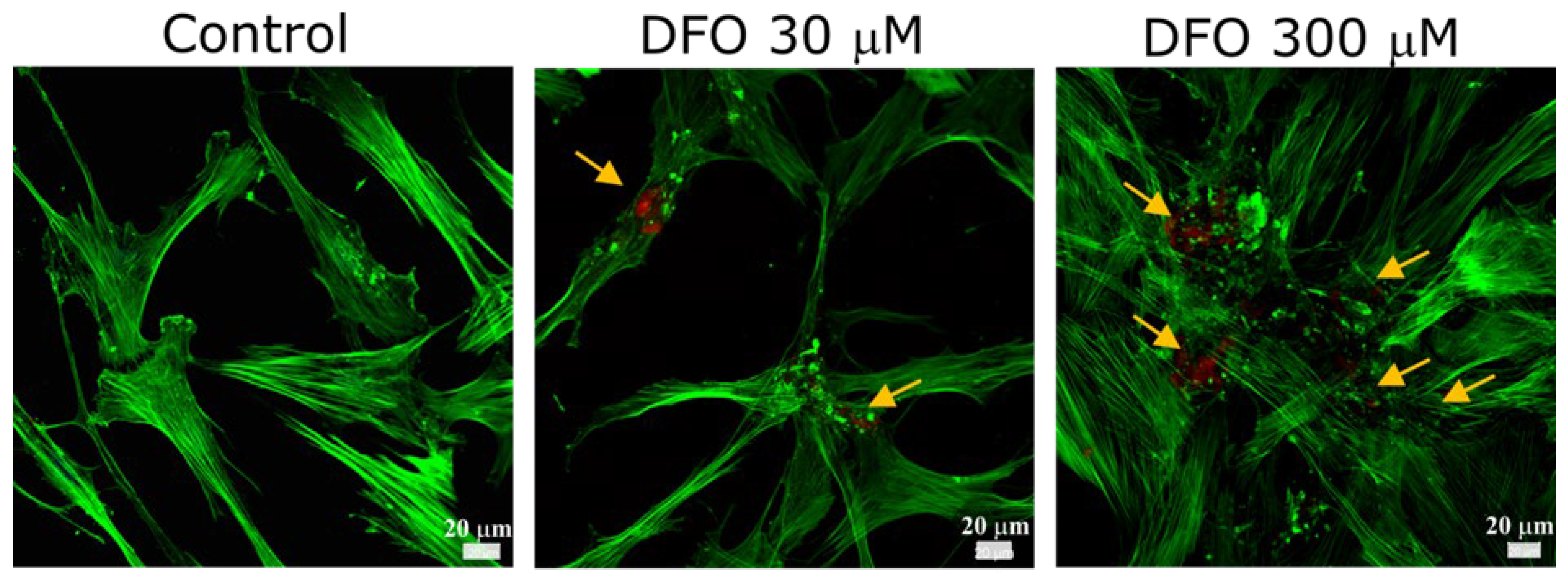
The identification of collagen fibers was based on the immunocytochemical studies and CLSM observation. In Figure 2, yellow arrows indicate the antibody-stained red fluorescence of collagen in hMSC-derived ECM. Images were captured for hMSC samples treated with 30 and 300 µM of DFO, representing lower and higher concentrations, respectively. Therefore, the presence of collagen in the hMSC-derived ECM in our experiments was confirmed. hMSC treated with a higher DFO concentration showed more collagen type I compared to the sample treated with 30 µM.

3.3. Quantitative Evaluation of Collagen Type I in hMSC-Derived ECM Under Various Culture Conditions

Next, additional culturing conditions after cell seeding were tested to quantitatively evaluate the content of collagen type I in hMSC-derived ECM under hypoxia (Figure 3) (please see also Tables S2 and S2a–c in the Supplementary Materials). To investigate the influence of cell adhesion on ECM deposition, we extended the experimental time points to 0 h 24 h, 3 h 24 h, and 24 h 24 h. These correspond to DFO being added immediately after cell seeding (before cell adhesion), 3 h after cell seeding (during initial cells attachment), and 24 h after cell seeding (after focal adhesion formation and actin organization), respectively.
Figure 3 presents the quantitative evaluation of collagen type I content in hMSC-derived ECM under these cell culture conditions. For 0 h 24 h, DFO concentrations of 100 and 500 µM were significantly different compared to the control. For 3 h 24 h, DFO concentrations of 300 and 500 µM showed significant differences, and for 24 h 24 h, only 3 µM of DFO showed significance compared to controls. Additionally, between time points, significant differences were found for 100 µM (0 h 24 h vs. 3 h 24 h, and 0 h 24 h vs. 24 h 24 h), 300 µM (3 h 24 h vs. 24 h 24 h), and 500 µM (0 h 24 h vs. 24 h 24 h). Overall, the results showed that collagen type I content in ECM was high at 24 h 24 h with 3 µM DFO, at 0 h 24 h with 100 µM DFO, and at 3 h 24 h with 300 µM DFO. However, the differences in variance among these conditions (3, 100, and 300 µM at 24 h 24 h, 0 h 24 h, and 3 h 24 h, respectively) were not statistically significant.

3.4. SEM Morphological Assessment of hMSC-Derived ECM Under DFO-Induced Hypoxia

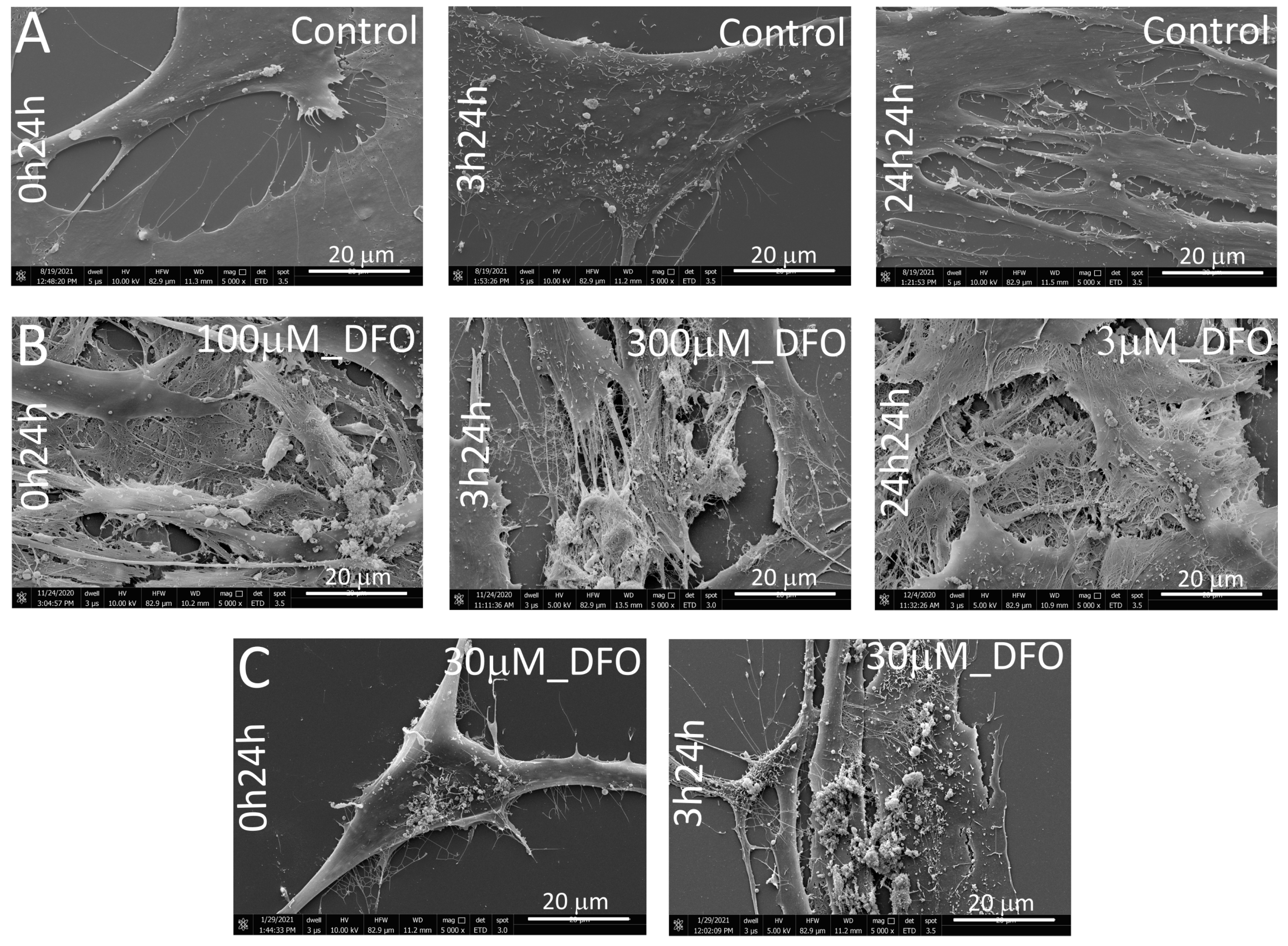
Based on the quantitative evaluation of collagen type I content in ECM under various culture conditions in hMSC exposed to DFO-induced hypoxia, SEM images of the ECM were obtained for the corresponding conditions (Figure 4). Figure 4A shows control, untreated hMSCs, in which no ECM secretion was observed. Figure 4B presents cases of increased ECM secretion under 100 µM DFO (0 h 24 h), 300 µM DFO (3 h 24 h), and 3 µM DFO (24 h 24 h). In contrast, Figure 4C shows reduced ECM production in hMSCs treated with 30 µM DFO at 0 h 24 h, and 3 h 24 h. These results are consistent with the data shown in Figure 3. Increased collagen type I secretion correlated with enhanced ECM production (Figure 3 and Figure 4B), whereas reduced collagen type I secretion correlated with lower ECM production (Figure 3 and Figure 4C).
hMSCs treated with 300 µM DFO secreted ECM at time points 3 h 24 h, providing evidence of enhanced ECM production (Figure 5B,C). In Figure 5B, the assembly of collagen fibrils (yellow arrow) was shown. Collagen fibers of assembled fibrils are presented in Figure 5C (yellow arrow). In the control sample (Figure 5A), the ECM synthesis was not observed (yellow arrow). Unexpectedly, the microvilli structure in the control samples was observed. Some microvilli were branched (red arrow), some extended from pre-existing microvilli (purple arrows), and others developed separately (green arrow) (Figure 5A). A Similar morphology of microvilli was presented in SEM images by Gaeta 2021 et al. [27].

3.5. Collagen Distribution Analysis in hMSC-Derived ECM

The most notable observation was collagen fibers projecting from the membrane of MSCs exposed to DFO treatment for 3 h 24 h. Figure 6 illustrates this phenomenon. Both SEM and AFM images suggested that the projecting structures resembled collagen fibers. Therefore, the diameters of the secreted collagen fibers were measured in these images using ImageJ software (https://imagej.nih.gov/ij). We distinguished and measured three different structures: the ‘head’ (a structure just emerging from the membrane, very short), thin collagen fibers (green arrow), and thicker collagen fibers (yellow arrow). The values obtained are presented in Table 1. The ‘head’ (red arrow in Figure 6) appeared slightly wider than the thin collagen fibers (red circle in Figure 6).
AFM image shows the increasing collagen diameter: 241 ± 34, 362 ± 18, and 689 ± 69 nm, indicating the collagen cross-linking process (fibrils for the first two structures and collagen fibers for the third one in Figure 6C). To further identify collagen fibers in the hMSC-ECM samples, collagen type I was labeled with specific antibodies and imaged with CLSM.

4. Discussion

Deferoxamine is known as a hypoxia-inducing agent that stimulates hMSC growth. According to the literature, DFO increases hMSC growth at 1 to 3 µM [28]. This study investigated a wider range of DFO concentrations on hMSC viability. DFO was used within a non-toxic range of DFO 0–300 µM, consistent with previous studies [29,30,31]. The presence of collagen fibers in the DFO-derived ECM was confirmed with anti-collagen antibodies under CLSM.
Adhesion is a complex process. It engages mechanical interlocking, intramolecular bonding, chain entanglement, and electrostatic binding. Intramolecular bonding is the principal mechanism, engaging in ionic, covalent, metallic bonds, and dipole–dipole interaction, London dispersion, and van der Waals forces [32]. The influence of initial and late adhesion on collagen type I deposition was tested [33,34]. DFO was added at three different time points: just after cell seeding 0 h 24 h (before cell adhesion), 3 h after cell seeding 3 h 24 h (during initial cell adhesion), and 24 h after cell seeding 24 h 24 h (after focal adhesion formation and actin organization) was investigated. The results indicated that collagen type I content in ECM obtained after treatment with 3, 100, and 300 µM of DFO was the highest at 24 h 24 h, 0 h 24 h, and 3 h 24 h, respectively. At higher DFO concentrations (100 and 300 µM), the no-adhesion and initial adhesion stages had the strongest impact on collagen type I secretion, whereas at the lower concentration (3 µM), the late adhesion stage was most significant.
The morphology of various stages of collagen formation was visible in SEM and AFM images. According to the literature, the molecule of the nascent collagen is transformed into pro-collagen. It is achieved by removing the signal peptide coming from its N-terminus after transcription [35,36]. The hydroxylation of lysine and glycosylation of the hydroxylysine are two key steps in creating the triple-helical structure typical of pro-collagen [37]. Its further processing and maturation are enabled after stabilization in the Golgi apparatus. Pro-collagen is then encapsulated into secretory vesicles (exocytosis) and extruded into the extracellular space, where pro-collagen is enzymatically converted into tropocollagen [36,38,39]. Covalent cross-linking is used to assemble the final collagen fibril. This cross-linking mechanism is responsible for the mechanical properties such as elasticity and reversible deformation of fibrillar collagens [40,41].
According to Ambati et al., collagen fibril structures ranging from 25 to 300 nm are interwoven into bundles with a diameter of 500–600 nm [42]. The results in Figure 6 and Table 1 demonstrate that the protruding collagen and thin structures are fibrils, while the thicker ones are fibers. The results from both SEM and AFM images showed the same collagen diameter and confirmed the presence of both fibers and fibrils in hMSC-derived ECM. Therefore, it was confirmed that tropocollagen is not emerging from the membrane as was described earlier. According to Jensen et al. [37], collagen fibril formation directly from the membrane could be considered as a possible mechanism. A similar suggestion was reported by Stearns et al. [43] and Trelstad et al. [44].
Hypoxia often induces hMSC differentiation. However, this study examined ECM deposition under pharmaceutically induced hypoxia only 24–48 h after cell seeding. Hypoxia influences hMSC-derived extracellular vehicles (EVs) through activation of HIF-1α, leading to protein and miRNA remodeling. This remodeling enhances the therapeutic properties of EVs compared with those derived from EVs from normoxic cells [45]. hMSC-derived EVs are known to promote ECM secretion and tissue regeneration by modulating ECM components and their signaling pathways [46]. Based on this, we can hypothesize that hypoxia may also enhance extracellular vesicle production, thereby supporting ECM deposition. However, the exact mechanism remains unclear and requires further investigation.

5. Conclusions

Our preliminary study demonstrated that DFO at concentrations of 3 µM and 100–300 µM stimulated ECM production in hMSCs within a short cultivation period of 24–48 h, without significantly affecting cell viability. These findings are promising, as they suggest that mimicking physiological processes can be used to stimulate endogenous tissue regeneration, for example, on the site of injury. This approach may be important for the future of regenerative medicine. Further studies will be carried out to investigate why DFO at concentrations of 30 and 50 µM did not have the same effect as 3 µM and 100–300 µM. Detailed studies on concentration, timing relevant to cell growth, and stage of adhesion process will be investigated to gain insight into DFO’s mechanism of action.

Supplementary Materials

The following supporting information can be downloaded at: https://www.mdpi.com/article/10.3390/app151910701/s1, Table S1: Figure_1_data. The hMSC viability after treatment with DFO at different concentrations for 24 hours (% of control ± SD); Table S1a: Figure 1_Raw data. The hMSC viability after treatment with DFO at different concentrations for 24 hours (Fluorescence a.u.). Cells in culture media; Table S1b: Figure 1_Raw data. The hMSC viability 24 hours after DFO removal from cell culture (Fluorescence a.u.). Cells in culture media; Table S2: Figure 3_data, Quantitative evaluation of collagen type I in ECM under various culturing conditions in hMSC under DFO-induced hypoxia. * Significant difference set at p < 0.05; Table S2a: Figure 3. Raw data. Quantitative evaluation of collagen type I in ECM under various culturing conditions in hMSC under DFO-induced hypoxia 0 h to 24 h. Cells in PBS buffer; Table S2b: Figure 3. Raw data. Quantitative evaluation of collagen type I in ECM under various culturing conditions in hMSC under DFO-induced hypoxia 3 h to 24 h. Cells in PBS buffer; Table S2c: Figure 3. Raw data. Quantitative evaluation of collagen type I in ECM under various culturing conditions in hMSC under DFO-induced hypoxia 24 h 24 h. Cells in PBS buffer.

Author Contributions

Conceptualization, A.N.-S. and E.A.T.; methodology, A.N.-S. and P.N.O.; formal analysis, A.N.-S., E.A.T., and P.N.O.; investigation, A.N.-S. and P.N.O.; resources, H.F. and E.A.T.; writing—original draft preparation, A.N.-S.; writing—review and editing, A.N.-S. and E.A.T.; visualization, A.N.-S. and P.N.O.; supervision, E.A.T. and H.F.; funding acquisition, H.F. All authors have read and agreed to the published version of the manuscript.

Funding

This research was partially funded by LASERLAB-EUROPE, grant number 872224, and LASER4EU, grant number 101131771.

Institutional Review Board Statement

Not applicable.

Informed Consent Statement

Not applicable.

Data Availability Statement

The raw data used in this study are available from the corresponding authors upon reasonable request.

Conflicts of Interest

There are no conflicts of interest to declare.

Abbreviations

The following abbreviations are used in this manuscript:
ECMExtracellular matrix
hMSCHuman mesenchymal stem cells
DFODeferoxamine
Col1Collagen type I
α2β1 integrinCollagen binding integrin
MMCMacromolecular crowding
CoCl2Cobalt (II) chloride
VEGFVascular endothelial growth factor
bFGFFibroblast growth factor-2
TGF-βTransforming growth factor-β
HIF-1αHypoxia inducible factor-1α
LDHALactate dehydrogenase A
HK2Hexokinase-2
PDK1Pyruvate dehydrogenase kinase-1
ETDEverhart-Thornley detector

References

  1. Mangani, S.; Vetoulas, M.; Mineschou, K.; Spanopoulos, K.; Vivanco, M.d.; Piperigkou, Z.; Karamanos, N.K. Design and Applications of Extracellular Matrix Scaffolds in Tissue Engineering and Regeneration. Cells 2025, 14, 1076. [Google Scholar] [CrossRef] [PubMed]
  2. Ding, X.; Xie, S.; Zhang, W.; Zhu, Y.; Xu, D.; Xian, S.; Sun, H.; Guo, X.; Li, Y.; Lu, J.; et al. Current application of tissue-engineered dermal scaffolds mimicking the extracellular matrix microenvironment in wound healing. Regen. Ther. 2025, 16, 371–382. [Google Scholar] [CrossRef] [PubMed] [PubMed Central]
  3. Hanai, H.; Jacob, G.; Nakagawa, S.; Tuan, R.S.; Nakamura, N.; Shimomura, K. Potential of Soluble Decellularized Extracellular Matrix for Musculoskeletal Tissue Engineering—Comparison of Various Mesenchymal Tissues. Front. Cell Dev. Biol. 2020, 8, 581972. [Google Scholar] [CrossRef] [PubMed]
  4. Turner, N.J. Special Issue: Application of Extracellular Matrix in Regenerative Medicine. Appl. Sci. 2021, 11, 3262. [Google Scholar] [CrossRef]
  5. Xu, Y.; Liu, X.; Ahmad, M.A.; Ao, Q.; Yu, Y.; Shao, D.; Yu, T. Engineering cell-derived extracellular matrix for peripheral nerve regeneration, Mater. Today Bio 2024, 27, 101125. [Google Scholar] [CrossRef]
  6. Gołębiowska, A.A.; Intravaia, J.T.; Sathe, V.M.; Kumbar, S.G.; Nukavarapu, S.P. Decellularized extracellular matrix biomaterials for regenerative therapies: Advances, challenges and clinical prospects. Bioact. Mat. 2024, 32, 98–123. [Google Scholar] [CrossRef]
  7. Bourget, J.M.; Gauvin, R.; Larouche, D.; Lavoie, A.; Labbé, R.; Auger, F.A. Human fibroblast-derived ECM as a scaffold for vascular tissue engineering. Biomaterials 2012, 33, 9205–9213. [Google Scholar] [CrossRef]
  8. Hoshiba, T. Cultured cell-derived decellularized matrices: A review towards the next decade. R. Soc. Chem. 2017, 5, 4322–4331. [Google Scholar] [CrossRef]
  9. Langenbach, F.; Handschel, J. Effects of dexamethasone, ascorbic acid and b-glycerophosphate on the osteogenic differentiation of stem cells in vitro. Stem Cell Res. Ther. 2013, 4, 117. [Google Scholar] [CrossRef]
  10. Vater, C.; Kasten, P.; Stiehler, M. Culture media for the differentiation of mesenchymal stromal cells. Acta Biomater. 2010, 7, 463–477. [Google Scholar] [CrossRef]
  11. Franceschi, R.T.; Iyer, B.S. Relationship between collagen synthesis and expression of the osteoblast phenotype in MC3T3-E1 cells. J. Bone Miner. Res. 1992, 7, 235–246. [Google Scholar] [CrossRef] [PubMed]
  12. Chen, X.D.; Dusevich, V.; Feng, J.Q.; Manolagas, S.C.; Jilka, R.L. Extracellular matrix made by bone marrow cells facilitates expansion of marrow-derived mesenchymal progenitor cells and prevents their differentiation into osteoblasts. J. Bone Miner. Res. 2007, 22, 1943–1956. [Google Scholar] [CrossRef] [PubMed]
  13. Decaris, M.L.; Leach, J.K. Design of Experiments Approach to Engineer Cell-Secreted Matrices for Directing Osteogenic Differentiation. Annals Biomed. Eng. 2011, 39, 1174–1185. [Google Scholar] [CrossRef] [PubMed]
  14. Alfano, C.; Fichou, Y.; Huber, K.; Weiss, M.; Spruijt, E.; Ebbinghaus, S.; De Luca, G.; Morando, M.A.; Vetri, V.; Temussi, P.A.; et al. Molecular Crowding: The History and Development of a Scientific Paradigm. Chem. Rev. 2024, 124, 3186–3219. [Google Scholar] [CrossRef]
  15. Chen, C.; Loe, F.; Blocki, A.; Peng, Y.; Raghunath, M. Applying macromolecular crowding to enhance extracellular matrix deposition and its remodeling in vitro for tissue engineering and cell-based therapies. Adv. Drug Del. Rev. 2011, 63, 277–290. [Google Scholar] [CrossRef]
  16. Lareu, R.R.; Arsianti, I.; Subramhanya, H.K.; Peng, Y.; Raghunath, M. In Vitro Enhancement of Collagen Matrix Formation and Crosslinking for Applications in Tissue Engineering: A Preliminary Study. Tissue Eng. 2007, 13, 385–391. [Google Scholar] [CrossRef]
  17. Blocki, A.; Wang, Y.; Koch, M.; Goralczyk, A.; Beyer, S.; Agarwal, N. Sourcing of an alternative pericyte-like cell type from peripheral blood in clinically relevant numbers for therapeutic angiogenic applications. Mol. Ther. J. 2015, 23, 510–522. [Google Scholar] [CrossRef]
  18. Lu, H.; Hoshiba, T.; Kawazoe, N.; Koda, I.; Song, M.; Chen, G. Cultured cell-derived extracellular matrix scaffolds for tissue engineering. Biomaterials 2011, 32, 9658–9666. [Google Scholar] [CrossRef]
  19. Tsiapalis, D.; Zeugolis, D.I. It is time to crowd your cell culture media—Physicochemical considerations with biological consequences. Biomaterials 2021, 275, 120943. [Google Scholar] [CrossRef]
  20. Wana, H.Y.; Shina, R.L.; Chend, J.C.H.; Assunçãoa, M.; Wanga, D.; Nilssone, S.K.; Tuana, R.R.; Blocki, A. Dextran sulfate-amplified extracellular matrix deposition promotes osteogenic differentiation of mesenchymal stem cells. Acta Biomater. 2022, 140, 163–177. [Google Scholar] [CrossRef]
  21. Gaspar, D.; Fuller, K.P.; Zeugolis, D.I. Polydispersity and negative charge are key modulators of extracellular matrix deposition under macromolecular crowding conditions. Acta Biomater. 2019, 88, 197–210. [Google Scholar] [CrossRef] [PubMed]
  22. Ryana, C.N.M.; Pugliese, E.; Shologua, N.; Gaspar, D.; Rooney, P.; Islamb, N.; O’Riordane, A.; Biggs, M.J.; Griffinb, M.D.; Zeugolis, D.I. Physicochemical cues are not potent regulators of human dermal fibroblast trans-differentiation. Biomater. Biosys. 2023, 11, 100079. [Google Scholar] [CrossRef] [PubMed]
  23. Satyam, A.; Kumar, P.; Cigognini, D.; Pandit, A.; Zeugolis, D.A. Low, but not too low, oxygen tension and macromolecular crowding accelerate extracellular matrix deposition in human dermal fibroblast culture. Acta Biomater. 2016, 15, 221–231. [Google Scholar] [CrossRef] [PubMed]
  24. Du, H.C.; Jiang, L.; Geng, W.X.; Li, J.; Zhang, R.; Dang, J.G.; Shu, M.G.; Li, L.W. Growth Factor-Reinforced ECM Fabricated from Chemically Hypoxic MSC Sheet with Improved In Vivo Wound Repair Activity. Hindawi BioMed Res. Intern. 2017, 2017, 2578017. [Google Scholar] [CrossRef]
  25. Noronha, N.D.; Mizukami, A.; Caliári-Oliveira, C.; Cominal, J.G.; Rocha, J.L.M.; Covas, D.T.; Swiech, K.; Malmegrim, K.C.R. Priming approaches to improve the efficacy of mesenchymal stromal cell-based therapies. Stem Cell Res. Ther. 2019, 10, 131. [Google Scholar] [CrossRef]
  26. Shen, H.; Ma, Y.; Qiao, Y.; Zhang, C.; Chen, J.; Zhang, R. Application of Deferoxamine in tissue regeneration attributed to promoted angiogenesis. Molecules 2024, 29, 20250. [Google Scholar] [CrossRef]
  27. Gaeta, I.M.; Meenderink, L.M.; Postema, M.M.; Cencer, C.S.; Tyska, M.J. Direct visualization of epithelial microvilli biogenesis. Cur. Biol. 2021, 31, 2561–2575. [Google Scholar] [CrossRef]
  28. Matsunaga, K.; Fujisawa, K.; Takami, T.; Burganova, G.; Sasai, N.; Matsumoto, T.; Yamamoto, N.; Sakaida, I. Nupr1 acts as a pro-survival factor in human bone marrow-derived mesenchymal stem cells and is induced by the hypoxia mimetic reagent deferoxamine. J. Clin. Biochem. Nutr. 2019, 64, 209–216. [Google Scholar] [CrossRef]
  29. Peyvandi, A.A.; Abbaszadeh, H.A.; Roozbahany, N.A.; Pourbakht, A.; Khoshsirat, S.; Niri, H.H.; Peyvandi, H.; Niknazar, S. Deferoxamine promotes mesenchymal stem cell homing in noise-induced injured cochlea through PI3K/AKT pathway. Cell Prolif. 2018, 51, e12434. [Google Scholar] [CrossRef]
  30. Liu, G.S.; Peshavariya, H.M.; Higuchi, M. Pharmacological priming of adipose derived stem cells for paracrine VEGF production with deferoxamine. J. Tissue Eng. Reg. Med. 2016, 10, E167–E176. [Google Scholar] [CrossRef]
  31. Nowak-Stępniowska, A.; Osuchowska, P.N.; Fiedorowicz, H.; Trafny, E.A. Insight in hypoxia-mimetic agents as potential tools for mesenchymal stem cell priming in regenerative medicine. Hindawi Stem Cells Inter. 2022, 2022, 8775591. [Google Scholar] [CrossRef] [PubMed]
  32. Bal-Ozturk, A.; Cecen, B.; Avci-Adali, M.; Topkaya, S.N.; Alarcin, E.; Yasayan, G.; Li, Y.-C.E.; Bulkurcuoglu, B.; Akpek, A.; Avci, H.; et al. Tissue adhesives: From research clinical translation. Nano Today 2021, 36, 1010492021. [Google Scholar] [CrossRef] [PubMed]
  33. Monteiro, G.A.; Fernandes, A.V.; Sundararaghavan, H.G.; Shreiber, D.I. Positively and Negatively Modulating Cell Adhesion to Type I Collagen Via Peptide Grafting. Tissue Eng. Part A 2011, 17, 1663–1673. [Google Scholar] [CrossRef] [PubMed]
  34. Rosell-García, T.; Rodriguez-Pascual, T.F. Techniques to Assess Collagen Synthesis, Deposition, and Cross-Linking In Vitro. Methods Mol. Biol. 2021, 2299, 115–122. [Google Scholar] [CrossRef]
  35. Sorushanova, A.; Delgado, L.M.; Wu, Z.; Shologu, N.; Kshirsagar, A.; Raghunath, R.; Mullen, A.M.; Bayon, Y.; Pandit, A.; Raghunath, M.; et al. The Collagen Suprafamily: From Biosynthesis to Advanced Biomaterial Development. Adv. Mater. 2019, 31, e1801651. [Google Scholar] [CrossRef] [PubMed]
  36. Kadler, K.E. Collagen fibril formation in vitro and in vivo. Inter. Exper. Pathol. 2017, 98, 4–16. [Google Scholar] [CrossRef]
  37. Revell, C.K.; Jensen, O.E.; Shearer, T.; Lu, Y.; Holmes, D.F.; Kadler, K.E. Collagen fibril assembly: New approaches to unanswered questions. Matrix Biol. Plus 2021, 12, 100079. [Google Scholar] [CrossRef]
  38. Pickard, A.; Garva, R.; Adamson, A.; Calverley, B.C.; Hoyle, A.; Hayward, C.E.; Spiller, D.; Lu, Y.; Hodson, N.; Mandolfo, O.; et al. Collagen fibril formation at the plasma membrane occurs independently from collagen secretion. BioRxiv 2024. [Google Scholar] [CrossRef]
  39. Ambrock, K.; Grohe, B.; Mittler, S. Oriented Type I Collagen—A Review on Artificial Alignment Strategies. IJSEIMS 2021, 9, 96–123. [Google Scholar] [CrossRef]
  40. Holmes, D.F.; Lu, Y.; Starborg, T.; Kadler, K.E. Collagen fibril assembly and function. Cur. Top Devel. Biol. 2018, 130, 107–142. [Google Scholar] [CrossRef]
  41. Zhang, Y.; Zhou, X.; Liu, Q.; Shen, M.; Liu, Y.; Zhang, X. Simultaneous co-assembly of collagen and glycosaminoglycans to build a biomimetic extracellular matrix for bone regeneration. Inter. J. Biol. Macromol. 2024, 279, 135535. [Google Scholar] [CrossRef]
  42. Ambati, J.; Adamis, A.P. Transscleral drug delivery to the retina and choroid. Prog. Retinal Eye Res. 2002, 21, 145–151. [Google Scholar] [CrossRef]
  43. Stearns, M.L. Studies on the development of connective tissue in transparent chambers in the rabbit’s ear. II. Am. J. Anatom. 1940, 67, 55–97. [Google Scholar] [CrossRef]
  44. Trelstad, R.L.; Kimiko, H. Tendon collagen fibrillogenesis: Intracellular subassemblies and cell surface changes associated with fibril growth. Develop. Biol. 1979, 71, 228–242. [Google Scholar] [CrossRef]
  45. Yuan, F.; Liu, J.; Zhong, L.; Liu, P.; Li, T.; Yang, K.; Gao, W.; Zhang, G.; Sun, J.; Zou, X. Enhanced therapeutic effects of hypoxia-preconditioned mesenchymal stromal cell-derived extracellular vesicles in renal ischemic injury. Stem Cell Res. Ther. 2025, 16, 39. [Google Scholar] [CrossRef]
  46. Wang, P.; Husch, J.; Amtz, O.A.; van der Kraan, P.; van de Loo, F.; van den Beucken, J.J. ECM-binding properties of extracellular vesicles: Advanced delivery strategies for therapeutic application in bone and joint diseases. Cell Commun. Sign. 2025, 23, 161. [Google Scholar] [CrossRef] [PubMed] [PubMed Central]
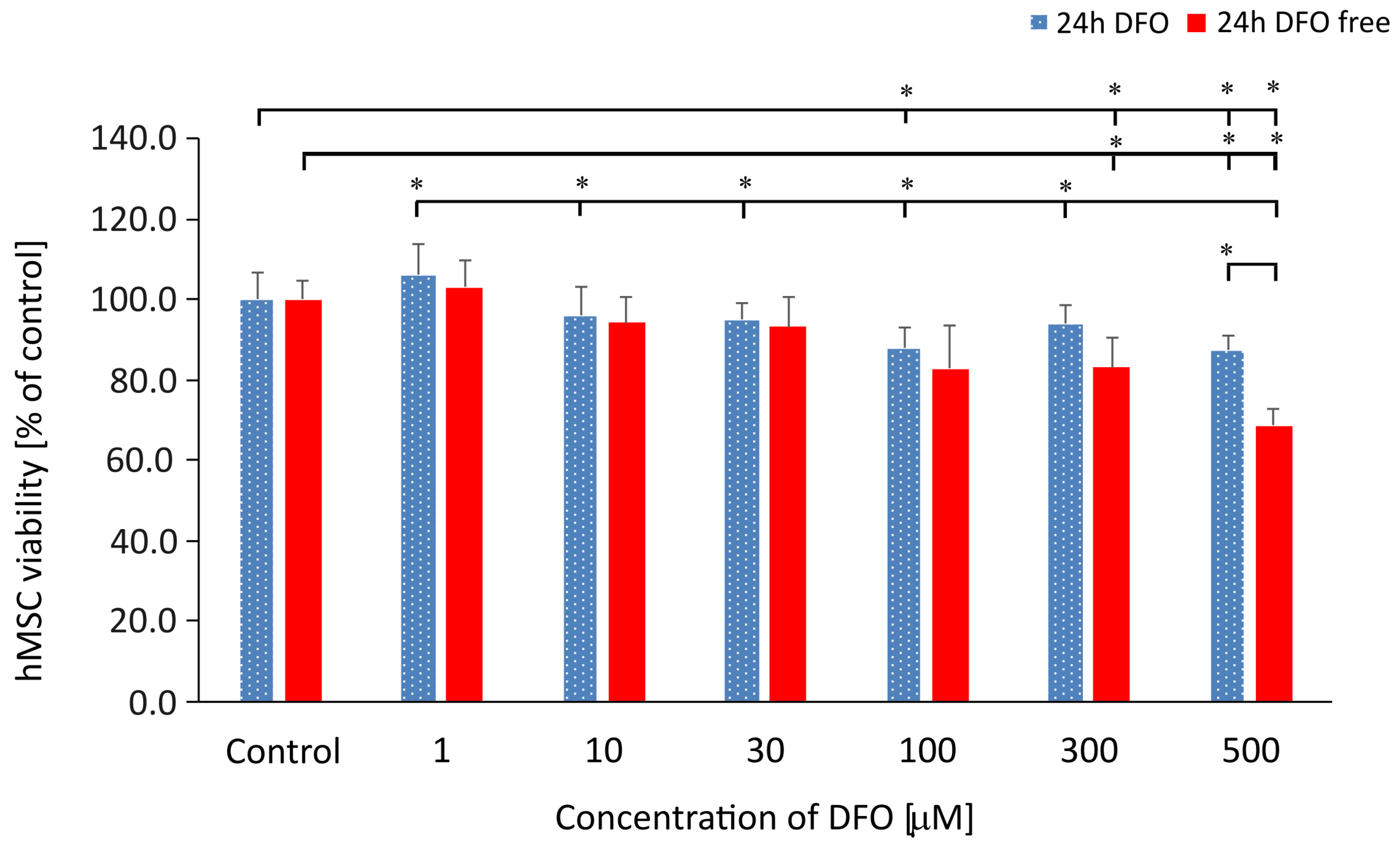
Figure 1. The hMSC viability after treatment with DFO at different concentrations (1 to 500 µM): (Blue series) immediately after 24 h incubation with DFO; (Red series) after growth for an additional 24 h following DFO removal. Control is an untreated sample. Data was normalized to control. * Significant difference set at p < 0.05.
Applsci 15 10701 g001
Figure 2. Identification of collagen type I in hMSC-derived ECM under 30 and 300 µM DFO. DFO treatment for time points 3 h and 24 h. Images were captured using CLSM. F-actin is labeled with AlexaFluor 488 Phalloidin (green fluorescence) and anti-collagen type I mouse anti-human antibody followed by goat anti-mouse IgG Alexa Fluor 647 nm secondary antibody (red fluorescence). The immunofluorescence protocol is described in the Section 2, Section 2.5. Control is an untreated sample. The scale bar is 20 µm.
Applsci 15 10701 g002
Figure 3. Quantitative evaluation of collagen type I in ECM under various culturing conditions in hMSC under DFO-induced hypoxia. The time points 0 h 24 h, 3 h 24 h, and 24 h 24 h refer to DFO being added immediately after cell seeding (before cell adhesion), 3 h after cell seeding (during initial cell attachment), and 24 h after cell seeding (after focal adhesions formation and actin organization), respectively. Then, cells were exposed to DFO at a concentration in the range of 0–500 µM. Control is an untreated sample. * Significant difference set at p < 0.05.
Applsci 15 10701 g003
Figure 4. Comparison of ECM production in hMSCs under DFO-induced hypoxia at different conditions. (A): control (untreated hMSC) at 0 h 24 h, 3 h 24 h and 24 h 24 h (from left to right); (B): enhanced ECM production under 100 µM DFO (0 h 24 h), 300 µM DFO (3 h 24 h) and 3 µM DFO (24 h 24 h) (from left to right); (C): 30 µM DFO (0 h 24 h), 30 µM DFO (3 h 24 h). Scale bar 20 µm. Magnification: ×5000.
Applsci 15 10701 g004
Figure 5. Enhancement of the ECM production under DFO-induced hypoxia in hMSCs compared to untreated, control cells after 27 h incubation: (A) The surface of hMSC in the control (untreated) sample—no ECM fibers are visible (yellow arrows), only microvilli protrude from the membrane branched microvilli (red arrow); microvilli grow from pre-existing microvilli (purple arrows), separated microvilli (green arrow), (B) collagen fibrils (yellow arrows) on the surface of hMSC under hypoxia, (C) collagen fibers (yellow arrows) on the surface of hMSC grown under hypoxia conditions. hMSC were treated with 300 µM DFO for time points 3 h 24 h (description in Section 2). Magnification: Left ×30,000, Right ×100,000. Scale bar 1 µm.
Applsci 15 10701 g005
Figure 6. Collagen distribution analysis of hMSC-derived ECM under DFO-induced hypoxia: (A) collagen fibers protruding from the membrane (SEM image: the ‘head’ (red arrow), thin collagen fibers (green arrow), thicker collagen fibers (yellow arrow)), (B) collagen fiber assembly (SEM image, purple arrow), (C) collagen assembly (AFM image, white arrows). DFO at a concentration of 300 µM for time points 3 h 24 h was used.
Applsci 15 10701 g006
Table 1. Diameter analysis and identification of hMSC-derived collagen structures based on SEM images.
Head (Coming Out Collagen)
(Red Arrow) [nm]
Thin Collagen (Green Arrow)
[nm]
Thicker Collagen (Yellow Arrow)
[nm]
Structure diameter67 ± 755 ± 7166 ± 14
Disclaimer/Publisher’s Note: The statements, opinions and data contained in all publications are solely those of the individual author(s) and contributor(s) and not of MDPI and/or the editor(s). MDPI and/or the editor(s) disclaim responsibility for any injury to people or property resulting from any ideas, methods, instructions or products referred to in the content.