Article

Agro-Industrial Residues as Cost-Effective and Sustainable Substrates for the Cultivation of Epicoccum nigrum, with Insights into Growth Kinetic Characteristics and Biological Activities

1
Department of Microbiology and Biotechnology, University of Food Technologies, 26 Maritsa Blvd., 4000 Plovdiv, Bulgaria
2
Department of Organic Chemistry and Inorganic Chemistry, University of Food Technologies, 26 Maritsa Blvd., 4000 Plovdiv, Bulgaria
3
Department of Biochemistry and Nutrition, University of Food Technologies, 26 Maritsa Blvd., 4000 Plovdiv, Bulgaria
4
Department of Mathematics, Physics and Information Technologies, University of Food Technologies, 26 Maritsa Blvd., 4000 Plovdiv, Bulgaria
*
Author to whom correspondence should be addressed.
Appl. Sci. 2025, 15(19), 10571; https://doi.org/10.3390/app151910571
Submission received: 27 August 2025 / Revised: 24 September 2025 / Accepted: 28 September 2025 / Published: 30 September 2025

Abstract

A significant quantity of agro-industrial waste is generated globally across various agricultural sectors and food industries. Composed primarily of cellulose, hemicellulose, and lignin—known as lignocellulosic materials—this waste holds significant potential and can be repurposed as a nutrient-rich substrate for mushroom cultivation. Therefore, mushroom cultivation can be regarded as a promising biotechnological approach for the reduction and valorization of agro-industrial waste. This investigation is the first to explore the utilization of agro-industrial waste- and by-products for the cultivation of Epicoccum nigrum for the production of extracts with valuable biological activities. The logistic curve and autocatalytic growth models were applied to study the kinetics of the growth process on wheat bran, sunflower cake, wheat straw, pine sawdust, and steam-distilled lavender straw substrates. Through mathematical modeling, the optimal composition of a nutrient medium containing the selected substrates was determined and successfully validated in experimental conditions. Biologically active water extracts were obtained after solid-state cultivation with α-amylase and cellulase activity up to 10.6 ± 0.6 U/mL and 0.52 ± 0.03 U/g, respectively. The extracts exhibited antimicrobial activity against fungal strains from six different species, and the most susceptible was the phytopathogen Sclerotinia sclerotiorum, with a minimum inhibitory concentration of 0.156–0.313 mg/mL.

1. Introduction

In recent years, the efficient use and valorization of agro-industrial residues and by-products have gained increasing attention and importance due to the environmental issues linked to their improper disposal [1,2]. Agricultural crop residues, including cereal straws, stovers, and hulls, as well as agro-industrial by-products such as oilseed meals, distillery wastes, and fruit and vegetable processing residues, are produced in substantial quantities across the globe. Many of these residues are inedible and inevitable by-products characterized by a low bulk density, a low ash melting point, a fibrous structure, high volatile matter content, high calorific value, and low moisture content [3,4]. Agro-industrial side-streams are often regarded as low-value materials and are typically used in animal feed, incinerated, sent to landfills, or, in some cases, disposed of improperly. Such practices can lead to significant environmental degradation and pose serious health risks [5,6,7,8,9]. At the same time, increasingly stringent waste disposal regulations are making conventional treatment methods more costly and energy-intensive, highlighting the need for alternative solutions [10,11].
Agro-industrial residues are structurally and chemically complex, containing mainly cellulose, hemicellulose, lignin, proteins, and polyphenolic compounds, and are often referred to as lignocellulosic substrates. These substrates represent low-cost, abundant, renewable, and environmentally friendly natural resources [12]. The microbial valorization of lignocellulose residues, utilizing them as nutrient sources for the synthesis of high-value bioproducts, offers a promising strategy for promoting environmental sustainability, substantially reducing production costs while improving the environmental efficiency of biotechnological processes. This approach aligns with the core principles of the circular bioeconomy by facilitating resource recovery and waste minimization. In addition to economic advantages, such bioprocesses are readily implementable and contribute to ecological preservation and public health enhancement [13].
A fungal platform for utilizing various agro-industrial by-products as cost-effective and sustainable substrates in submerged or solid-state cultivation (SSC) represents a highly promising strategy for the sustainable production of diverse high-value bioproducts with broad bioactivities and wide-ranging applications [2]. The technical and economic feasibility of agro-industrial waste valorization is primarily influenced by the physicochemical characteristics of the substrates and the metabolic capacity of the fungal strains, particularly their ability to produce the requisite enzymes for efficient biodegradation and conversion of lignocellulosic materials [13]. Comprehensive physicochemical characterization and formulation of standardized mixtures are essential prior to their application as substrates for fungal cultivation.
Fungi are efficient biotechnological agents due to their high mycelial growth rate and response to metabolic regulators in large-scale production processes [14,15]. Many fungal strains have the ability to grow on various agro-industrial by-product streams. The composition and physical properties of the substrate, as well as the environmental growth conditions, influence the efficiency of colonization and biomass formation [16]. Filamentous fungi digest before they ingest. Upon colonizing a substrate, fungal cells secrete a range of extracellular enzymes—such as cellulases, amylases, pectinases, inulinases, proteases, and lipases—into the surrounding environment. These enzymes hydrolyze complex plant polymers, including cellulose, starch, pectin, inulin, proteins, and lipids, into smaller molecules. The resulting degradation products, such as monosaccharides and oligosaccharides, are then absorbed into the hyphae via specific sugar transporters. The enzymatic products the fungi produce are utilized across a wide range of major industries, including food and feed, detergents, pulp and paper, biofuels, pharmaceuticals, and biochemicals manufacture [17]. The biosynthetic capabilities of fungi extend well beyond enzyme production, encompassing an exceptionally diverse and unparalleled range of metabolic functions found in nature. Fungal cell factories play a central role in the sustainable production of biofuels and a plethora of biochemicals [18]. Fungal endophytes—fungi that colonize the internal tissues of asymptomatic plants—and fungicolous fungi—those that establish associations with other fungi—have garnered increasing scientific attention in recent years, particularly those isolated from unconventional or extreme environments. These organisms are regarded as promising sources of structurally diverse and biologically active secondary metabolites [19]. Many researchers have pointed out that these fungi are capable of synthesizing a wide array of chemically diverse secondary metabolites [20], many of which exhibit significant biological activities, including antibacterial, antifungal, antiparasitic, insecticidal, antiprotozoal, anthelmintic activities, anticancer, and antiviral properties [21,22,23,24]. Endophytic fungi belonging to the genera Penicillium, Epicoccum, Trichoderma, Chaetomium, Piriformospora, and Curvularia are well-documented for their ability to suppress fungal pathogens and promote plant growth and yield under both biotic and abiotic stress conditions [25,26,27].
Epicoccum nigrum is an anamorphic ascomycete organism, belonging to the family Didymellaceae, which is widely distributed in diverse ecosystems. It is primarily involved in the initial decomposition of plant tissues and is generally classified as a saprotrophic fungus; however, in certain cases, it has also been reported to exhibit weak pathogenicity toward plants [28]. It is a ubiquitous fungus that can adopt an endophytic lifestyle and inhabits the internal tissues of various plant species [29,30,31]. It is hypothesized that E. nigrum elicits an enhanced defense response in the host by reprogramming its metabolic pathways, thus causing the biosynthesis and accumulation of distinct classes of secondary metabolites [32]. In a previous study, we reported, for the first time, the isolation of E. nigrum from the fruiting body of a Bulgarian Dryad’s Saddle mushroom (Polyporaceae) and referred E. nigrum to fungicolous fungi [33]. E. nigrum has attracted considerable scientific interest due to its ability to produce a broad spectrum of bioactive secondary metabolites, such as polycyclic alkaloids (epicoccins, epicorazins, epicoccarines, epipyridone, flavipin, and epirodins) with antibacterial properties [10], and epicoccamides with anticancer properties [10]. Epicocconone—a fluorescent pigment—is the most characteristic metabolite produced by E. nigrum. It has applications as a protein-binding dye and possesses broad antifungal activity [11]. Due to its beneficial properties, E. nigrum is considered a valuable organism for use as a biological control agent and a potential source of bioactive compounds.
Solid-state cultivation refers to the cultivation of microorganisms under conditions with little to no free water, closely mimicking the natural environment of many fungal species. This method of cultivation has been shown to enhance fungal resistance to catabolite repression, promote increased biomass, and stimulate the production of secondary metabolites [34,35,36]. In most of the studies, E. nigrum is cultivated in nutrient media containing conventional synthetic or refined substrates [30,37,38,39,40,41]. However, these substrates are often costly and may not be compatible with current trends toward sustainable and cost-effective bioproduction. Only a handful of investigations have explored its growth on complex agro-industrial residues, and these were largely restricted to pigment production [33,42]. For example, Elnaggar et al. (2024) employed rice-based substrates to obtain specific naphtho- and benzofuran derivatives [30].
By integrating fungal biotechnology, mathematical modeling, and sustainability principles, this study introduces a new platform for transforming low-value agro-industrial residues into high-value bioresources. Two mathematical models were applied to determine the maximum specific growth rate (μmax), and other key kinetic parameters, enabling the formulation of an optimized nutrient medium. The most suitable substrates identified were then employed for large-scale biomass production under solid-state conditions. Furthermore, the bioactivity of water extracts from the resulting fungal biomass was assessed, establishing a foundation for the novel valorization of agro-industrial residues into biologically active products.
The novelty of this research lies not only in the cultivation of E. nigrum into unconventional substrates but also in the kinetic modeling and the establishment of a foundation for the novel valorization of agro-industrial residues into biologically active products, thus advancing the field toward scalable, environmentally friendly, and economically viable bioprocesses.

2. Materials and Methods

2.1. Fungal Strain

The fungicolous Epicoccum nigrum used in this study is a part of the fungal collection of the Department of Microbiology and Biotechnology, University of Food Technologies, Plovdiv, Bulgaria. The strain was maintained on a mushroom complete medium (MCM) with the following composition (g/L): Glucose—20.0; KH2PO4—0.5; K2HPO4—1.0; MgSO4 × 7H2O—0.5; peptone—2.0; yeast extract—2.0; agar–agar—20.0, supplied by Merck KGaA (Darmstadt, Germany). The pH prior to sterilization was adjusted to 6.00. The cultures were incubated at 25 °C for 5 days and stored afterwards at 4 °C.

2.2. Agro-Industrial Substrates and Preparation Procedure

Wheat straw (WS), pine sawdust (PS), wheat bran (WB), sunflower cake (SC), and steam-distilled lavender straw (Lavandula angustifolia Mill.) (SDLS) were used as feeding substrates for fungal strain cultivation. According to a previously published protocol, the substrates were sourced and prepared as follows: the SC, WS, and PS were sourced from agricultural areas near Plovdiv, Bulgaria, from 2020 crops and stored at room temperature. SDLS crop (2020) was provided by the company Galen-N, based at Zelenikovo distillery, Brezovo region, Bulgaria. The SDLS was collected from the area near the distillery and was immediately air-dried at 40 °C. All studied substrates were milled and sieved (particle size 1–5 mm) prior to use as components of nutrient media [43].

2.3. Proximate Composition Characterization of the Substrates

The anhydrouronic acid (GalA) was determined by the m-hydroxybiphenyl method [44].
The polyuronide content and degree of esterification (DE) of polyuronides were determined according to Marovska et al. (2024) [45].
The quantity of dietary fibers (total, soluble, and insoluble) was determined using a Total Dietary Fiber Assay kit (K-TDFR-100A, Megazyme, Bray, Co. Wicklow, Ireland) according to the AOAC method [46] and the instructions provided by the kit manufacturer.
Total (non-cellulosic) carbohydrates, uronic acids, cellulose, and lignin were evaluated, as described by Escalada Pla et al. [47].

2.4. Cultivation and Modeling of the Kinetics of the Process

For the cultivation of E. nigrum, we prepared five different agar media by dissolving WS, PS, WB, SC, or SDLS in a concentration of 40 g/L in potable water, and adding agar–agar in a concentration of 20 g/L. The media were sterilized for 30 min at 121 °C. The sterilized media were cooled to 45 °C and poured into 90 mm Petri dishes. Inoculation was made with agar disks (10 mm), containing mycelium of a 5-day-old E. nigrum culture, which were placed in the center of the Petri dishes. The cultivation was carried out at 25 °C to a fully grown culture, and the diameters of the growing colonies were measured daily.
To model the kinetics of the cultivation process, the logistic curve model (Equation (1)) and the reverse autocatalytic growth model (Equation (2)) were used [48,49,50].
dD d σ = μ max . D δ . D 2
dD d σ = k 1 S 0 D k 1 S 0 D D m D 2 D = D 0 S 0 K K + 1 D 0 S 0 K K + 1 D 0 e k 1 S 0 σ
D m = S 0 K K + 1
where µmax—specific growth rate, d−1; D0, D, and Dm—initial, current, and maximum diameter of the mycelium, mm; δ—growth inhibition coefficient, mm.d−1; K1—rate constant of biomass formation, d−1; S0—initial amount of substrate in cell units, expressed by the diameter of the mycelium, mm; K/1 + K—coefficient of substrate utilization; τ-cultivation time, day.
The logistic curve model was solved numerically using the 4th order Runge–Kutta method, and the identification of the parameters in the used models was performed by minimizing the square of the difference between the experimental data and those obtained from the corresponding model with the Solver function in Microsoft Excel Office 2019 [51,52].
To plan the experiment and optimize the composition of the nutrient medium, Simplex–lattice designs from the Statgraphics Centurion® 18 (Version 18.1.12) software package, which are characterized by a symmetrical arrangement of points in the triangular lattice (the experimental area), was used, and the number of parameters in the polynomial model was equal to the number of selected points in the lattice [53].
The chosen model for describing the response surface is a special cubic model (Equation (3)), which has the following form [54]:
Y = i = 1 q β i x i + i = 1 q i < j q β ijk x i x j + k = 1 q j < k q i < j q β ijk x i x j x k

2.5. Solid-State Cultivation and Post-Cultivation Treatment

Three nutrient media were used as feeding substrate for the SSC of E. nigrum—WS, WB, and the newly formulated optimal nutrient medium. A total of 25 g of the feeding substrates was transferred to 500 mL Erlenmeyer flasks and moisturized to a final humidity of 65–75% with potable water, sterilized at 121 °C for 30 min, and subsequently cooled to room temperature. The inoculum was prepared by suspending the biomass of a 5-day slant agar culture of E. nigrum in 10 mL of distilled water, and the cooled substrates were inoculated, thoroughly mixed, and incubated at 25 ± 2 °C for 14 days. After the cultivation process ended, sterile distilled water was added to each Erlenmeyer flask in a ratio of 1:1 (w/w). The mixtures were blended in a sterile laboratory blender and placed on a laboratory shaker at 150 rpm for 2 h at 25 °C. Following incubation, the mixtures were filtered on a Buhner’s funnel and centrifuged at 13,000 rpm for 10 min at 4 °C. The resulting supernatants were collected and used to determine their enzyme activities. A part of the supernatants was subjected to evaporation under vacuum at 40 °C until completely dry. The dried extracts were then dissolved in 50% dimethyl sulfoxide (DMSO) to a final concentration of 10 mg dry weight (DW)/mL and were sterilized through membrane filtration, using sterile 0.45 µm PTFE syringe filters (Merck KGaA, Darmstadt, Germany). These extracts were then used to determine their antimicrobial activity.

2.6. Determination of Enzyme Activities

2.6.1. Total Cellulolytic Activity (Filter Paper Assay, FPA)

The analysis was performed according to the IUPAC procedure with modifications. The determination is based on the quantity of glucose liberated as a result of the degradation of 0.05 g of filter paper by the enzymes present in the extract. For this purpose, 0.05 ± 0.002 g of filter paper was measured on an analytic scale (Kern & Sohn GmbH, Balingen-Frommern, Germany) and placed in a glass test tube, and 2 mL of appropriately diluted extract was placed in the test tube. The control test tube contained 0.05 g of filter paper and 2 mL of inactivated extract. The inactivation was performed by incubating the extract at 100 °C for 10 min. All test tubes (test and control) were incubated for 60 min at 50 °C, and at the end of the hydrolysis, the reducing sugars were determined according to Lever [55]. One unit of total cellulolytic activity was determined as the amount of enzyme that liberated 1 µg glucose from the substrate for 1 min at the reaction conditions.

2.6.2. Amylolytic Activities

The activity of glucoamylase (GLA) was established by the measurement of the produced free glucose after starch hydrolysis. The reaction mixture contained 1 mL 1% starch with a pH of 5.0 and 200 µL of extract, and the process took place in a thermostat at 60 °C for 10 min. The quantity of the reducing sugars was evaluated using the Lever method [55]. One unit of glucoamylase activity was defined as the quantity of the enzyme needed to release 1 µg glucose from starch for 1 min at the reaction conditions.
The α-amylase activity (AAA) was determined by the Fuwa method [56] with modifications. The reaction mixture contained 500 µL 1% starch with pH 5.0 as substrate and 100 µL extract, and the hydrolysis continued for 10 min at 30 °C. The hydrolysis was terminated by the addition of 10 µL of hydrolysate to a mixture containing 100 µL of 0.1 N HCl, 100 µL of 0.01 N I2, and 800 µL of distilled H2O. The color of the formed starch–iodine complex was measured at 690 nm on a UV-VIS spectrophotometer (BMG Labtech, Ortenberg, Germany). One unit of α-amylase activity was determined as the amount of enzyme that degraded 1 mg of starch for 1 min under the reaction conditions.

2.6.3. Ligninolytic Activities

The laccase activity was determined using syringaldazyne as substrate according to the method of Ride [57] with modifications. The 0.216 mM substrate solution was prepared in 99% methanol and sonification at 45 Hz for 30 min. The reaction mixture with a total volume of 300 µL contained a 220 µL 50 mM potassium–phosphate buffer (pH 5.0) and 50 µL extract with appropriate dilution, and the reaction was initiated by the addition of 30 µL substrate solution. The changes in the absorption at 530 nm were monitored for 5 min, and one unit of enzyme activity was defined as the enzyme quantity required for achieving a change of 0.001 in the absorbance value and was expressed as U/mL.
The determination of the activity of the manganese-dependent peroxidase was based on the measurement of the oxidation of Mn (II) to Mn (III) at 270 nm according to the method of Warshii [58]. The reaction mixture contained 40 µL 1.0 mM MnSO4, 100 µL sodium–malonate buffer (pH 5.0), and 20 µL extract. The oxidation was initiated by the addition of 40 µL 0.5 mM H2O2 and was monitored at 270 nm for 10 min. One unit of enzyme activity was defined as the amount of enzyme required to oxidase 1 µmol substrate for 1 min under the reaction conditions.
The lignin peroxidase activity was determined according to Tien and Kirk [59], based on the measurement of the oxidation of 3,4-dimethoxybenzyl alcohol (DMBA) in the presence of H2O2. The reaction mixture included 100 µL 0.1 M citrate buffer (pH 5.0), 50 µL 1 mM 3,4-DMBA, and 50 µL extract. The oxidation was initiated by the addition of 20 µL 0.2 mM H2O2 and was monitored at 310 nm for 15 min. One unit of enzyme activity was determined as the quantity of enzyme required for the transformation of 1 µmol substrate for 1 min under the reaction conditions.

2.7. Determination of Antifungal Activities

The fungal strains Penicillium chrysogenum, Alternaria sp., Alternaria alternata, Mucor sp., Fusarium verticillioides, Fusarium oxysporum, Aspergillus flavus, Botrytis cinerea, and Sclerotinia sclerotiorum were provided from the collection of the Department of Microbiology and Biotechnology at the University of Food Technologies, Plovdiv, Bulgaria.

2.7.1. In Vitro Antagonism Determination

The antifungal activity of E. nigrum was screened using the dual culture method. Wort agar plates were inoculated with mycelium plugs (6 mm in diameter) from separate cultures of E. nigrum and the test microorganisms. The mycelium plugs were cut from the growing margins of a 10-day culture for S. sclerotiorum and B. cinerea and a 5-day culture for the rest of the fungi and placed on the opposite sides of the 90 mm diameter Petri dishes. The plates were incubated at 25  ±  2 °C for 7 days.

2.7.2. Minimal Inhibitory Concentration (MIC) Determination

The minimal inhibitory concentration (MIC) of the water extracts was determined according to the CLSI method [60]. The extract was subjected to serial two-fold dilutions in RPMI-1640 broth (Merck KGaA, Darmstadt, Germany) in 96-well microtiter plates. Each well was inoculated with a microbial suspension at a concentration of 5 × 10⁵ CFU/mL. Two controls were used—a growth control (positive control) and a sample controlfor each dilution without inoculation (negative control). After mixing, the plates were incubated at 25 °C for 48 h, after which they were examined visually. The MIC was the lowest concentration that prevented any discernible growth (100% inhibition).

2.8. Statistical Analysis

All the experiments were conducted in triplicate, and the values were expressed as mean values ± SD. The statistical significance was determined by the analysis of variance (ANOVA, Tukey’s test, and Games–Howell test); the value of p < 0.05 indicated a statistical difference.

3. Results and Discussion

3.1. Characterization of the Agro-Industrial Substrates

Wheat straw (WS), pine sawdust (PS), wheat bran (WB), sunflower cake (SC), and steam distilled lavender straw (Lavandula angustifolia Mill.) (SDLS) were chosen as substrates for this study primarily because of their widespread availability in Bulgaria, where they are generated in a significant amount as by-products of the essential oil and agricultural industries. In order to determine their suitability as substrates for the cultivation of E. nigrum, their proximate composition was determined (Table 1). In a previous study, we published the data for SDLS [43].
The presented results reveal significant differences in the composition of the selected substrates. PS is highest in total dietary fibers, followed by SDLS [43], WS, WB, and SC. The highest content of polyuronides was determined for WB, followed by SC, WS, PS, and SDLS [43]. For all substrates, the content of lignocellulosic carbohydrates was significantly higher than that of non-cellulosic ones. WB and SDLS [43] had the highest content of non-cellulosic polysaccharides—15.80 ± 0.15 and 13.79 ± 0.15 g/100 g DW, respectively. The largest quantity of cellulose was detected in PS and WS, while SC was the richest in lignin. In addition, according to data published by other authors, SC is a good source of crude protein (37.10 g), crude fat (0.69 g), ash (7.49 g), carbohydrate (23.97 g), iron (6.44 mg), calcium (650 mg), phosphorus (711.33 mg), and phytic acid (700 mg) per 100 g DW [61].
According to Ogórek et al. (2020), who investigated 18 strains of E. nigrum from various host plant sources, 61% of the strains produced amylases and proteases, 44% produced cellulases, and 33% pectinases [62]. Other authors identified the synthesis of hydrolytic enzymes such as polygalacturonase, pectin-lyase, pectinase, protease, cellulase, xylanase, arabanase, lipase, laccase, gelatinase, glucoamylase, and amylase [63,64,65]. These findings, combined with the determined proximate composition of the selected substrates (Table 1), ensure that they are sufficient and varied carbon sources for the growth of E. nigrum.

3.2. Cultivation of E. nigrum and Modeling of the Kinetics of the Process

The fungicolous Epicoccum nigrum used in this study was isolated from the basidiocarp of a local Dryad’s Saddle mushroom (Polyporaceae). The molecular identification of the isolate was performed by the amplification of the ITS1-5.8S-ITS2 region, and the strain was identified with 100.00% confidence as Epicoccum nigrum [37].
In a series of experiments, the growth of E. nigrum on complex nutrient media—WB, SDLS, WS, PS, and SC—was studied. The change in the mycelial diameter was the main observed parameter by which the growth of the strain was evaluated. For this reason, the change in the mycelial diameter was monitored during the cultivation of E. nigrum for 14 days on the individual nutrient media, and the results of these experiments are depicted in Figure 1.
The data shown in Figure 1 indicate that on wheat bran and wheat straw media, the strain reached the highest diameter of the mycelium mass—85 mm—and this value on WB media was reached after 12 days of cultivation and remained constant until the end of the process. In addition, on the complex WB medium, the strain developed with the highest intensity compared to the other complex media. During the growth of E. nigrum, a continuous increase in the diameter of the mycelium was observed until the 14th day, where it reached its maximum value (85 mm). A comparable intensity of growth to that on WB media E. nigrum was exhibited on SC, but this trend persisted only until the seventh day, after which a slowdown in growth was observed; and on the 10th day, the growth of the strain stopped, reaching a maximum diameter of the mycelium mass of 63 mm. This was probably due to the rapid depletion of potential substrates from the SC that were necessary for its growth. The data from Figure 1 also show that the growth of E. nigrum on the complex media SDLS and PS proceeded with lower intensity, compared to its cultivation on WB and WS. On SDLS medium, a continuous increase in the diameter of the mycelial mass was observed from the beginning to the end of the process, when it reached 75 mm. The studied strain was characterized by the lowest intensity on the PS medium, and also on this medium, the smallest mycelial growth was observed (the diameter at the end of the cultivation reached 51 mm). For a more complete study of the reproductive ability of the strain on different nutrient media, its growth kinetics were determined. In modeling the growth kinetics, the logistic curve models (Verhulst model) and reversible autocatalytic growth were used. The parameters in the models were identified, and the results are reflected in Table 2.
The data presented in Table 2 demonstrates that both models are characterized by high correlation coefficients, which indicates that the selected models describe the experimental results very well and allow the reliable modeling of the growth kinetics. According to the logistic curve model, the highest maximum specific growth rate was established for WB medium—0.523 ± 0.029 h−1. Close values of µmax were observed for the WS, SC, and SDLS media—0.439 ± 0.015 h−1, 0.438 ± 0.085 h−1, and 0.421 ± 0.017 h−1, respectively. The lowest maximum specific growth rate was established for E. nigrum, cultivated on PS medium—0.375 ± 0.005 h−1. The growth inhibition coefficient δ varied in the range of 0.0052 ± 0.0002 mm.d−1 to 0.0080 ± 0.0007 mm.d−1. These values are significantly lower than one, which indicates that the used complex media did not contain factors inhibiting the growth of Epicoccum nigrum, and also that there was no strong competition between the culture cells for the substrate, which would lead to their death. The results shown in Table 2 indicate that the model of reversible autocatalytic expansion produced close values of the rate constant of biomass formation k1, which, for the different media, varied in the range from 0.0036 ± 0.0002 d−1 to 0.0041 ± 0.0006 d−1. The advantage of the reversible autocatalytic growth model over the Verhulst model is that it allows the determination of the substrate supply in cell units (S0), as well as the substrate utilization factor K/1 + K. S0 illustrates the nutrient content of the medium, expressed as biomass units, i.e., how much biomass could be obtained in theory from a particular nutrient medium. K/1 + K varies between 0 and 1, and values closer to 1 show that the composition of the nutrient medium is optimal and balanced, and the cultivation conditions are appropriate. In the WB, WSS, and SC media, a high substrate supply in the medium in cell units was observed. It was expressed as the mycelial diameter, which was 99 ± 0 mm, 100 ± 0 mm, and 98 ± 0 mm, respectively. This indicates that in these media a larger amount of biomass is expected to accumulate, compared to SDLS and PS media, in which S0 was 81 ± 1 mm and 79 ± 1 mm, respectively. This was confirmed for the WB and WS media, in which the highest diameter of the mycelial mass of 85 mm was recorded. A smaller diameter of the mycelial mass was recorded in PS and SDLS media, which had lower S0. In the SDLS medium, a mycelium diameter of 75 mm was recorded, which is close to the theoretical one that the medium allows—81 ± 1 mm. A similar trend was observed for the PS medium, where mycelium growth of the order of 51 mm was recorded, while theoretically, this medium allows for a mycelium diameter of 79 ± 1 mm. Despite the high value of S0, for the SC medium, a maximum mycelium diameter of the order of 63 mm was observed, which indicates that the strain utilized completely certain substrates, and after their depletion, the growth of the culture stopped. This was confirmed by the coefficient of substrate utilization of the substrate (K/1 + K). For the WB and WS media, this parameter had the highest value—0.9516 ± 0.0022 and 0.8849 ± 0.0178, respectively. This indicates that they contain many substances that are utilized by Epicoccum nigrum, which was also the cause of the highest value of the mycelium diameter in these media. This theory is supported by the determined proximate composition of WB and WS, which contain higher amounts of non-cellulosic polysaccharides and dietary fibers. In terms of the parameter K/1 + K, the SDLS medium occupied an intermediate position, while the lowest efficiency of substrate utilization was observed in the other two media (PS and SC), which were expressed as low values of the mycelial diameter. For the latter media, K/1 + K was 0.7727 ± 0.0123 and 0.6932 ± 0.0717, respectively. SC, according to reference literature, contains high amounts of crude fat [61], and PS has the lowest concentration of NCP, which probably hinders the growth of the strain.
From the kinetic study results, it can be concluded that the complex nutrient media SC, SDLS, and PS contain higher amounts of substrates, which are more difficult for E. nigrum to utilize, and a smaller amount of readily digestible compounds, which were probably quickly depleted as a result of the relatively high growth rate. This, in turn, led to a stop in the growth of the strain, and from there, a smaller mycelial mass, which indirectly affected the amount of synthesized metabolic products. In order to more fully and effectively use the complex media SC, SLDS, and PS for solid-state cultivation of E. nigrum, an optimal multicomponent mixture of WB, WS, SC, SDLS, and PS was made. For this purpose, the Simplex method from the Statgraphics Centurion® 18 (Version 18.1.12) package was used. Table S1 presents the composition of the individual mixtures, as well as the maximum diameter of the mycelial mass that was obtained.
After mathematical processing of the obtained results, a mathematical model was derived to determine the influence of the mixture components on the mycelium diameter:
D = 84.7436 · WB + 86.4161 · WS   + 70.4856 · SDLS   + 58.0398 · PS   + 59.475 · SC   + 19.4196 · WB · SDLS + 55.3367 · WB · PS   66.01 · WS · SC   27.3778 · SDLS · PS   16.0365 · PS · SC   + 286.44 · WB · SDLS · PS   + 312.426 · SPDS · PS · SC
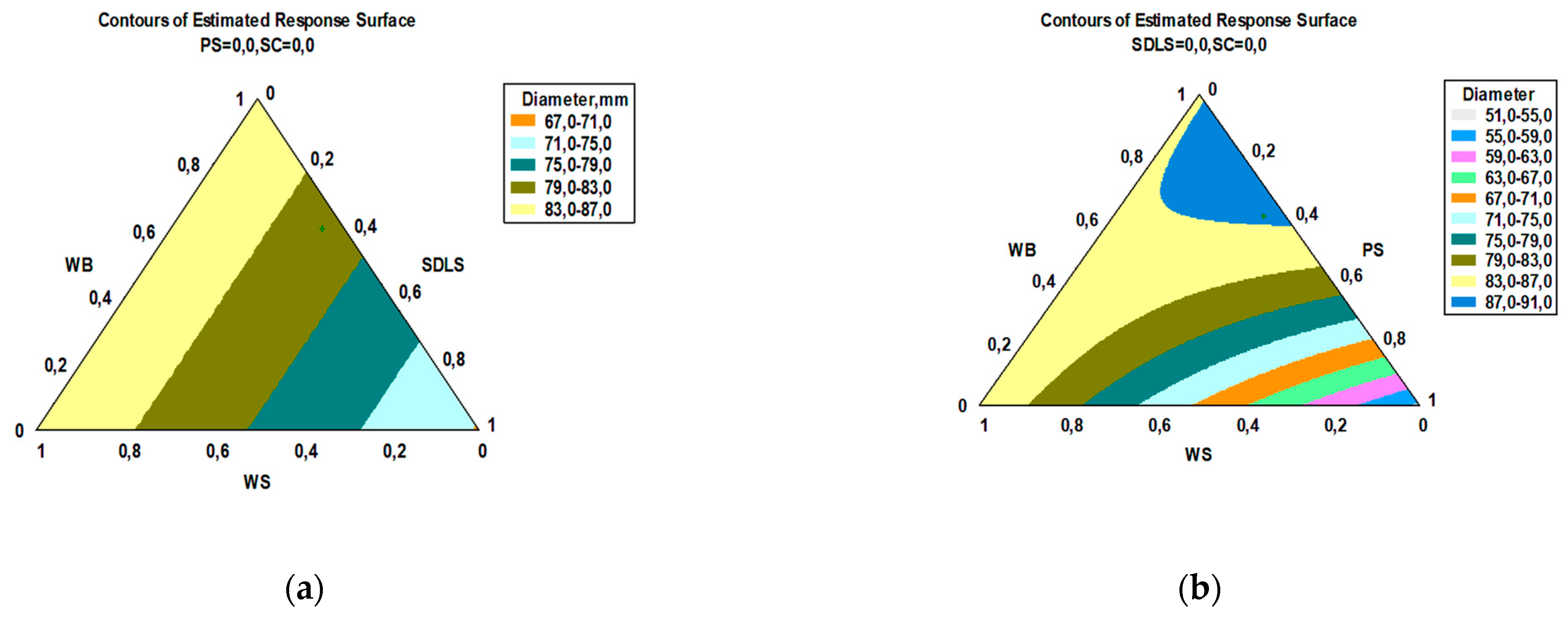
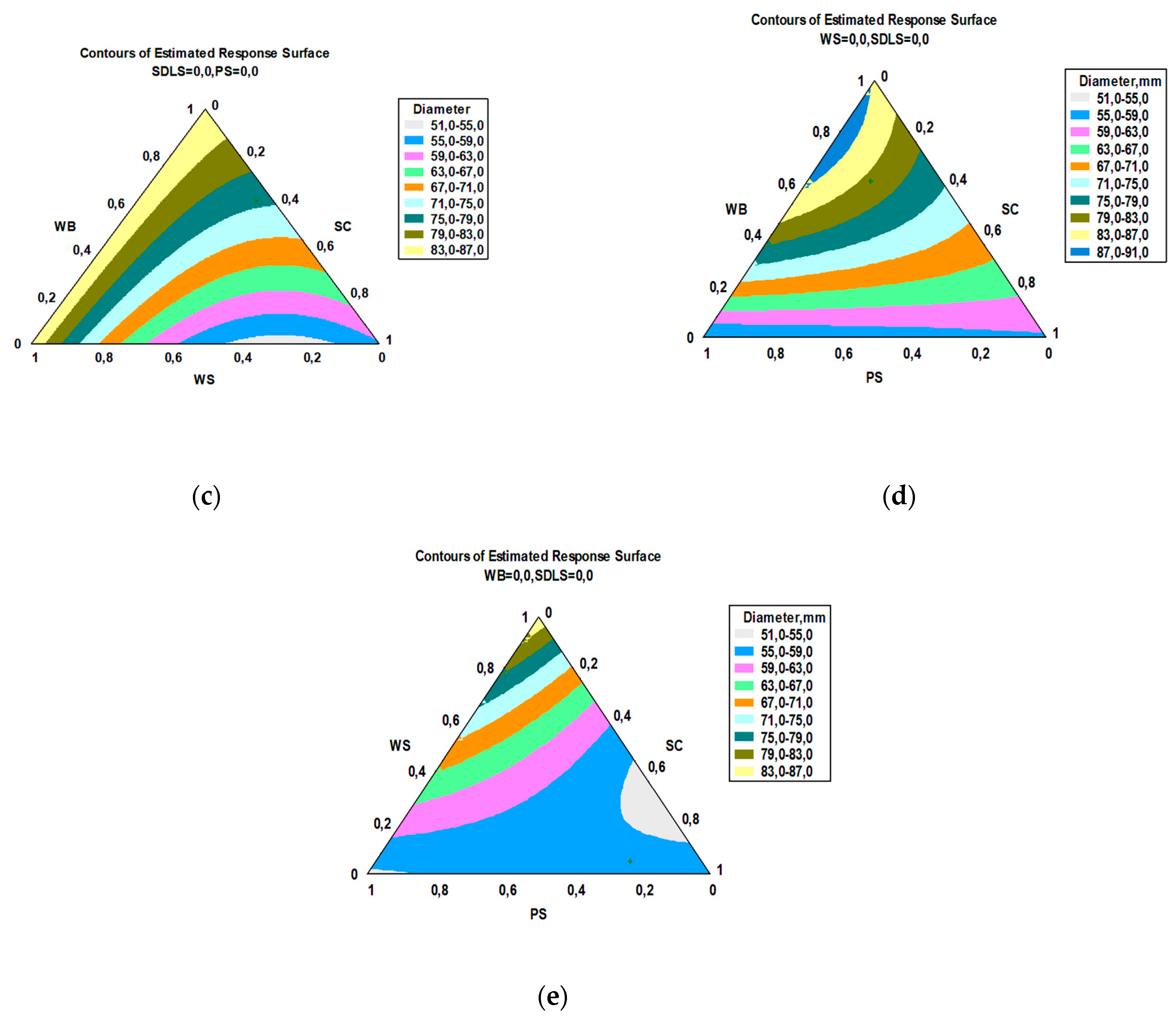
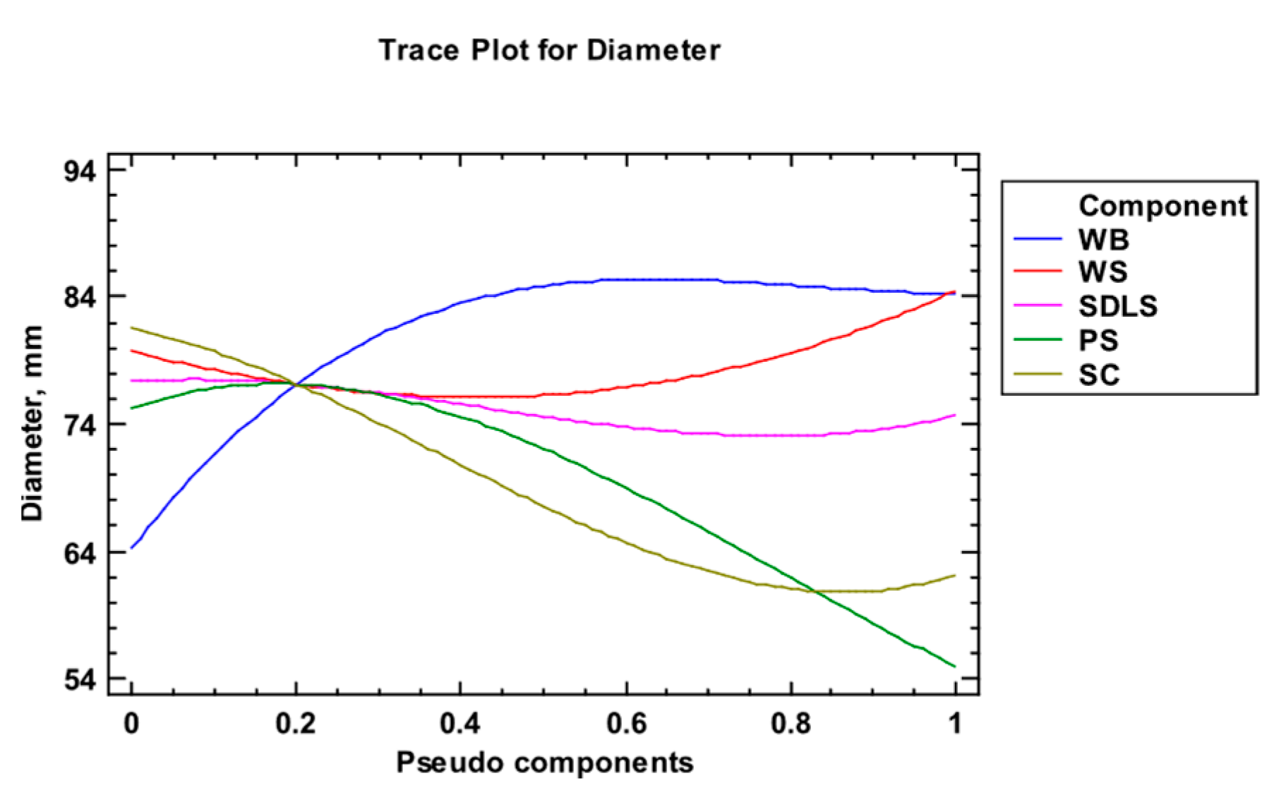
Figure 2a–e present the multitarget optimization response surfaces, and Figure 3 presents the dependence of the diameter of the micelle mass on the pseudo components in the mixture.
Figure 3 shows that as the amount of WB in the mixture increases, the mycelial diameter increases, and this trend is observed until reaching an amount of 0.6, after which a further increase in WB does not lead to an increase in biomass. In the case of WS, from the beginning, with an increase in its amount in the mixture, a decrease in biomass is expected, which is observed up to an amount of 0.4, then with its increase, the mycelial mass also increases. The diagram also shows that the SDLS component does not significantly affect the increase in the mycelial mass, while the PS and SC components have a significant impact. The graph shows that high mycelial growth is expected with a small amount of these components in the medium, and with an increase in their share and a decrease in WB, WS, and, to some extent, SDLS, will lead to a decrease in mycelial growth.
After an optimization procedure, the optimal composition of the multicomponent mixture was determined. It is presented in Table 3. During the mathematical processing of the obtained results, it was calculated that the amount of SC was 2.4 × 10⁻⁸%, which had no statistically significant effect on the growth of the mycelium.
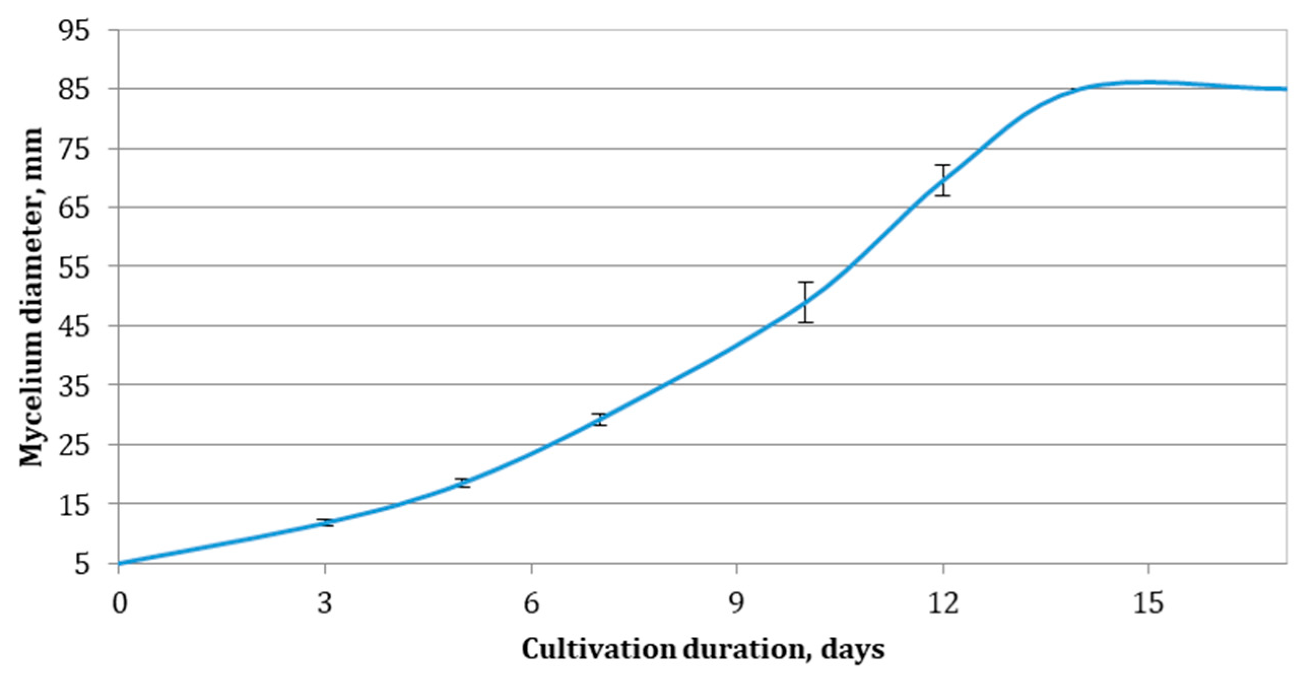
A process of cultivation of E. nigrum on the optimized multicomponent medium was carried out for the purpose of its experimental validation. The dynamics of the change in the mycelial mass were again monitored, and the results of these experiments are presented in Figure 4.
The data presented in Figure 4 show that on this medium, the strain reached a maximum diameter of 85 mm on the 14th day from the beginning of the process, and this value remained constant until the 17th day. The kinetics of growth of the strain in the multicomponent mixture were studied. The identification of the parameters of the models was made, and the results of these studies are shown in Table 4.
From the results presented in Table 4, it can be seen that in the composed medium, the culture developed with a relatively high growth rate—0.325 ± 0.028 d−1, and also a low value of the parameter δ—0.0034 ± 0.0004 mm.d−1—was observed, which proves that the multicomponent mixture has an optimal composition. This is evidenced by the high coefficient of substrate utilization, which was 0.9851 ± 0.0166, and also by S0, which was 90 ± 1 mm, which is close to the experimentally recorded mycelial diameter—85 mm.

3.3. Bioactivity

Nutrient medium content influences both the biomass accumulation of a fungal organism and its bioactivity. In order to study the effect of the selected substrates on E. nigrum bioactivity, and based on the results from the kinetic modelling, three nutrient media were selected for solid-state cultivation of the strain—WB, WS, and the optimized multicomponent medium—without the addition of agar–agar. After 14 days of cultivation, water extracts were prepared in a ratio of 1:1 (w/w) in order to minimize the dilution of the bioactive components. The obtained extracts were analyzed for biological activities.

3.3.1. Determination of Enzyme Activities

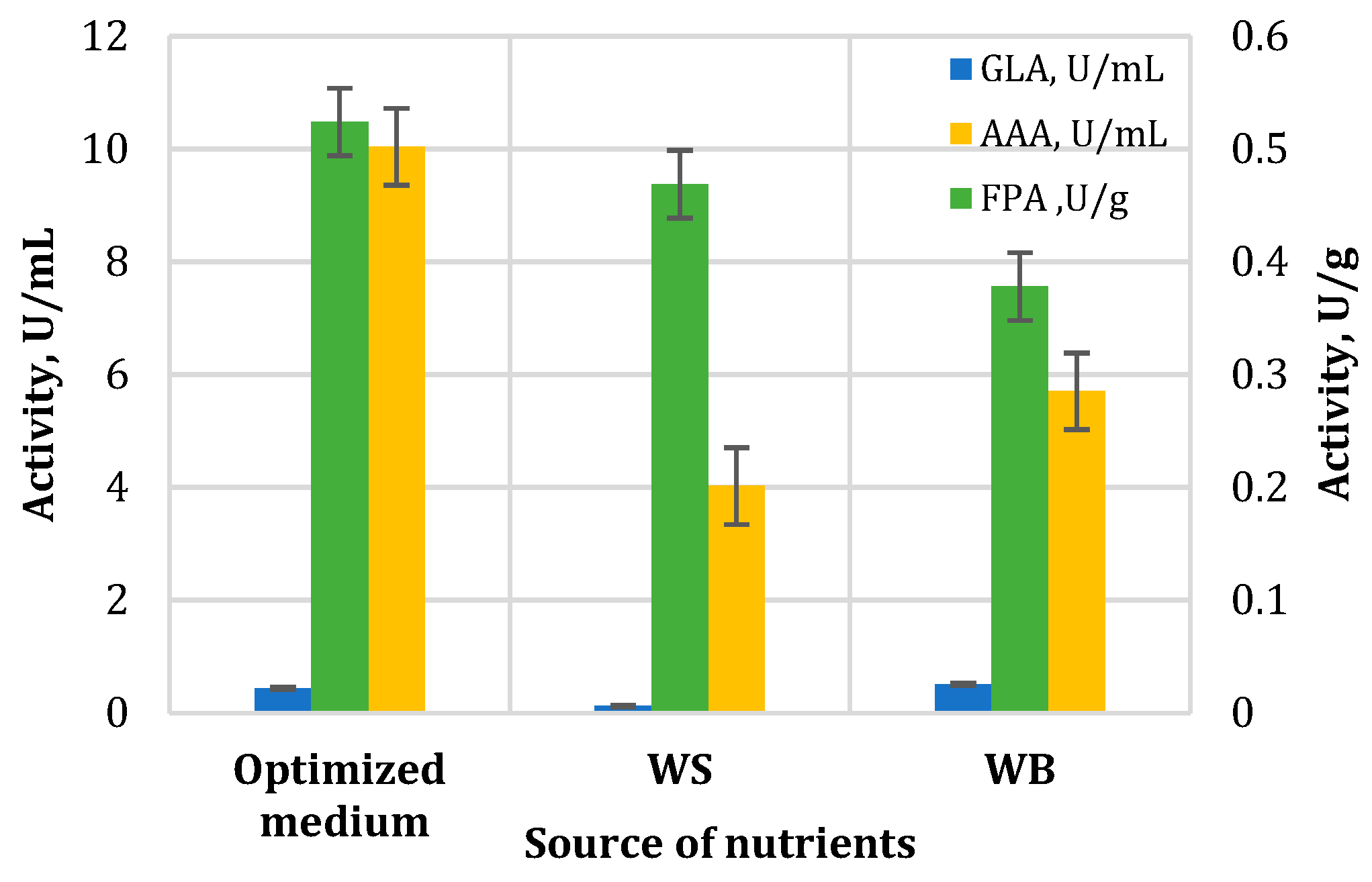
The extract obtained after the cultivation of E. nigrum on the selected substrates was analyzed for the presence of various enzymes, including cellulases, amylases, and ligninolytic enzymes. The results are presented in Figure 5. The strain demonstrated the ability to secrete cellulolytic enzymes on all substrates used for cultivation, which is not surprising, given the fact that they are necessary for providing nutrients. The highest activity was measured when the optimized medium was used as a substrate for the cultivation of E. nigrum (0.52 ± 0.03 U/g), and the lowest activity was measured in the WB medium extract (0.38 ± 0.019 U/g). Unfortunately, in the available literature, there is almost no data regarding the synthesis of cellulolytic enzymes by the Epicoccum species. In one study, just two E. nigrum strains out of 30 Epicoccum isolates exhibited cellulolytic enzyme activity, as evidenced by their growth on specific media and the presence of degradation halos [65]. Brown (1984) reported cellulase activity of 40 µg/mL glucose equivalent [63]. In the screening of 18 E. nigrum strains for cellulose activity, another study identified 8 strains with an average activity of 0.27 × 10⁻⁵ EU/mL [62].
The extracts were analyzed for the presence of amylolytic enzymes—glucoamylase and α-amylase in particular. These enzymes play an important role in the degradation of starch, with the α-amylase acting on α-1,4-linked glycosyl polysaccharides and the glucoamylase acting on single glycosidic residues from the non-reducing end of the polysaccharide chain in a stepwise matter. The latter enzyme is also capable of hydrolyzing the 1,6-α bonds at the branching point of amylopectin, but with much lower efficiency [66,67]. In our study, the highest α-amylase activity was measured when the optimized medium was used as a feeding substrate for the SSC (10.6 ± 0.6 U/mL), and the lowest activity (4.02 ± 0.08 U/mL) was measured with WS used as a substrate. The best substrate for the synthesis of glucoamylase was identified to be WB, where the measured activity was 0.51 ± 0.03 U/mL. The lowest activity (0.12 ± 0.01 U/mL) belonged to the extract obtained after cultivation on WS.
Even though there are several researchers who state the ability of Epicoccum species to produce lignin-degrading enzymes [64,65], the strain used in this research did not demonstrate such ability when cultivated on the used lignocellulose substrates at solid-state conditions.
The results from the enzyme activity analyses explain the ability of E. nigrum to grow and develop on agro-industrial substrates. It is not unusual for the Epicoccum species to produce amylolytic enzymes, and there are few papers on that matter [65,68]. E. nigrum PG16 was capable of producing 1 U/mL of amylase activity under optimized submerged culturing conditions [64]. This observation highlights the potential of the strain used in our study to produce high yields of amylase after optimization of the culture medium.

3.3.2. Determination of Antifungal Activity

In a previous study, a controlled submerged cultivation was carried out, and the untreated cultural liquid demonstrated antiphytopathogenic activity against Sclerotinia sclerotiorum, Botrytis cinerea, and Aspergillus flavus [37]. To further investigate the antifungal activities of E. nigrum, a dual culture study was performed against nine fungal strains for antagonistic effects between the fungi (Figure 6).
The most significant antagonism was detected between Epicoccum nigrum and Fusarium verticillioides, Fusarium oxysporum, Alternaria alternate, and Sclerotinia sclerotiorum.
Other authors also report on the antifungal activity of E. nigrum against some of the tested fungi. Hoyte et al. (2006) studied kiwifruit diseases and found that an E. nigrum biocontrol agent reduced Sclerotinia sclerotiorum petal infections by 100% in field tests [69]. In different field experiments, it significantly reduced infections with Sclerotinia sclerotiorum in white beans [70]. In field tests, a culture filtrate of E. nigrum was more effective than the fungicide penconazole at the recommended concentrations in preventing powdery mildew on squash caused by Botrytis cinerea [71]. In another study, Alcock et al. (2015) identified five strains producing epirodin, a polyene antibiotic, which inhibited the same pathogen in vitro [72]. A dual culture assay proved that Fusarium oxysporum, causing root rot and wilting in plants, was inhibited by E. nigrum [73]. Yellowing of cabbage in vitro and in vivo was reduced by inhibiting the growth of F. oxysporum [74]. E. nigrum also exhibited antagonism against Fusarium verticillioides on sugarcane, and an ethyl acetate extract of its biomass at a 2.0 mg/mL concentration reduced the pathogen colony diameter by more than 30% [29]. Alternaria triticimaculans, causing leaf spots of wheat, was inhibited under greenhouse conditions [75].
Most of these experiments are in field, where the authors used spore suspensions of E. nigrum, or they are in vitro dual culture studies. Other investigations focus on the isolation of individual compounds with antimicrobial activity, synthesized by E. nigrum, such as epicoccolides (polyketides) [76], diketopiperazines [77], and polyenes (epipyrone A) [39]. Our goal was to quantify the antifungal activity of the strain and evaluate the influence of the different substrates for SSC; in order to achieve this rapidly and efficiently, we determined the minimal inhibitory concentration of the extracts (Table 5).
The extracts exhibited activity against all fungal strains, unlike in the dual culture assay. This fact is probably due to the higher concentration of antimicrobial substances in the extract and the hindered diffusion of the antifungal substances in the agar medium. In our previous study on the effect of culture liquid after submerged cultivation in liquid medium, no inhibitory effect was detected against F. verticillioides [37], which could also be attributed to the lower concentration of antimicrobials in that case. The most sensitive strain against all extracts was S. sclerotiorum with MICs ranging between 0.156 and 0.313 mg/mL, followed by B. cinerea, Alternaria sp., and F. verticillioides, with MICs between 0.313 and 0.625. The least susceptible was A. alternata, with MICs of 1.25 mg/mL for all three extracts, while A. flavus exhibited the highest overall resistance against the WB extract (MIC of 2.5 mg/mL). Such values for MIC are significantly higher than those of purified antifungals, like amphotericin B, with MICs lower than 0.008 µg/mL against 106 Mucor sp. isolates [78], or an overall MIC of 0.002 µg/mL against 118 Penicillium sp. isolates [79]. The same antibiotic demonstrated MICs between 0.001 and 0.004 µg/mL against 58 clinical A. flavus isolates [80]. Commercial fungicides, such as Prochloraz, are also more potent in comparison. Xu et al. (2020) reported an MIC of 0.016 µg/mL against A. alternata and 0.008 µg/mL against B. cinerea and F. oxysporum [81]. In terms of antifungal activity, WS proved to be the best substrate for SSC since the extract exhibited the lowest MIC (0.156 mg/mL against P. chrysogenum), and no MIC was higher than 1.25 mg/mL (against A. alternata). There are no reports from other authors of extracts with antifungal activity obtained after SSC of agro-industrial waste- and by-products with Epicoccum nigrum.

4. Conclusions

The application of mathematical modeling for nutrient medium optimization led to the first successful solid-state cultivation of E. nigrum on agro-industrial substrates for the production of water extracts with valuable biological activities. The obtained extracts demonstrated amylolytic and cellulase activities, as well as inhibitory activity against fungal pathogens. The proposed optimized nutrient medium and method for solid-state cultivation could be used for the manufacturing of environmentally friendly biocontrol agents since no harmful chemicals were used for extraction. Further experiments are needed for individual antifungal compounds to be identified and their production through biotechnological methods to be optimized. Our findings suggest that this method of cultivation could effectively be used for the ecologically friendly, sustainable, and cost-effective production of biologically active compounds, such as amylolytic enzymes and antimicrobials.

Supplementary Materials

The following supporting information can be downloaded at https://www.mdpi.com/article/10.3390/app151910571/s1, Table S1: Composition of the individual mixtures and mycelium diameter.

Author Contributions

Conceptualization, G.A. and M.B.; methodology, G.A., M.B., P.S., D.B. and B.G.; software, B.G. and S.B.; validation, B.G., Z.G. and R.B.; formal analysis, Z.G., R.B., B.G., P.S., R.D.-K., A.S., D.B., M.B. and G.A.; investigation, Z.G., R.B., B.G., P.S., R.D.-K., D.B., M.B., A.S. and G.A.; resources, G.A., Z.G., D.B. and M.B.; data curation, D.B., M.B., B.G., A.S. and S.B.; writing—original draft preparation, G.A., B.G., A.S., M.B., Z.G., S.B. and D.B.; writing—review and editing, M.B., D.B. and G.A.; visualization, Z.G.; supervision, G.A.; project administration, Z.G.; funding acquisition, Z.G. All authors have read and agreed to the published version of the manuscript.

Funding

This research is supported by the Bulgarian Ministry of Education and Science under the National program “Young Scientists and Postdoctoral Students—2”, DCM #206/07.04.2022.

Institutional Review Board Statement

Not applicable.

Informed Consent Statement

Not applicable.

Data Availability Statement

The original contributions presented in this study are included in the article. Further inquiries can be directed to the corresponding author.

Conflicts of Interest

The authors declare no conflicts of interest.

Abbreviations

The following abbreviations are used in this manuscript:
SSCsolid-state cultivation
WSwheat straw
PSpine sawdust
WBwheat bran
SCsunflower cake
SDLSsteam-distilled lavender straw
GalAanhydrouronic acid
TDF total dietary fibers
IDFinsoluble dietary fibers
SDFsoluble dietary fibers
DEdegree of esterification
MICminimal inhibitory concentration
DMBA3,4-dimethoxybenzyl alcohol
FPAfilter paper assay
GLAglucoamylase activity
AAAα-amylase activity

References

  1. Madeira, J.; Contesini, F.J.; Calzado, F.; Rubio, M.V.; Zubieta, M.P.; Lopes, D.B.; de Melo, R.R. Agro-industrial residues and microbial enzymes: An overview on the eco-friendly bioconversion into high value-added products. In Biotechnology of Microbial Enzymes; Brahmachari, G., Ed.; Elsevier: London, UK, 2017; pp. 475–511. [Google Scholar]
  2. Vučurović, D.; Bajić, B.; Trivunović, Z.; Dodić, J.; Zeljko, M.; Jevtić-Mučibabić, R.; Dodić, S. Biotechnological Utilization of Agro-Industrial Residues and By-Products—Sustainable Production of Biosurfactants. Foods 2024, 13, 711. [Google Scholar] [CrossRef]
  3. Kassim, F.O.; Thomas, C.L.P.; Afolabi, O.O.D. Integrated conversion technologies for sustainable agri-food waste valorization: A critical review. Biomass Bioenergy 2022, 156, 106314. [Google Scholar] [CrossRef]
  4. Shah, A.M.; Zhang, H.; Shahid, M.; Ghazal, H.; Shah, A.R.; Niaz, M.; Naz, T.; Ghimire, K.; Goswami, N.; Shi, W.; et al. The vital roles of agricultural crop residues and agro-industrial by-products to support sustainable livestock productivity in subtropical regions. Animals 2025, 15, 1184. [Google Scholar] [CrossRef] [PubMed]
  5. Pilafidis, S.; Diamantopoulou, P.; Gkatzionis, K.; Sarris, D. Valorization of agro-industrial wastes and residues through the production of bioactive compounds by macrofungi in liquid state cultures: Growing circular economy. Appl. Sci. 2022, 12, 11426. [Google Scholar] [CrossRef]
  6. Georganas, A.; Giamouri, E.; Pappas, A.C.; Papadomichelakis, G.; Galliou, F.; Manios, T.; Tsiplakou, E.; Fegeros, K.; Zervas, G. Bioactive compounds in food waste: A review on the transformation of food waste to animal feed. Foods 2020, 9, 291. [Google Scholar] [CrossRef]
  7. Sarsaiya, S.; Jain, A.; Kumar Awasthi, S.; Duan, Y.; Kumar Awasthi, M.; Shi, J. Microbial dynamics for lignocellulosic waste bioconversion and its importance with modern circular economy, challenges and future perspectives. Bioresour. Technol. 2019, 291, 121905. [Google Scholar] [CrossRef] [PubMed]
  8. Parfitt, J.; Barthel, M.; Macnaughton, S. Food waste within food supply chains: Quantification and potential for change to 2050. Philos. Trans. R. Soc. B Biol. Sci. 2010, 365, 3065–3081. [Google Scholar] [CrossRef]
  9. Ferronato, N.; Torretta, V. Waste mismanagement in developing countries: A review of global issues. Int. J. Environ. Res. Public Health 2019, 16, 1060. [Google Scholar] [CrossRef] [PubMed]
  10. Hamelin, L.; Borzęcka, M.; Kozak, M.; Pudełko, R. A spatial approach to bioeconomy: Quantifying the residual biomass potential in the EU-27. Renew. Sustain. Energy Rev. 2019, 100, 127–142. [Google Scholar] [CrossRef]
  11. Matei, E.; Râpă, M.; Predescu, A.M.; Țurcanu, A.A.; Vidu, R.; Predescu, C.; Bobirica, C.; Bobirica, L.; Orbeci, C. Valorization of agri-food wastes as sustainable eco-materials for wastewater treatment: Current state and new perspectives. Materials 2021, 14, 4581. [Google Scholar] [CrossRef]
  12. Azelee, N.I.W.; Manas, N.H.A.; Dailin, D.J.; Ramli, A.N.M.; Shaarani, S.M. Biological Treatment of Agro-Industrial Waste. In Valorisation of Agro-Industrial Residues—Volume I: Biological Approaches; Zakaria, Z., Boopathy, R., Dib, J., Eds.; Springer: Cham, Switzerland, 2020; pp. 59–80. [Google Scholar]
  13. Javourez, U.; O’Donohue, M.; Hamelin, L. Waste-to-nutrition: A review of current and emerging conversion pathways. Biotechnol. Adv. 2021, 53, 107857. [Google Scholar] [CrossRef]
  14. Foong, L.C.; Loh, C.W.L.; Ng, H.S.; Lan, J.C. Recent development in the production strategies of microbial carotenoids. World J. Microbiol. Biotechnol. 2021, 37, 12. [Google Scholar] [CrossRef]
  15. El-Sayed, E.R.; Gach, J.; Olejniczak, T.; Boratyński, F. A new endophyte Monascus ruber SRZ112 as an efficient production platform of natural pigments using agro-industrial wastes. Sci. Rep. 2022, 12, 12611. [Google Scholar] [CrossRef] [PubMed]
  16. Pelkmans, J.F.; Patil, M.B.; Gehrmann, T.; Reinders, M.J.; Wösten, H.A.; Lugones, L.G. Transcription factors of Schizophyllum commune involved in mushroom formation and modulation of vegetative growth. Sci. Rep. 2017, 7, 310. [Google Scholar] [CrossRef] [PubMed]
  17. Meyer, V.; Andersen, M.R.; Brakhage, A.A.; Braus, G.H.; Caddick, M.X.; Cairns, T.C.; de Vries, R.P.; Haarmann, T.; Hansen, K.; Hertz-Fowler, C.; et al. Current challenges of research on filamentous fungi in relation to human welfare and a sustainable bio-economy: A white paper. Fungal Biol. Biotechnol. 2016, 3, 6. [Google Scholar] [CrossRef] [PubMed]
  18. Meyer, V.; Basenko, E.Y.; Benz, J.P.; Braus, G.H.; Caddick, M.X.; Csukai, M.; de Vries, R.P.; Endy, D.; Frisvad, J.C.; Gunde-Cimerman, N.; et al. Growing a circular economy with fungal biotechnology: A white paper. Fungal Biol. Biotechnol. 2020, 7, 5. [Google Scholar] [CrossRef]
  19. Hashem, A.H.; Attia, M.S.; Kandil, E.K.; Fawzi, M.M.; Abdelrahman, A.S.; Khader, M.S.; Khodaira, M.A.; Emam, A.E.; Goma, M.A.; Abdelaziz, A.M. Bioactive compounds and biomedical applications of endophytic fungi: A recent review. Microb. Cell Factories 2023, 22, 107, Erratum in Microb. Cell Factories 2023, 22, 122. [Google Scholar] [CrossRef]
  20. Frisvad, J.C.; Andersen, B.; Thrane, U. The use of secondary metabolite profiling in chemotaxonomy of filamentous fungi. Mycol. Res. 2008, 112, 231–240. [Google Scholar] [CrossRef]
  21. Attia, M.S.; Salem, M.S.; Abdelaziz, A.M. Endophytic fungi Aspergillus spp. reduce fusarial wilt disease severity, enhance growth, metabolism and stimulate the plant defense system in pepper plants. Biomass Convers. Biorefin. 2024, 14, 16603–16613. [Google Scholar] [CrossRef]
  22. Zhang, P.; Yuan, X.-L.; Du, Y.-M.; Zhang, H.-B.; Shen, G.-M.; Zhang, Z.-F.; Liang, Y.-J.; Zhao, D.-L.; Xu, K. Angularly prenylated indole alkaloids with antimicrobial and insecticidal activities from an endophytic fungus Fusarium sambucinum TE-6L. J. Agric. Food Chem. 2019, 67, 11994–12001. [Google Scholar] [CrossRef]
  23. Manganyi, M.C.; Ateba, C.N. Untapped potentials of endophytic fungi: A review of novel bioactive compounds with biological applications. Microorganisms 2020, 8, 1934. [Google Scholar] [CrossRef] [PubMed]
  24. Hawas, U.W.; Abou El-Kassem, L.T. Anticancer and antiviral diketopiperazine produced by the Red Sea endophytic fungus Penicillium chrysogenum. Lett. Org. Chem. 2019, 16, 409–414. [Google Scholar] [CrossRef]
  25. Mastouri, F.; Björkman, T.; Harman, G.E. Seed treatment with Trichoderma harzianum alleviates biotic, abiotic, and physiological stresses in germinating seeds and seedlings. Phytopathology 2010, 100, 1213–1221. [Google Scholar] [CrossRef]
  26. Murphy, B.R.; Doohan, F.M.; Hodkinson, T.R. Yield increase induced by the fungal root endophyte Piriformospora indica in barley grown at low temperature is nutrient limited. Symbiosis 2014, 62, 29–39. [Google Scholar] [CrossRef]
  27. Taguiam, J.D.; Evallo, E.; Balendres, M.A. Epicoccum species: Ubiquitous plant pathogens and effective biological control agents. Eur. J. Plant Pathol. 2021, 159, 713–725. [Google Scholar] [CrossRef]
  28. Kou, Z.-A.; Zhang, Z.-K.; Zhang, W.-X.; Wang, X.-F.; Liu, L.; Teng, G.-X.; Tian, Y.-Q. First report of Epicoccum nigrum causing leaf spot of endive (Cichorium endivia L.) in China. Crop Prot. 2024, 177, 106534. [Google Scholar] [CrossRef]
  29. Fávaro, L.C.; Sebastianes, F.L.; Araújo, W.L. Epicoccum nigrum P16, a sugarcane endophyte, produces antifungal compounds and induces root growth. PLoS ONE 2012, 7, e36826. [Google Scholar] [CrossRef]
  30. Elnaggar, M.S.; Fayez, S.; Anwar, A.; Ebada, S.S. Cytotoxic naphtho- and benzofurans from an endophytic fungus Epicoccum nigrum Ann-B-2 associated with Annona squamosa fruits. Sci. Rep. 2024, 14, 4940. [Google Scholar] [CrossRef]
  31. Li, T.; Im, J.; Lee, J. Genetic Diversity of Epicoccum nigrum and its effects on Fusarium graminearum. Mycobiology 2022, 50, 457–466. [Google Scholar] [CrossRef]
  32. Musetti, R.; Grisan, S.; Polizzotto, R.; Martini, M.; Paduano, C.; Osler, R. Interactions between ‘Candidatus Phytoplasma mali’ and the apple endophyte Epicoccum nigrum in Catharanthus roseus plants. J. Appl. Microbiol. 2011, 110, 746–756. [Google Scholar] [CrossRef] [PubMed]
  33. Sawinder, K.; Panesar, P.S.; Gurumayum, S.; Singh, J.; Assouguem, A.; Lazraq, A.; Ullah, R.; Ali, E.A.; Skender, A.; Rasane, P. Stability kinetics of orevactaene pigments produced by Epicoccum nigrum in solid-state fermentation. Open Chem. 2023, 21, 20230166. [Google Scholar] [CrossRef]
  34. El-Sayed, E.R.; Ahmed, A.S.; Al-Hagar, O.E.A. Agro-industrial wastes for production of paclitaxel by irradiated Aspergillus fumigatus under solid-state fermentation. J. Appl. Microbiol. 2020, 128, 1427–1439. [Google Scholar] [CrossRef]
  35. Manan, M.A.; Webb, C. Design aspects of solid-state fermentation as applied to microbial bioprocessing. J. Appl. Biotechnol. Bioeng. 2017, 4, 511–532. [Google Scholar] [CrossRef]
  36. Zaki, A.G.; El-Sayed, E.R. New and potent production platform of the acetylcholinesterase inhibitor Huperzine A by gammairradiated Alternaria brassicae under solid-state fermentation. Appl. Microbiol. Biotechnol. 2021, 105, 8869–8880. [Google Scholar] [CrossRef]
  37. Baldzhieva, R.; Brazkova, M.; Blazheva, D.; Goranov, B.; Stefanova, P.; Ganeva, Z.; Teneva, D.; Denev, P.; Angelova, G. Growth kinetics modeling and evaluation of antiphytopathogenic activity of newly isolated fungicolous Epicoccum nigrum associated with Dryad’s Saddle (Polyporaceae). Agriculture 2024, 14, 2179. [Google Scholar] [CrossRef]
  38. Ali, S.A.-B.; Abdelmoaty, H.; Ramadan, H.; Salman, Y. The endophytic fungus Epicoccum nigrum: Isolation, molecular identification and study its antifungal activity against phytopathogenic fungus Fusarium solani. J. Microbiol. Biotechnol. Food Sci. 2024, 13, e10093. [Google Scholar] [CrossRef]
  39. Lee, A.J.; Cadelis, M.M.; Kim, S.H.; Swift, S.; Copp, B.R.; Villas-Boas, S.G. Epipyrone A, a broad-spectrum antifungal compound produced by Epicoccum nigrum ICMP 19927. Molecules 2020, 25, 5997. [Google Scholar] [CrossRef] [PubMed]
  40. Dzoyem, J.P.; Melong, R.; Tsamo, A.T.; Maffo, T.; Kapche, D.G.W.F.; Ngadjui, B.T.; McGaw, L.J.; Eloff, J.N. Cytotoxicity, antioxidant and antibacterial activity of four compounds produced by an endophytic fungus Epicoccum nigrum associated with Entada abyssinica. Rev. Bras. Farmacogn. 2017, 27, 251–253. [Google Scholar] [CrossRef]
  41. Nzabanita, C.; Zhang, L.; Zhao, H.; Wang, Y.; Wang, Y.; Sun, M.; Wang, S.; Guo, L. Fungal endophyte Epicoccum nigrum 38L1 inhibits in vitro and in vivo the pathogenic fungus Fusarium graminearum. Biol. Control 2022, 174, 105010. [Google Scholar] [CrossRef]
  42. Carboué, Q.; Besaury, L.; Petracco, E.; Fournier, R.; Dosso, A.; Godon, B.; Sayagh, C.; Martinez, A.; Legin, E.; Alavoine, G.; et al. Pigments production from a newly isolated psychrotrophic strain of Epicoccum nigrum grown on non-sterile exhausted sugar beet pulp through solid state fermentation. Chem. Eng. J. 2025, 507, 160682. [Google Scholar] [CrossRef]
  43. Angelova, G.; Yemendzhiev, H.; Zaharieva, R.; Brazkova, M.; Koleva, R.; Stefanova, P.; Baldzhieva, R.; Vladev, V.; Krastanov, A. Mycelium-Based Composites Derived from Lignocellulosic Residual By-Products: An Insight into Their Physico-Mechanical Properties and Biodegradation Profile. Appl. Sci. 2025, 15, 6333. [Google Scholar] [CrossRef]
  44. Blumenkrantz, N.; Asboe-Hansen, G. New method for quantitative determination of uronic acids. Anal. Biochem. 1973, 54, 484–489. [Google Scholar] [CrossRef]
  45. Marovska, G.I.; Hambarliyska, I.P.; Petkova, N.T.; Ivanov, I.G.; Georgiev, V.; Topuzova, M.G.; Slavov, A.M. General characteristics of lavender biomass (Lavandula angustifolia Mill.) before and after industrial distillation. Food Sci. Appl. Biotechnol. 2024, 7, 133–144. [Google Scholar] [CrossRef]
  46. AOAC. Official Method 991.43. Total, Soluble, and Insoluble Dietary Fiber in Foods, En-Zymatic-Gravimetric Method, MES-TRIS Buffer; Association of Official Analytical Chemists International: Gaithersburg, MD, USA, 1994. [Google Scholar]
  47. de Escalada Pla, M.F.; González, P.; Sette, P.; Portillo, F.; Rojas, A.M.; Gerschenson, L.N. Effect of processing on physico-chemical characteristics of dietary fibre concentrates obtained from peach (Prunus persica L.) peel and pulp. Food Res. Int. 2012, 49, 184–192. [Google Scholar] [CrossRef]
  48. Bouguettoucha, A.; Balannec, B.; Amrane, A. Unstructured models for lactic acid fermentation—A review. Food Technol. Biotechnol. 2011, 49, 3–12. [Google Scholar]
  49. Ibarz, A.; Augusto, P.E.D. An autocatalytic kinetic model for describing microbial growth during fermentation. Bioprocess Biosyst. Eng. 2015, 38, 199–205. [Google Scholar] [CrossRef]
  50. Gaponov, K.P. Processes and Apparatus for Microbiological Industry; Legkaya I Pishchevaya Promyshlenmost: Moscow, Russia, 1981; p. 240. (In Russian) [Google Scholar]
  51. Kemmer, G.; Keller, S. Nonlinear least-squares data fitting in Excel spreadsheets. Nat. Protoc. 2010, 5, 267–281. [Google Scholar] [CrossRef]
  52. Choi, M.; Al-Zahrani, S.M.; Lee, S.Y. Kinetic model-based feed-forward controlled fed-batch fermentation of Lactobacillus rhamnosus for the production of lactic acid from Arabic date juice. Bioprocess Biosyst. Eng. 2014, 37, 1007–1015. [Google Scholar] [CrossRef]
  53. Granato, D.; Calado, V.; Pinto, E.R. Optimization of food processes using mixture experiments. In Mathematical and Statistical Applications in Food Engineering, 1st ed.; Sevdav, S., Singh, A., Eds.; CRC Press: Boca Raton, FL, USA, 2020; p. 15. [Google Scholar] [CrossRef]
  54. NIST/SEMATECH e-Handbook of Statistical Methods; National Institute of Standards and Technology: Gaithersburg, MD, USA, 2012. Available online: https://www.itl.nist.gov/div898/handbook/ (accessed on 22 August 2025). [CrossRef]
  55. Lever, M. A New Reaction for Colorimetric Determination of Carbohydrates. Anal. Biochem. 1972, 47, 273–279. [Google Scholar] [CrossRef]
  56. Fuwa, H. A new method of microdetermination of amylase activity by the use of amylase as the substrate. J. Biochem. 1954, 41, 583–603. [Google Scholar] [CrossRef]
  57. Ride, J.P. The effect of induced lignification on the resistance of wheat cell walls to fungal degradation. Physiol. Plant Pathol. 1970, 16, 187–196. [Google Scholar] [CrossRef]
  58. Wariishi, H.; Dunford, H.B.; McDonald, I.D.; Gold, M.H. Manganese Peroxidase from the lignin-degrading Basidiomycette Phanerochaete chrysosporium. J. Biol. Chem. 1989, 264, 3335–3340. [Google Scholar] [CrossRef]
  59. Tien, M.; Kirk, T.K. Lignin-Degrading Enzyme from the Hymenomycete Phanerochaete chrysosporium. Burds Sci. 1983, 221, 661–663. [Google Scholar] [CrossRef]
  60. CLSI. Method for Broth Dilution Antifungal Susceptibility Testing of Filamentous Fungi, Approved Standard, 2nd ed.; CLSI document M38-A2; Clinical and Laboratory Standards Institute: Wayne, PA, USA, 2008. [Google Scholar]
  61. Vasudha, C.; Sarla, L. Nutritional quality analysis of sunflower seed cake (SSC). Pharma Innov. J. 2021, 10, 720–728. [Google Scholar]
  62. Ogórek, R.; Przywara, K.; Piecuch, A.; Cal, M.; Lejman, A.; Matkowski, K. Plant-fungal interactions: A case study of Epicoccoum nigrum Link. Plants 2020, 9, 1691. [Google Scholar] [CrossRef] [PubMed]
  63. Brown, A.E. Epicoccum nigrum, a primary saprophyte involved in the retting of flax. Trans. Br. Mycol. Soc. 1984, 83, 29–35. [Google Scholar] [CrossRef]
  64. Martim, D.B.; dos Santos, F.C.; Barbosa-Tessmann, I.P. Production, partial purification, and characterization of a glucoamylase from Epicoccum nigrum. Acta Scientiarum. Biol. Sci. 2023, 45, e61179. [Google Scholar] [CrossRef]
  65. Xu, X.; Li, J.; Yang, X.; Zhang, L.; Wang, S.; Shen, G.; Hui, B.; Xiao, J.; Zhou, C.; Wang, X.; et al. Epicoccum spp. causing maize leaf spot in Heilongjiang Province, China. Plant Dis. 2022, 106, 3050–3060. [Google Scholar] [CrossRef] [PubMed]
  66. Zhang, J.; Cui, J.H.; Yin, T.; Sun, L.; Li, G. Activated effect of lignin on α-amylase. Food Chem. 2013, 141, 2229–2237. [Google Scholar] [CrossRef]
  67. Soccol, C.R.; Rojan, P.J.; Patel, A.K.; Woiciechowski, A.L.; Vandenberghe, L.P.; Pandey, A. Glucoamylase. In Enzyme Technology; Pandey, A., Webb, C., Soccol, R.C., Larroche, C., Eds.; Asiatech Publishers, Inc.: New Delhi, India, 2006; pp. 221–237. [Google Scholar]
  68. De Paula, N.M.; da Silva, K.; Brugnari, T.; Haminiuk, C.W.I.; Maciel, G.M. Biotechnological potential of fungi from a mangrove ecosystem: Enzymes, salt tolerance and decolorization of a real textile effluent. Microbiol. Res. 2022, 254, 126899. [Google Scholar] [CrossRef]
  69. Hoyte, S.M.; Elmer, P.A.G.; Parry, F.J.; Taylor, J.T.; Marsden, R.S. Biological suppression of Sclerotinia sclerotiorum in kiwifruit. Acta Hortic. 2007, 753, 661–668. [Google Scholar] [CrossRef]
  70. Huang, H.C.; Bremer, E.; Hynes, R.K.; Erickson, R.S. Foliar application of fungal biocontrol agents for the control of white mold of dry bean caused by Sclerotinia sclerotiorum. Biol. Control 2000, 18, 270–276. [Google Scholar] [CrossRef]
  71. Elkot, G.A.E.; Derbalah, A.S.H. Use of cultural filtrates of certain microbial isolates for powdery mildew control in squash. J. Plant Prot. Res. 2011, 51, 252–260. [Google Scholar] [CrossRef]
  72. Alcock, A.; Elmer, P.; Marsden, R.; Parry, F. Inhibition of Botrytis cinerea by epirodin: A secondary metabolite from New Zealand isolates of Epicoccum nigrum. J. Phytopathol. 2015, 163, 841–852. [Google Scholar] [CrossRef]
  73. Ogórek, R.; Plaskowska, E. Epicoccum nigrum for biocontrol agents in vitro of plant fungal pathogens. Commun. Agric. Appl. Biol. Sci. 2011, 76, 691–697. [Google Scholar]
  74. Park, J.Y.; Okada, G.; Takahashi, M.; Oyaizu, H. Screening of fungal antagonists against yellows of cabbage caused by Fusarium oxysporum f. sp. conglutinans. Mycoscience 2002, 43, 447–451. [Google Scholar] [CrossRef]
  75. Perelló, A.; Simón, M.R.; Arambarri, A.M.; Cordo, C.A. Greenhouse screening of the saprophytic resident microflora for control of leaf spots of wheat (Triticum aestivum). Phytoparasitica 2001, 29, 341–351. [Google Scholar] [CrossRef]
  76. Talontsi, F.M.; Dittrich, B.; Schüffler, A.; Sun, H.; Laatsch, H. Epicoccolides: Antimicrobial and antifungal polyketides from an endophytic fungus Epicoccum sp. associated with Theobroma cacao. Eur. J. Org. Chem. 2013, 15, 3174–3180. [Google Scholar] [CrossRef]
  77. Qader, M.M.; Hamed, A.A.; Soldatou, S.; Abdelraof, M.; Elawady, M.E.; Hassane, A.S.I.; Belbahri, L.; Ebel, R.; Rateb, M.E. Antimicrobial and antibiofilm activities of the fungal metabolites isolated from the marine endophytes Epicoccum nigrum M13 and Alternaria alternata 13A. Mar. Drugs 2021, 19, 232. [Google Scholar] [CrossRef] [PubMed]
  78. Badali, H.; Cañete-Gibas, C.; McCarthy, D.; Patterson, H.; Sanders, C.; David, M.P.; Mele, J.; Fan, H.; Wiederhold, N.P. Epidemiology and antifungal susceptibilities of mucoralean fungi in clinical samples from the United States. J. Clin. Microbiol. 2021, 59, JCM0123021. [Google Scholar] [CrossRef]
  79. Guevara-Suarez, M.; Sutton, D.A.; Cano-Lira, J.F.; García, D.; Martin-Vicente, A.; Wiederhold, N.; Guarro, J.; Gené, J. Identification and antifungal susceptibility of penicillium-like fungi from clinical samples in the United States. J. Clin. Microbiol. 2016, 54, 2155–2161. [Google Scholar] [CrossRef] [PubMed]
  80. Gonçalves, S.S.; Stchigel, A.M.; Cano, J.; Guarro, J.; Colombo, A.L. In vitro antifungal susceptibility of clinically relevant species belonging to Aspergillus section Flavi. Antimicrob. Agents Chemother. 2013, 57, 1944–1947. [Google Scholar] [CrossRef] [PubMed]
  81. Xu, K.; Wei, X.-L.; Xue, L.; Zhang, Z.-F.; Zhang, P. Antimicrobial meroterpenoids and erythritol derivatives isolated from the marine-algal-derived endophytic fungus Penicillium chrysogenum XNM-12. Mar. Drugs 2020, 18, 578. [Google Scholar] [CrossRef] [PubMed]
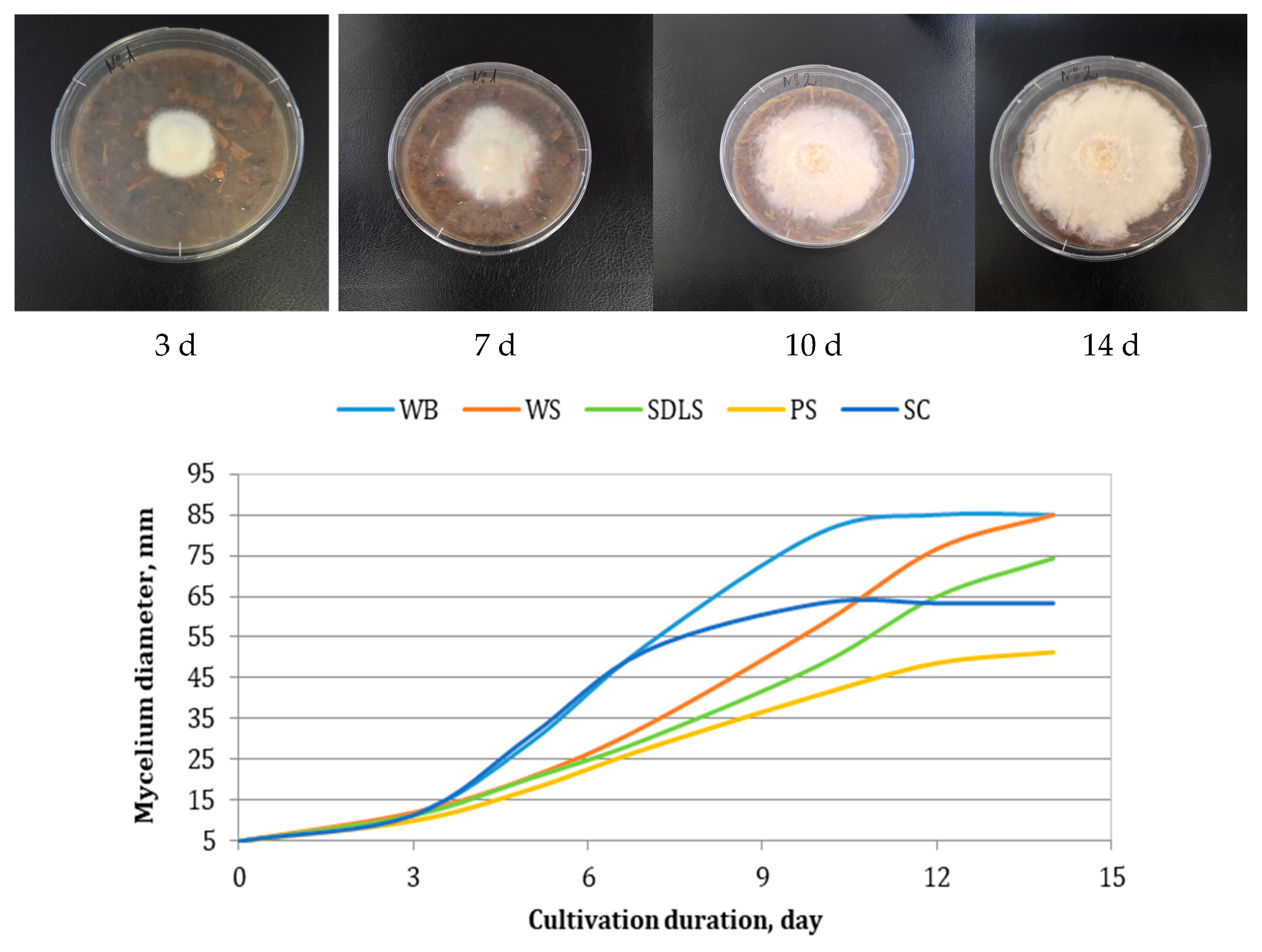
Figure 1. Mycelial growth of E. nigrum on different complex nutrient media.
Applsci 15 10571 g001
Figure 2. Multitarget optimization response surfaces for components: (a) WB, WS, and SDLS; (b) WB, WS, and PS; (c) WB, WS, and SC; (d) WB, PS, and SC; (e) WS, PS, and SC.
Applsci 15 10571 g002aApplsci 15 10571 g002b
Figure 3. Dependence of the diameter of the micelle mass on the pseudo components in the mixture.
Applsci 15 10571 g003
Figure 4. Mycelial growth of E. nigrum on the optimized multicomponent nutrient medium.
Applsci 15 10571 g004
Figure 5. Enzyme activities in the extract obtained after cultivation of E. nigrum on different substrates. GLA—glusoamylase activity; AAA—α-amylase activity; FPA—cellulase activity.
Applsci 15 10571 g005
Figure 6. Dual culture assay of Epicoccum nigrum against (a) Fusarium verticillioides; (b) Fusarium oxysporum; (c) Alternaria alternata; (d) Sclerotinia sclerotiorum.
Applsci 15 10571 g006
Table 1. Proximate composition of pine sawdust, wheat bran, sunflower cake, and wheat straw.
ComponentContent, g/100 g DW
PSSUMWBSUMSCSUMWSSUM
TDF98.10 ± 0.41 a-61.64 ± 0.24 c-46.81 ± 0.73 d-91.21 ± 0.71 b-
IDF97.80 ± 0.41 a-55.32 ± 0.18 c-43.45 ± 0.73 d-89.75 ± 0.71 b-
SDF0.30 ± 0.04 d-6.32 ± 0.24 a-3.36 ± 0.34 b-1.46 ± 0.01 c-
Polyuronides11.00 ± 0.60 d-26.00 ± 0.25 a-20.10 ± 0.21 b-13.00 ± 0.50 c-
DE80.00 ± 1.24 a-26.81 ± 1.32 c-42.60 ± 1.47 b-83.01 ± 1.69 a-
GalA0.55 ± 0.07 d97.53 ± 1.21 b2.14 ± 0.12 a85.49 ± 1.33 d1.72 ± 0.11 b94.57 ± 1.67 c1.01 ± 0.08 c98.41 ± 1.39 a
NCP4.32 ± 0.21 c15.80 ± 0.15 a5.13 ± 0.34 b5.67 ± 0.28 b
Cellulose66.69 ± 0.66 a39.49 ± 0.59 d42.66 ± 0.56 c61.81 ± 0.68 b
Lignin25.97 ± 0.49 d28.06 ± 0.28 c45.06 ± 0.38 a29.92 ± 0.45 b
Data are expressed as mean ± SD (n = 3); a, b, c, d—different letters in the rows indicate significantly different values (Tuckey’s HSD test, p < 0.05); TDF—total dietary fibers; IDF—insoluble dietary fibers; SDF—soluble dietary fibers; DE—degree of esterification; GalA—anhydrouronic acid; NCP—non-cellulosic polysaccharides.
Table 2. Kinetic parameters of the models for cultivation of Epicoccum nigrum on different complex nutrient media.
Nutrient MediumLogistic Curve ModelReversible Autocatalytic Growth Model
µmax, d−1δ, mm.d−1R2k1, d−1S0, mmK/1 + KR2
WB0.523 ± 0.029 a0.0062 ± 0.0003 ab0.99650.0038 ± 0.0000 a99 ± 0 a0.9516 ± 0.0022 a0.9912
WS0.439 ± 0.015 ab0.0052 ± 0.0002 a0.99810.0037 ± 0.0000 a100 ± 0 a0.8849 ± 0.0178 a0.9950
SDLS0.421 ± 0.017 bc0.0056 ± 0.0002 ab0.98330.0041 ± 0.0006 a81 ± 1 b0.7907 ± 0.1650 ab0.9983
PS0.375 ± 0.005 c0.0080 ± 0.0007 c0.98290.0036 ± 0.0002 a79 ± 1 c0.7727 ± 0.0123 b0.9851
SC0.438 ± 0.085 abc0.0067 ± 0.0008 bc0.99350.0039 ± 0.0001 a98 ± 0 a0.6932 ± 0.0717 ab0.9868
Data are expressed as mean ± SD (n = 3); a, b, c,—different letters in the rows indicate significantly different values (Games–Howell and Tukey HSD tests, p < 0.05).
Table 3. Optimal composition of the multicomponent mixtures.
FactorLowHighOptimum
WB0.01.00.588
WS0.01.00.065
SDLS0.01.00.193
PS0.01.00.154
Table 4. Kinetic parameters of the models for cultivation of Epicoccum nigrum on the multicomponent mixture.
Logistic Curve ModelReversible Autocatalytic Growth Model
µmax, d−1δ, mm.d−1R2k1, d−1S0, mmK/1 + KR2
0.325 ± 0.0280.0034 ± 0.00040.98480.0034 ± 0.000190 ± 10.9851 ± 0.01660.9887
Data were expressed as mean ± SD (n = 3).
Table 5. Minimal inhibitory concentration (MIC) of Epicoccum nigrum extract after solid-state cultivation.
Test MicroorganismMIC, mg/mL
WBWSOptimized
Medium
Alternaria sp.0.6250.3130.625
Alternaria alternata1.251.251.25
Aspergillus flavus2.50.3131.25
Botrytis cinerea0.6250.3130.625
Fusarium verticillioides0.6250.6250.625
Fusarium oxysporum1.250.6251.25
Mucor sp.1.250.6251.25
Penicillium sp.1.250.1561.25
Sclerotinia sclerotiorum0.3130.3130.156
Disclaimer/Publisher’s Note: The statements, opinions and data contained in all publications are solely those of the individual author(s) and contributor(s) and not of MDPI and/or the editor(s). MDPI and/or the editor(s) disclaim responsibility for any injury to people or property resulting from any ideas, methods, instructions or products referred to in the content.