Review

Peri-Implant Soft Tissue Augmentation with Connective Tissue Graft Substitutes

by
Ilham Mounssif
1,
Valentina Bentivogli
1,
Alexandra Rendón
1,
Claudio Mazzotti
1,
Isabella De Rubertis
2,
Giovanni Zucchelli
3 and
Martina Stefanini
1,*
1
Periodontal Unit, Department of Biomedical and Neuromotor Sciences, University of Bologna, 40126 Bologna, Italy
2
Unit of Periodontics, Department of Medical Biotechnologies, University of Siena, 53100 Siena, Italy
3
Department of Periodontology, Dental School of Dental Medicine, University of Bern, 3012 Bern, Switzerland
*
Author to whom correspondence should be addressed.
Appl. Sci. 2025, 15(18), 10178; https://doi.org/10.3390/app151810178
Submission received: 27 July 2025 / Revised: 7 September 2025 / Accepted: 15 September 2025 / Published: 18 September 2025

Abstract

The soft tissue around dental implants plays a crucial role in achieving successful rehabilitation outcomes related to aesthetics, peri-implant health, and bone stability. These tissues, made up of mucosa and keratinized tissue, serve different functions based on specific clinical objectives. Improving mucosal thickness enhances soft tissue volume, emergence profile, and aesthetic results. Historically, autogenous connective tissue grafts (CTGs) have been considered the gold standard for soft tissue augmentation; however, this approach has drawbacks, including patient morbidity and limited availability. Due to these issues, developing connective tissue graft substitutes (CTGSs) has gained considerable interest in the field. CTGSs may provide a predictable and minimally invasive option for increasing soft tissue volume and quality. This paper aims to outline key strategies for managing soft tissues around implants across three distinct surgical stages. It highlights the anticipated outcomes and underscores the importance of connective tissue graft substitutes in achieving these goals.

1. Introduction

The success of dental implants is contingent upon the integrity and health of peri-implant soft tissues. Not only is the presence of adequate soft tissue volume and quality around implants critical for achieving optimal aesthetic outcomes, but soft tissues also serve as a biological barrier against microbial invasion, resulting in stability of the peri-implant tissues [1]. Recent systematic reviews have identified a correlation between reduced keratinized tissue (KT) width and biological complications such as inflammation, increased biofilm accumulation, marginal bone loss, and a higher prevalence of peri-implantitis [2,3,4,5,6]. Soft-tissue augmentation at implant sites has been proposed as a preventive approach to mitigate the onset of these complications [7], and it has been proven to enhance aesthetic outcomes by contributing to the stability of the mucosal margin [8] and masking of tissue discoloration caused by metal abutments in thin biotypes (<2 mm) [9,10]. Current evidence also suggests that soft-tissue augmentation supports peri-implant health by reducing bleeding and plaque indices, and by stabilizing marginal bone levels [11,12,13].
Autogenous connective tissue grafts (CTGs) are to date considered the gold standard for soft tissue augmentation. Typically harvested from the palate, CTGs offer unmatched biocompatibility and provide predictable clinical outcomes, including increased tissue thickness, enhanced keratinized tissue, and improved aesthetic results. Nevertheless, their use entails several limitations, like morbidity from the donor site, extended surgical times, and restricted availability of donor tissue [14]. In light of these limitations, and given that over the past decades, advancements have been made in tissue engineering, the development of connective tissue graft substitutes (CTGSs) has garnered significant attention within the field. The main advantage of CTGSs is eliminating the need for a second surgical site, consequently reducing patient morbidity and shortening operative time while potentially providing a limitless graft supply. However, their efficacy in relation to autogenous tissue remains an area of ongoing research and debate, with numerous studies evaluating their efficacy in improving clinical outcomes. While certain studies emphasize their reduced capacity for volume stability and integration, others underscore their potential to achieve satisfactory aesthetic outcomes [15,16]. However, while short-term results are generally favorable, the medium- and long-term stability of these therapies remains a critical area of investigation.
Biomaterials used as CTGSs can be sourced from allogenic, xenogenic, or synthetic origins. This paper is a narrative review with case descriptions focusing primarily on dermal and collagen matrices, which are among the most prevalent CTGSs utilized in dentistry [17], exploring the indications, outcomes, and limitations of their use in peri-implant soft tissue augmentation. By synthesizing current evidence, we aim to provide a comprehensive understanding of these approaches, aiding clinicians in making informed treatment decisions tailored to individual patient needs and according to the timing of the intervention: pre-implant, intra-operative, and post-implant installation.

2. Connective Tissue Substitutes: Properties and Applications

Ideal characteristics of connective tissue substitute biomaterials include their straightforward adaptation and placement at the treatment site and their ability to stabilize the blood clot, while achieving seamless integration with host tissues and minimizing pain and recovery time typically associated with the harvesting of autologous grafts. These materials are particularly advantageous in cases where autogenous tissue is limited and multiple recession defects or major soft tissue defects are to be treated [17].
CTGS biomaterials are derived from three main sources: allogenic and xenogeneic tissues (indicated also as natural polymers), and alloplastic (synthetic polymers) [18]. Human-derived tissue products mainly include acellular dermal matrices (ADM), which are one of the most utilized soft tissue graft substitutes. Conversely, animal-derived tissue products encompass both acellular dermal matrices as well as a diverse range of xenogeneic collagen matrices, predominantly from porcine origin [18]. Alloplastic products are synthetic polymers that offer well-controlled and reproducible molecular structures, allowing for modifications of their mechanical properties as needed. However, they exhibit lower biocompatibility and safety compared to natural products [18].

2.1. Acellular Dermal Matrices

Acellular dermal matrix (ADM), a processed soft tissue graft derived from human (hADM) or porcine (pADM) dermis, holds significant promise in the field of soft tissue regeneration. Created through a decellularization process, in order to remove cellular components while retaining the extracellular matrix, ADM serves as a scaffold, facilitating revascularization and cell migration from adjacent host tissues [19]. Preclinical studies have demonstrated its effectiveness in promoting three-dimensional soft tissue thickening by preserving the space necessary for new periodontal tissue formation, which provides potential for successful periodontal regeneration [20].
In clinical practice, ADM is frequently utilized for soft tissue augmentation and root coverage, particularly in scenarios with the unavailability of donor tissue, or where minimizing patient morbidity and avoiding the need for a secondary surgical site are of paramount importance. Given its advantageous color-matching properties and biocompatibility, ADM’s potential for horizontal tissue gain is being considered as a promising option for treating ridge deformities, as it has been shown to effectively enhance buccal tissue thickness within the first three months [19].
Nonetheless, current evidence suggests that consistent tissue gain with ADM has been achieved in only a limited number of cases [19] and this is linked to some of its limitations like: susceptibility to shrinkage, which leads to tissue thinning or an appearance reminiscent of scarring, and significant relapse of the gingival margin position, attributed to ADM’s constrained capacity to induce keratinization of the overlying epithelium [21]. Despite these challenges, the integration of keratinocytes and/or fibroblasts into ADM scaffolds has been shown to enhance blood vessel formation and promote cell migration through the secretion of growth factors, speaking to its valuable regenerative properties.
Similarly, a porcine-derived acellular dermal matrix (pADM; Mucoderm (Botiss Gmbh, Germany), supports the proliferation of fibroblasts and endothelial cells, thereby enhancing vascularization and integration with adjacent tissues [22]. Its structure facilitates the cross-talk between fibroblasts and keratinocytes, which is vital for epithelial growth and efficient wound healing. While Mucoderm serves as a substrate for keratinocyte differentiation, its integration significantly depends on cellular infiltration and vascularization [23].

2.2. Collagen Matrices

The matrices in this category, often sourced from porcine or bovine tissue, obtained through extraction and purification processes, have emerged as promising alternatives to allogeneic donor tissues, presenting several advantages like increased availability, cost-effectiveness, and mitigated ethical concerns. These xenogenic-origin collagen matrices (CM), predominantly constituted of types I and III collagen, facilitate tissue remodeling and promote connective tissue formation [24]. They are characterized by rapid vascularization, biocompatibility, and hemostatic properties; they enhance fibroblast adhesion, osteoblast proliferation, and wound healing via enzymatic degradation [25].
Specifically, a bilayered non-cross-linked CM (Mucograft, Geistlich Pharma AG, Switzerland) can also be used in an open environment, thus enhancing the width of KT. This matrix has demonstrated potential for promoting fibroblast and keratinocyte proliferation, stimulating the release of growth factors such as VEGF, PDGF-BB, and FGF-2, instrumental in angiogenesis and re-epithelialization, without eliciting foreign body reactions. However, there are ongoing concerns regarding its lack of cellular components that are crucial for effective tissue formation [26].
Another type of cross-linked volume-stable collagen matrix (VCMX; Fibro-Gide, Geistlich Pharma AG, Switzerland) has demonstrated enhanced tensile strength and slower degradation, providing elasticity, tissue stability and volume increase. In contrast to the bilayer CM, which is suitable for use in open environments, the VCMX necessitates submerged healing. However, the cross-linking may provoke inflammatory responses and foreign body reactions, potentially impairing clinical outcomes [25]. This matrix encourages fibroblast ingrowth, angiogenesis, and biosynthetic activity with the caveat of requiring submerged healing to achieve optimal performance [21].
The integration process of CTGS is characterized by biodegradation and subsequent replacement by connective tissue. A notable volume loss typically occurs within the initial month following grafting, which is succeeded by the generation of connective tissue. In the literature, outcomes fluctuate based on several factors, including the type of matrix (cross-linked versus native), matrix thickness, treatment indication, clinical context, method of placement, and duration of study follow-up [27].
A recent systematic review and network meta-analysis [28] comparing autogenous soft tissue grafts with different matrices used to increase peri-implant soft tissues showed that CTG is the most effective material for increasing peri-implant soft tissue thickness at 180 and 360 days post-surgery. While CTG consistently outperforms other soft tissue grafts, ADM and VCMX are viable alternatives for horizontal soft tissue augmentation with reduced patient morbidity. However, the authors of said review have recognized the multiple limitations including the small number of trials to be analyzed and significant heterogeneity in their study designs, patient populations, surgical techniques, and follow-up protocols.
Histological comparisons of CTG, ADM, and VCMX offer valuable insights into their integration characteristics. In a preclinical dog study, Schmitt et al. [27] showed that CTG produced a more structured and extensive collagen fiber network, along with better vascular infiltration, in comparison to xenogeneic CM. ADM, while supportive of neovascularization, showed signs of moderate resorption and less favorable integration. Conversely, volume-stable collagen matrices (VCMX) revealed a slower but more controlled biodegradation pattern and facilitated fibroblast infiltration with good biocompatibility, albeit with reduced early vascular density [27]. These differences underscore the importance of matching the biomaterial properties to clinical objectives—such as prioritizing volume stability in posterior regions or rapid epithelialization in esthetic zones (Table 1).

3. Clinical Applications of CTGS in Peri-Implant Soft Tissue Augmentation

The clinical success of peri-implant soft tissue augmentation with CTGSs largely depends on the timing of the intervention, the anatomical characteristics of the recipient site, and the biological behavior of the selected graft material. CTGSs can be utilized at different phases of implant therapy: before implant placement, alongside the installation of the implant, or during the second-stage surgery. Each phase presents unique benefits and drawbacks. By adopting a timing-focused strategy, clinicians can customize the use of CTGSs to fit the clinical situation, thereby enhancing both functional and aesthetic results. The following sections provide a structured overview of these applications through clinical cases explained step by step.

3.1. Soft Tissue Augmentation Before Implant Installation

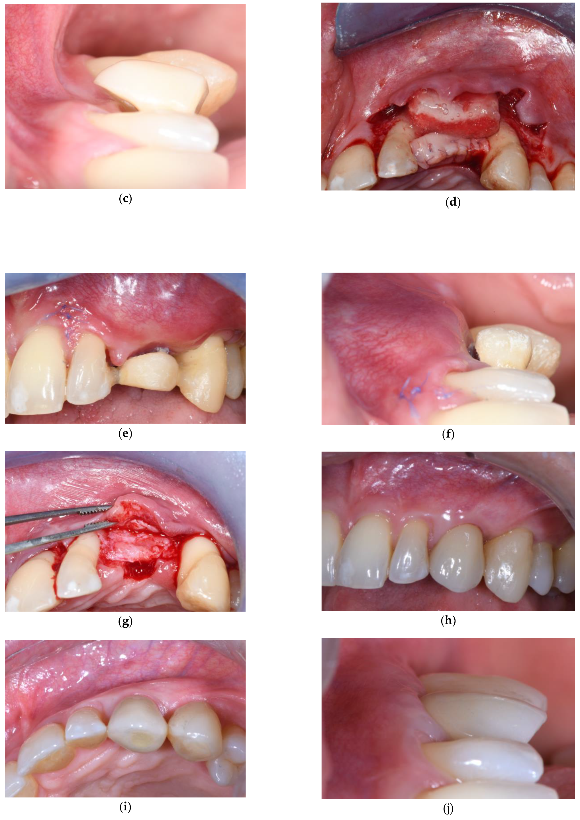
There are two primary methods to enhance soft tissues in a healed edentulous site prior to implant placement depending on the need for augmenting keratinized tissue or buccal soft tissue thickness. The first method is a free gingival graft [29,30], which increases the width of keratinized mucosa, usually used in the posterior region. The second method is the Platform Technique, aimed at augmenting soft tissue volume around implants in the case of ridge deficiency. This technique (Figure 1), originally described with the use of a CTG, has been shown to be successful in recreating healthy and esthetically compliant peri-implant soft tissues.
The technique (Figure 1d) consists of two horizontal crestal incisions, one buccal and another palatal, on the occlusal aspect of the edentulous area. Elevation of a buccal and palatal flap is performed in a way to isolate a central area of supracrestal soft tissues in the shape of a truncated pyramid that will be de-epithelialized. The buccal horizontal incision is connected to an envelope-type coronally advanced flap that will be full thickness for 2–3 mm and then split thickness to allow for the advancement that will be greatest at the level of the connective tissue platform. The palatal flap is elevated in a partial-thickness manner, starting from the horizontal crestal incision and creating a long bevel by keeping the blade at an angle to reach the palatal bone in the most apical position possible. The purpose of this flap is to expose the palatal surface of the connective tissue platform to aid in the suturing and stabilization of matrix; additionally, it will serve as anchorage for the buccal flap to ensure first intention wound healing over the connective tissue platform (Figure 1d). In the traditional technique that usually employs a CTG for both vertical and horizontal augmentation, CTGS can be used in instances of mild horizontal deficiency. After a CTG is fixed in the occlusal area, the matrix is then fixed to the buccal surface of the platform with two internal mattress sutures placed at the mesial and distal edges of the edentulous site. Lastly, the coronally advanced flap is secured at the adjacent de-epithelialized anatomical papillae with sling sutures suspended around the palatal cingulum of the neighboring teeth and with simple interrupted sutures at the level of the edentulous area, without engaging the graft or matrix. This way, the buccal flap slides over the CTGS and CTG until it reaches the palatal flap and achieves primary intention wound closure (Figure 1e,f). To ensure adequate soft tissue volume, it is advisable to wait for a period of 4–6 months of soft tissue maturation, before proceeding with implant installation (Figure 1g).

3.2. Soft Tissue Augmentation During Implant Installation

Strong evidence indicates that undergoing soft tissue grafting during implant installation can help decrease the likelihood of post-surgical contour changes and remodeling of hard and soft tissues [31]. Specifically, utilizing CTGs permits the enhancement of the height and thickness of supracrestal soft tissues, thereby improving the appearance and bone stability of the implant while simultaneously reducing peri-implant soft tissue dehiscence occurrence [31,32].
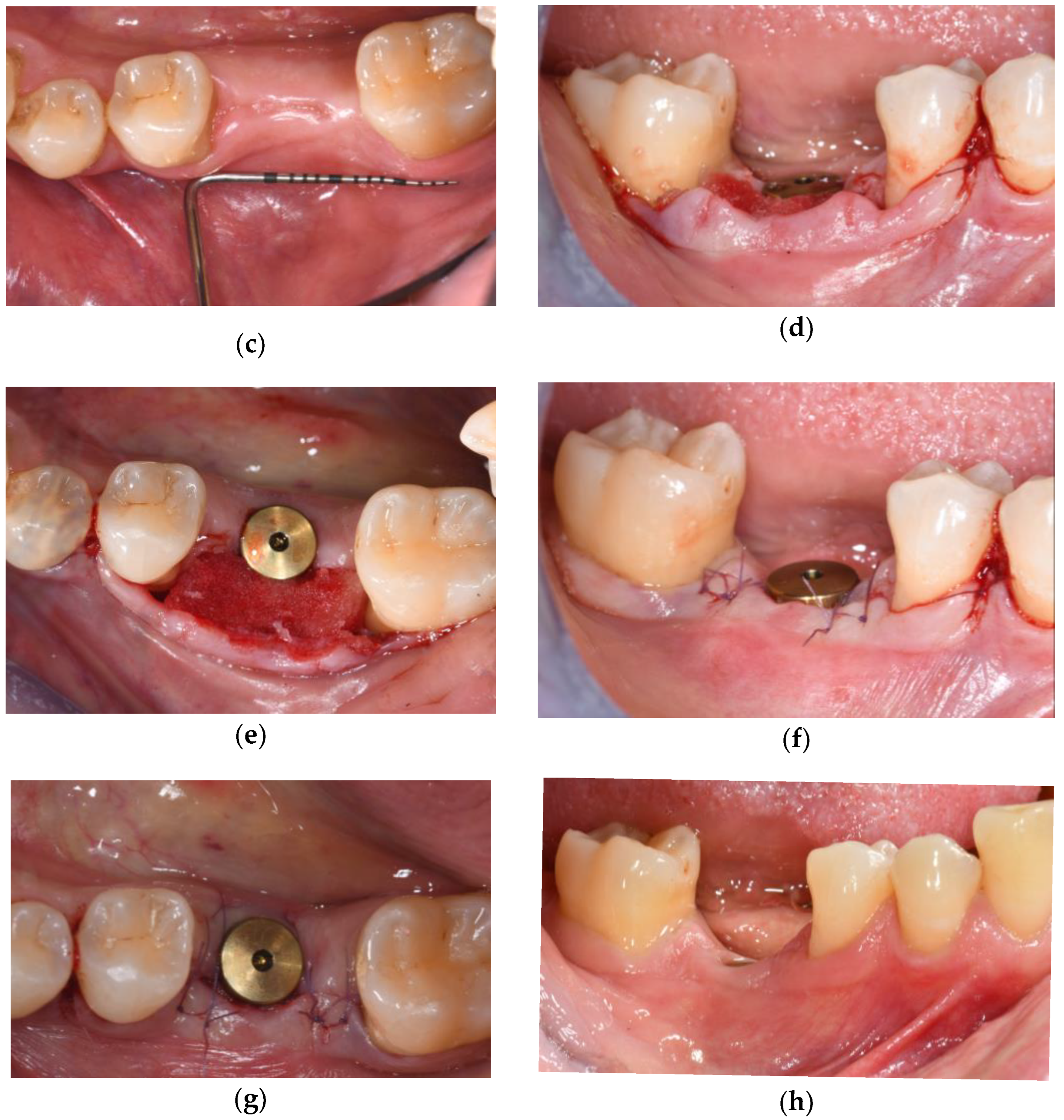
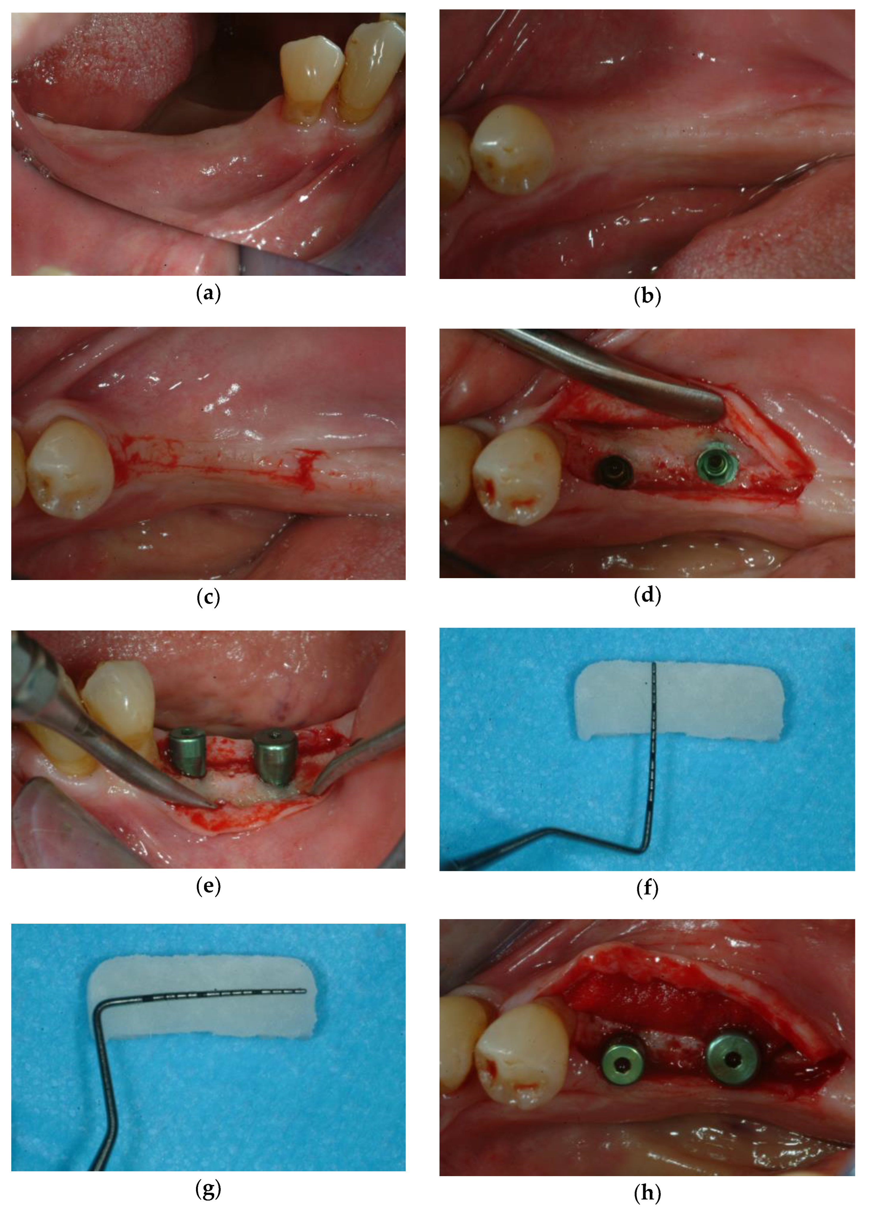
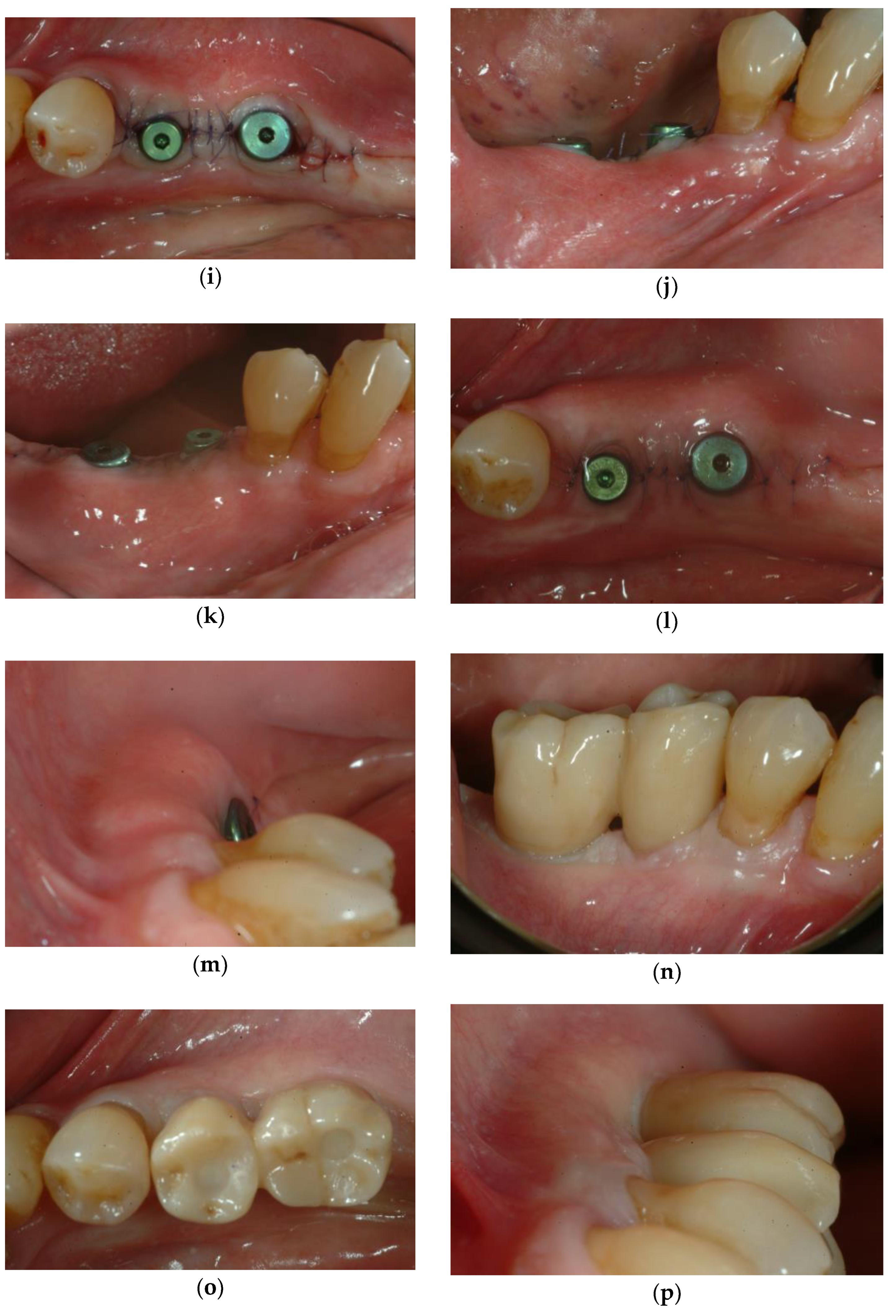
When there is an adequate band of KT, but a mild buccal bone concavity is present, addressing this deficiency can significantly enhance the long-term stability and aesthetics of the implant. In such cases, a CTGS can be strategically applied buccally to augment the soft tissue volume and improve the overall contour of the area (Figure 2).
This approach can be applied with either immediate or delayed implant installation, and the implementation of guided implant placement is crucial since this allows the sole elevation of a buccal flap, sparing disruption to the crestal, interproximal, and palatal/lingual tissues.
For posterior areas, the technique entails the elevation of an envelope-type coronally advanced flap (lateral approach) with a buccally displaced crestal incision at the edentulous site (Figure 2d). The dimensions of the CTGS should span from 1 mm coronal to the ideal level of the soft tissue margin and up to 2–3 mm apical to the buccal bone crest and mesio-distally they should cover the area corresponding to the implant site. After implant insertion, a tall healing abutment is placed and the CTGS is positioned buccally; CTGS can be placed without suturing (Figure 2e). The flap is coronally advanced and fixed with sling sutures suspended around the neighboring teeth and with simple interrupted sutures mesial and distal to the implant’s healing abutment (Figure 2f). A final sling suture can be performed, suspended around the healing abutment and without engaging the matrix, to improve adaptation of the flap and ensure first intention wound closure in the grafted area.
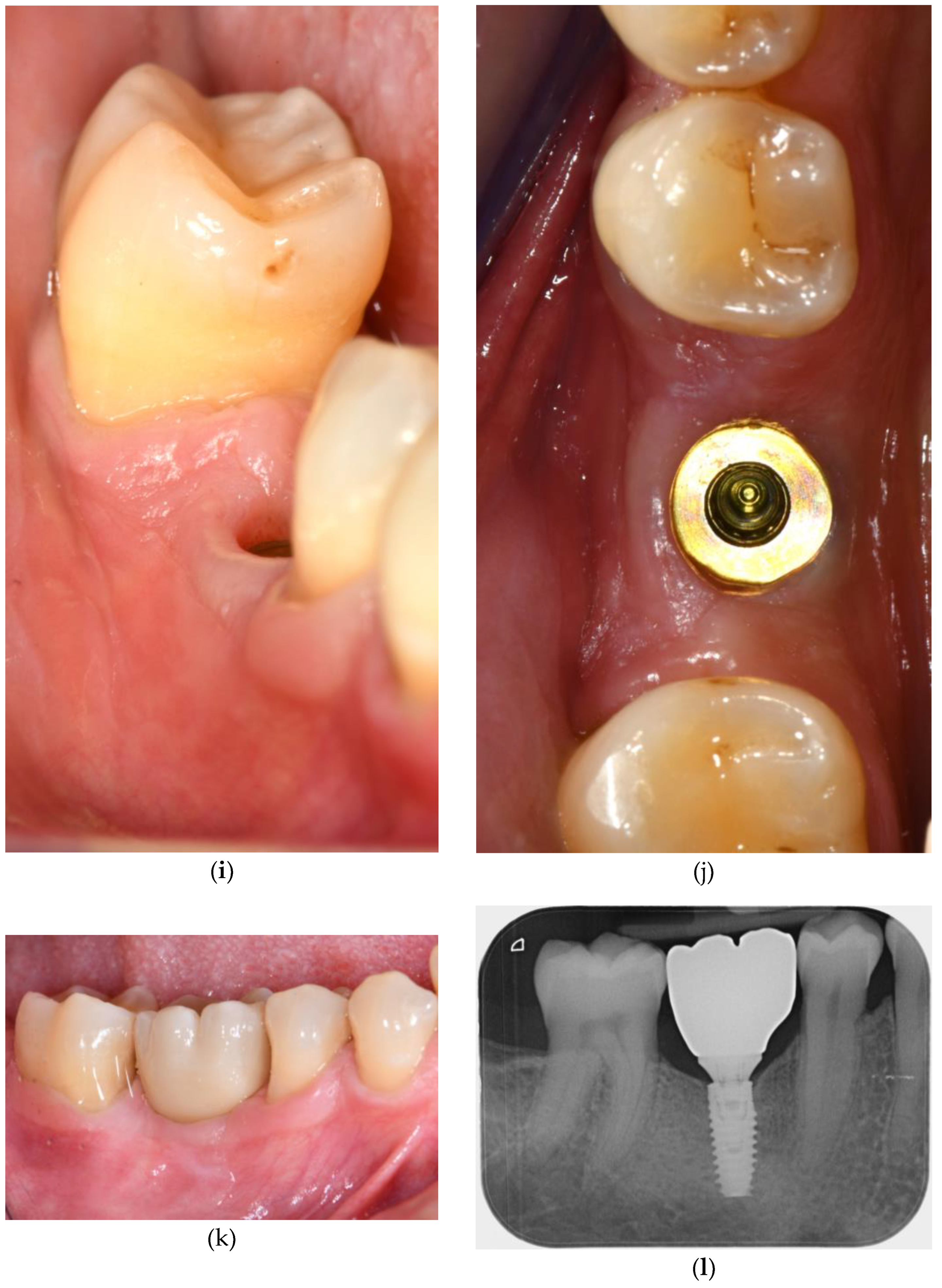
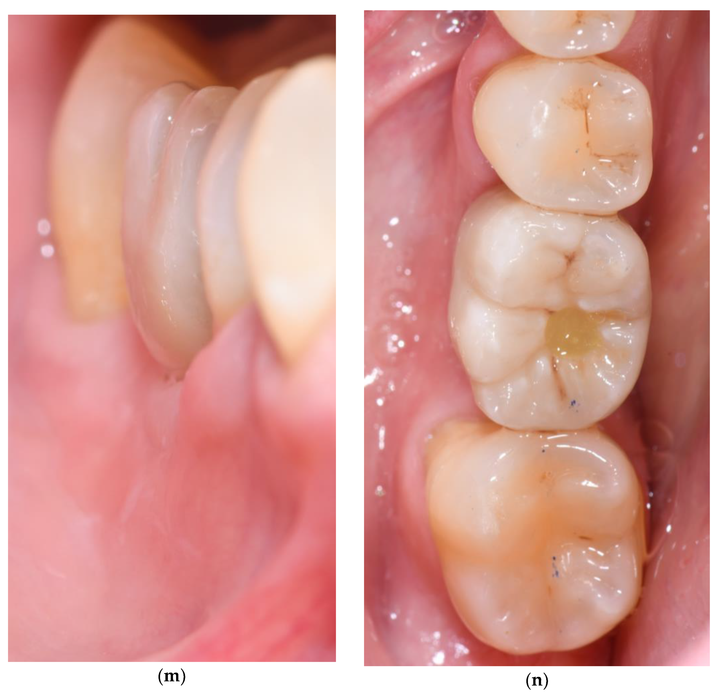
Research has indicated that the utilization of this particular technique can significantly improve the height and thickness of the soft tissues surrounding dental implants while remaining stable over an extended duration of time (Figure 2).

3.3. Soft Tissue Augmentation After Implant Installation

In instances in which implants must undergo submerged healing (Figure 3), the surgical intervention required for the abutment connection presents a favorable opportunity to address concerns regarding ridge contour discrepancies. In the presence of adequate KT height with small horizontal ridge deficiencies, CTGS could be added on the buccal aspect during implant uncovering.
The technique resembles the one described for soft tissue augmentation simultaneously with implant installation. In detail, a crestal incision is performed at the implant site taking care to leave at least 2 mm of keratinized tissue on the buccal and lingual flaps and an envelope-type coronally advanced flap is elevated buccally extending one tooth mesial and distal to the implant site (Figure 3c–e). The vertical dimensions of the CTGS should cover 1 mm coronal to the ideal level of the soft tissue margin and up to 2–3 mm apical to the buccal bone crest; the matrix should cover the mesio-distal area corresponding to the implant site After flap elevation, a tall healing abutment is placed (Figure 3e). and the CTGS is positioned buccally (without suturing in case of VCMX) (Figure 3h). The flap is coronally advanced and fixed with sling sutures suspended around the neighboring teeth and with simple interrupted sutures mesial and distal to the implant’s healing abutment (Figure 3i,l). If needed, a final sling suture can be performed as previously described to improve flap adaptation and ensure first intention wound closure.

4. Discussion and Clinical Recommendations

The management of peri-implant soft tissues is a cornerstone of long-term implant success, influencing not only biological stability but also aesthetic integration. In recent years, CTGSs have gained increasing clinical relevance as alternatives to autogenous CTGs, offering the advantages of reduced surgical morbidity, elimination of a second surgical site, and broader availability.
A recent consensus published by the Italian Academy of Osseointegration [33] was focused on the importance of soft tissues around implants, and one of the main objectives was to evaluate the efficacy of CTGS (ADM, CM, VCMX). Based on the systematic review conducted before the consensus [28], CTGs provide the best outcomes for increasing soft tissue thickness, while free gingival grafts remain the preferred option for gaining KT height, despite some esthetic drawbacks. ADM and VCMX offer a less invasive alternative with lower morbidity, though they are less effective than CTG. Similarly, the results from the systematic review and meta-analyses by Montero et al. [16] for the Consensus of DGI/SEPA/OF demonstrated that autogenous soft tissue grafts are more effective than CTGS for the augmentation of peri-implant KT, achieving ~1 mm greater increase. The authors also emphasized that XCM, combined with apically positioned flaps, can be an alternative to CTG, achieving clinically relevant gains in keratinized mucosa of >2 mm, which suggests its potential role in selected clinical scenarios. Additionally, Rotundo et al. [17] in their review reported that CTG and free gingival grafts remain superior in enhancing KT width, and CTGS provided satisfactory aesthetic outcomes, improving patient-reported comfort.
Current research is limited by high heterogeneity among studies, different surgical techniques and measurement methods, and when it comes to long-term results, the evidence remains scarce. In a randomized clinical trial, Thoma et al. [15] compared a VCMX with CTG for soft tissue augmentation at single implants with 5 years follow-up: data showed that both CTG and VCMX resulted in increased peri-implant mucosal thickness and favorable esthetic, with no statistical difference. Similarly, Fischer et al. [19] showed that ADM used at second-stage surgery gained significant thickness with respect to the baseline and was stable over 24 months, with minimal changes after the initial healing period (0.83 ± 0.64 mm at 6 months vs. 0.77 ± 0.65 mm at 24 months; p = 0.19).
CTGSs’ clinical performance is closely linked to the timing of their application—before, during, or after implant placement—as well as to the specific anatomical and biological context of the treated site.
In the pre-implant phase, soft tissue augmentation aims to optimize mucosal conditions in healed edentulous ridges, particularly in the presence of horizontal deficiencies or reduced KT. The platform technique described in this paper (Figure 1) exemplifies how CTGSs can be integrated in a staged approach to improve tissue volume before implant placement. In the illustrated case, a collagen matrix was used in combination with a CTG to compensate for a buccal deficiency, achieving notable volumetric gains after 6 months of healing. This protocol highlights how CTGSs can effectively complement autogenous grafts, especially when tissue demands exceed the availability of palatal donor tissue. However, the remodeling dynamics of CTGSs—particularly the tendency for early resorption and contraction—must be taken into account, and outcomes may vary depending on matrix thickness and integration capacity. While the pre-implant phase offers optimal conditions for matrix healing and maturation, the overall treatment time is inevitably extended, and the final tissue architecture remains subject to variability.
When CTGSs are employed during implant placement, as shown in Figure 2, they can serve as adjunctive tools to simultaneously manage both hard and soft tissue dimensions. This timing is particularly efficient, as it reduces the number of surgical procedures and enhances patient comfort. In the clinical case illustrated, a VCMX was used buccally in conjunction with guided implant placement and flap advancement, leading to stable horizontal tissue volume 1 year post-surgery. The flap design, which preserved the interproximal and palatal tissues, contributed to favorable healing. Nonetheless, the use of CTGSs in this context poses specific challenges: the surgical site is biologically active, and competition between healing processes—bone remodeling, osseointegration, and soft tissue integration—can affect matrix performance. Furthermore, certain matrices, such as volume-stable cross-linked collagen scaffolds, require submerged healing for optimal results, limiting their use in immediate loading protocols or in esthetic areas where soft tissue mobility is constrained. Thus, while effective in mild-to-moderate defects, simultaneous application demands precise control over surgical variables, including flap tension and closure.
In the post-implant phase, particularly during second-stage surgery, CTGSs can be employed to refine peri-implant tissue contours or correct minor deficiencies prior to prosthetic restoration, in presence of an adequate height of KT. The clinical case reported in Figure 3 demonstrates how a VCMX was placed at the time of implant uncovering to augment buccal soft tissue thickness. This approach allowed for improved emergence profile and satisfactory aesthetic outcomes after 1 year. Because the matrix is applied after osseointegration, this timing offers a minimally invasive way to correct and enhance tissue volume. The limited vascular supply and flap thickness in this phase may restrict the regenerative potential of CTGSs, making them more suitable for small corrections rather than for substantial volume augmentation. The reduced envelope flap must be carefully elevated without any tension, since it may increase the risk of graft exposure or partial resorption if the matrix is not adequately adapted and stabilized.
Across all phases, the application of CTGSs must be cautiously tailored to the specific clinical scenario. The cases presented in this manuscript underscore the importance of surgical planning and technique, from the platform design in the pre-implant phase, to guided flap management during implant insertion, to delicate tissue handling during uncovering. While the short-term outcomes are encouraging, especially when CTGSs are used in combination with autogenous grafts or in well-selected cases, long-term data on their volumetric stability and integration remain limited. Therefore, CTGSs should not be considered direct substitutes for CTGs in all indications, but rather valuable adjuncts within a broader surgical toolkit.
As the field of soft tissue regeneration continues to evolve, further studies are warranted to clarify the long-term performance of these materials in various clinical contexts. Future advancements in biomaterial engineering, including the incorporation of bioactive molecules or cellular components, may enhance the regenerative capabilities and clinical reliability of CTGSs, allowing for more predictable outcomes in peri-implant soft tissue augmentation.

5. Conclusions

Within the limits of this paper (case description), for peri-implant soft tissue management, the use of CTGS represents a valuable alternative to autogenous CTGs to increase soft tissue volume: in case of mild buccal ridge deficiencies, in presence of combined horizontal and vertical defects (in conjunction with a CTG), in case of limited availability of the palatal donor site, and when minimizing patient morbidity is a priority.
While CTG remains the gold standard due to its long-term predictability and tissue regeneration, CTGSs offer the advantage of avoiding a second surgical site, reducing operative time, and improving patient comfort and satisfaction. However, the variability in outcomes of CTGSs due to the biomaterial’s properties, surgical technique and individual case characteristics underscores the critical need for careful individualized case assessment and matrix selection, in addition to its tailored application (i.e., matrix dimensions and suturing protocols). It is essential to consider specific clinical scenarios as well as patient needs when choosing whether to use autologous CTG or matrices, emphasizing the necessity for clinicians to have precision and expertise in the field of tissue regeneration.
Further long-term randomized controlled trials are needed to confirm the stability of CTGS outcomes, particularly beyond the first year of healing.

Author Contributions

Conceptualization, I.M. and M.S.; methodology, I.M., A.R., V.B., M.S., C.M. and I.D.R.; validation, M.S. and G.Z.; writing—original draft preparation, review and editing, I.M., A.R., V.B. and M.S.; supervision, M.S. and G.Z. All authors have read and agreed to the published version of the manuscript.

Funding

This research received no external funding.

Informed Consent Statement

All patients provided informed consent for the use and publication of their clinical photographs in this manuscript.

Conflicts of Interest

The authors declare no conflicts of interest.

Glossary and Abbreviations

“Connective tissue graft substitutes”: materials developed to replace autogenous connective tissue grafts, used for peri-implant soft tissue augmentation.
“Matrices”: general term describing structural scaffolds derived from allogenic, xenogenic, or synthetic sources (e.g., collagen matrix, dermal matrix).
“Biomaterials”: broad category encompassing natural or synthetic substances engineered to interact with biological systems, including matrices.
The following abbreviations are used in this manuscript:
CTGConnective Tissue Graft
CTGSConnective Tissue Graft Substitute
KTKeratinized Tissue
pADMPorcine Acellular Dermal Matrices
hADMHuman Acellular Dermal Matrices
CMCollagen Matrix
VCMXVolume-stable Collagen Matrix

References

  1. Reddy, K.S.; Biswas, S.; Sarangi, S.; Chaurasia, A.; Reddy, M.P.; Jose, A.T.; Kashwani, R. Impact of Smoking on Dental Implant: A Review. Bioinformation 2024, 20, 1750–1753. [Google Scholar] [CrossRef]
  2. Heydari, M.; Ataei, A.; Riahi, S.M. Long-Term Effect of Keratinized Tissue Width on Peri-Implant Health Status Indices: An Updated Systematic Review and Meta-Analysis. Int. J. Oral Maxillofac. Implants 2021, 36, 1065–1075. [Google Scholar] [CrossRef] [PubMed]
  3. Longoni, S.; Tinto, M.; Pacifico, C.; Sartori, M.; Andreano, A. Effect of Peri-Implant Keratinized Tissue Width on Tissue Health and Stability: Systematic Review and Meta-Analysis. Int. J. Oral Maxillofac. Implants 2019, 34, 1307–1317. [Google Scholar] [CrossRef] [PubMed]
  4. Mahardawi, B.; Jiaranuchart, S.; Damrongsirirat, N.; Arunjaroensuk, S.; Mattheos, N.; Somboonsavatdee, A.; Pimkhaokham, A. The Lack of Keratinized Mucosa as a Risk Factor for Peri-Implantitis: A Systematic Review and Meta-Analysis. Sci. Rep. 2023, 13, 3778. [Google Scholar] [CrossRef] [PubMed]
  5. Ramanauskaite, A.; Schwarz, F.; Sader, R. Influence of Width of Keratinized Tissue on the Prevalence of Peri-Implant Diseases: A Systematic Review and Meta-Analysis. Clin. Oral Implants Res. 2022, 33 (Suppl. 23), 8–31. [Google Scholar] [CrossRef]
  6. Ravidà, A.; Arena, C.; Tattan, M.; Caponio, V.C.A.; Saleh, M.H.A.; Wang, H.L.; Troiano, G. The Role of Keratinized Mucosa Width as a Risk Factor for Peri-Implant Disease: A Systematic Review, Meta-Analysis, and Trial Sequential Analysis. Clin. Implant. Dent. Relat. Res. 2022, 24, 287–300. [Google Scholar] [CrossRef]
  7. Tavelli, L.; Barootchi, S.; Avila-Ortiz, G.; Urban, I.A.; Giannobile, W.V.; Wang, H.L. Peri-Implant Soft Tissue Phenotype Modification and Its Impact on Peri-Implant Health: A Systematic Review and Network Meta-Analysis. J. Periodontol. 2021, 92, 21–44. [Google Scholar] [CrossRef]
  8. Barootchi, S.; Mancini, L.; Heck, T.; Zucchelli, G.; Stefanini, M.; Kazarian, E.; Rasperini, G.; Wang, H.L.; Tavelli, L. Reliability Assessment of the Classification for Facial Peri-Implant Soft Tissue Dehiscence/Deficiencies (PSTDs): A Multi-Center Inter-Rater Agreement Study of Different Skill-Level Practitioners. J. Periodontol. 2022, 93, 1173–1182. [Google Scholar] [CrossRef]
  9. Jung, R.E.; Holderegger, C.; Sailer, I.; Khraisat, A.; Suter, A.; Hämmerle, C.H. The Effect of All-Ceramic and Porcelain-Fused-to-Metal Restorations on Marginal Peri-Implant Soft Tissue Color: A Randomized Controlled Clinical Trial. Int. J. Periodontics Restor. Dent. 2008, 28, 357–365. [Google Scholar]
  10. Thoma, D.S.; Ioannidis, A.; Cathomen, E.; Hämmerle, C.H.; Hüsler, J.; Jung, R.E. Discoloration of the Peri-Implant Mucosa Caused by Zirconia and Titanium Implants. Int. J. Periodontics Restor. Dent. 2016, 36, 39–45. [Google Scholar] [CrossRef]
  11. Oh, S.L. Peri-Implantitis Associated with a Pre-Existing Pathology. J. Oral Implantol. 2017, 43, 232–236. [Google Scholar] [CrossRef]
  12. Oh, S.L.; Chung, M.K. Creeping Attachment Following Free Gingival Grafts around Dental Implants Exhibiting Mucosal Recession with a Lack of Keratinised Mucosa: A Case Series. Int. J. Oral Implantol. 2020, 13, 401–409. [Google Scholar]
  13. Roccuzzo, M.; Grasso, G.; Dalmasso, P. Keratinized Mucosa around Implants in Partially Edentulous Posterior Mandible: 10-Year Results of a Prospective Comparative Study. Clin. Oral Implants Res. 2016, 27, 491–496. [Google Scholar] [CrossRef] [PubMed]
  14. Cairo, F.; Pagliaro, U.; Nieri, M. Soft Tissue Management at Implant Sites. J. Clin. Periodontol. 2008, 35, 163–167. [Google Scholar] [CrossRef] [PubMed]
  15. Thoma, D.S.; Gasser, T.J.W.; Hämmerle, C.H.F.; Strauss, F.J.; Jung, R.E. Soft Tissue Augmentation with a Volume-stable Collagen Matrix or an Autogenous Connective Tissue Graft at Implant Sites: Five-year Results of a Randomized Controlled Trial Post Implant Loading. J. Periodontol. 2023, 94, 230–243. [Google Scholar] [CrossRef]
  16. Montero, E.; Molina, A.; Matesanz, P.; Monje, A.; Sanz-Sánchez, I.; Herrera, D. Efficacy of Soft Tissue Substitutes, in Comparison with Autogenous Grafts, in Surgical Procedures Aiming to Increase the Peri-implant Keratinized Mucosa: A Systematic Review. Clin. Oral Implants Res. 2022, 33, 32–46. [Google Scholar] [CrossRef]
  17. Rotundo, R.; Pancrazi, G.L.; Grassi, A.; Ceresoli, L.; Di Domenico, G.L.; Bonafede, V. Soft Tissue Substitutes in Periodontal and Peri-Implant Soft Tissue Augmentation: A Systematic Review. Materials 2024, 17, 1221. [Google Scholar] [CrossRef]
  18. Toledano, M.; Toledano-Osorio, M.; Carrasco-Carmona, Á.; Vallecillo, C.; Lynch, C.D.; Osorio, M.T.; Osorio, R. State of the Art on Biomaterials for Soft Tissue Augmentation in the Oral Cavity. Part I: Natural Polymers-Based Biomaterials. Polymers 2020, 12, 1850. [Google Scholar] [CrossRef]
  19. Fischer, K.R.; Testori, T.; Wachtel, H.; Mühlemann, S.; Happe, A.; Del Fabbro, M. Soft Tissue Augmentation Applying a Collagenated Porcine Dermal Matrix during Second Stage Surgery: A Prospective Multicenter Case Series. Clin. Implant. Dent. Relat. Res. 2019, 21, 923–930. [Google Scholar] [CrossRef]
  20. Wolff, J.; Farré-Guasch, E.; Sándor, G.K.; Gibbs, S.; Jager, D.J.; Forouzanfar, T. Soft Tissue Augmentation Techniques and Materials Used in the Oral Cavity: An Overview. Implant. Dent. 2016, 25, 427–434. [Google Scholar] [CrossRef]
  21. Tavelli, L.; McGuire, M.K.; Zucchelli, G.; Rasperini, G.; Feinberg, S.E.; Wang, H.L.; Giannobile, W.V. Extracellular Matrix-Based Scaffolding Technologies for Periodontal and Peri-Implant Soft Tissue Regeneration. J. Periodontol. 2020, 91, 17–25. [Google Scholar] [CrossRef] [PubMed]
  22. Pabst, A.M.; Happe, A.; Callaway, A.; Ziebart, T.; Stratul, S.I.; Ackermann, M.; Konerding, M.A.; Willershausen, B.; Kasaj, A. In Vitro and in Vivo Characterization of Porcine Acellular Dermal Matrix for Gingival Augmentation Procedures. J. Periodontal Res. 2014, 49, 371–381. [Google Scholar] [CrossRef] [PubMed]
  23. Rothamel, D.; Benner, M.; Fienitz, T.; Happe, A.; Kreppel, M.; Nickenig, H.-J.; Zöller, J.E. Biodegradation Pattern and Tissue Integration of Native and Cross-Linked Porcine Collagen Soft Tissue Augmentation Matrices—An Experimental Study in the Rat. Head Face Med. 2014, 10, 10. [Google Scholar] [CrossRef] [PubMed]
  24. Thoma, D.S.; Zeltner, M.; Hilbe, M.; Hämmerle, C.H.F.; Hüsler, J.; Jung, R.E. Randomized Controlled Clinical Study Evaluating Effectiveness and Safety of a Volume-stable Collagen Matrix Compared to Autogenous Connective Tissue Grafts for Soft Tissue Augmentation at Implant Sites. J. Clin. Periodontol. 2016, 43, 874–885. [Google Scholar] [CrossRef]
  25. Wang, J.; Wang, L.; Zhou, Z.; Lai, H.; Xu, P.; Liao, L.; Wei, J. Biodegradable Polymer Membranes Applied in Guided Bone/Tissue Regeneration: A Review. Polymers 2016, 8, 115. [Google Scholar] [CrossRef]
  26. Yu, S.H.; Tseng, S.C.; Wang, H.L. Classification of Soft Tissue Grafting Materials Based on Biologic Principles. Int. J. Periodontics Restor. Dent. 2018, 38, 849–854. [Google Scholar] [CrossRef]
  27. Schmitt, C.M.; Schlegel, K.A.; Gammel, L.; Moest, T. Gingiva Thickening with a Porcine Collagen Matrix in a Preclinical Dog Model: Histological Outcomes. J. Clin. Periodontol. 2019, 46, 1273–1281. [Google Scholar] [CrossRef]
  28. Tommasato, G.; Del Fabbro, M.; Oliva, N.; Khijmatgar, S.; Grusovin, M.G.; Sculean, A.; Canullo, L. Autogenous Graft versus Collagen Matrices for Peri-Implant Soft Tissue Augmentation. A Systematic Review and Network Meta-Analysis. Clin. Oral Investig. 2024, 28, 300. [Google Scholar] [CrossRef]
  29. Nabers, J.M. Free Gingival Grafts. Periodontics 1966, 4, 243–245. [Google Scholar]
  30. Sullivan, H.C.; Atkins, J.H. Free Autogenous Gingival Grafts. I. Principles of Successful Grafting. Periodontics 1968, 6, 121–129. [Google Scholar]
  31. Thoma, D.S.; Gil, A.; Hämmerle, C.H.F.; Jung, R.E. Management and Prevention of Soft Tissue Complications in Implant Dentistry. Periodontol. 2000 2022, 88, 116–129. [Google Scholar] [CrossRef]
  32. Zucchelli, G.; Mazzotti, C. Mucogingival Esthetic Surgery Around Implants, 1st ed.; Quintessence Publishing: New Malden, UK, 2022. [Google Scholar]
  33. Bressan, E.; Zucchelli, G.; Tommasato, G.; Pesce, P.; Canullo, L.; Grusovin, M.G. Consensus Report by the Italian Academy of Osseointegration on the Importance of Peri-Implant Soft Tissues. Medicina 2024, 60, 1393. [Google Scholar] [CrossRef]
Figure 1. (ac) Buccal, occlusal and lateral view of the edentulous site of a maxillary left canine displaying horizontal ridge resorption, insufficient gingival thickness and keratinized band. The site was temporarily restored with an adhesive Maryland bridge; (d) Surgery (Platform Technique) to compensate for the deficient soft tissue volume of the healed edentulous site: CTG is fixed at the occlusal platform with interrupted sutures. The CTGS is placed in the apical buccal position and fixed to the platform with two internal mattress sutures placed at the mesial and distal edges of the edentulous site; (e,f) At 14 days suture removal: stability of the flap and volume increase; (g) After 6 months of follow-up, at the time of implant placement, there was a significant increase in soft tissue thickness (3 mm); (hj) One-year post-delivery of the definitive crown, the implant rehabilitation obtained satisfactory esthetic outcomes for both the patient and clinician.
Applsci 15 10178 g001aApplsci 15 10178 g001b
Figure 2. (ac) Baseline: buccal, lateral and occlusal view of the edentolous site of a mandibular right first molar. Note the mild buccal bone concavity and the insufficient gingival thickness in the buccal aspect; (d,e) An immediate guided implant insertion has been performed along with an envelope coronally advanced flap. A tall healing abutment is placed and the CTGS is positioned buccally without suturing; (f,g) Frontal and occlusal views at the end of the surgery. The flap is coronally advanced and fixed with sling sutures suspended around the neighboring teeth and the healing abutment and with single interrupted sutures mesial and distal to the implant; (hj) Clinical situation at definitive prosthesis delivery: buccal, lateral and occlusal views 1 year after surgery. It is possible to appreciate that the horizontal defect has been successfully treated: a significant soft tissue thickness has been created; (kn) Clinical and radiographic pictures at 1-year follow up. The definitive implant-supported crown was delivered satisfactorily by both the patient and the clinicians involved.
Applsci 15 10178 g002aApplsci 15 10178 g002bApplsci 15 10178 g002cApplsci 15 10178 g002d
Figure 3. (a,b) Clinical views of the edentulous area at baseline; (c) Flap design of implant uncovering; (d,e) Flap elevation and tall healing abutments placement; (f,g) CTGS trimming; (h) Positioning of the on the inner part of the vestibular flap; (i,j) Frontal and occlusal views of the flap closure with single interrupted sutures; (km) Clinical aspect at suture removal 14 days after surgery (frontal, occlusal and lateral views); (np) Clinical situation of the definitive crown restoration 1 year after implant uncovering: note the soft tissue integration and correct crown emergence profile.
Applsci 15 10178 g003aApplsci 15 10178 g003b
Table 1. Summary of CTGSs properties and clinical indications.
Matrix TypeSourceResorptionFibroblasts ProliferationAngiogenesisVolume StabilityKeratinizationIndications
hADM/pADM
[22,25]
Human/PorcineModerateModerateModerateModerateLimitedEsthetic zones, thin biotype
VCMX (Crosslinked)
[26,27]
PorcineSlowModerate-highModerateHighModeratePosterior sites, soft tissue bulking
Non-Crosslinked CM [27,29]PorcineFastStrongHighLow-moderateHigh-moderateKT gain, minor thickness
Disclaimer/Publisher’s Note: The statements, opinions and data contained in all publications are solely those of the individual author(s) and contributor(s) and not of MDPI and/or the editor(s). MDPI and/or the editor(s) disclaim responsibility for any injury to people or property resulting from any ideas, methods, instructions or products referred to in the content.