Review

Microplastics in Dairy Products: Occurrence, Characterization, Contamination Sources, Detection Methods, and Future Challenges

by
Hüseyin Ender Gürmeriç
1 and
Burhan Basaran
2,*
1
Department of Food Processing, Şiran Mustafa Beyaz Vocational School, Gümüşhane University, 29000 Gumushane, Türkiye
2
Department of Nutrition and Dietetics, Faculty of Health Sciences, Recep Tayyip Erdoğan University, 53100 Rize, Türkiye
*
Author to whom correspondence should be addressed.
Appl. Sci. 2025, 15(17), 9411; https://doi.org/10.3390/app15179411
Submission received: 28 July 2025 / Revised: 26 August 2025 / Accepted: 27 August 2025 / Published: 27 August 2025
(This article belongs to the Special Issue Advanced Research on Microplastics, Human Exposure and Food Safety)

Abstract

In this study, data from 17 studies reporting the presence of microplastics in milk and dairy products in the literature were examined with a product-based systematic approach. In addition, geographical comparisons were made between different countries. In milk and dairy products, the concentration of microplastics has been reported to exhibit a broad range, extending from non-detectable levels to as high as 10,040 MPs per kilogram, contingent upon the specific product types. Milk powder (especially baby milk powder) stands out as the riskiest product group in terms of microplastic content. Although the sizes and colors of the detected microplastics vary significantly, the fiber form is generally predominant. While polyethylene, polypropylene, polyamide and polyester are among the polymers frequently detected, high-temperature-resistant industrial polymers such as polytetrafluoroethylene, polysulfone, polyurethane were also encountered. In addition, the presence of some polymers (such polyvinyl chloride, polyurethane) that are toxicologically risky for human health was reported in the studies. In addition, the study evaluated the chemical, enzymatic and physical methods used for the separation and identification of MPs; the advantages and limitations of FT-IR, Raman and other analysis techniques were revealed. This study reveals that MP contamination in milk and dairy products is a multidimensional problem. The findings show that milk and dairy products are highly susceptible to plastic contamination at every stage of production.

1. Introduction

Plastics are now widely used across a broad range of industrial and consumer applications, including packaging, food processing, automotive manufacturing, transportation systems, electronics, construction materials, medical equipment, textiles, and infrastructure components such as water and sewage pipes [1,2]. The widespread use of plastics is primarily attributed to their versatile properties, including low cost, light weight, mechanical strength, and flexibility [3,4]. The production and use of plastics has increased significantly in recent years. In 2023, global plastic production reached approximately 420 million metric tons [5,6]. Approximately 40% of global plastic production consists of single-use products. High production volumes of plastic materials increase environmental risk factors due to their non-biodegradable polymeric structures, which persist in ecosystems over the long term [1]. These effects lead to an increase in physicochemical pollution parameters and ecotoxicological interactions, as well as an increased risk of environmental contamination in biotic and abiotic areas, depending on the accumulation of plastics in hydrological and soil systems [7]. Today, plastics are recognized as one of the most widespread and concerning anthropogenic threats to the environment [8]. This extensive level of pollution has led to the characterization of the current era as the “Plastic Age” in literature [9].
Microplastics (MPs) are heterogeneous particles formed by the degradation of plastic materials due to physical abrasion, chemical reactions, ultraviolet (UV) radiation, and biological activity [10]. MPs vary in size from 1 μm to 5 mm, are morphologically classified as fragments (irregular shapes), fibrils (fibrous structures), spheres (spherical forms) or filaments (thread-like structures) and can be of various colors and polymer types [11,12,13]. MPs are generally classified into two main categories. Primary MPs are intentionally manufactured microscopic particles, commonly found in personal care products such as cosmetics and toothpaste, as well as in synthetic microfibers released from textile materials. In contrast, secondary MPs are generated through the environmental degradation of larger plastic items, including packaging materials, plastic bags, and polyethylene terephthalate (PET) bottles [14]. In recent years, numerous studies have demonstrated that MPs are pervasive environmental pollutants found across virtually all ecosystems in nearly all ecosystems, including soil, glaciers, deserts, oceans, rivers, and the atmosphere [15,16,17,18,19,20]. Over the past few years, it has been reported that the food chain—from primary production to final consumer products—is also under threat from MPs. This indicates that MPs can enter the human body through dietary intake [21,22].
Although the full effects of MPs on human health remain unclear, current studies on MPs smaller than 1 µm have identified several potential risks, including cellular damage, oxidative stress, inflammation, the transport of toxic substances, and impairments to the immune and reproductive systems [23,24,25]. Although potential health risks have been suggested for submicron MPs, the possibility of similar mechanisms in larger MPs remains hypothetical and warrants further investigation. Even though most orally administered particles are excreted in feces, those smaller than 150 µm are believed to penetrate tissues, while particles smaller than 1.5 µm may enter organs via systemic circulation [3,26]. MPs have even been detected in various human tissues, including the placenta [25], bone [27], heart [28], lung [29], testis [30], brain [31], liver [32] and blood [33]. These findings demonstrate that MPs can enter the human body via dietary exposure and potentially cause potential health issues.
MPs can enter the human body through inhalation, dermal contact, and ingestion, primarily via the gastrointestinal tract through food consumption. Recently, MPs have been found in many foods, including water, mineral water, sparkling soft drinks, cold tea, turnip juice [34], table salt, soy sauce, fish sauce, salted seafood, seaweed, honey, processed drinks [35,36], fish, sugar, eggs, chicken, strawberry, tomato [37], milk [38], yoghurt, buttermilk [39], and milk powder [40]. The present study focuses on milk and dairy products, which are widely consumed—particularly by children—and are considered a fundamental part of the human diet. MP pollution has been documented in agricultural and livestock production environments that form part of the milk and dairy supply chain. As a result, ruminants are directly exposed to MPs through contaminated grazing pastures, drinking water sources, and ambient air [41]. Moreover, plastic wrapping of silage bales and packaging materials for pellet feed can serve as a potential source of MP contamination, thereby increasing the likelihood of MP ingestion by livestock [42,43]. In this context, the presence of MPs in the blood [44], feces [45], meat [46], and milk [47] of ruminant animals, as reported in various studies, indicates that this exposure poses a risk not only to the environment but also to human health. In addition, the risk of MP contamination increases when plastic packaging materials, extensively used in the food industry, interact directly with food products [48].
Nutrition is fundamental to human survival, with milk and dairy products serving as important sources of beneficial nutrients. However, contamination of essential foods like milk and dairy products with MPs may contribute to adverse health effects. Therefore, a comprehensive investigation into the presence of MPs in milk and dairy products is warranted. This study also assessed the methodologies employed for detecting MPs in milk and dairy products. Due to the potential health risks, it is crucial that MPs in foods are detected accurately and reliably. To identify MPs in food samples, microscopy, chemical analysis, and/or spectroscopic techniques are typically applied following chemical digestion and density separation procedures. These techniques must be further developed and standardized to enable accurate determination of MP particle size, morphology, color, and chemical composition [40,49,50]. However, the methods used to detect MPs in milk and dairy products have not been sufficiently evaluated in the current literature.
This study aims to: (i) evaluate the presence of MPs in milk and dairy products, (ii) evaluate the sources of MP contamination in milk and milk products, (iii) compare existing methods for the detection and identification of MPs, and (iv) assess current policies and future challenges related to MP contamination in the dairy sector.

2. Methodology

Relevant articles were manually collected by the researchers from the Google Scholar, Web of Science, Scopus, and PubMed databases, with no starting date limitation and covering the period up to May 2025. Appropriate keywords were identified based on preliminary literature review and used during the search process. The keywords used are as follows: “microplastic and micro-plastic and dairy”, “microplastic and micro-plastic and milk”, “microplastic and micro-plastic and yoghurt”, microplastic and micro-plastic and yogurt”, “microplastic and micro-plastic and buttermilk”, “microplastic and micro-plastic and ayran”, “microplastic and micro-plastic and powder and milk”, “microplastic and micro-plastic and cream”, “microplastic and micro-plastic and butter”, “microplastic and micro-plastic and cheese”, and “microplastic and micro-plastic and whey”. Following a comprehensive review and elimination process, only original research articles written in English and specifically addressing the detection of MPs in milk and dairy products were included in this study. To ensure reliability and minimize potential bias, two authors independently screened and evaluated the articles. In this process, review papers, conference proceedings, books, book chapters, and non-English publications were excluded. The selection procedure was carried out in two stages: initially, the titles and abstracts of the articles were examined, and subsequently, the full texts of the remaining studies were carefully assessed for eligibility. Although broad keywords were used during the search, the number of eligible studies was limited. This is mainly because research on microplastics in food, particularly milk and dairy products, has only gained attention in recent years. Additionally, exclusion criteria further reduced the number of eligible articles. After a rigorous screening and full-text evaluation, only 17 original research articles met the inclusion criteria and were therefore considered suitable for analysis in this study.

3. Results and Discussions

3.1. Subsection Occurrence and Characterization of MPs in Dairy Products

In recent years, the presence of MPs in the environment has extended beyond marine ecosystems and is now being detected in staple food products intended for direct human consumption. In this context, detection of MPs in milk and dairy products represents an emerging concern with potential implications for food safety and public health. Table 1 summarizes the findings of 17 studies, providing a comparative analysis of MPs found in dairy products from various countries based on product type, polymer type, particle size, shape, and color.

3.2. Evaluation of Studies in Terms of MPs Properties and the Possibility of MP Contamination

3.2.1. Raw Milk

The available data on MPs in raw milk are based on three primary studies: one comprehensive analysis from Türkiye and more limited studies from Switzerland and Romania.
In the first study, conducted by Da Costa Filho et al. [62] in Switzerland, two raw milk samples were analyzed. The amount of MPs in these samples ranged from 2040 to 6250 MPs/kg. Six distinct polymer types were identified, with polyethylene (PE) being the most prevalent among the detected MPs.
A total of 588 raw milk samples obtained from cattle, buffalo, sheep, and goats in 11 cities in the Marmara Region of Türkiye were examined. This study stands out due to the large number of samples examined, especially compared to similar studies in literature, including the study of Da Costa Filho et al. [62]. The study identified MPs in approximately 90% of the milk samples analyzed. MP concentrations ranged from 84 to 128 MPs/kg. The detected MPs varied in color, shape, and size; however, fibers were the most dominant form, comprising 52% of the total. Among the seven different polymer types identified, ethylene propylene copolymer (EPC) was the most prevalent, accounting for 77% of the MPs [47].
The last study was carried out in Romania on 4 raw milk samples. In the samples examined, particles were detected in the range of 7–23 MPs/kg. The MPs detected in the study, in which fiber form MPs were dominant, had 5 different colors and two different polymer properties. This study draws attention to its limited polymer properties compared to other studies [52].
The substantial differences observed in MP concentrations among the studies conducted in Switzerland [62], Türkiye [47], and Romania [52] can be attributed to several factors. Variations in analytical methodologies, including sampling strategies, filtration techniques, and spectroscopic identification, directly influence the number of particles detected. Furthermore, discrepancies in sample size and representativeness, as well as regional and environmental conditions such as air quality, feed handling, and the extent of plastic use in farming practices, may contribute to the reported differences. These findings highlight the importance of developing standardized protocols to enable more reliable cross-study comparisons.
Raw milk samples are unprocessed dairy products that represent the initial stage of the direct supply chain. Therefore, with environmental factors, the on-farm environment, milking equipment, animal feed, and transport containers should be considered priority sources of the detected MPs. The most common form encountered in the study was fiber, which indicates that materials, especially those from milking hoses, milk collection pipes, and tank inner surfaces, may contribute to this contamination. The wide spectrum of colors observed in MP particles suggests that environmental sources—such as dust, airborne suspended microplastics, and industrial pollutants—together with plastic surfaces in animal shelters, may contribute to microplastic contamination in raw milk. The most striking finding regarding chemical content is that industrial polymers, such as polyethyl acrylate (PEA), hydrogenated nitrile butadiene rubber (HNBR), and polytetrafluoroethylene (PTFE), which have more specific purposes, were detected instead of the common polymers often reported in literature. This suggests that contamination may originate not only from the farm environment but also from the technical equipment used during milk collection and storage processes. The findings indicate that MP contamination begins at the early stages of milk production and that the product is exposed to MPs even before it reaches the processing phase. Therefore, food safety practices should account for the risk of MP exposure at every stage of the food supply chain from farm to table, not just at the processed product stage.

3.2.2. Milk

The products evaluated under this category include processed or packaged milk samples. Nine studies reported in various countries, employing different sample sizes and methodological approaches, reported detailed information on MP density, physical forms, and chemical compositions.
In a study performed by Kutralam-Muniasamy et al. [38] in Mexico, 23 milk samples were examined, and an average of 6.5 ± 2.3 (range 3–11) MPs/kg was reported. The highest rate of total MPs detected was in particles sized <0.5 mm at 40%. This was followed by particles sized 0.5–1 mm at 28% and 1–2 mm at 25%. MPs of different sizes were reported to be in the form of fiber (97.5%) and fragments (2.5%), and they had four different colors (the dominant color was blue at 72.5%). Polyethersulfone (PES) (dominant polymer) and polysulfone (PSU) polymers were detected in the milk samples. MP concentrations were found to be higher in commercially packaged processed milk products such as lactose-free, semi-skimmed, and low-fat/light milk compared to packaged whole milk samples. It was estimated that MP exposure through milk consumption in Mexico could occur at a level of approximately 858 MPs per year.
Diaz-Basantes et al. [63] carried out another study in Ecuador that detected significant levels of MP contamination in commercially sold milk samples. The average amount of MPs in the examined samples was reported as 40 (16–53) MPs/kg. Most of the particles were composed of MP fibers and fragments, and their sizes were found to range from 2.5 to 6742 µm. The particles were also found to be five different colors. Polymer analyses performed with FT-IR spectroscopy determined that the MPs were made of high-density polyethylene (HDPE), low-density polyethylene (LDPE), polyacrylamide (PAM), and polypropylene (PP).
In a study conducted in Switzerland involving the analysis of four milk samples, the concentration of MPs was notably high compared to similar studies, ranging from 1720 to 3480 MPs/kg. The detected MPs were present in the form of fibers and fragments. In addition to the polymers identified by Kutralam-Muniasamy et al. [38], six additional polymer types were reported in the study. This study is notable for reporting a wide range of polymers, with PTFE identified as the dominant polymer type [62].
Another study by [61] in India analyzed 10 milk samples, confirming 72 of 560 suspected particles as MPs. Unlike other studies, all detected MPs were smaller than 500 µm. Fragment-shaped MPs were dominant in this study, alongside fibers and pellets. Notably, this was the first study to report pellet-form MPs in milk. The color variation observed was lower than in other studies, with only three polymer types identified: PP at 52%, PE at 29%, and polyamide (PA) at 21%. The study estimated that individual MP exposure through milk consumption ranged from 35 to 80 MPs.
Basaran et al. [57] reported MP contamination in all 14 ultra-high-temperature (UHT) milk samples from various brands. The average MP concentration in the samples was 6 ± 5 MPs/kg, ranging from 3 to 48 MPs/kg. The size distribution of detected MPs was approximately similar across three categories: <500 µm, 500–1000 µm, and >1000 µm. The MPs were predominantly in fiber and fragment forms and exhibited six different colors. Identified polymer types included poly(ethylene-vinyl acetate) (PEVA, 53%), PA (32%), PET (24.9%), PP, 12.5%), and polyurethane (PU, 3.1%). The milk samples were categorized based on fat content: ≤1.5% and >1.5%. The average MP concentrations for these groups were 6 ± 5 MPs/kg and 7 ± 5 MPs/kg, respectively. The study also performed risk analyses, estimating average daily MP exposure from milk consumption for individuals aged 15 and older at 0.21 MPs/mL, with a cumulative exposure of 5289 MPs/mL over a 70-year lifespan. The study reported that the mean MP contamination factor (MCF) values ranged from 0.60 to 9.52, with 64% of the analyzed samples classified as exhibiting a moderate level of contamination (MCF < 1 = low contamination; 1 ≤ MCF < 3 = moderate contamination; 3 ≤ MCF < 6 = significant contamination; MCF ≥ 6 = very high contamination [64]). The average MP polymer hazard index (PHI) was 255 ± 733, indicating a medium risk level (PHI < 150 = low risk; 150 < PHI < 300 = medium risk; 300 < PHI < 600 = considerable risk; 600 < PHI < 1200 = high risk; PHI > 1200 = very high risk [65]).
In a study conducted by Chakraborty et al. [55] in Bangladesh, six milk samples were analyzed. The concentration of MPs ranged from 95 to 250 MPs/kg, with an average of 183 ± 60 MPs/kg. Most detected MPs (65%) were larger than 100 µm. Regarding morphology, 81% of MPs were fibers, 11% fragments, and 8% films exhibiting eight different colors. The most prevalent colors were black (34%), blue (17%), and red (16%). Polymer identification was confirmed using fourier-transform infrared spectroscopy (FT-IR) spectroscopy, revealing the following distribution: PE 44%, PP 26%, Nylon-6 13%, PA 12%, and polystyrene (PS) 5%. The study estimated MP exposure from milk consumption as 0.31 ± 0.09 MPs/day for children and 0.09 ± 0.03 MPs/day for adults, indicating that children are exposed to approximately three times more MPs than adults. The MCF for the samples ranged from 1.01 to 2.59 (mean: 1.92 ± 0.62), indicating moderate contamination. The PHI values, ranging from 11.9 to 23.7, were lower in this study compared to others.
A recent study conducted in Romania revealed the presence of MPs in both organic and conventional milk samples. The study reported that the total number of MPs detected in organic milk samples was higher than in conventional milk, and a wide variety of colors were observed, with black MPs being dominant. In both milk types, fibers were the dominant MP form, and fragmented MPs were also detected. Among the polymer types, polymethyl methacrylate (PMMA), PA, PU and polyester were observed in both groups, while PE was detected only in conventional milk samples [52].
A study performed in India examined 21 milk samples for MP content and detected between 375 and 1500 MPs/kg. The MPs’ colors were blue, red, pink, and brown, and PS was the most detected MP, followed by polyvinyl chloride (PVC), PS and PP. The MPs detected using the FT-IR method were mostly fragments and fibers [53].
Santonicola et al. [51] analyzed 20 different UHT milk samples and confirmed the presence of MPs in 67.5% of them in Italy. On average, 38.5 MPs/kg were detected, with 53.5% of the samples containing 10–40 MPs/kg, 32.1% containing 50–80 MPs/kg, and 10.7% containing 100–130 MPs/kg. The size distributions of the detected MPs were as follows: <350 µm (23%), 350–1000 µm (37%), 1000–2000 µm (26%), 2000–3000 µm (9%), and 3000–5000 µm (5%). All detected particles were fibers. The colors ranged widely: blue (dominant, at 32%), black, red, pink, purple, yellow, green, orange, transparent, and light blue. Polymer analysis identified PE and polyester polymers.
The amount of MPs detected in packaged milk varies significantly across studies, likely due to geographical differences, the technological complexity of the production chain, and the variety of equipment used. The frequent presence of fiber-type particles indicates that such contamination commonly occurs during dairy product processing. Contact with plastic is inevitable, particularly during processes such as filtering, pumping, and pasteurization. While most particles are smaller than 500 µm, some have been observed to reach sizes up to 5 mm, which may reflect physical degradation caused by unhygienic or aging equipment. Color variation among MPs serves as an important indicator of production environment control. The presence of colored plastics—such as blue, pink, and brown—suggests that environmental contaminants may also be introduced into the system, highlighting the role of environmental contamination at this stage. Polymer diversity is likely directly related to the types of equipment used in milk production, including hoses, sealing materials, and tank surfaces. The detection of high-performance industrial polymers such as PSU, polyvinyl alcohol (PVA), and PTFE indicates the use of plastics designed to withstand high heat and pressure. Because dairy products typically have a short shelf life, their contact time with packaging is limited compared to other food products. Therefore, MP contamination in milk is more likely related to the production environment and processing equipment than to packaging materials. These findings demonstrate that MP contamination in milk is a multifaceted issue involving the environment, equipment and materials, and the production chain. Consequently, comprehensive evaluation and control of the entire process are necessary to prevent MP contamination.

3.2.3. Yoghurt

MP content of yoghurt was examined in two studies conducted within similar geographic regions.
The first MP analysis of yoghurt was conducted in Türkiye. This study reported the presence of MPs at all stages of the production process, from raw milk intake to the final yoghurt product. The concentration of MPs in samples collected from 12 different process stages ranged from 20 to 580 particles per kilogram. MPs smaller than 500 µm were predominant among the detected particles. The study identified MPs in four different shapes and 11 distinct colors, as well as various polymers, including ethylene propylene (EP), neoprene (NP), PAM, and PTFE. The estimated daily average intake of MPs from yoghurt consumption was calculated to be 202 ± 14.4 MPs/day [60].
Another study on yoghurt, performed in Iran, analyzed 16 yoghurt samples and found MP concentrations ranging from 0 to 1660 MPs/kg, indicating substantial variation among products. Size distribution analysis revealed that MPs larger than 1000 µm were predominant, and all detected particles were in fiber form. The observed colors included transparent, red, brown, black, gray, blue, and green. Chemical characterization identified seven different polymer types: PET (25%-dominant polymers), PA, PVC, PE, PU, polycarbonate (PC), and PMMA. These included polymers commonly used in food packaging materials (e.g., PET and PE) as well as those with toxicological concerns (e.g., PVC and PU). Estimated MP exposure from yoghurt consumption was reported as 2.20–2.65 MPs/day for adult men and 2.40–2.89 MPs/day for adult women [39].
The last study was conducted in Romania on organic and conventional yoghurt samples. It was reported that the number of MPs detected in organic yoghurts (1155 MPs/kg) was approximately 1.5 times higher than in conventional yoghurts (693 MPs/kg). It was reported that fiber and black colored MPs were dominant in the study and that the detected MPs had four different polymer types. According to the pollution load index (PLI) values, it was explained that the samples in both yoghurt types had low, medium and very levels of contamination (PLI < 10 = very low contamination; 10 < PLI < 20 = low contamination; 20 < PLI < 30 = medium contamination; PLI > 30 = high contamination) [56].
The findings indicate that yoghurt is significantly contaminated with MPs, both in terms of quantity and polymer diversity. As a fermented dairy product typically stored in plastic containers, yoghurt has high potential for prolonged plastic exposure during production and storage. The exclusive presence of fiber-form particles suggests that contamination likely originates from mechanical components of the production line, such as filtration systems, plastic mixing equipment, and filling units. Additionally, water used in the manufacturing process may serve as an important source of MP contamination at this stage. The diversity of particle colors points to contributions from environmental sources or packaging materials. Likewise, the wide range of detected polymers suggests that contamination arises from multiple points of contact rather than a single source. From a food safety perspective, the detection of polymers such as PVC and PU—both associated with potential health risks—raises concerns about long-term consumption. Consequently, the equipment and packaging materials used in the production of dairy products should be critically re-evaluated to minimize MP exposure.

3.2.4. Buttermilk

Only one study investigated the MP content of buttermilk products. The study, conducted in Iran, analyzed 14 buttermilk samples. MP concentrations ranged from 0 to 2000 particles per kilogram, with 51% of the particles exceeding 1000 μm (µm) in size. All detected MPs were in fiber form, and seven different polymer types were identified, with PA being the dominant polymer (33.4%). The color distribution of MPs in these buttermilk samples was consistent with findings from a previous yoghurt study by the same researchers. Observed colors included transparent (the most dominant), red, brown, black, gray, blue, and green. The study also estimated MP exposure from buttermilk consumption in adults, reporting values of 3.63–4.88 and 3.96–5.33 MPs/kg/day for males and females, respectively [39].

3.2.5. Butter and Sour Cream

The study, conducted in Romania, examined butter and sour cream together. Black, blue, red, yellow, gray, and purple were the colors of MPs detected in sour cream, while red and brown were also found in butter. Black was the dominant color in both butter (46%) and sour cream (71%). The highest distribution of synthetic microparticles was found in butter (80%), while the highest distribution of natural microparticles (e.g., cotton, wool, raffia, cellulose, etc.) was found in sour cream (78%). The MPs detected by micro-FT-IR were found to range from 375 to 1500 MPs/kg. In the same study, butter samples were reported to have low-very high contamination levels according to PLI levels, while soy cream samples were reported to have low-medium contamination levels [54].

3.2.6. Ayran (Traditional Fermented Dairy Product)

MP content of ayran was investigated in one study. The study was carried out in Türkiye to cover all production stages of ayran and 180 ayran samples were analyzed. Microplastic density in these samples varied between 0–430 particles/L. Detected MPs had 4 different shapes (dominant: fiber) and 12 different colors (dominant: blue). In the analyzed ayran samples, the size distribution of the detected MPs was reported as <150 µm (37%), 151–1000 µm (37%), and >1000 µm (26%). MPs polymer types were reported as EP (39.3%), PTFE (25.9%), PA (3.2%), NP (10.2%), PP (7.3%), PAM (11.2%) and PE (2.9%). The study also explained that the amount of MPs intake from ayran consumption of children, adolescents, pregnant-lactating women and postmenopausal women varied between 173 ± 14 and 346 ± 7 MPs/day [59].
The high proportion of small particles in the ayran samples implies that contamination may be widespread and difficult to control during the production process. The variety of MP morphologies, particularly the presence of films and spherical particles, indicates that plastic release likely occurs through friction and abrasion at the microscopic level during manufacturing. Furthermore, the broad range of observed colors suggest contributions from secondary contamination sources such as environmental dust, packaging materials, and plastic components used in processing equipment. The chemical diversity of the detected MPs is also notable. The presence of heat- and chemical-resistant polymers such as EP and PTFE, which are commonly used in food processing machinery, supports the conclusion that contamination originates from production line components. The consistent detection of polymers like PA, PET, PE, and PP across samples suggests leaching from standard plastic materials used throughout the ayran production chain. Additionally, the detection of elastomers such as NP points to equipment parts—such as gaskets, lid seals, and hose nozzles—as potential sources of MP contamination.

3.2.7. Milk Powder

The content of MPs in milk powders was evaluated in studies conducted in four different countries. These studies included samples of skimmed milk powder, whole milk powder, and infant milk powder.
In this context, the study by Da Costa Filho et al. [62], which involved a limited number of samples, reported a notably high range of MP concentrations (10,040–35,060 MPs/kg). The study identified a total of five different polymer types, with PTFE being the dominant polymer.
Zhang et al. [58] conducted a study on MP contamination in infant milk products in China, analyzing a total of 13 infant milk powder samples. The amount of MPs detected in the study ranged from 0 to 180 particles per kilogram. On average, more MPs were found in boxed milk powder (7 ± 3 MPs/100 g) than in canned milk powder (4 ± 3 MPs/100 g). The particle size distribution was equal. The detected forms were classified as fiber (dominant), film, and fragment. However, the colors of the MPs were not specified in the study. In terms of chemical composition, common and toxicologically controversial polymers, such as PE (dominant), PET, PP, PS, and PVC, were detected. The study further estimated that infants aged 1–12 months may ingest, on average, between 305 and 580 MPs per year through the consumption of milk powder.
In a European study, 16 skimmed milk powder samples were analyzed for MP contamination. MP concentrations ranged from 466 to 5765 MPs/kg. Approximately 80% of the detected MPs were smaller than 99 µm. The identified physical forms included fibers, spheres, and irregular particles. A wide variety of colors was observed among the detected MPs. The most frequently detected polymers in the study were PP, PE, PS and PET [40].
In a study analyzing 19 milk powder samples, Chakraborty et al. [55] reported an average MP concentration of 280 ± 134 MPs/kg. The particle size distribution of the detected MPs differed significantly from that reported by Visentin et al. [40]. Notably, 69% of the MPs were larger than 100 µm. The MPs exhibited three distinct shapes: fibers (78%), fragments (14%), and films (8%). Seven different colors were identified, with black (33%), transparent (26%), and red (13%) being the most prevalent. The identified polymer types included PE, (48%), PP, PA, PET, PA, and PS, which are commonly reported in similar studies. The study also estimated MP exposure from milk powder consumption to be 0.48 ± 0.23 MPs/day for children and 0.14 ± 0.07 MPs/day for adults. The MCF ranged from 1 to 4.75, with a mean value of 2.33 ± 1.11. Based on this metric, 74% of the samples were classified as moderately contaminated, while 26% were considered significantly contaminated. The PHI values in the analyzed samples were found to range between 2.76 and 20.2. Based on the established classification criteria, 19% of the samples fell into the low polymeric risk category, whereas the remaining 81% were classified as representing a moderate level of polymeric risk. These results suggest that, although no samples indicated a very high level of risk, the majority of the analyzed milk samples exhibited contamination levels that warrant attention.
Due to the production process (especially spray drying) and storage conditions after packaging, milk powder products are susceptible to MP contamination. Three independent studies have found that the MP density in milk powder products may be higher than in other dairy products. Notably, the level of 5765 MPs/kg reported in the European study demonstrates the extent of contamination. The exceptionally high MP concentrations reported for Switzerland’s milk powder samples (10,040–35,060 MPs/kg) should be interpreted with caution. Such discrepancies may result from methodological differences in sample preparation, filtration pore sizes, and particle size detection limits, or from potential laboratory contamination. These findings may also represent outliers related to product-specific or regional factors. Therefore, further research employing standardized protocols is necessary to improve the comparability and reliability of results across studies. The small size of the MPs (<50 µm) detected in infant milk powders is striking. Additionally, preparing these products with hot water may not dissolve the polymer chains themselves but can accelerate the migration of additives or low-molecular-weight compounds from the plastic matrix [66]. In terms of polymer profiles, substances commonly used in the food industry, such as PP, PE, and PET, dominate. However, the finding of toxicologically controversial polymers such as PVC, especially in baby products, raises serious product safety concerns. In addition, spherical particles may also carry traces of environmental particles carried by air flow during processing and settled on the product surface. Unlike liquid products, milk powders remain in packaging for extended periods throughout their shelf life, increasing their vulnerability to MP contamination. The diversity of colors and variety of particle forms suggest that, besides environmental contamination, the physical wear and degradation of processing equipment contribute significantly to microscopic particle release.

3.3. Product-Based Comparative Assessment of MP Properties

The level of MP contamination in milk and dairy products varies significantly depending on the product type, as well as differences in particle count, size distribution, physical morphology, and polymer composition. This section aims to compare MPs across different categories of milk and dairy products in terms of their number as well as their physical and chemical characteristics (Figure 1).
The highest MP concentrations were detected in milk powder products. In skimmed milk powder samples analyzed in Europe, MP loads of 5765 and 10,040 MPs/kg were reported, likely reflecting the significant impact of drying systems and plastic-based equipment used during processing. Although infant milk powder exhibited lower MP counts, it is considered a higher-risk category due to the vulnerability of its target consumer group. In general, products made from raw milk tend to contain higher levels of MPs. Dairy products such as yoghurt and buttermilk have been found to contain more MPs than both raw and packaged milk, suggesting that processing methods contribute substantially to MP contamination. Moreover, geographic variation is evident: milk samples from Switzerland contained up to 3480 MPs/kg, while samples from Mexico, Romania and Türkiye showed lower concentrations, ranging from 10 to 50 MPs/kg. These findings indicate that country-specific production practices, the degree of processing, and the use of closed versus semi-open systems are likely influential factors in MP contamination.
Particle size is one of the most critical factors influencing the biological effects of MPs. In milk and infant formula samples, the high proportion of particles smaller than 100 µm (50–80%) suggests an increased likelihood of absorption within the gastrointestinal tract. Notably, in infant milk powder, approximately 50% of detected MPs were smaller than 50 µm, indicating a potential risk of systemic circulation uptake. In contrast, fermented dairy products such as yoghurt and ayran are dominated by larger particles (>1000 µm), which account for more than 50% of the total MPs. These larger particles are generally attributed to surface abrasion from processing equipment or contamination during filling operations. In raw and UHT milk samples, particle sizes were more evenly distributed, with both small (<500 µm) and large (>1000 µm) particles present. Plastic particles can enter the bloodstream and be transported to various organs [67]. Ragusa et al. [25] reported the presence of 12 MPs, measuring 5–10 μm and displaying either spherical or irregular morphologies, in 4 human placentas, including 5 located in the fetal side, 4 in the maternal side, and 3 in the chorioamniotic membranes. Animal studies support these findings: nanopolystyrene (20 nm) crossed into the placenta and fetus in rats [68], while in mice, polystyrene MPs accumulated in the liver, kidney, and intestine after oral exposure [69,70]. More recently, a Nature Communications study demonstrated that MPs can be transported by immune cells through the bloodstream to the brain, where they may induce vascular occlusion [71]. PC, PET, and PS have been identified as exhibiting the highest toxic effects on enzymes. These microplastics can directly alter enzymatic activity and pose significant risks to the placenta through the inhibition of key enzymes [72]. Exposure to MPs in zebrafish, along with their accumulation in different organs, has been shown to cause multiple toxic effects, including structural damage in the gonads, increased oxidative stress, developmental abnormalities, impaired gastrointestinal function, and reduced locomotor activity [73]. Exposure to MPs induces oxidative stress through the generation of reactive oxygen species, a process that plays a central role in the development of lung diseases while also contributing to DNA damage, disruption of cell membrane integrity, and inflammatory responses, ultimately leading to significant harm in living organisms and laboratory models [74]. Another study evaluated the cytotoxic and genotoxic effects of cryo-milled particles derived from commercial fork and cup plastics across eight distinct human cell-based models, demonstrating that MPs from consumer-grade plastics pose significant risks to cellular health, with endothelial and microglial cells being particularly vulnerable [75].
The morphology of MPs can provide valuable insights into their sources of contamination. The fiber form is predominant across nearly all product categories and is typically attributed to the friction-induced degradation of plastic components in hoses, filters, or filling systems. Film- and fragment-shaped particles are generally associated with packaging materials or the abrasion of plastic surfaces during processing. These particle types have been reported more frequently in buttermilk, UHT milk, and milk powder samples. Notably, pellet-type MPs were exclusively detected in milk samples from India [61].
The chemical composition of MPs provides direct insight into the types of plastic materials that come into contact with food products. The most detected polymers throughout the studies were PE, PP, and PET. This is consistent with global production and usage patterns, as these polymers are among the most widely manufactured and are frequently used in food packaging and dairy-related applications. They are also approved for food contact use in most regulatory jurisdictions. However, the detection of more controversial or industrial-grade polymers, such as PVC, PU, PTFE, and PSU, in milk, milk powder, and infant products highlights potential gaps in quality control within the food processing industry. The presence of PVC in infant formulas is concerning and may indicate non-compliance with existing food safety regulations.

3.4. Comparative Assessment of Countries Based on Product MP Properties

The levels of MPs detected in milk and dairy products may vary not only by product, but also by country (Figure 2). These differences are directly related to the variety of raw materials used, environmental exposure, the complexity of production technology, and the variety of materials employed. A comparative analysis of studies conducted in different countries is provided below.
In terms of both product diversity and sample size, Türkiye stands out as the most comprehensively studied country. The highest MP concentrations were detected in raw milk, regular milk, and milk powder products from Switzerland, suggesting that advanced industrial systems and complex production lines may increase plastic contact and thus contamination levels. In contrast, samples from Mexico and Türkiye generally showed low to moderate MP concentrations. The number of MPs detected in different types of yoghurt in Romania is also quite remarkable. Although infant milk powders analyzed in China contained relatively low MP counts, they present unique risks due to the presence of potentially hazardous polymers and smaller particle sizes. A comparative ranking of countries based on the maximum MP levels reported in different dairy products is as follows: for raw milk Switzerland > Türkiye > Romania, for milk Switzerland > Ecuador > India > Italy > Bangladesh > Romania > Türkiye > Mexico, for yoghurt Iran > Romania > Türkiye.
When considering particle size, a significant proportion of most MPs detected in infant milk powder samples from China and Europe were smaller than 100 µm. These smaller particles may pose a higher biological risk due to their potential to cross the gastrointestinal barrier and enter the circulatory system. Conversely, the dominance of particles >1000 µm in samples from Iran, Italy, and Switzerland is notable.
In almost all countries, fiber-shaped particles were the predominant MP form. This is likely due to the widespread use of plastic components in production line equipment, such as hoses, filters, and filling heads. Film and fragment types of MP particles were particularly prevalent from Türkiye, China, and Bangladesh. In contrast, pellet-type MPs were detected only in samples from India, suggesting a higher likelihood of raw material contamination. Furthermore, greater diversity in particle morphology may reflect insufficient environmental controls and an elevated risk of external (extra-system) contamination, particularly in developing countries.
The greatest diversity of polymers was observed in samples from Switzerland, Türkiye, and Iran. Products from these countries contained both commonly used food-grade polymers—such as PE, PP, and PET—as well as industrial or potentially hazardous polymers, including PVC, PU, PTFE, PSU, and PMMA. In contrast, samples from Mexico and India exhibited fewer and structurally simpler polymer types. The range of polymers detected reflects not only the complexity of the production processes but also the degree of oversight and control in the use of plastic materials. Notably, polymers PVC and PU were common in samples from China, Türkiye, Romania and Iran, raising significant concerns regarding public health safety. The ranking of countries based on polymer variety is as follows: Switzerland ≈ Türkiye ≈ Iran > China > Bangladesh > Italy > India ≈ Romania > Ecuador > Mexico.
This comparative analysis shows that MP contamination is directly related not only to the level of technological development but also to production culture, packaging preferences, hygiene practices, and environmental plastic pollution. While the data from countries such as Türkiye and China is more detailed due to the variety of products and number of samples, the European and Swiss examples demonstrate the extent of the contamination. The presence of MPs in sensitive products, such as infant milk powder, shows that this contamination penetrates the early stages of life and may lead to greater public health problems in the future. Therefore, countries need to develop MP regulation policies based on human health that are suitable for their production process.

3.5. Sampling and Analytical Techniques for MP Determining and Defining in Dairy Products

The detection and evaluation of MPs in food matrices generally requires a three-stage methodology. Typical methodology for characterizing MPs in food matrices:
(1)
Biological matrix elimination: Organic components in the food sample—such as proteins, lipids, and carbohydrates—are removed through enzymatic (e.g., protease, lipase) or chemical digestion methods using agents like hydrogen peroxide (H2O2) or potassium hydroxide (KOH).
(2)
MP isolation: MP particles are separated from the food matrix using techniques such as centrifugation, density separation with hypertonic solutions, or filtration.
(3)
Polymer identification: The chemical composition of isolated particles is confirmed using analytical techniques such as FT-IR, Raman spectroscopy and pyrolysis–gas chromatography/mass spectrometry (Py-GC/MS).
This process eliminates organic content from food matrices, allowing for the determination of the morphological (shape and size) and chemical (polymer type) properties of MPs with high specificity. The analytical methods employed for the detection and identification of microplastics in milk and dairy products are presented in Table 2.

3.5.1. Digestion PROCESSES

The initial step in MP characterization involves removing organic matter. This is achieved through various digestion protocols designed to enhance analytical sensitivity. These protocols are typically classified as alkaline, acidic, enzymatic, or oxidative. Although chemical and enzymatic digestion methods degrade organic components (proteins, lipids, and carbohydrates) through thermal or reactive processes, it is crucial to optimize the selected reagents to preserve the structural integrity of polymeric MPs. Commonly used chemicals for the removal of organic matter include KOH, sodium hydroxide (NaOH), H2O2, hydrochloric acid (HCl), nitric acid (HNO3), iron (II) sulfate (FeSO4), and perchloric acid (HClO4) [82]. The Fenton reaction facilitates the oxidative degradation of organic compounds through the catalytic action of H2O2 and ferrous ions (Fe2+). Strong acid mixtures, such as HNO3 and HClO4, can also be used but should be applied with caution due to the risk of depolymerization in polymers with low pH tolerance, including PS and PA. Therefore, KOH, an alkaline reagent effective across a broad pH range, is often preferred in organic digestion protocols due to its ability to preserve the chemical stability of polymers [49]. The digestion of milk and dairy products generally involves the use of 30% H2O2 [39,51], 10% KOH [40], and a mixture of sodium dodecyl sulfate and NaOH [54] solutions. H2O2 and KOH are the preferred chemicals for digesting milk and dairy products.
Depending on the characteristics of the food matrix and substrate, various agents can be used to simulate intestinal digestion during enzymatic treatment. These include cellulase, proteinase-K (which hydrolyzes casein and whey proteins), chitinase, artificial gastric juice (which contains dilute acid and pepsin), and commercial pancreatic enzymes (which contain lipase, amylase, and protease). However, in the enzymatic digestion of milk and dairy products, multi-enzymatic detergents [59,78] and commercial pancreatic enzymes [58], which contain protease, amylase, and lipase, are generally used. A study was conducted to detect MPs in buttermilk and milk samples. Sample digestion was performed using an enzymatic protocol involving a combination of protease, amylase, lipase, and cellulase enzymes [59]. Another study involving flavored yoghurt employed a combination of multi-enzymatic detergent and alkaline digestion for sample preparation [78]. In a study aimed at developing method for detecting MPs in cheese, various digestion protocols were tested, including 1 M and 5 M KOH for alkaline digestion, 65% HNO3 for acidic digestion, and 30% H2O2 and Fenton’s reagent for oxidative digestion. Among these, alkaline digestion with KOH demonstrated the highest digestion efficiency [81].
Owing to the distinct optimal operating conditions (e.g., pH, temperature) required by each enzyme and the associated high costs, enzymatic digestion is generally more expensive and procedurally complex than other digestion methods. Furthermore, the use of multiple enzymes can further complicate the process. Nevertheless, enzymatic digestion offers notable advantages, including environmental friendliness and effective isolation of MP particles [49,83]. Consequently, improvements in enzymatic protocols, including the application of immobilized enzymes or tailored enzyme mixtures, hold promise for standardizing MP analysis while minimizing procedural complexity [59].

3.5.2. Density Separation

The principle of density-based separation involves mixing MP-containing sediments with a high-density solution to exploit the differences in particle buoyancy. Given that MPs have an average density between 0.8 and 1.6 g/cm3, a solution with a density around 1.4 g/cm3 is commonly considered sufficient to achieve efficient separation. However, the density of plastics can vary depending on the type of polymer and the additives it contains. Therefore, the selection of a separation medium in density-based protocols must be tailored to the characteristics of the target polymer. While water can be adequate for recovering low-density polymers like PE and PP from simple matrices such as soil, it is generally ineffective in more complex organic matrices, including dairy products [84]. Following digestion, the sample is typically transferred to a flotation medium to facilitate the separation of MPs based on density. The choice of flotation solution depends on the density of the target polymers. Zinc chloride (ZnCl2), with a density of approximately 1.5 g/cm3, is commonly used for medium-density MPs; sodium iodide (NaI), which has a higher density (~1.6–1.8 g/cm3), is suitable for recovering high-density MPs such as PET or PVC. Sodium chloride (NaCl), calcium chloride (CaCl2), sodium bromide (NaBr, 1.4 g/cm3), zinc bromide (ZnBr2), and sodium polytungstate (SPT) are commonly used in density separation methods. NaCl is often preferred due to its low cost, environmental friendliness, and non-toxic nature. After waiting a while in the mixing medium, phase separation occurs. The particles that rise to the surface can then be identified using advanced analysis techniques [83]. Similarly, various flotation-based methods for isolating MP particles continue to be developed. These techniques are collectively referred to as sediment-MP isolation [85]. A study was conducted to separate MPs in milk and dairy products based on density differences. For the study, 21 milk samples were examined to determine the presence of MPs. NaCl with a density of 1.17 g/cm3 was used for density-based separation [53].

3.5.3. Filtration Techniques

Filtration is a common method used to separate MPs from environmental samples following enzymatic or chemical digestion. However, certain aqueous and liquid food samples can be filtered directly without prior digestion [57]. However, foods such as dairy products have complex structures containing carbohydrates, proteins, fats, vitamins, and mineral compounds. Therefore, digesting these foods first provides more effective results in determining MPs [86]. Membrane-based filter systems are widely employed for the isolation of MP particles from food matrices. Commonly used filter materials include glass fiber, cellulose nitrate (CN), and cellulose acetate membranes; polycarbonate mesh filters; and metal-coated filters such as gold (Au) or aluminum (Al) coated membranes. Gold- and aluminum-coated filters offer low background interference, enabling rapid and sensitive identification of MPs through techniques such as Raman or FT-IR spectroscopy by providing high signal-to-noise ratios. However, these filters are limited in long-term applications due to their susceptibility to corrosion and relatively high cost [87]. he stepwise filtration approach (e.g., 100 μm→20 μm→5 μm pore array) concentrates MPs with a heterogeneous size distribution within a certain range (e.g., 5–50 μm). Additionally, chemical inertness is a critical parameter in filter selection. Membrane stability and pore integrity must be maintained, particularly when in contact with organic solvents or acidic/basic digestion solutions (e.g., H2O2, HNO3) [49].
In the filtration of milk and dairy products, vacuum filtration with membrane filters is preferred. Commonly, these membrane filters are composed of CN [39], cellulose [54], glass fiber [78], glass microfiber [55], silver [40], microfiber [59], polycarbonate [58]. The preferred pore diameter of the filters ranges between 0.45 μm and 15 μm [39,54].

3.5.4. Extraction Techniques

Ultrasound extraction: Compared to chemical and enzymatic methods, ultrasound application offers several advantages: it is non-destructive to samples, effective with small sample sizes, and cost-efficient. However, a major drawback is that it can fragment fragile plastic components into smaller particles, complicating the accurate determination of their melting points [83].
Solid phase microextraction (SPME): One of the key advantages of this technique is that it integrates sampling, extraction, concentration, and sample introduction into a single streamlined process. It also offers significant benefits in terms of time efficiency and cost-effectiveness. Furthermore, this sampling approach facilitates the fragmentation of both monomers and polymers. For instance, it enables the fragmentation of PS fibers within polymer matrices commonly used in food packaging under varying temperature conditions [83].
Magnetic extraction: In this technique, hydrophobic iron nanoparticles interact with plastic particles in the sample, causing the plastic to become magnetized. This magnetization enables efficient separation of MPs within the 10–20 μm size range [83].

3.5.5. Detection and Identification Methods of MPs

Microscopy Techniques
Microscopes, which are used to obtain detailed images by magnifying surfaces, have been employed in detection and identification studies across numerous fields since their invention. Light microscopy, stereomicroscopy, fluorescent microscopy, electron microscopy, transmission electron microscopy and scanning electron microscopy (SEM) are commonly used microscopy techniques for detecting MPs. Microscopy techniques such as hyperspectral imaging, energy-dispersive X-ray spectroscopy (EDS), and optical microscopy can be combined to enhance identification accuracy. While plastic particles in the macro size range of 1–5 mm can be directly observed at the macroscopic level, visualization of submicrometer-sized particles requires specialized analytical techniques such as spectroscopic, microscopic, or chemical analyses. However, precise differentiation of plastics from organic or inorganic polymers using these visual methods may be limited due to morphological and optical similarities [88]. Most microscopy methods rely on visual assessment and particle counting, which are time-consuming and prone to error due to operator subjectivity [89]. In some cases, these methods are not sensitive enough to accurately identify polymers. Additionally, it is not possible to determine the type of polymer. They only allow the detection of MPs that are visible to the human eye [49]. Therefore, imaging techniques are often combined with other analytical methods, such as FT-IR and Raman spectroscopy, to enhance the identification of MPs.
When studies on MP detection in milk and dairy products were examined, optical microscopy was found to be the most used imaging technique. Additionally, epifluorescence microscopy [61], SEM [60], field emission scanning electron microscope (FESEM) [76], transmission electron microscopy (TEM) [78] have also been employed in some investigations.
Thermal Techniques
Thermal analysis methodologies quantitatively evaluate changes in the chemical and physical properties of substances based on their thermal stability. The polymeric composition of MPs can be determined by characterizing their thermal degradation profiles using techniques such as thermogravimetric analysis (TGA) or differential scanning calorimetry (DSC). This method enables the differentiation of plastics from organic and inorganic polymers based on differences in their thermal degradation kinetics and phase transition behaviors. Thermal analysis techniques can generally be used to identify MPs such as PE, PP, PVC, PS, PA, and PET [88]. Methods used in thermal analysis, such as Py-GC-MS, TGA-DSC, thermal desorption gas chromatography-mass spectrometry (TD-GC-MS), and thermal extraction desorption gas chromatography-mass spectrometry (TED-GC/MS), can also be used to determine MPs. Although thermal methods can identify plastic polymers, these polymers might degrade at high temperatures. As a result, information about the shape, size, and quantity of MPs may not be obtainable [49].
Among the studies conducted on the determination of MP in milk and dairy products, three studies using thermal methods were found. Two of these studies employed thermogravimetric analysis coupled with FT-IR (TGA-FT-IR) [76,77], while the other utilized Py–GC–MS [81]. These studies aimed to detect MPs likely to be present in dairy products by externally adding MPs such as PE, PS, PP, and PET [76] to the food matrix. They also sought to develop new methods for MP detection. The limited number of studies underscores the need to establish and standardize reliable methods for detecting and identifying MPs in milk and dairy products.
Spectroscopy Techniques
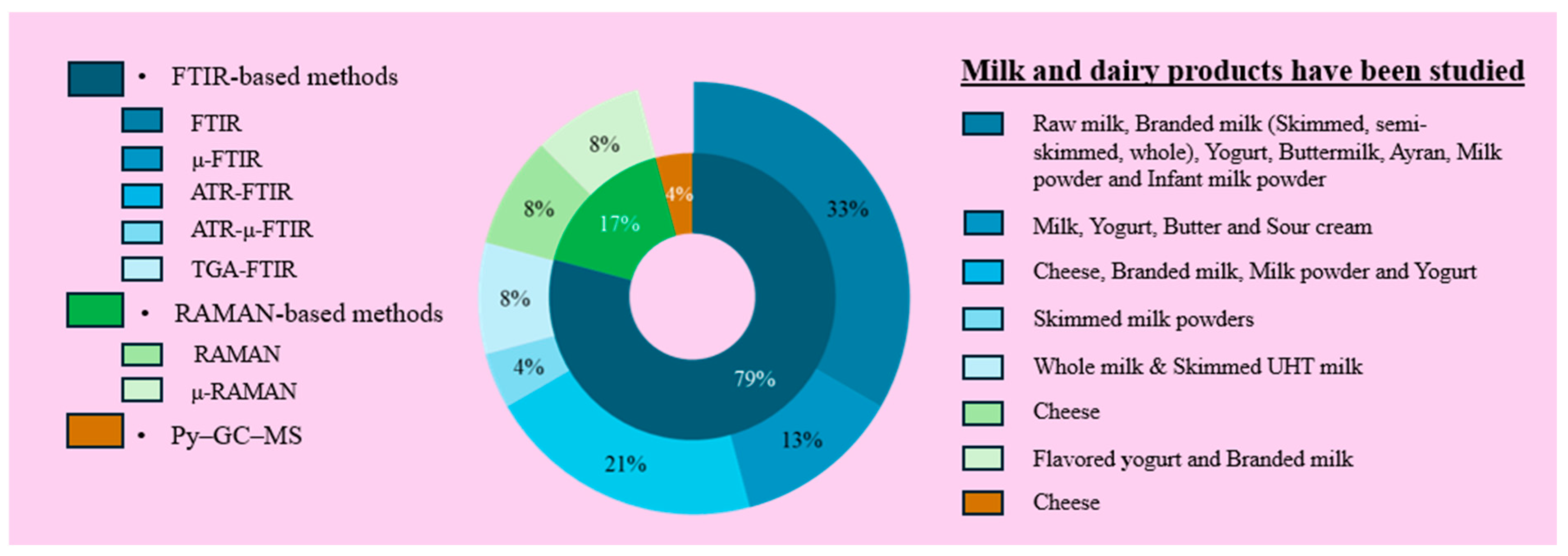
Spectroscopic techniques, such as FT-IR spectroscopy and Raman spectroscopy, are alternative analytical methods that enable sensitive, non-destructive determination, identification, and quantification of various polymer types and chemical structures in food samples. In FT-IR analysis, target particles are identified by examining the samples under a microscope. The infrared (IR) beam is directed by pointing the microscope objective toward the target. The detector detects the light reflected or transmitted by the particles, and the spectrum is recorded. The obtained spectrum is compared with reference values to determine the polymer type. The FT-IR spectroscopy technique can also be used in conjunction with other methods. The most used methods include attenuated attenuated total reflectance-FT-IR (ATR-FT-IR), micro-FT-IR (µ-FT-IR), focal plane array-FT-IR (FPA-FT-IR), and TGA-FT-IR. Nuclear magnetic resonance (NMR) spectroscopy can also be used for MP identification. Currently, FT-IR based method is the most used technique for both identifying and quantifying MPs in milk and dairy products (Figure 3). The FT-IR method enables simultaneous identification and quantification of polymer types while providing detailed information on their chemical properties and structures. The high cost and time-consuming nature of analyzing individual particles are the disadvantages of this method. Additionally, samples must be clean, dry, and free from contaminants to ensure accurate results [40,49].
In Raman technology, a monochromatic laser beam with a wavelength ranging from 500 to 800 nanometers (nm) is directed at the target area. This beam interacts with the vibrational and rotational energy levels, as well as the low-frequency interactions, of the molecules in the sample. These interactions result in energy differences, called Raman shifts, between the backscattered photons and the incident laser frequency. These shifts produce Raman spectra (in units of cm−1) that are directly related to the force constants and symmetry properties of the molecular bonds. The unique spectral signature of each polymer, which depends on its chemical bond structure and degree of crystallinity, allows for the identification of MPs. Raman technology can be used in conjunction with microscopy [90]. Raman spectroscopy can determine the polymer type of MPs, while morphology is assessed by complementary microscopy. Additionally, due to its high light-scattering efficiency, Raman spectroscopy enables the direct analysis of MPs as small as ~2 µm on filtration membranes [1]. The laser direct infrared imaging system can be used to detect MPs. It provides high-quality imaging and spectral data through rapid scanning optics. However, the automatic analysis methodology requires improvement, and the identification time should be reduced. In addition, reliable and appropriate methods for the identification and characterization of MPs in foods need to be standardized [50].
Four studies were identified that investigated the presence of MPs in milk and dairy products. Two of these focused on developing detection methods for MPs in cheese [79,80]. Of the remaining studies, one examined MPs in flavored yoghurt [78], and the other analyzed branded milks [38]; both utilized micro-Raman spectroscopy. Additionally, micro-Raman spectroscopy was employed in two studies investigating MPs in human breast milk [1,91].
Despite the growing number of studies investigating MPs in milk and dairy products, several critical limitations remain. First, there is substantial variability in detection limits across analytical methods, with FT-IR generally less sensitive than Raman spectroscopy for smaller particles. Such methodological differences complicate cross-study comparisons and may partially explain inconsistencies in reported concentrations. Second, the complex composition of dairy matrices, particularly the presence of fats and proteins, can cause matrix interference that increases the likelihood of both false positives and false negatives. Third, standardized protocols for MP extraction, isolation, and detection in dairy products have not yet been established, limiting reproducibility and comparability across research groups. Finally, the specific pathways of MP contamination remain insufficiently quantified. While packaging materials, processing equipment, and environmental sources are all likely contributors, their relative importance has not been systematically evaluated. Addressing these knowledge gaps through standardized methodologies and source apportionment studies will be critical to advancing the field and guiding effective food safety policies.

3.6. Other Studies on MP Determination and Method Development for Milk and Dairy Products

Katsara et al. [79] studied the migration of LDPE from food packaging into three types of cheese: Edam (soft), Kefalotyri (semi-hard), and Parmesan (hard). ATR-FT-IR and Raman spectroscopy were employed for analysis. The cheese samples were cut into small squares and vacuum-sealed in LDPE bags. LDPE migration was first observed after 14 days of cheese ripening, and LDPE detection became difficult after 21 days due to microbial growth.
In another study, Katsara et al. [80] investigated the migration of LDPE and PP into Cretan graviera cheese ripened for four and eight months. The cheese was sliced and stored in LDPE or PP bags or boxes at 7 °C for 21 days in the study conducted in Greece. ATR-FT-IR and Raman spectroscopy analyses were performed every three days to determine the migration of MPs in cheese. MP migration into the cheese was detected starting on the third day of storage, with LDPE exhibiting a higher tendency to migrate than PP.
Di Fiore et al. [81] conducted a study to develop an analytical protocol focused on sample preparation before chemical analysis for the detection of MPs in cheese. The study also aimed to identify the most effective digestion method. PS microbeads were added to cheese samples, and alkaline, acidic, and oxidative digestion techniques were applied to determine the most suitable approach. Alkaline digestion using 5 M KOH at 50 °C for 48 h was identified as the most effective method, achieving a digestion efficiency of 97.5%. Py-GC–MS was subsequently employed to confirm the polymeric structure of the microparticles.
Dang et al. [77] developed a method to rapidly detect and quantify MPs in milk, water, and coffee without pretreatment. The researchers employed TGA and FT-IR to detect 1 μm monodisperse PS spheres added in various food matrices. The method was reported to be suitable for the rapid detection of MPs/NPs (nanoplastics) of any size and shape. The study indicated that the results reliably estimated the PS content in milk, water, and coffee, with an R2CV above 0.96 and Root Mean Square Error of Cross-Validation (RMSECV) between 0.045 and 0.07 mg.
In a recent study, a novel method was developed for the determination of MPs and NPs in flowered yoghurt. PS microparticles were first spiked into the yoghurt samples. Following enzymatic digestion using a multi-enzymatic detergent, microscopic Raman imaging was employed to visualize and identify PS particles ranging in size from 400 to 2600 nm. The technique was reported to provide high analytical accuracy (R2 = 0.9946), detailed imaging, and precise chemical characterization, offering superior lateral resolution (1 µm) compared to FT-IR. The imaging sequence of MPs was found to be acquirable within a matter of minutes [78].
Another study investigated the behavior, transformations, and interactions of MPs with milk components during in vitro digestion. Four types of plastics (PE, PS, PP, and PET) were used at a concentration of 1 mg/mL in milk. Thermal decomposition of the MPs was assessed using TGA, their morphology was characterized by FESEM, and their chemical composition was analyzed using gas chromatography–mass spectrometry (GC–MS). Simulated digestion with gastric and intestinal fluids, both with and without milk, led to notable changes in MP size and surface morphology. The digestion process was also found to increase MP aggregation [76].

4. Regulatory of MPs: Current Policies and Future Challenges

With the implementation of appropriate policies, studies should be conducted to prevent environmental pollution, food contamination, and the potential adverse effects of MPs on human health. The impact of MPs on both human health and ecosystems can only be minimized through policies grounded in scientific evidence and reinforced by global collaboration. During this transition, raising consumer awareness, ensuring industrial transparency, and the governments’ determined policies will lay the foundation for a sustainable economy. To support this goal, research should focus on developing biodegradable packaging materials that decompose easily in the environment, replacing conventional fossil-based plastics that persist in nature for many years.
In this context, specific policy frameworks provide useful guidance for mitigating MP contamination. For instance, the European Union’s Directive (EU) 2019/904 on the reduction of the impact of certain plastic products on the environment—commonly referred to as the Single-Use Plastics Directive—sets clear restrictions on the use of conventional plastics and encourages the development of sustainable alternatives [92]. In parallel, partnerships between academia, industry, and regulatory authorities could accelerate the design and commercialization of biodegradable packaging materials, such as polylactic acid (PLA)-based polymers or starch-derived bioplastics, which are already being piloted in the dairy sector [93,94]. Moreover, integrating extended producer responsibility (EPR) schemes would ensure that producers remain accountable for the entire lifecycle of plastic products, thereby reducing the risk of environmental leakage. These strategies move beyond general recommendations by linking global policy initiatives with actionable industry practices.
The identification of health problems caused by MPs is also a critical issue that requires further attention. In this context, studies should be conducted to improve MP identification methods and to establish the necessary standards. Rather than relying on techniques that require visual identification and manual counting, such as microscopy, the development of faster methods with lower error rates should be encouraged. Artificial intelligence-supported software can be utilized to enhance MP detection and analysis. Additionally, portable sensors with advanced on-site identification capabilities can be developed to detect MPs commonly present in environmental samples.
The lack of knowledge regarding the potential effects of MPs on human health poses a significant challenge to effective risk management. In particular, the absence of clear data on parameters such as cytotoxic effects on cell viability and systemic absorption levels hinders the establishment of safe exposure thresholds. Therefore, characterizing the sources, polymer composition, and exposure levels of foodborne MP contamination through interdisciplinary research is essential for accurate risk assessment. For instance, studies should include monitoring plastic use in agriculture and food processing, modeling the chronic effects on human health, and determining toxicological thresholds.

5. Conclusions

This review evaluates the prevalence, characteristics, potential sources and possible effects on human health of MP contamination in milk and dairy products and the methods used for MP detection from an interdisciplinary perspective. The results clearly show that MP contamination begins not only in processed dairy products but also at the earliest stages of the production chain. When comparing between product types, it is seen that the highest MP levels are detected in milk powder products. However, the detection of small-sized and toxicologically controversial polymers (PVC, PU, etc.) in products targeting sensitive consumer groups such as baby food is a potential concern in terms of public health. This underscores the necessity of prioritizing the regulation of hazardous polymers within national food safety frameworks. Establishing clear legal thresholds and monitoring mechanisms for these polymers should therefore be regarded as an immediate policy priority to protect vulnerable consumer groups. Comparisons between different countries reveal that the technological level of production systems, hygiene standards, equipment quality and environmental exposure levels play an important role in determining MP variations. Due to the limited number of available studies and the heterogeneity of methodologies applied, no statistical analyses were performed. Therefore, the comparisons presented in this study should be interpreted as descriptive rather than inferential. When evaluated in terms of analytical methods, spectroscopic and microscopic techniques such as FT-IR, Raman and SEM are seen to be the most frequently used techniques for MP detection and characterization. However, the fact that these techniques are often time-consuming, costly and prone to manual errors brings the need for standardization to the agenda.
Considering the results, it is essential to adopt a holistic and systematic approach to prevent MP contamination in the milk and dairy products sector. In addition, it is critical for countries to develop national MP regulation policies in accordance with their own production chains, both to ensure food safety and to protect public health. In conclusion, this study reveals that MP contamination in milk and dairy products is at a level that cannot be ignored and emphasizes that food safety policies should be restructured in line with this new threat. Future research focusing on bioavailability, toxicity profile and long-term health effects of MPs will deepen scientific knowledge and guide policy makers and industry representatives.

Author Contributions

Conceptualization, H.E.G. and B.B.; methodology, H.E.G. and B.B.; validation, H.E.G. and B.B.; investigation, H.E.G. and B.B.; data curation, H.E.G. and B.B.; writing—original draft preparation, H.E.G. and B.B.; writing—review and editing, H.E.G. and B.B.; visualization, H.E.G. and B.B.; supervision, B.B. All authors have read and agreed to the published version of the manuscript.

Funding

This research received no external funding.

Institutional Review Board Statement

Not applicable.

Informed Consent Statement

Not applicable.

Data Availability Statement

Not applicable.

Conflicts of Interest

The authors declare that they have no known competing financial interests or personal relationships.

Abbreviations

ATR-FT-IRAttenuated total reflectance FT-IR
CNCellulose nitrate
CPEChlorinated polyethylene
CRPolychloroprene
DSCDifferential scanning calorimetry
EPEthylene-propylene
EPCEthylene-propylene copolymer
EDSEnergy-dispersive X-ray spectroscopy
PEVAPoly(ethylene-vinyl acetate)
FESEMField emission SEM
FPA-FT-IRFocal plane array FT-IR
FT-IRFourier-transform infrared spectroscopy
HDPEHigh-density polyethylene
HNBRHydrogenated nitrile butadiene rubber
IRInfrared
LDPELow-density polyethylene
MCFMicroplastic contamination factor
MPsMicroplastics
NMRNuclear magnetic resonance
NPNeoprene
PAPolyamide
PAMPolyacrylamide
PARAPolyaramid
PCPolycarbonate
PEPolyethylene
PEAPolyethyl acrylate
PETPolyethylene terephthalate
PHIPolymer hazard index
PLIPollution load index
PMMAPolymethyl methacrylate
PPPolypropylene
PSPolystyrene
PESPolyethersulfone
PSUPolysulfone
PTFEPolytetrafluoroethylene
PUPolyurethane
PVAPolyvinyl alcohol
Py-GC/MSPyrolysis–gas chromatography/mass spectrometry
RMSECVRoot mean square error of cross-validation
SEMScanning electron microscopy
SPMESolid-phase microextraction
TD-GC-MSThermal desorption gas chromatography–mass spectrometry
TED-GC/MSThermal extraction and desorption gas chromatography/mass spectrometry
TEMTransmission electron microscopy
TGAThermogravimetric analysis
TGA-FT-IRThermogravimetric analysis coupled with FT-IR
UVUltraviolet
µ-FT-IRMicro-Fourier transform infrared spectroscopy

References

  1. Ragusa, A.; Notarstefano, V.; Svelato, A.; Belloni, A.; Gioacchini, G.; Blondeel, C.; Zucchelli, E.; De Luca, C.; D’avino, S.; Gulotta, A.; et al. Raman Microspectroscopy Detection and Characterisation of Microplastics in Human Breastmilk. Polymers 2022, 14, 2700. [Google Scholar] [CrossRef]
  2. Bucci, K.; Tulio, M.; Rochman, C.M. What Is Known and Unknown about the Effects of Plastic Pollution: A Meta-Analysis and Systematic Review. Ecol. Appl. 2020, 30, e02044. [Google Scholar] [CrossRef]
  3. Cverenkárová, K.; Valachovičová, M.; Mackul’ak, T.; Žemlička, L.; Bírošová, L. Microplastics in the Food Chain. Life 2021, 11, 1349. [Google Scholar] [CrossRef]
  4. Tian, W.; Song, P.; Zhang, H.; Duan, X.; Wei, Y.; Wang, H.; Wang, S. Microplastic Materials in the Environment: Problem and Strategical Solutions. Prog. Mater. Sci. 2023, 132, 101035. [Google Scholar] [CrossRef]
  5. Edirisooriya, E.M.N.T.; Senanayake, P.S.; Ahasan, T.; Xu, P.; Wang, H. Comprehensive Insights into Photoreforming of Waste Plastics for Hydrogen Production. Catalysts 2025, 15, 453. [Google Scholar] [CrossRef]
  6. Trueba, D.; Palos, R.; Crespo, I.; Veloso, A.; Azkoiti, M.J.; Bilbao, J.; Gutiérrez, A. Production of Plastic-Derived Fuel by Cohydrocracking of Different Polyethylene Terephthalate (PET) with Vacuum Gas Oil (VGO). Energy Fuels 2025, 39, 3598–3610. [Google Scholar] [CrossRef]
  7. Wan, L.; Cheng, H.; Liu, Y.; Shen, Y.; Liu, G.; Su, X. Global Meta-Analysis Reveals Differential Effects of Microplastics on Soil Ecosystem. Sci. Total Environ. 2023, 867, 161403. [Google Scholar] [CrossRef] [PubMed]
  8. Ziani, K.; Ioniță-Mîndrican, C.B.; Mititelu, M.; Neacșu, S.M.; Negrei, C.; Moroșan, E.; Drăgănescu, D.; Preda, O.T. Microplastics: A Real Global Threat for Environment and Food Safety: A State of the Art Review. Nutrients 2023, 15, 617. [Google Scholar] [CrossRef] [PubMed]
  9. Mishra, A.K.; Singh, J.; Mishra, P.P. Microplastics in Polar Regions: An Early Warning to the World’s Pristine Ecosystem. Sci. Total Environ. 2021, 784, 147149. [Google Scholar] [CrossRef]
  10. Özçifçi, Z.; Basaran, B.; Akçay, H.T. Microplastic Contamination and Risk Assessment in Table Salts: Turkey. Food Chem. Toxicol. 2023, 175, 113698. [Google Scholar] [CrossRef]
  11. Frias, J.P.G.L.; Nash, R. Microplastics: Finding a Consensus on the Definition. Mar. Pollut. Bull. 2019, 138, 145–147. [Google Scholar] [CrossRef]
  12. Adjama, I.; Dave, H.; Balarabe, B.Y.; Masiyambiri, V.; Marycleopha, M. Microplastics in Dairy Products and Human Breast Milk: Contamination Status and Greenness Analysis of Available Analytical Methods. J. Hazard. Mater. Lett. 2024, 5, 100120. [Google Scholar] [CrossRef]
  13. Skaf, D.W.; Punzi, V.L.; Rolle, J.T.; Kleinberg, K.A. Removal of Micron-Sized Microplastic Particles from Simulated Drinking Water via Alum Coagulation. Chem. Eng. J. 2020, 386, 123807. [Google Scholar] [CrossRef]
  14. Khan, A.; Qadeer, A.; Wajid, A.; Ullah, Q.; Rahman, S.U.; Ullah, K.; Safi, S.Z.; Ticha, L.; Skalickova, S.; Chilala, P.; et al. Microplastics in Animal Nutrition: Occurrence, Spread, and Hazard in Animals. J. Agric. Food Res. 2024, 17, 101258. [Google Scholar] [CrossRef]
  15. Zhang, Y.; Kang, S.; Allen, S.; Allen, D.; Gao, T.; Sillanpää, M. Atmospheric Microplastics: A Review on Current Status and Perspectives. Earth Sci. Rev. 2020, 203, 103118. [Google Scholar] [CrossRef]
  16. Wang, F.; Lai, Z.; Peng, G.; Luo, L.; Liu, K.; Huang, X.; Xu, Y.; Shen, Q.; Li, D. Microplastic Abundance and Distribution in a Central Asian Desert. Sci. Total Environ. 2021, 800, 149529. [Google Scholar] [CrossRef] [PubMed]
  17. Bodor, A.; Feigl, G.; Kolossa, B.; Mészáros, E.; Laczi, K.; Kovács, E.; Perei, K.; Rákhely, G. Soils in Distress: The Impacts and Ecological Risks of (Micro)Plastic Pollution in the Terrestrial Environment. Ecotoxicol. Environ. Saf. 2024, 269, 115807. [Google Scholar] [CrossRef]
  18. Nakano, H.; Alfonso, M.B.; Jandang, S.; Imai, K.; Arakawa, H. Microplastic Pollution Indexes in the Coastal and Open Ocean Areas around Japan. Reg. Stud. Mar. Sci. 2024, 69, 103287. [Google Scholar] [CrossRef]
  19. Rosso, B.; Scoto, F.; Hallanger, I.G.; Larose, C.; Gallet, J.C.; Spolaor, A.; Bravo, B.; Barbante, C.; Gambaro, A.; Corami, F. Characteristics and Quantification of Small Microplastics (<100 Μm) in Seasonal Svalbard Snow on Glaciers and Lands. J. Hazard. Mater. 2024, 467, 133723. [Google Scholar] [CrossRef] [PubMed]
  20. Zhao, W.; Li, J.; Liu, M.; Wang, R.; Zhang, B.; Meng, X.Z.; Zhang, S. Seasonal Variations of Microplastics in Surface Water and Sediment in an Inland River Drinking Water Source in Southern China. Sci. Total Environ. 2024, 908, 168241. [Google Scholar] [CrossRef]
  21. Kadac-Czapska, K.; Knez, E.; Gierszewska, M.; Olewnik-Kruszkowska, E.; Grembecka, M. Microplastics Derived from Food Packaging Waste—Their Origin and Health Risks. Materials 2023, 16, 674. [Google Scholar] [CrossRef]
  22. Nakat, Z.; Dgheim, N.; Ballout, J.; Bou-Mitri, C. Occurrence and Exposure to Microplastics in Salt for Human Consumption, Present on the Lebanese Market. Food Control 2023, 145, 109414. [Google Scholar] [CrossRef]
  23. Gautam, R.; Jo, J.H.; Acharya, M.; Maharjan, A.; Lee, D.E.; Pramod, P.B.; Kim, C.Y.; Kim, K.S.; Kim, H.A.; Heo, Y. Evaluation of Potential Toxicity of Polyethylene Microplastics on Human Derived Cell Lines. Sci. Total Environ. 2022, 838, 156089. [Google Scholar] [CrossRef]
  24. Rochman, C.M.; Hoh, E.; Kurobe, T.; Teh, S.J. Ingested Plastic Transfers Hazardous Chemicals to Fish and Induces Hepatic Stress. Sci. Rep. 2013, 3, 3263. [Google Scholar] [CrossRef] [PubMed]
  25. Ragusa, A.; Svelato, A.; Santacroce, C.; Catalano, P.; Notarstefano, V.; Carnevali, O.; Papa, F.; Rongioletti, M.C.A.; Baiocco, F.; Draghi, S.; et al. Plasticenta: First Evidence of Microplastics in Human Placenta. Environ. Int. 2021, 146, 106274. [Google Scholar] [CrossRef]
  26. Zhang, N.; Li, Y.B.; He, H.R.; Zhang, J.F.; Ma, G.S. You Are What You Eat: Microplastics in the Feces of Young Men Living in Beijing. Sci. Total Environ. 2021, 767, 144345. [Google Scholar] [CrossRef]
  27. Yang, Q.; Peng, Y.; Wu, X.; Cao, X.; Zhang, P.; Liang, Z.; Zhang, J.; Zhang, Y.; Gao, P.; Fu, Y.; et al. Microplastics in Human Skeletal Tissues: Presence, Distribution and Health Implications. Environ. Int. 2025, 196, 109316. [Google Scholar] [CrossRef]
  28. Yang, Y.; Xie, E.; Du, Z.; Peng, Z.; Han, Z.; Li, L.; Zhao, R.; Qin, Y.; Xue, M.; Li, F.; et al. Detection of Various Microplastics in Patients Undergoing Cardiac Surgery. Environ. Sci. Technol. 2023, 57, 10911–10918. [Google Scholar] [CrossRef] [PubMed]
  29. Chen, Q.; Gao, J.; Yu, H.; Su, H.; Yang, Y.; Cao, Y.; Zhang, Q.; Ren, Y.; Hollert, H.; Shi, H.; et al. An Emerging Role of Microplastics in the Etiology of Lung Ground Glass Nodules. Environ. Sci. Eur. 2022, 34, 25. [Google Scholar] [CrossRef]
  30. Hu, C.J.; Garcia, M.A.; Nihart, A.; Liu, R.; Yin, L.; Adolphi, N.; Gallego, D.F.; Kang, H.; Campen, M.J.; Yu, X. Microplastic Presence in Dog and Human Testis and Its Potential Association with Sperm Count and Weights of Testis and Epididymis. Toxicol. Sci. 2024, 200, 235–240. [Google Scholar] [CrossRef]
  31. Nihart, A.J.; Garcia, M.A.; El Hayek, E.; Liu, R.; Olewine, M.; Kingston, J.D.; Castillo, E.F.; Gullapalli, R.R.; Howard, T.; Bleske, B.; et al. Bioaccumulation of Microplastics in Decedent Human Brains. Nat. Med. 2025, 31, 1114–1119. [Google Scholar] [CrossRef]
  32. Horvatits, T.; Tamminga, M.; Liu, B.; Sebode, M.; Carambia, A.; Fischer, L.; Püschel, K.; Huber, S.; Fischer, E.K. Microplastics Detected in Cirrhotic Liver Tissue. EBioMedicine 2022, 82, 104147. [Google Scholar] [CrossRef]
  33. Lee, D.W.; Jung, J.; Park, S.A.; Lee, Y.; Kim, J.; Han, C.; Kim, H.C.; Lee, J.H.; Hong, Y.C. Microplastic Particles in Human Blood and Their Association with Coagulation Markers. Sci. Rep. 2024, 14, 30419. [Google Scholar] [CrossRef] [PubMed]
  34. Basaran, B.; Aytan, Ü.; Şentürk, Y.; Özçifçi, Z.; Akçay, H.T. Microplastic Contamination in Some Beverages Marketed in Türkiye: Characteristics, Dietary Exposure and Risk Assessment. Food Chem. Toxicol. 2024, 189, 114730. [Google Scholar] [CrossRef]
  35. Pham, D.T.; Kim, J.; Lee, S.H.; Kim, J.; Kim, D.; Hong, S.; Jung, J.; Kwon, J.H. Analysis of Microplastics in Various Foods and Assessment of Aggregate Human Exposure via Food Consumption in Korea. Environ. Pollut. 2023, 322, 121153. [Google Scholar] [CrossRef] [PubMed]
  36. Özçifçi, Z.; Dizman, S.; Basaran, B.; Aliu, H.; Akçay, H.T. Occurrence and Evaluation of Microplastics in Honeys: Dietary Intake and Risk Assessment. J. Food Compos. Anal. 2025, 145, 107831. [Google Scholar] [CrossRef]
  37. Jasso–Salcedo, A.B.; Díaz–Cruz, C.A.; Rivera–Vallejo, C.C.; Jiménez–Regalado, E.J.; Aguirre–Loredo, R.Y. Human Consumption of Microplastics via Food Type and Habits: Recent Review. Water Air Soil Pollut. 2024, 235, 139. [Google Scholar] [CrossRef]
  38. Kutralam-Muniasamy, G.; Pérez-Guevara, F.; Elizalde-Martínez, I.; Shruti, V.C. Branded Milks—Are They Immune from Microplastics Contamination? Sci. Total Environ. 2020, 714, 136823. [Google Scholar] [CrossRef]
  39. Abedi, D.; Niari, M.H.; Ramavandi, B.; De-la-Torre, G.E.; Renner, G.; Schmidt, T.C.; Dobaradaran, S. Microplastics and Phthalate Esters in Yogurt and Buttermilk Samples: Characterization and Health Risk Assessment. J. Environ. Health Sci. Eng. 2025, 23, 14. [Google Scholar] [CrossRef] [PubMed]
  40. Visentin, E.; Manuelian, C.L.; Niero, G.; Benetti, F.; Perini, A.; Zanella, M.; Pozza, M.; De Marchi, M. Characterization of Microplastics in Skim-Milk Powders. J. Dairy Sci. 2024, 107, 5393–5401. [Google Scholar] [CrossRef]
  41. Wu, R.T.; Cai, Y.F.; Chen, Y.X.; Yang, Y.W.; Xing, S.C.; Liao, X. Di Occurrence of Microplastic in Livestock and Poultry Manure in South China. Environ. Pollut. 2021, 277, 116790. [Google Scholar] [CrossRef]
  42. van der Veen, I.; van Mourik, L.M.; van Velzen, M.J.M.; Groenewoud, Q.R.; Leslie, H.A. Plastic Particles in Livestock Feed, Milk, Meat and Blood; Amsterdam, 2022. Available online: https://vakbladvoedingsindustrie.nl/storage/app/media/Rapporten/rapporten%202022/07-juli/VOE-2022-JUL-PLASTICSOUP.pdf (accessed on 5 July 2025).
  43. Meyer, G.; Puig-Lozano, R.; Fernández, A. Anthropogenic Litter in Terrestrial Flora and Fauna: Is the Situation as Bad as in the Ocean? A Field Study in Southern Germany on Five Meadows and 150 Ruminants in Comparison with Marine Debris. Environ. Pollut. 2023, 323, 121304. [Google Scholar] [CrossRef]
  44. Chang, X.; Li, Y.; Han, Y.; Fang, Y.; Xiang, H.; Zhao, Z.; Zhao, B.; Zhong, R. Polystyrene Exposure Induces Lamb Gastrointestinal Injury, Digestive Disorders and Inflammation, Decreasing Daily Gain, and Meat Quality. Ecotoxicol. Environ. Saf. 2024, 277, 116389. [Google Scholar] [CrossRef] [PubMed]
  45. Shelver, W.L.; McGarvey, A.M.; Billey, L.O. Disposition of [14C]-Polystyrene Microplastics after Oral Administration to Lactating Sheep. Food Addit. Contam.—Part A 2024, 41, 1132–1143. [Google Scholar] [CrossRef]
  46. Bahrani, F.; Mohammadi, A.; Dobaradaran, S.; De-la-Torre, G.E.; Arfaeinia, H.; Ramavandi, B.; Saeedi, R.; Tekle-Röttering, A. Occurrence of Microplastics in Edible Tissues of Livestock (Cow and Sheep). Environ. Sci. Pollut. Res. 2024, 31, 22145–22157. [Google Scholar] [CrossRef]
  47. Rbaibi Zipak, S.; Muratoglu, K.; Buyukunal, S.K. Microplastics in Raw Milk Samples from the Marmara Region in Turkey. J. Fur Verbraucherschutz Und Leb. 2024, 19, 175–186. [Google Scholar] [CrossRef]
  48. Kedzierski, M.; Lechat, B.; Sire, O.; Le Maguer, G.; Le Tilly, V.; Bruzaud, S. Microplastic Contamination of Packaged Meat: Occurrence and Associated Risks. Food Packag. Shelf Life 2020, 24, 100489. [Google Scholar] [CrossRef]
  49. Guo, X.; Lin, H.; Xu, S.; He, L. Recent Advances in Spectroscopic Techniques for the Analysis of Microplastics in Food. J. Agric. Food Chem. 2022, 70, 1410–1422. [Google Scholar] [CrossRef] [PubMed]
  50. Liu, S.; Lin, G.; Liu, X.; Yang, R.; Wang, H.; Sun, Y.; Chen, B.; Dong, R. Detection of Various Microplastics in Placentas, Meconium, Infant Feces, Breastmilk and Infant Formula: A Pilot Prospective Study. Sci. Total Environ. 2023, 854, 158699. [Google Scholar] [CrossRef]
  51. Santonicola, S.; Volgare, M.; Cocca, M.; Colavita, G. Study of Fibrous Microplastic and Natural Microfiber Levels in Branded Milk Samples from Italy. Ital. J. Food Saf. 2025, 14, 13523. [Google Scholar] [CrossRef] [PubMed]
  52. Banica, A.L.; Radulescu, C.; Dulama, I.D.; Bucurica, I.A.; Stirbescu, R.M.; Stanescu, S.G. Microplastics, Polycyclic Aromatic Hydrocarbons, and Heavy Metals in Milk: Analyses and Induced Health Risk Assessment. Foods 2024, 13, 3069. [Google Scholar] [CrossRef]
  53. Badwanache, P.; Dodamani, S. Qualitative and Quantitative Analysis of Microplastics in Milk Samples. Indian J. Health Sci. Biomed. Res. KLEU 2024, 17, 150–154. [Google Scholar] [CrossRef]
  54. Banica, A.L.; Radulescu, C.; Stirbescu, R.M.; Dulama, I.D.; Bucurica, I.A.; Stanescu, S.G.; Stirbescu, N.M. Microplastics Contamination of Dairy Products with High-Fat Content-Occurrence and Associated Risks. UPB Sci. Bull. Ser. B Chem. Mater. Sci. 2024, 86, 2024. [Google Scholar]
  55. Chakraborty, T.K.; Hasan, M.J.; Netema, B.N.; Rayhan, M.A.; Asif, S.M.H.; Biswas, A.; Sarker, S.; Ahmmed, M.; Nice, M.S.; Islam, K.R.; et al. Microplastics in the Commercially Available Branded Milk in Bangladesh: An Emerging Threat for Human Health. J. Hazard. Mater. 2024, 477, 135374. [Google Scholar] [CrossRef] [PubMed]
  56. Banica, A.L.; Radulescu, C.; Dulama, I.D.; Bucurica, I.A.; Stirbescu, R.M.; Stanescu, S.G. Microplastic Debris in Yogurt: Occurrence, Characterization, and Implications for Human HealthHEALTH. J. Sci. Arts 2024, 24, 223–248. [Google Scholar] [CrossRef]
  57. Basaran, B.; Özçifçi, Z.; Akcay, H.T.; Aytan, Ü. Microplastics in Branded Milk: Dietary Exposure and Risk Assessment. J. Food Compos. Anal. 2023, 123, 105611. [Google Scholar] [CrossRef]
  58. Zhang, Q.; Liu, L.; Jiang, Y.; Zhang, Y.; Fan, Y.; Rao, W.; Qian, X. Microplastics in Infant Milk Powder. Environ. Pollut. 2023, 323, 121225. [Google Scholar] [CrossRef] [PubMed]
  59. Buyukunal, S.; Rbaibi Zipak, S.; Muratoglu, K. Microplastics in a Traditional Turkish Dairy Product: Ayran. Pol. J. Food Nutr. Sci. 2023, 73, 139–150. [Google Scholar] [CrossRef]
  60. Rbaibi Zipak, S.; Muratoglu, K.; Buyukunal, S.K. Evaluation of Microplastic Presence in Yogurt Production Process. Kafkas Univ. Vet. Fak. Derg. 2022, 28, 633–641. [Google Scholar] [CrossRef]
  61. Kiruba, R.; Preethi, M.; Aganasteen, R.; Rithick Raj, M.; Hannah Thabitha, C.; Monica, P.; Sakthivel, J.; Levince, C.; Naseera Banu, I. Identification of Microplastics as Emerging Contaminant in Branded Milk of Tamil Nadu State, India. Asian J. Biol. Life Sci. 2022, 11, 181–187. [Google Scholar] [CrossRef]
  62. Da Costa Filho, P.A.; Andrey, D.; Eriksen, B.; Peixoto, R.P.; Carreres, B.M.; Ambühl, M.E.; Descarrega, J.B.; Dubascoux, S.; Zbinden, P.; Panchaud, A.; et al. Detection and Characterization of Small-Sized Microplastics (≥ 5 Μm) in Milk Products. Sci. Rep. 2021, 11, 24046. [Google Scholar] [CrossRef] [PubMed]
  63. Diaz-Basantes, M.F.; Conesa, J.A.; Fullana, A. Microplastics in Honey, Beer, Milk and Refreshments in Ecuador as Emerging Contaminants. Sustainability 2020, 12, 5514. [Google Scholar] [CrossRef]
  64. Ibeto, C.N.; Enyoh, C.E.; Ofomatah, A.C.; Oguejiofor, L.A.; Okafocha, T.; Okanya, V. Microplastics Pollution Indices of Bottled Water from South Eastern Nigeria. Int. J. Environ. Anal. Chem. 2023, 103, 8176–8195. [Google Scholar] [CrossRef]
  65. Enyoh, C.E.; Verla, A.W.; Rakib, M.R.J. Application of Index Models for Assessing Freshwater Microplastics Pollution. World News Nat. Sci. 2021, 38, 37–48. [Google Scholar]
  66. Koelmans, A.A.; Redondo-Hasselerharm, P.E.; Nor, N.H.M.; de Ruijter, V.N.; Mintenig, S.M.; Kooi, M. Risk Assessment of Microplastic Particles. Nat. Rev. Mater. 2022, 7, 138–152. [Google Scholar] [CrossRef]
  67. Leslie, H.A.; Van Velzen, M.J.; Brandsma, S.H.; Vethaak, A.D.; Garcia-Vallejo, J.J.; Lamoree, M.H. Discovery and Quantification of Plastic Particle Pollution in Human Blood. Environ. Int. 2022, 163, 107199. [Google Scholar] [CrossRef]
  68. Fournier, S.B.; D’Errico, J.N.; Adler, D.S.; Kollontzi, S.; Goedken, M.J.; Fabris, L.; Yurkow, J.E.; Stapleton, P.A. Nanopolystyrene Translocation and Fetal Deposition After Acute Lung Exposure during Late-stage Pregnancy. Part. Fibre Toxicol. 2020, 17, 55. [Google Scholar] [CrossRef]
  69. Deng, Y.; Zhang, Y.; Lemos, B.; Ren, H. Tissue Accumulation of Microplastics in Mice and Biomarker Responses Suggest Widespread Health Risks of Exposure. Sci. Rep. 2017, 7, 46687. [Google Scholar] [CrossRef]
  70. Lu, L.; Wan, Z.; Luo, T.; Fu, Z.; Jin, Y. Polystyrene Microplastics Induce Gut Microbiota Dysbiosis and Hepatic Lipid Metabolism Disorder in Mice. Sci. Total Environ. 2018, 631, 449–458. [Google Scholar] [CrossRef]
  71. Mallapaty, S. Microplastics Block Blood Flow in the Brain, Mouse Study Reveals. Nature 2015, 638, 20. [Google Scholar] [CrossRef]
  72. Niu, H.; Liu, S.; Jiang, Y.; Hu, Y.; Li, Y.; He, L.; Xing, M.; Li, X.; Wu, L.; Chen, Z.; et al. Are Microplastics Toxic? A Review from Eco-toxicity to Effects on the Gut Microbiota. Metabolites 2023, 13, 739. [Google Scholar] [CrossRef]
  73. Yön Ertuğ, N.D.; Koçak, Ş.; Bağdatli, S.; Dinç, T.; Ikican, K.; Canik, M.; Kayhan, F.E. Recent Advances of Microplastics Toxicity and Fate on Zebrafish-a Review. Int. J. Environ. Sci. Technol. 2025, 22, 5143–5158. [Google Scholar] [CrossRef]
  74. Fard, N.J.H.; Jahedi, F.; Khaksar, M.A.; Shenavar, B. Systematic Review of Pulmonary Toxicity Induced by Microplastics and Nanoplastics: Insights from in Vivo and in Vitro Studies. Toxicol. Anal. Et Clin. 2025, 37, 223–241. [Google Scholar] [CrossRef]
  75. Janiga-MacNelly, A.; Hoang, T.C.; Lavado, R. Comparative Toxicity of Microplastics Obtained from Human Consumer Products on Human cell-based Models. Food Chem. Toxicol. 2025, 196, 115194. [Google Scholar] [CrossRef]
  76. Mummaleti, G.; Feng, J.; Mohan, A.; Suh, J.; Kong, Z.L.; Kong, F. Microplastics Interactions and Transformations during in Vitro Digestion with Milk. Food Res. Int. 2024, 197, 115247. [Google Scholar] [CrossRef] [PubMed]
  77. Dang, T.T.; Sogut, E.; Uysal-Unalan, I.; Corredig, M. Quantification of Polystyrene Microplastics in Water, Milk, and Coffee Using Thermogravimetry Coupled with Fourier Transform Infrared Spectroscopy (TGA-FTIR). Chemosphere 2024, 368, 143777. [Google Scholar] [CrossRef] [PubMed]
  78. Ling, X.; Cheng, J.; Yao, W.; Qian, H.; Ding, D.; Yu, Z.; Xie, Y.; Yang, F. Identification and Visualization of Polystyrene Microplastics/Nanoplastics in Flavored Yogurt by Raman Imaging. Toxics 2024, 12, 330. [Google Scholar] [CrossRef]
  79. Katsara, K.; Kenanakis, G.; Viskadourakis, Z.; Papadakis, V.M. Polyethylene Migration from Food Packaging on Cheese Detected by Raman and Infrared (ATR/FT-IR) Spectroscopy. Materials 2021, 14, 3872. [Google Scholar] [CrossRef] [PubMed]
  80. Katsara, K.; Viskadourakis, Z.; Kenanakis, G.; Papadakis, V.M. Microplastic Migration from Food Packaging on Cheese. Microplastics 2025, 4, 17. [Google Scholar] [CrossRef]
  81. Di Fiore, C.; Carriera, F.; Iannone, A.; Paris, E.; Gallucci, F.; Avino, P. First Approach for Defining an Analytical Protocol for the Determination of Microplastics in Cheese Using Pyrolysis–Gas Chromatography–Mass Spectrometry. Appl. Sci. 2024, 14, 5621. [Google Scholar] [CrossRef]
  82. Bhavsar, P.S.; Dongare, P.R.; Shimada, Y.; Gore, A.H. A Critical Review on Current Challenges in the Analysis of Microplastics in Food Samples. ACS Food Sci. Technol. 2023, 3, 2001–2017. [Google Scholar] [CrossRef]
  83. Sridhar, A.; Kannan, D.; Kapoor, A.; Prabhakar, S. Extraction and Detection Methods of Microplastics in Food and Marine Systems: A Critical Review. Chemosphere 2022, 286, 131653. [Google Scholar] [CrossRef]
  84. Prata, J.C.; da Costa, J.P.; Duarte, A.C.; Rocha-Santos, T. Methods for Sampling and Detection of Microplastics in Water and Sediment: A Critical Review. TrAC Trends Anal. Chem. 2019, 110, 150–159. [Google Scholar] [CrossRef]
  85. Coppock, R.L.; Cole, M.; Lindeque, P.K.; Queirós, A.M.; Galloway, T.S. A Small-Scale, Portable Method for Extracting Microplastics from Marine Sediments. Environ. Pollut. 2017, 230, 829–837. [Google Scholar] [CrossRef]
  86. Dehaut, A.; Cassone, A.L.; Frère, L.; Hermabessiere, L.; Himber, C.; Rinnert, E.; Rivière, G.; Lambert, C.; Soudant, P.; Huvet, A.; et al. Microplastics in Seafood: Benchmark Protocol for Their Extraction and Characterization. Environ. Pollut. 2016, 215, 223–233. [Google Scholar] [CrossRef] [PubMed]
  87. Oßmann, B.E.; Sarau, G.; Schmitt, S.W.; Holtmannspötter, H.; Christiansen, S.H.; Dicke, W. Development of an Optimal Filter Substrate for the Identification of Small Microplastic Particles in Food by Micro-Raman Spectroscopy. Anal. Bioanal. Chem. 2017, 409, 4099–4109. [Google Scholar] [CrossRef]
  88. Baruah, A.; Sharma, A.; Sharma, S.; Nagraik, R. An Insight into Different Microplastic Detection Methods. Int. J. Environ. Sci. Technol. 2022, 19, 5721–5730. [Google Scholar] [CrossRef]
  89. Giardino, M.; Balestra, V.; Janner, D.; Bellopede, R. Automated Method for Routine Microplastic Detection and Quantification. Sci. Total Environ. 2023, 859, 160036. [Google Scholar] [CrossRef] [PubMed]
  90. Löder, M.G.J.; Gerdts, G. Methodology Used for the Detection and Identification of Microplastics—A Critical Appraisal. In Marine Anthropogenic Litter; Springer: Cham, Switzerland, 2015; pp. 201–227. [Google Scholar] [CrossRef]
  91. Saraluck, A.; Techarang, T.; Bunyapipat, P.; Boonchuwong, K.; Pullaput, Y.; Mordmuang, A. Detection of Microplastics in Human Breast Milk and Its Association with Changes in Human Milk Bacterial Microbiota. J. Clin. Med. 2024, 13, 4029. [Google Scholar] [CrossRef] [PubMed]
  92. European Commission. Directive (EU) 2019/904 of the European Parliament and of the Council of 5 June 2019 on the Reduction of the İmpact of Certain Plastic Products on the Environment (Text with EEA Relevance). 2019. Available online: https://eur-lex.europa.eu/legal-content/EN/TXT/HTML/?uri=CELEX:32019L0904 (accessed on 14 July 2025).
  93. Kossalbayev, B.D.; Belkozhayev, A.M.; Abaildayev, A.; Kadirshe, D.K.; Tastambek, K.T.; Kurmanbek, A.; Toleutay, G. Biodegradable Packaging from Agricultural Wastes: A Comprehensive Review of Processing Techniques, Material Properties, and Future Prospects. Polymers 2025, 17, 2224. [Google Scholar] [CrossRef]
  94. Shaikh, S.; Yaqoob, M.; Aggarwal, P. An Overview of Biodegradable Packaging in Food Industry. Curr. Res. Food Sci. 2021, 4, 503–520. [Google Scholar] [CrossRef] [PubMed]
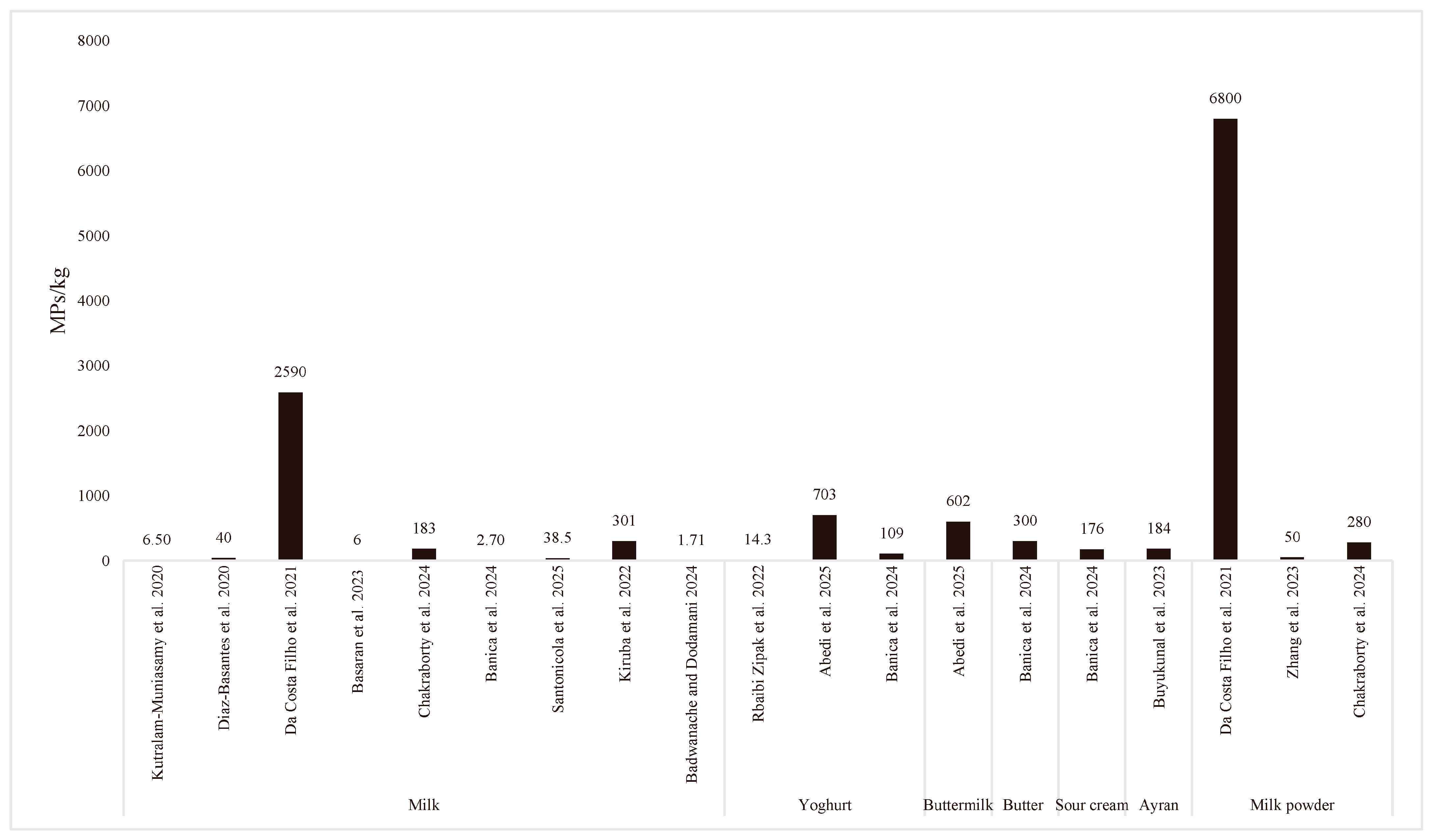
Figure 1. Average amounts of MP detected in milk and dairy products [38,39,51,52,53,54,55,56,57,58,59,60,61,62,63].
Applsci 15 09411 g001
Figure 2. Comparison of countries reporting MP presence in milk and dairy products.
Applsci 15 09411 g002
Figure 3. Methods used to detect microplastics in milk and dairy products.
Applsci 15 09411 g003
Table 1. Amounts and characterization of MPs milk and dairy products.
Dairy ProductAmount of MPPhysicalChemicalMethodCountryReferences
Size (µm)ShapeColorPolymers
Yoghurt (n = 16)0–1660 MPs/kg<500: 20%, 500–1000: 30%, >1000: 50%FibersTransparent, red, brown, black, gray, blue and greenPA, PET, PVC, PE, PC, PMMA, PUFT-IRIran[39]
Buttermilk (n = 14)0–2000 MPs/kg<500: 13%, 500–1000: 36%, >1000: 51%Transparent, red, brown, black, gray, blue and greenPA, PET, PVC, PC, PMMA, PU
Milk (skimmed, semi-skimmed, and
whole UHT) (n = 20)
10–270 MPs/kg<350: 23%,
350–1000: 37%, >1000: 40%
FibersBlue, black, red, pink, violet, yellow, green, orange, transparent, blue skyPE, polyesterFT-IRItaly[51]
Skim-milk powder (n = 16)466–5765 MPs/kg<99: 80%,
99–1444: 20%
Fibers, sphere, fragmentsBlack, blue, brown, fuchsia, green, and grayPEVA, PE, CPE, PP, PVC, PA, PC, polyisoprene, polyvinylidene fluorideFT-IRSpain[40]
Raw milk (n = 588)84–128 MPs/kg<500: 56%,
500–1000: 15%, >1000: 29%
Fibers, fragments, film, SphereBlack, blue, brown, grey, green, orange, pink, purple, red, transparent, and yellowPEA, EPC, HNBR, PAM, PARA, CR, PTFEFT-IRTürkiye[47]
Raw milk (n = 4)7–23 MPs/kg115–798Fibers, fragmentsBlack, blue, red, brown, yellowPMMA, PAµ-FT-IRRomania[52]
Organic milk (n = 6)10–99 MPs/kg68–2152Fibers, fragmentsBlack, blue, red, brown, gray, yellow, greenPMMA, PA, PU, polyester
Conventional milk (n = 10)2–36 MPs/kg76–1507Fibers, fragmentsBlack, blue, red, brown, gray, golden, turquoisePMMA, PA, PE, PU, polyester
Milk (n = 21)Not availableNot availableFibers, fragmentsBlue, red, violet, and
green
PS, PP, PVCFT-IRIndia[53]
Butter (n = 8)375–1500 MPs/kg41–1444Irregular, fiber oval, square, film, triangle, trapezoid, rectangular and diamondBlack, blue, red, brown, yellow, gray, purplePA, acrylicμ-FT-IRRomania[54]
Sour cream (n = 7)400–1200 MPs/kg47–1748Irregular, film, fiber, oval, triangleBlack, blue, green, yellow, gray, purplePA, acrylic
Milk (n = 6)95–250 MPs/kg<100: 35%,
>100: 65%
Fibers, fragments, filmBlack, red, blue, white, green, yellow, and violetPE, PP, Nylon-6, PS, PAATR-FT-IRBangladesh[55]
Milk powder (n = 25)120–570 MPs/kg<100: 31%,
>100: 69%
Fibers, fragments, filmBlack, red, blue, white, green, yellow, and violetPE, PP, PET, PA, PS, PA
Yoghurt (n = 17)693–1155 MPs/kgNot availableFibers, fragmentsBlack, blue, red, brown, grey, yellow, purplePA, PE, PU, polyesterµ-FT-IRRomania[56]
UHT Milk (n = 14)3–48 MPs/kg<500: 30%,
500–1000: 37%, >1000: 33%
Fibers, fragmentsBlack, red, green, blue, brown, and grayPA, PET, PEVA, PP, PUATR-FT-IRTürkiye[57]
Infant milk powder (n = 13)0–180 MPs/kg<50: 50%
50–608: 50%
Fibers, fragments, filmNot availablePE, PET, PP, PA, PVCFT-IRChina[58]
Ayran (traditional fermented dairy product) (n = 180)0–430 MPs/kg<150: 37%,
151–1000: 37%, >1000: 26%
Fibers, fragments, film, sphereBlack, blue, brown, dark blue, grey, green, orange, pink, purple, red, transparent, and yellowEP, PTFE, PA, NP, PP, PAM, PEFT-IRTürkiye[59]
Yoghurt (n = 12)20–580 MPs/kg<500: 70%,
500–1000: 11%,
>1000: 19%
Fibers, fragments
film, sphere
Black, blue, brown, gray, green, orange, pink, red, purple, reddish brown, and transparent whitePP, PS, PE, PVCFT-IRTürkiye[60]
Milk (n = 10)164–427 MPs/kg<500 Fibers, fragments pelletPink, purple, and bluePE, PP, PAMFT-IRIndia[61]
Raw milk (n = 2)2040–6250 MPs/kg≥5Fibers, fragmentNot availablePE, PES, PP, PU, PA, PTFEμ-RamanSwitzerland[62]
Milk (n = 4)1720–3480 MPs/kgPE, PS, PES, PVA, PTFE, PU, PP, PSU
Milk powder (n = 2)3560–10,040 MPs/kgPE, PES, PP, PA, PTFE
Milk (n = 23)3–11 MPs/kg<500: 40%,
500–1000: 28%, >1000: 32%
Fibers, FragmentsBlue, brown, red and pinkPES, PSUμ-RamanMexico[38]
Skim milk (n = 10)134–444 MPs/kg2.48–6742Fibers, fragmentsGreen, yellow, red, violet and blueHDPE, LDPE, PAM, PPFT-IREquator[63]
Polyamide (PA), Polyethylene terephthalate (PET), Polyvinyl chloride (PVC), Polyvinyl alcohol (PVA), Polyethylene (PE), Polycarbonate (PC), Polymethyl methacrylate (PMMA), Polyurethane (PU), poly(ethylene-vinyl acetate) (PEVA), Polyethylene chlorinated (CPE), Polyethyl acrylate (PEA), Ethylene propylene copolymer (EPC), Hydrogenated nitrile butadiene rubber (HNBR), Polyacrylamide (PAM), Polyaramid (PARA), Polychloroprene (CR), Polystyrene (PS), Ethylene propylene (EP), Neoprene (NP), Polyethersulfone (PES), Polysulfone (PSU), Polytetrafluoroethylene (PTFE), High-density polyethylene (HDPE), Low-density polyethylene (LDPE), Polypropylene (PP), Fourier-transform infrared spectroscopy (FT-IR).
Table 2. Analytical techniques used for MP determining and defining in dairy products.
Identification MethodsSpectroscopy Device Brand and ModelImaging MethodsFiltration Methods
Filter Type and Pore Size
DigestionReference
FT-IRAgilent Cary 630 (Santa Clara, CA, USA)Binocular biological microscope, SEM–EDSVacuum filtration
(1 μm)
Multi-enzymatic detergent [47]
FT-IRNot specifiedMicroscopyVacuum filtration
(11 µm)
Density separation: NaCl (1.17 g/cm3)[53]
µ-FT-IRVertex 80 v, Bruker (Ettlingen, Germany)Optical Microscopy—SEM-EDSVacuum filtration
Membrane filter (5–13 µm)
Ultrasonic bath[52]
μ-RamanHoriba (Longjumeau, France)Epifluorescence microscope + SEMVacuum filtration
(11 µm)
No[38]
FT-IRNot specifiedEpifluorescence microscopeVacuum filtration
(11 µm)
No[61]
FT-IRPerkinElmer (Hopkinton, MA, USA)Digital microscopeVacuum filtration
(10 µm)
No[57]
FT-IRNicolet 6700, Thermo Fisher Scientific (Middleton, WI, USA)FESEMFiltrationSalivary digestion, gastric digestion and intestinal digestion[76]
FT-IRNot specifiedOptical microscopyVacuum filtration Membrane filter (10 µm)H2O2 (30%)[63]
FT-IRNicolet iMX10, Thermo Fisher Scientific (Waltham, MA, USA)Optical microscopyVacuum filtration Cellulose nitrate membrane filter (8 µm)H2O2 (30%)[51]
TGA- FT-IRMettler Toledo (Zurich, Switzerland)NoNoNo[77]
ATR-FT-IRFTIR- 4600, JASCO Inc. (Tokyo, Japan)MicroscopyVacuum filtration
Glass microfiber filter
No[55]
FT-IRPerkinElmer (Milan, Italy)MicroscopyVacuum filtration
Silver membrane microfilter (3 µm)
KOH (10%)[40]
FT-IRHyperion 2000, Bruker
(Ettlingen, Germany)
Not mentionedVacuum filtration, Polycarbonate filter (8 μm)Pancreatic enzymes [58]
µ-FT-IRVertex 80, Bruker (Billerica, MA, USA)Optical microscopyVacuum filtration, Cellulose membrane filter (12–15 µm)1% mix solution of Sodium dodecyl sulfate & NaOH[54]
FT-IRShimadzu AIM-9000 (Kyoto, Japan)Binocular microscope,
SEM-EDS
Vacuum filtration, Cellulose nitrate membrane filter (0.45 µm)H2O2 (30%)[39]
FT-IRAgilent Cary 630 (Santa Clara, CA, USA)Binocular microscope, SEMVacuum filtration, Microfiber filters (1 μm)Multi-enzymatic detergent[60]
µ-FT-IRVertex 80 v, Bruker (Massachusetts, USA)Optical microscopyVacuum filtration, Cellulose membrane filter (12–15 µm)Ultrasonic bath[56]
μ-RamanHoriba (Longjumeau, France)Optical microscopyMicrofiltration, Silicon (Si) filter (5 μm)Multi-enzymatic and alkaline digestion[62]
μ-RamanThermo Fisher Scientific
(Wisconsin, USA)
Optical microscopy,
SEM-TEM
Vacuum filtration,
Glass fiber filter (0.22 µm)
Multi-enzymatic detergent and alkali[78]
FT-IRAgilent Cary 630 (Santa Clara, CA, USA)Optical microscope,
SEM
Vacuum filtration,
Glass microfiber filter (1 µm)
Multi-enzymatic detergent [59]
ATR-FT-IR & RamanVertex 70 v, Bruker (Rosenheim, Germany) & LabRAM HR, Horiba (Longjumeau, France)Optical microscopyNoNo[79]
ATR-FT-IR & RamanVertex 70 v, Bruker (Rosenheim, Germany) & LabRAM HR, Horiba (Longjumeau, France)Optical microscopyNoNo[80]
Py–GC–MSAgilent 7890 A (Santa Clara, CA, USA)Optical microscopyVacuum filtration,
Glass fiber filter (1.6 µm)
HNO3 (65%), H2O2 (5.4 M), KOH (1 and 5 M), and Fenton’s reagent.[81]
Fourier-transform infrared spectroscopy (FT-IR), micro-FT-IR (μ-FT-IR), Thermogravimetric analysis FT-IR (TGA-FT-IR), Attenuated total reflectance-FT-IR (ATR-FT-IR), Pyrolysis–gas chromatography–mass spectrometry (Py-GC-MS), Scanning electron microscopy (SEM), Field Emission SEM (FESEM), Energy-dispersive X-ray spectroscopy (EDS), Transmission electron microscopy (TEM), Hydrogen peroxide (H2O2), Nitric acid (HNO3), Potassium hydroxide (KOH), Salt (NaCl).
Disclaimer/Publisher’s Note: The statements, opinions and data contained in all publications are solely those of the individual author(s) and contributor(s) and not of MDPI and/or the editor(s). MDPI and/or the editor(s) disclaim responsibility for any injury to people or property resulting from any ideas, methods, instructions or products referred to in the content.