Review

Exposure Pathways, Systemic Distribution, and Health Implications of Micro- and Nanoplastics in Humans

1
Department of Civil Engineering, Lassonde School of Engineering, York University, North York, Toronto, ON M3J 1P3, Canada
2
Biology Department, York University, North York, Toronto, ON M3J 1P3, Canada
*
Author to whom correspondence should be addressed.
Appl. Sci. 2025, 15(16), 8813; https://doi.org/10.3390/app15168813
Submission received: 6 July 2025 / Revised: 1 August 2025 / Accepted: 6 August 2025 / Published: 9 August 2025
(This article belongs to the Special Issue Exposure Pathways and Health Implications of Environmental Chemicals)

Abstract

Plastics have become integral to modern life; however, their widespread use and persistent nature have resulted in significant environmental contamination, especially by microplastics (MPs < 5 mm) and nanoplastics (NPs < 100 nm). These plastic particles can enter the human body via ingestion, inhalation, or dermal absorption, raising substantial concerns about their potential health impacts. Recent studies using zebrafish, rodent models, and human cell lines have begun to elucidate the mechanisms underlying micro- and nanoplastics (MNPs)-induced toxicity. These mechanisms include oxidative stress, inflammation, disruption of metabolic processes, neurotoxicity, reproductive dysfunction, and carcinogenicity. Despite these advances, significant knowledge gaps remain. There remains a lack of comprehensive reviews that systematically evaluate these effects across major human organ systems and address how MNPs cross biological barriers in the human body. This review addresses these gaps by summarizing the available evidence on MNPs’ toxicity, critically discussing their absorption, distribution, metabolism, and the associated cellular and molecular mechanisms of action. Furthermore, it outlines urgent research priorities, emphasizing the need for standardized analytical protocols, realistic exposure models, and extended epidemiological research to evaluate human health risks posed by MNPs accurately. In addition, the adoption of precautionary regulatory actions is recommended to mitigate exposure and safeguard public health.

1. Introduction

The widespread use and improper disposal of plastic materials have led to the emergence of micro- and nanoplastics (MNPs) as significant and persistent environmental contaminants [1]. MPs (<5 mm) and NPs (<100 nm) are either produced intentionally for commercial use or formed through the fragmentation of larger plastic debris [2,3]. Due to their high persistence and mobility in the environment, they have become globally distributed pollutants, detected in marine and freshwater systems, soil, air, and even polar and alpine environments [4]. Recent estimates suggest that between 0.013 and 25 million metric tons of MNPs are transported within the marine environment and deposited into ocean waters each year, highlighting the widespread nature of plastic pollution, with some coastal and river sediments containing tens of thousands of particles per cubic metre [5].
Their omnipresence in the environment is closely tied to inefficient waste management systems and the durability of plastic polymers, which can persist for hundreds of years without degrading. Recent studies have identified atmospheric transport as a key pathway for the long-distance movement of MNPs, highlighting the global scale of this emerging issue [6]. The increasing prevalence of MNPs in diverse ecosystems has raised significant concerns within the field of environmental toxicology. Due to their small size, MNPs are easily taken up by organisms across trophic levels, where they can cause internal abrasions, gastrointestinal blockage, reduced nutrient uptake, and impaired reproduction. Additionally, their large surface area-to-volume ratio enables them to adsorb and transport toxic chemicals, which may then bioaccumulate through the food web [7].
The potential implications for human health are also increasingly evident. MNPs have been detected in drinking water, food, and air, making human exposure virtually unavoidable [8]. Alarmingly, plastic particles have recently been found in human placentas and blood, suggesting that these particles can be absorbed systemically and reach internal organs [9,10]. Experimental studies have shown that MNPs may induce cytotoxic effects, oxidative stress, inflammation, and possibly endocrine disruption in mammalian systems [11,12]. Understanding the toxicological impacts of these contaminants is therefore essential for assessing both environmental and public health risks. This review discusses the sources, distribution, exposure pathways, and mechanisms of toxicity of MNPs, with a focus on their effects on human health. It also highlights current knowledge gaps and emphasizes the need for multidisciplinary strategies to mitigate their impact and to develop effective monitoring and regulatory approaches.

2. Sources, Transport, and Environmental Distribution of MNPs

The sources of MNPs can be classified as primary and secondary sources. Primary sources include intentionally engineered MNPs for specific commercial and industrial uses [13]. Among these are microbeads found in personal care products (e.g., face scrubs, toothpaste), plastic powders in industrial abrasives and coatings, and NPs used in paints, printing inks, drug delivery systems, and biomedical devices [14,15]. Accidental spills during manufacturing or transportation, as well as direct release during product use or disposal, contribute to their environmental impact [16]. Conversely, secondary MNPs are generated through the progressive fragmentation of larger plastic debris due to a combination of physical (mechanical abrasion, wind), chemical (UV radiation, photooxidation), and biological (microbial degradation) processes [17]. Common sources of macroplastic debris that eventually break down into MNPs include plastic bags, packaging films, containers, fishing nets, synthetic textiles, and tire wear [18]. Once introduced into the environment, MNPs are transported and redistributed through air, water, and soil, forming interconnected pathways that facilitate their movement across different ecosystems [19]. This distribution is illustrated in Figure 1. These pathways deliver particles that can be inhaled, ingested, or absorbed through the skin to humans, making the tracking of MNPs from where they originate (source) to where they accumulate (sink) a critical first step in safeguarding human health.

2.1. Aquatic Transport

In aquatic environments, MNPs are introduced through multiple routes, including stormwater runoff from urban areas, effluents from wastewater treatment plants (WWTPs) [20], industrial discharges, and activities such as coastal tourism, shipping, and fishing [21]. Estimates suggest that the ocean currently holds around 50–75 trillion pieces of plastic and MNPs [22]. Rivers and estuaries act as major channels, transporting land-based plastics into the ocean. Once in marine or freshwater systems, the fate of MNPs depends on their density, size, and biofouling behaviour [23]. Typically, low-density polymers such as polyethylene (PE) and polypropylene (PP) tend to float and accumulate on the surface, where surface-feeding organisms readily ingest them. Denser polymers like polystyrene (PS), polyethylene terephthalate (PET), and polyvinyl chloride (PVC) sink and settle into sediments, forming long-term sinks [24]. Water currents, waves, and tidal forces can redistribute these particles both horizontally across the surface and vertically through the water column [23]. In deep-sea environments, MPs have been found embedded in sediment cores, indicating their potential for long-term deposition [25]. These aquatic reservoirs subsequently feed into human exposure pathways, emphasizing the health stakes of tracking MNPs in marine and freshwater systems.

2.2. Atmospheric Transport

Atmospheric transport has been identified as a significant pathway for the dispersal of MNPs [17]. An estimated 124 kilotons of MNPs are suspended globally. Sources include road traffic emissions, synthetic fibre shedding, industrial emissions, and the resuspension of particles from land and sea surfaces. Once airborne, these particles can be carried across regional and even continental scales [26]. Previous studies reported MNPs in remote regions, for example, the Arctic and the Alps, transported by wind currents and deposited through wet (rain, snow) and dry (gravitational settling) processes [27,28]. Their small size allows them to remain air-suspended for longer durations, and they have been found in urban dust, indoor air, and atmospheric fallout. Indoor environments, where synthetic materials are abundant, often exhibit higher concentrations, contributing to chronic human inhalation exposure [29].

2.3. Soil and Terrestrial Transport

In terrestrial ecosystems, MNPs enter soils through several anthropogenic practices [30]. Research estimates that over 1.5 million metric tons of MNPs infiltrate terrestrial ecosystems each year, profoundly affecting soil health and biodiversity [31]. Land application of sewage sludge (biosolids), compost containing plastic fragments, plastic mulching in agriculture, and landfill leachates contribute to soil contamination [32]. Agricultural soils, in particular, can accumulate significant quantities of MNPs due to the repeated application of contaminated organic fertilizers [32]. These particles may interact with soil biota, alter soil structure and function, and potentially affect nutrient cycling and crop productivity [33]. Once in the soil, MNPs can be transported vertically through bioturbation, percolation with rainwater, or root uptake [34]. Lighter particles may also be resuspended into the atmosphere via wind erosion or mobilized by surface runoff into nearby aquatic systems [35]. Such terrestrial reservoirs create additional human contact points, such as contaminating crops and drinking water catchments.

3. Human Exposure Pathways to MNPs

Human exposure to MNPs has become an emerging concern due to their increasing presence in environmental systems. MNPs have been detected in drinking water, food, and air; however, current evidence regarding their health impacts remains limited. Currently, there are no established regulatory thresholds or permissible limits for MNP concentrations in environmental or biological samples, making it difficult to interpret the health significance of detected levels. The World Health Organization acknowledges these significant data gaps and emphasizes the need for further research into the potential toxicological effects and long-term human exposure risks [36]. Addressing these challenges requires a thorough understanding of human exposure pathways, as this knowledge is fundamental to the accurate assessment and effective management of associated health risks.
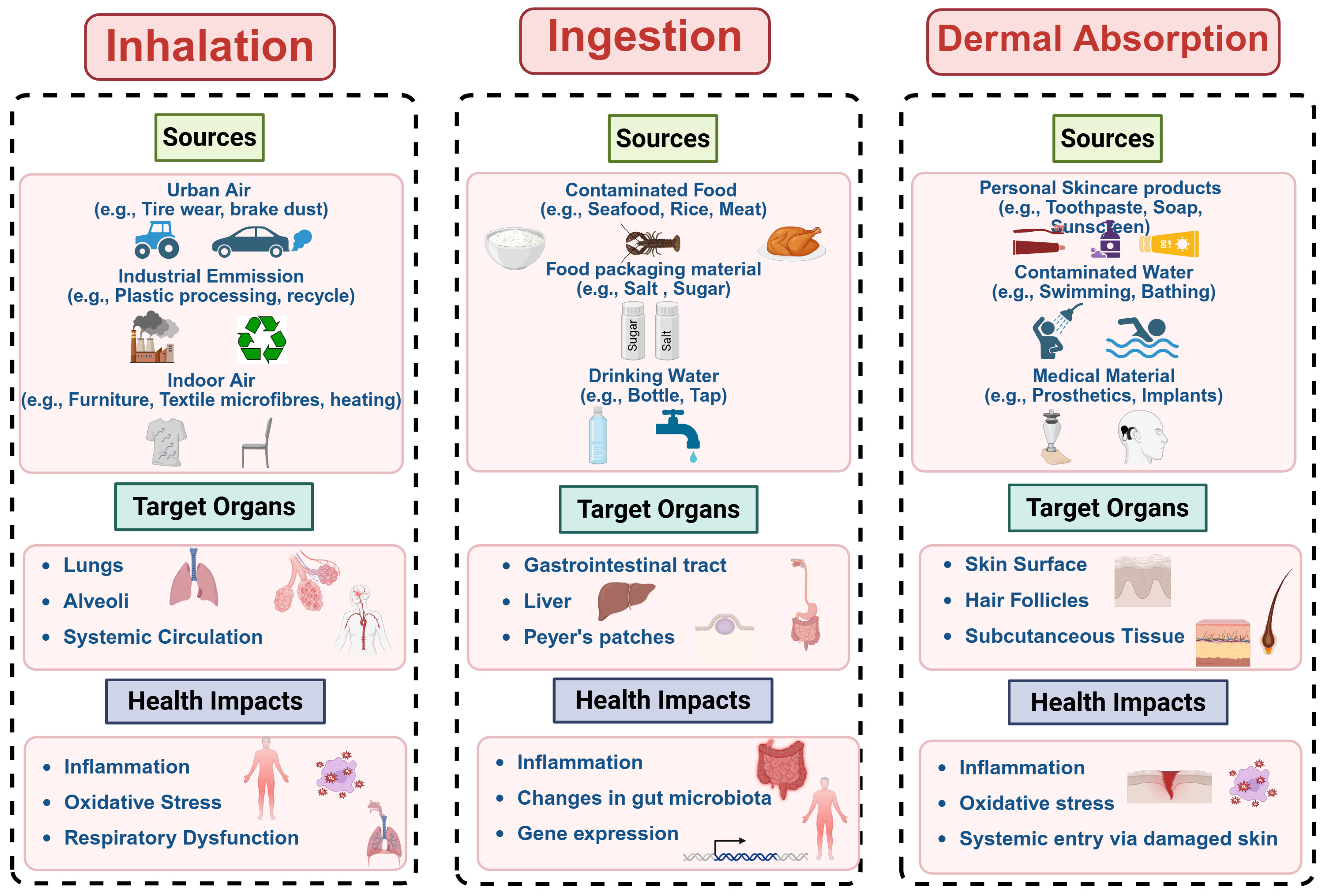
Human exposure to MNPs occurs via three primary pathways: inhalation, ingestion, and dermal absorption [21,37]. Figure 2 provides an overview of each route, visually mapping how airborne, dietary, and skin contact exposures originate from various environmental sources, such as air, water, food, consumer products, and occupational settings. The figure further illustrates the target human organs affected by these exposure pathways and the subsequent health impacts associated with MNP contact.

3.1. Inhalation of Airborne MNPs

Inhalation is particularly concerning in densely populated or industrial regions where airborne MNPs are widespread [38]. Environmental monitoring has confirmed the omnipresence of airborne MNPs; for example, Dris et al. reported outdoor concentrations of 0.3–1.5 particles/m3 and indoor levels up to 56.5 particles/m3. Approximately one-third of reported indoor particles were in the inhalable range [39]. Vianello et al. estimated that a person performing light physical activity could inhale approximately 272 MPs per day [40], whereas Prata et al. suggested a range of 26–130 particles/day depending on individual activity and environmental conditions [41].
The detection of MNPs in human lung tissues and sputum reinforces concerns about long-term respiratory exposure [42]. Amato-Lourenço et al. reported that 65% of tested human lung samples contain MNPs [43], while Jenner et al. confirmed MP fibres in human lung tissue [44], further validating chronic respiratory exposure. Some case studies have shown workers in the PVC, textile, and flock industries have reported lung inflammation, fibrosis, and interstitial disease attributable to chronic microplastic inhalation [11,45].
As depicted in Figure 2, the inhalation pathway illustrates how these airborne particles originate from urban dust, industrial activities, and household sources, entering the respiratory tract, primarily targeting the lungs, and posing potential health risks. Ultrafine particles (<2.5 µm) may bypass mucociliary clearance mechanisms, penetrate deep into alveolar spaces, and translocate into circulatory or lymphatic systems [46,47]. Their deposition and clearance dynamics depend on particle size, density, and surface properties, with smaller and less dense particles having higher alveolar deposition potential.
Mechanistically, the large surface area of NPs can intensify oxidative stress and chemotactic signalling, impair macrophage activity, and lead to persistent inflammation [48]. Supporting this, studies have shown that PS NPs (~64 nm) induce neutrophil influx and pro-inflammatory gene expression in pulmonary epithelial cells [49], while PVC MPs (2 µm) cause cytotoxicity and hemolysis in both rat and human lung cells [50].
Collectively, this body of evidence reinforces the need to recognize airborne MNPs as a legitimate respiratory hazard, especially under high-concentration exposure scenarios or in vulnerable groups. Consistent detection of MNPs in human lung tissues, occupational case studies, and in vitro cytotoxicity data highlight the potential for chronic respiratory harm driven by deep pulmonary deposition and persistent inflammation. Although environmental concentrations vary, indoor exposure appears especially significant and the estimated daily inhalation rates underscore the relevance of routine, low-level exposure. Nevertheless, substantial gaps remain regarding dose–response relationships, long-term outcomes, and particle-specific toxicodynamics.

3.2. Ingestion of MNPs Through Dietary Intake

Ingestion constitutes a principal and extensively studied route of human exposure to MNPs. As illustrated in Figure 2, the ingestion pathway highlights multiple dietary sources through which MNPs enter the human body, including contaminated drinking water, processed foods, seafood, and other food staples. The estimated annual intakes range widely, from tens of thousands to over 35 million particles per individual, depending on diet, regional factors, and water source [8,51]. Human exposure to MNPs is closely tied to daily consumption patterns, particularly through drinking water, processed foods, and seafood [52].
Bivalves, crustaceans, and commercial fish species are frequently identified as high-risk commodities due to their direct interaction with MNP-contaminated aquatic environments [53,54]. Beyond seafood, bottled beverages and packaged foods further lead to widespread MNPs infiltration. Critically, MNP contamination is not restricted to aquatic products; foods such as salt [55], honey [56], tea [57], rice [58], and meat [59] have all shown a measurable contamination. For instance, MPs in rice were recorded at concentrations of 67 ± 26 μg/g dry weight [58], while packaged meat samples exhibited 4.0 to 18.7 particles/kg [59]. Similarly, drinking water, both tap and bottled, represents a significant ingestion route for MNPs, as shown in Figure 2, with studies showing widespread contamination. Kosuth et al. found MPs in 83% of global tap water samples (mean: 4.34 particles/L) [60], while Mason et al. reported a 93% contamination in bottled water, 325 particles/L [61], contributing up to 35.7 million particles ingested annually [62].
In summary, ingestion is a major route of MNP exposure, driven by contaminated food, water, and indoor dust. These findings show the cumulative and multifactorial nature of MNP ingestion and highlight the importance of considering not only environmental contamination but also food processing, packaging, and indoor environments in exposure assessments. Seafood shows the highest risk due to bioaccumulation, while bottled water and packaged foods also contribute significantly. These findings highlight the cumulative nature of MNP intake and the need to consider both environmental and post-harvest sources in exposure assessments.

3.3. MNP Exposure Through Dermal Contact

Dermal absorption, though less studied than ingestion and inhalation, remains a plausible route for MNP exposure. As shown in Figure 2, the dermal pathway highlights how MNPs present in personal care products (e.g., toothpaste, soap, and sunscreen), textiles, airborne particles, and medical materials can come into direct contact with human skin [63,64]. Before regulatory restrictions, personal care items commonly used MNPs as exfoliants, raising concerns about NPs (<100 nm) breaching the dermal barrier [52]. Previous studies have quantified dermal exposure: Napper et al. found 10–100 g/L MNPs in facial scrubs, with daily use estimated at 40.5–215 mg per person, while Gouin et al. reported 2.4 mg/day from liquid soaps [65,66]. Although direct evidence of transdermal absorption remains limited, the likelihood of dermal penetration depends on particle size, surface properties, and the presence of co-contaminants. Some in vitro studies suggest that NPs have the potential to infiltrate deeper layers of the skin and induce oxidative stress in epithelial cells [67].
Furthermore, Figure 2 incorporates additional dermal exposure routes such as contact with contaminated water or airborne MNPs settling on skin surfaces, which further elevate the exposure risk [68]. Additionally, plastic wear from medical devices, including joint replacements and dental and cosmetic implants, can generate MNPs with prolonged internal exposure implications [54]. These particles may trigger local inflammation or encapsulation, particularly in subcutaneous environments. Despite limited quantitative estimates of dermal uptake, studies support the plausibility of transdermal absorption and systemic effects, particularly where skin integrity is compromised.
Current evidence indicates that dermal contact with MNPs from products, textiles, and medical materials can lead to particle penetration into deeper skin layers, especially for NPs and when the skin barrier is compromised. Although quantitative data on uptake remain limited, studies suggest the potential for oxidative stress, local inflammation, and systemic distribution. These findings underscore dermal absorption as a plausible exposure route that warrants further investigation.

3.4. Disparities in Exposure and Vulnerability

Human contact with MNPs is unevenly distributed across regions, socioeconomic strata, and occupations, leading to marked differences in both exposure intensity and health risk.
Geographically, dietary intake models show Southeast Asia topping global rankings, with residents of Indonesia, Malaysia, and the Philippines ingesting up to 500 mg per person each day, largely from seafood that bioaccumulates plastics in polluted waters [69]. Inhalation burdens also vary: China and Mongolia lead worldwide estimates at roughly 2.8 million airborne particles inhaled per person per day, a reflection of dense urban industrial activity and poor emission controls [69]. Conversely, many African nations experience significant MNP contamination of drinking water and freshwater fisheries amid limited wastewater treatment capacity, compounding ingestion risks in already resource-constrained settings [70].
Socioeconomic factors further modulate exposure. Low-income communities frequently rely on bottled water when municipal supplies are unsafe, yet 93% of bottled brands sampled globally contain MPs [61]. Informal settlements situated near open dumps or ports that import plastic waste from wealthier countries face constant contact with plastic debris and its breakdown products, intensifying both inhalation and ingestion pathways. Indoor MP loads are also higher in poorly ventilated, overcrowded housing, increasing the incidental ingestion of dust that may rival foodborne sources [70].
Occupational settings create some of the most extreme exposure scenarios. Airborne concentrations in textile or flock factories can reach ~1 million fibres m−3, and up to 40% of workers develop interstitial or airway disease linked to synthetic microfibers [71]. Waste-management, recycling, and 3D-printing facilities have recorded particle counts orders of magnitudes higher than ambient urban air, occasionally exceeding 40 billion particles m−3 [72]. Plastic product workers exposed to PVC dust show elevated risks of lung impairment and even liver angiosarcoma, highlighting compound hazards from mixed polymer and chemical exposures [73]. These occupational burdens disproportionately affect labour forces in developing economies and low-wage sectors where personal protective equipment and engineering controls are often inadequate.
Children, pregnant women, and informal waste pickers constitute especially vulnerable sub-populations. Children may inhale three times more airborne particles than adults owing to higher ventilation rates and a closer proximity to contaminated floors, while pregnant women in high-exposure regions have shown placental MNP burdens that could portend developmental effects. Informal recyclers, estimated at >2 million workers in India alone, sort plastic waste by hand with minimal respiratory protection, placing them at the apex of combined inhalation and dermal risk [69].
Recognizing and quantifying these disparities is essential for targeted mitigation, ranging from region-specific dietary advisories and infrastructure investment to stringent workplace exposure standards and environmental justice policies that shield the most burdened communities.

4. MNPs’ Toxicity to Humans

MNPs are widely recognized for their ability to breach human physiological barriers and interact with cellular and molecular systems, leading to a cascade of toxicological events. Once internalized, MNPs can translocate across epithelial barriers and distribute systemically, accumulating in critical organs such as the lungs, liver, kidneys, placentas, and brain (Table 1). This section explores how MNPs are taken up by human cells, how they are transported and accumulated within the body, and the biological mechanisms through which they induce toxicity, including oxidative stress, inflammation, mitochondrial dysfunction, and apoptosis. Understanding these mechanisms is vital to elucidating the full spectrum of health risks posed by chronic and low-dose exposure to environmental MNPs.

4.1. Cellular Uptake, Translocation Pathways, and Accumulation Mechanisms of MNPs

4.1.1. Cellular Uptake of MNPs

The uptake of MNPs by cells involves multiple mechanisms that are influenced by particle size, surface properties, and morphology. MPs are primarily internalized via phagocytosis, especially in immune cells like macrophages, while NPs more quickly infiltrate cells via endocytosis, macropinocytosis (an actin-driven process where cells engulf fluid into large vesicles), or direct membrane penetration [74]. Surface chemistry also plays an important role in modulating the uptake efficiency. Functionalization with carboxyl or amino groups, whether through environmental ageing or during production, significantly influences cellular interactions. For example, Forte et al. demonstrated the enhanced uptake of carboxylated PS NPs in human gastric cells compared to their non-functionalized counterparts [75]. Positively charged particles exhibit a stronger electrostatic attraction to negatively charged cell membranes, further enhancing internalization rates [76].

4.1.2. Translocation and Systemic Distribution

Once MNPs are internalized, their ability to translocate to distant tissues raises a concern for systemic toxicity. Translocation depends on the particle size, route of exposure, and integrity of biological barriers. In the gastrointestinal tract, NPs can cross the intestinal epithelium via paracellular transport and endocytosis by Microfold cells (M cells) located in Peyer’s patches (specialized cells that transfer particles to immune tissues) [77]. Inhalation is another critical route, with studies indicating that inhaled NPs may penetrate the alveolar epithelium, traverse the air–blood barrier, and reach systemic circulation, potentially affecting vital organs such as the brain [47]. Supporting this, in vivo studies have shown the presence of MNPs in the liver, kidneys, spleen, and central nervous system, suggesting compromised intestinal, pulmonary, and even placental barriers [78,79]. Ragusa et al. showed clinical evidence of MPs in human placentas, raising serious concerns about fetal exposure during pregnancy [10]. Lymphatic transport is also believed to enhance systemic distribution, bypassing hepatic first-pass metabolism and enabling accumulation in secondary organs [80]. These findings show the capacity of MNPs, especially those at the nanoscale, to circulate beyond initial exposure sites.

4.1.3. Accumulation and Persistence

The systemic distribution of MNPs raises significant concerns regarding their bioaccumulation and long-term persistence in human tissues, as shown in Table 1. Due to their resistance to enzymatic degradation and strong affinity for binding biological macromolecules, MNPs can avoid immune detection by binding with proteins, forming a ‘protein corona’, a layer of adsorbed proteins that modulates their biological identity and cellular interactions. Recent studies, particularly using human intestinal models (an iPSC-derived IEC model), have shown that the composition of this corona can influence toxicity by enhancing oxidative stress and inflammatory signalling [81]. PS MPs preferentially accumulate in the liver, spleen, kidneys, and brain, persisting even weeks after exposure cessation [82]. This persistence shows the potential for chronic, low-level exposures to result in significant tissue loading over time. Furthermore, although direct evidence in humans remains limited, ecological studies suggest that MNPs can transfer across trophic levels. For instance, Farrell and Nelson et al. showed the movement of MPs from mussels to crabs [83], highlighting the risk of biomagnification and the potential for cumulative exposure through the food chain.
These findings highlight the need to consider chronic, low-dose accumulation and food chain transfer in human health risk assessments.
Table 1. Accumulation and abundance of MNPs in human biological samples.
SamplePolymer Type(s)Size RangeConcentration/
Abundance (n = number of particles)
Detection MethodStudy PopulationKey Finding on AccumulationReference
BloodPET, PE, PS, PMMA, and PP0.7–700 nm1.6 μg/mLPy-GC/MS22 healthy adults (Netherlands)Systemic circulation; binds to plasma proteins[9]
Body fluidsPP, PS, PTFE, PVB, PA, LDPE, and PVA2.15–103.27 μm702 n/LRaman micro-spectroscopy104 adults (24–96 years)Higher in individuals with vascular disease[84]
PlacentaPP, PE, PET, PS, PVC, PC, POM, and acrylic5–307 μm0.029–18 n/gμFTIR, LD-IR, and RamanPregnant women (Italy, China, Germany, and Iran)Fetal–maternal barrier penetration[10,85,86,87]
Breast MilkPA, PU, PE, PVC, PP, PET, and PMMA2–50 μm0.53–20.2 n/gLD-IR, RamanLactating women (Italy, China)Transfer to infants via lactation[10,87]
LungPP, PE, PET, PVC, PS, rayon, and cotton1.6–2475 μm0.56–1.42 n/gμFTIR, RamanAdults (Brazil, UK, and China)Fibres trapped in alveoli; higher in urban residents[43,44,88]
SputumPU, PVC, PE, alkyd varnish, and acrylates20–500 μm3.95–39.5 n/mLFTIR, LD-IR22 respiratory disease patients (China)Correlated with occupational exposure[89]
ColonPC, PA, PP, PS, and PET800–1600 μm (fibres)28.1 ± 15.4 n/gFTIR11 adults (Malaysia)Accumulation in the inflamed mucosa (IBD link)[90]
Feces (Adult)PET, PP, PE, PA, PVC, and PS20–1800 μm1–41.8 n/gμFTIR, Raman, and Py-GC/MSAdults (China, Indonesia, USA, and Germany)Higher in IBD patients vs. healthy[91,92,93,94,95]
Feces (Infant)PET, PC, PA, PU, and PE>20 μm26.6–54.1 n/gμFTIR, LD-IRNewborns/infants (China, Germany)Early-life exposure via ingestion[87,96]
LiverPET, PP, PS, POM, PMMA, and PVC4–30 μm0.70 (non-cirrhotic); 6.9 (cirrhotic) n/gRaman11 adults (Germany)Higher accumulation in cirrhotic livers[97]
ThrombiLDPE, PS, PVC, PET, PMMA, and POM1–26 μm5 n/thrombiRaman, SEM26 cardiovascular patients (China)Associated with vascular pathology[98]
UrinePE, PS, PTFE, PVA, PVC, and PP3–15 μm1–3 n/LRaman, μFTIRAdults (Italy, UK)Renal filtration evidence[99,100]
SalivaPE, PET, PS, PVC, PA, POM, PC, and PU1.5–500 μm0.33–2 n/individual/dayμRaman2000 adults (Iran)Oral cavity retention; daily shedding[101]
Seminal Fluid/TestisPET, PP, PS, PE, and PVC20–286 μm0.23–11.6 n/gPy-GC/MS, LD-IR30 semen/6 testis samplesGonadal accumulation[102,103,104]

4.2. Toxicodynamic Responses of MNPs: Oxidative, Inflammatory, and Cytotoxic Effects

The presence of MNPs in biological systems can trigger a cascade of cellular and molecular responses, with oxidative stress, inflammation, and cytotoxicity being key mechanisms of toxicity. As illustrated in Figure 3, MNPs uptake by cells induces reactive oxygen species (ROS)-driven oxidative damage, which further leads to downstream effects such as the activation of inflammatory pathways, organelle dysfunction, lipid and protein degradation, and DNA damage, which are mechanisms that underlie a wide range of adverse health outcomes associated with MNP exposure. To clarify the possible health effects of MNP exposure, it is essential to comprehend these mechanisms.

4.2.1. Oxidative Stress

Exposure to MNPs frequently induces oxidative stress, a condition in which the generation of reactive oxygen species (ROS) overwhelms the body’s antioxidant defences. This redox imbalance activates sensitive signalling pathways such as Nrf2 and MAPK, resulting in damage to cellular components, including lipids, proteins, and DNA [105]. NPs, in particular, are potent in driving ROS production, especially when they carry toxic additives or adsorbed contaminants like heavy metals and plasticizers [106]. For example, Schirinzi et al. found that PS NPs increased ROS generation in human cerebral and epithelial cells [12]. Similarly, Hwang et al. reported mitochondrial dysfunction in human retinal pigment epithelium cells exposed to PS NPs under oxidative stress conditions [107]. Additional studies confirm that PS NPs reduce glutathione levels and impair mitochondrial function in the liver and epithelial cells, highlighting both particle size and surface chemistry as key factors in oxidative damage mechanisms [108].
In contrast, MPs, characterized by their larger size and distinct cellular uptake mechanisms compared to NPs, commonly induce inflammation and oxidative imbalance in experimental models. These effects contribute to tissue damage such as liver fibrosis and other organ-specific toxicities. For instance, PE MPs have been shown to provoke inflammatory responses and oxidative stress, leading to liver fibrosis in animal studies. Similarly, PS and PVC MPs, alone or in combination, exacerbate liver toxicity by enhancing oxidative damage [109,110]. Although polymer type and particle size vary, these studies collectively emphasize oxidative stress and inflammation as key hallmarks of MP-induced toxicity across biological systems.

4.2.2. Inflammation

Inflammation is a key biological response to MNP exposure and plays a central role in mediating their toxicity. Human cell studies have demonstrated that MNPs stimulate the production of pro-inflammatory cytokines such as IL-8 and TNF-α and activate inflammatory signalling pathways including NF-κB [111,112]. For instance, the exposure of human alveolar epithelial cells (A549) to PS NPs induces increased the secretion of IL-8 and TNF-α, reduced cell viability, apoptosis, and activation of caspase-dependent inflammatory cascades [113]. Similarly, PS MPs elicit elevated TNF-α expression and activate the JNK1/2/3 pathway in human testicular cells, with dysregulation of the PI3K-AKT signalling pathway, reflecting oxidative stress-linked inflammatory processes [114]. These findings highlight inflammation as a principal mediator of MNP-induced cellular injury across human tissues.
Animal studies elucidate the underlying mechanisms by which MNPs may impact human health. For instance, PS NPs have been shown to induce pulmonary inflammation in rats, suggesting similar respiratory risks in humans [49]. Likewise, exposure to PS MPs in mice caused intestinal inflammation and altered gut microbiota, indicating the possible disruption of human gut health. Chronic exposure in murine models also resulted in persistent inflammatory changes in liver and gut tissues, highlighting risks of long-term tissue damage [115]. Complementing these findings, human renal epithelial cells exposed to PS MPs (~1.9 µm) exhibited increased ROS production and the upregulation of inflammatory mediators such as cPLA2 and COX-1, reflecting conserved inflammatory pathways across species [116]. MNP-induced inflammation often involves the activation of signalling pathways like NF-κB and the NLRP3 inflammasome, which create a feedback loop with oxidative stress that exacerbates tissue injury [117]. Furthermore, MNPs influence macrophage polarization and disrupt epithelial barrier integrity, amplifying both local and systemic inflammatory responses [118]. Collectively, these data highlight inflammation as a critical mechanistic pathway linking MNP exposure to cellular and tissue damage, with implications for human health risks that warrant further focused research. These findings highlight how inflammatory responses vary by particle size and polymer type and the role of both acute and chronic exposures in driving local and systemic inflammation.

4.2.3. Cytotoxicity

Cytotoxicity represents a key endpoint of MNP toxicity, often resulting in cell death via apoptosis, necrosis, or autophagy. Several studies have demonstrated the cytotoxic effects of MNPs across different cell types. Schirinzi et al. showed that PS MNPs induced significant cytotoxic effects in human cerebral and epithelial cells, with NPs exhibiting greater toxicity. Mechanisms include mitochondrial dysfunction, oxidative stress, and the activation of apoptotic pathways [12]. Wu et al. found that PS MPs caused ROS generation and apoptosis in hepatic cells [119], while Cheng et al. found that PS MPs elevated markers of oxidative damage and liver injury, including AST, ALT, LDH, ROS, and MDA [108]. Poma et al. highlighted MNP-induced genotoxicity, showing that both direct DNA interactions and ROS-mediated damage can contribute to potential carcinogenic outcomes [120].
Together, these mechanisms illustrate the multifaceted nature of MNP toxicity, which depends on physicochemical properties, exposure routes, and biological context. Oxidative stress frequently initiates cellular injury, triggering inflammation and further tissue damage; with ongoing exposure, these processes can disrupt normal cell functions and ultimately lead to cell death. The severity and manifestation of these effects vary according to particle type, entry route, and target organ. However, it should be noted that much of the mechanistic understanding is derived from animal and in vitro studies, which, while essential for revealing underlying pathways, may not fully replicate human physiological responses. As a result, caution is needed when extrapolating these findings to human health risks. Overall, the interplay between these mechanisms explains the broad spectrum of potential health impacts associated with MNP exposure and emphasizes the need for further research using human-relevant models to more accurately assess risks and inform harm-reduction strategies.

5. Health Effects of MNPs on Human Physiological Systems

MNPs are increasingly recognized as environmental contaminants capable of traversing physiological barriers and eliciting toxic effects in multiple human organ systems. Recent advancements have enabled the detection of MNPs in human biological matrices (Table 1), including lung tissues, blood, placenta, stool, and sputum, thereby providing direct evidence of internal exposure. Complementary in vitro studies have identified consistent patterns of cellular disturbance that help explain the biological effects observed and elucidate the underlying mechanistic pathways. However, while these studies are invaluable for mechanistic insight, their findings may not fully translate to human physiology due to differences in cell complexity, metabolism, and exposure conditions, underscoring the need for cautious interpretation and further validation in more human-relevant systems. This section synthesizes the current human-based evidence on MNP toxicity, critically examining the effects on major physiological systems by evaluating the underlying cellular mechanisms contributing to organ-specific vulnerability (Table 2).

5.1. Respiratory System

Inhalation of MNPs poses substantial respiratory health risks, evidenced by clinical, occupational, and experimental findings. Occupational exposure studies, notably in boatbuilding, fibreglass manufacturing, and PET production industries, have consistently reported increased incidences of respiratory diseases, including bronchial asthma, obstructive bronchiolitis, and hypersensitivity pneumonitis (OHP). Retrospective cohort analyses further suggest marginal elevations in lung cancer risks among workers chronically exposed to styrene, though definitive causation remains challenging due to confounding factors such as smoking [130,131]. Severe outcomes, including cases requiring lung transplantation, underline the critical occupational hazard posed by prolonged exposure to fibreglass and styrene resin materials [131].
The consistent deposition of MNPs in pulmonary tissues emphasizes their bioaccumulation potential. Surgical and autopsy studies explicitly identified PP, PET, and nylon microfibers in human lung tissues, validating occupational reports and suggesting chronic exposure implications [44,132]. Mechanistic studies using human alveolar epithelial cell models (A549 cells) consistently show oxidative stress, inflammation, and apoptotic pathways following MNP exposure, marked by elevated cytokines (TNF-α, IL-6, and IL-8) and impaired pulmonary surfactant dynamics [113,117].
Rodent inhalation studies provide additional mechanistic evidence, demonstrating pulmonary inflammation, epithelial barrier dysfunction, and fibrosis, indicative of chronic obstructive pulmonary disease (COPD)-like conditions following exposure to PS MPs [133,134,135]. Collectively, current evidence highlights oxidative stress, chronic inflammation, and epithelial barrier disruption as core mechanisms underpinning MNP-induced respiratory toxicity. These outcomes emphasize the urgent need for targeted regulatory interventions and preventive strategies, particularly in occupational and urban environments.

5.2. Digestive System

The digestive system represents a primary exposure route to MNPs, primarily via ingestion of contaminated food and water, leading to localized gastrointestinal and broader systemic effects. Clinical findings highlight elevated fecal concentrations of MNPs in patients with inflammatory bowel disease (IBD), correlating significantly with disease severity and suggesting a direct link between MP load and gastrointestinal inflammation [95]. Furthermore, occupational epidemiological studies reveal increased colorectal and pancreatic cancer risks among plastic and rubber industry workers, implying chronic gastrointestinal exposure as a potential carcinogenic factor [136,137].
Human-derived gastrointestinal cell line studies further elucidate toxicity mechanisms, demonstrating that PS NPs disrupt cell viability and induce oxidative stress and inflammatory responses [75,138]. These effects are compounded by gut microbiota dysbiosis, evidenced by human fecal analyses showing significant microbial community shifts in individuals with gastrointestinal conditions. Such dysbiosis potentially exacerbates intestinal inflammation and systemic toxicant exposure [139,140].
Emerging evidence suggests gastrointestinal MNP uptake compromises gut barrier integrity, facilitating systemic translocation and broader toxicological impacts. Experimental models show enhanced intestinal permeability and inflammatory responses, particularly under combined MNP and chemical co-exposures [141,142].
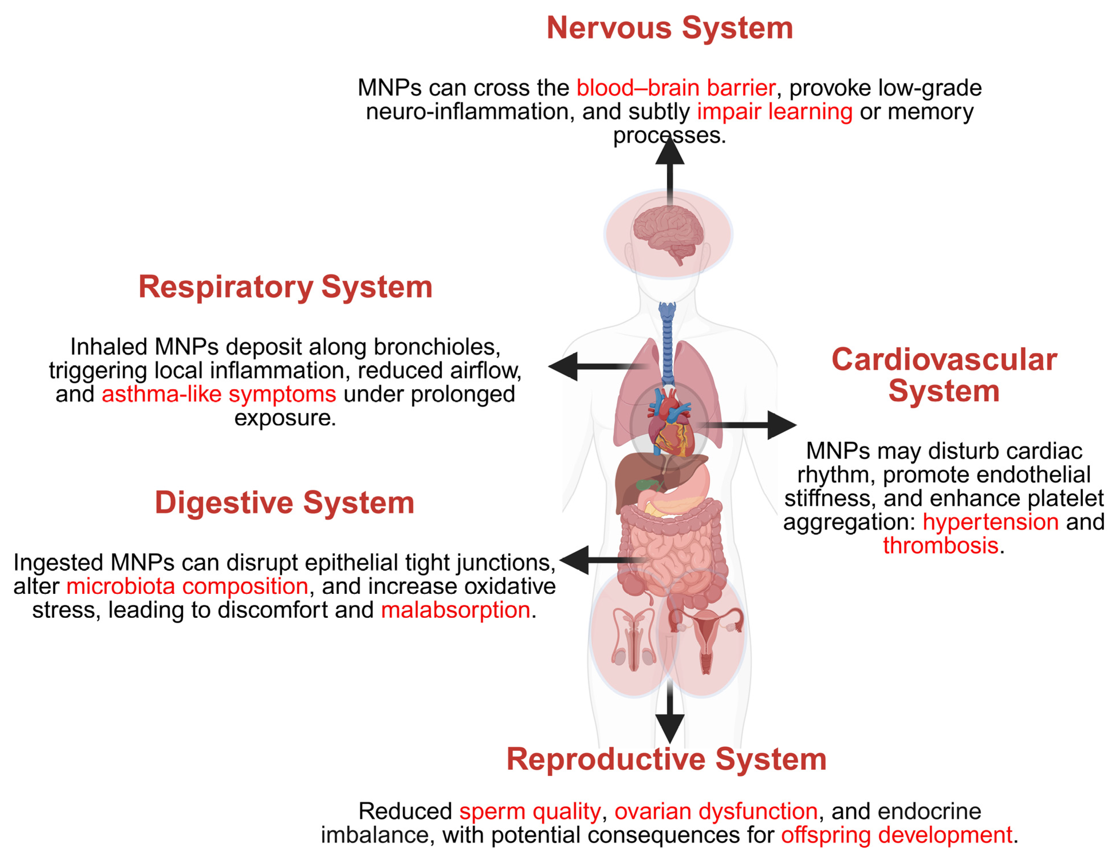
In summary, gastrointestinal exposure to MNPs presents critical health risks through inflammation, microbiota alterations, barrier dysfunction, and potential carcinogenesis, as summarized in Figure 4. Further human-specific research focusing on realistic exposure scenarios and vulnerable populations are urgently required to refine risk assessments and inform regulatory frameworks.

5.3. Cardiovascular System

Exposure to MNPs has significant implications for cardiovascular health, with evidence from human clinical data, in vitro studies, and animal models consistently highlighting associated risks. Human studies have identified MPs in cardiovascular tissues and circulating blood. For instance, the analysis of human atherosclerotic plaques from carotid endarterectomy patients revealed a notable accumulation of PE and PVC MPs, correlating with elevated inflammatory markers (IL-1β, IL-6, IL-18, and TNF-α), plaque instability, and increased cardiovascular event risks, including myocardial infarction and stroke [143,144].
Clinical observations further emphasize the relevance of MPs in cardiovascular pathology, demonstrating a correlation between PE particle concentrations in thrombi and disease severity, inflammation, and thrombosis markers (e.g., D-dimer levels) in patients with acute cardiovascular events [143]. Higher MP levels in coronary arteries and atherosclerotic plaques compared to non-atherosclerotic tissues reinforce their potential role in the pathogenesis of cardiovascular disease [145].
Mechanistically, human endothelial cell studies have shown that MNP exposure disrupts endothelial function, induces oxidative stress, and initiates inflammatory responses. For instance, the exposure of human umbilical vein endothelial cells (HUVECs) to PS NPs caused endothelial damage, autophagic dysfunction, and cellular senescence via oxidative stress and impaired nitric oxide signalling [127,146].
Experimental studies further confirm cardiovascular toxicity, reporting cardiac fibrosis, myocardial cell apoptosis, and functional impairment following MP exposure. Human-derived cardiac organoids exposed to MPs exhibited oxidative stress-induced apoptosis, fibrosis mediated by Wnt/β-catenin signalling activation, and inflammation [147,148]. Animal models provided supportive evidence, highlighting myocardial damage, mitochondrial dysfunction, increased cardiac enzyme levels, and impaired cardiac function following environmental-level MNP exposure [149]. Additionally, human erythrocyte studies identified hemolytic activity linked to the physicochemical properties of MNPs, suggesting increased thrombosis and cardiovascular complication risks [150,151].
Overall, current evidence robustly supports oxidative stress, inflammation, endothelial dysfunction, and thrombosis as key mechanisms responsible for MNP-induced cardiovascular toxicity (Figure 4). Further detailed research into particle characteristics and mechanistic pathways are crucial to accurately inform risk assessments and guide preventive strategies.

5.4. Nervous System

Exposure to MNPs has emerged as a significant concern for human neurological health due to their ability to breach critical barriers and induce neurotoxic responses. Epidemiological data highlight neurological symptoms, including headaches, dizziness, fatigue, and visual disturbances, among populations residing near plastic production zones, correlating these symptoms with elevated environmental styrene levels [152]. Occupational studies further report increased incidences of degenerative neurological conditions, such as Parkinson’s disease and multiple sclerosis, among workers frequently exposed to plastic materials; however, confounding occupational factors complicate direct causative associations [136].
Direct evidence from human autopsy analyses reveals substantial MP accumulation in brain tissues, notably higher in patients diagnosed with dementia, suggesting a potential contribution to neurodegenerative diseases [153]. Mechanistically, in vitro studies employing human cortical neuron models have demonstrated increased oxidative stress, neuroinflammation (e.g., TNF-α, IL-6 upregulation), mitochondrial dysfunction, and neuronal apoptosis following exposure to MNPs [154]. Additionally, the exposure of human neural progenitor cells to MPs resulted in significant disruptions in gene expression linked to neuronal differentiation, synaptic connectivity, and DNA repair mechanisms, indicating potential impacts on neuronal development and function [155,156].
Animal models have confirmed these findings, showing that MNP exposure can impair cognitive functions, disrupt neurotransmission (particularly cholinergic signalling), and increase neuroinflammation [157,158]. Furthermore, as shown in Figure 4, MNP exposure has been associated with compromised blood–brain barrier integrity, facilitating increased particle entry into the central nervous system, exacerbating neurotoxicity [159].
In summary, current evidence strongly supports the neurotoxic potential of MNPs through mechanisms involving oxidative stress, inflammation, barrier disruption, and impaired neurotransmission. Continued human-specific investigations are essential to fully elucidate these mechanisms and better define the neurological risks associated with MNP exposure.

5.5. Reproductive System

The reproductive system is highly sensitive to environmental contaminants, including MNPs, due to its essential role in species survival and its complex physiological regulation. Although human data remain limited, animal models and emerging human evidence highlight significant reproductive toxicity linked to MNP exposure.
In males, MNPs have been detected in seminal fluid and testicular tissues, indicating their ability to cross physiological barriers such as the blood–testis barrier [102,103,104]. MNP exposure induces oxidative stress, mitochondrial dysfunction, inflammation, apoptosis, and structural damage within testicular tissues. Key pathways include ROS production, the activation of MAPK signalling (notably p38 and JNK), suppression of steroidogenic acute regulatory protein (StAR)-mediated testosterone biosynthesis, and engagement of endoplasmic reticulum stress responses [114]. These molecular disturbances result in reduced sperm viability, motility, concentration, and increased morphological abnormalities. Furthermore, chronic exposure disrupts hormonal regulation by impairing luteinizing hormone (LH)-stimulated testosterone synthesis via the downregulation of LHR/cAMP/PKA signalling [160].
Female reproductive organs also accumulate MNPs, detected in ovarian follicular fluid, endometrial, and placental tissues [161]. Exposure provokes oxidative stress, inflammation, and apoptosis in granulosa cells, mediated through pathways such as NLRP3/Caspase-1 inflammasome activation, ER stress via PERK-eIF2α-ATF4-CHOP, and the dysregulation of Wnt/β-catenin signalling [162,163]. Consequences include diminished ovarian reserve, disrupted estrous cycles, reduced oocyte quality, impaired fertilization, and decreased embryonic developmental success [164]. Importantly, MNPs can cross the placental barrier, reach fetal tissues, and potentially induce prenatal toxic effects that impact offspring growth, neurodevelopment, immune function, and reproductive capacity even across generations [165,166].
At the cellular level, oxidative stress, inflammatory cascades, mitochondrial impairment, loss of cell junction integrity, and hormonal dysregulation are central to MNP-induced reproductive toxicity. Additionally, plastic-associated chemicals such as phthalates and bisphenol A may act synergistically to exacerbate adverse effects. Particle physicochemical characteristics such as, size, shape, and composition also modulate toxicity severity, underscoring the need for further mechanistic clarification.
Collectively, the evidence demonstrates that reproductive toxicity from MNP exposure arises through conserved mechanisms that overlap with other organ systems, translating cellular disturbances into functional impairments and pathological outcomes (see Figure 4). Future research should prioritize detailed mechanistic studies in humans, focus on vulnerable reproductive windows, and explore multigenerational impacts to inform protective policy and intervention strategies.

6. Challenges and Future Research Perspectives

MNPs have emerged as pervasive environmental contaminants with demonstrable impacts on human health. However, several critical challenges hinder comprehensive risk assessments. One of the major challenges in assessing MNPs lies in their reliable detection and quantification, especially for NPs (<100 nm). Although analytical techniques such as FT-IR, Py-GC/MS, and emerging methods integrating artificial intelligence have advanced detection capabilities, the lack of standardized protocols hampers consistent exposure assessments and a meaningful comparison of results. Compounding this issue, traditional toxicological frameworks have yet to adequately address the unique properties of MNPs. Established safety thresholds, including No Observed Adverse Effect Levels (NOAELs), remain undefined for most plastic polymers, while the complex interplay of particle size, shape, chemical composition, and additives further complicates the application of conventional dose–response paradigms. These challenges highlight the critical need for tailored analytical methodologies and risk assessment approaches specifically designed to address the distinct characteristics of MNPs.
Addressing these challenges requires coordinated advancements in both analytical methodology and study design. Promising analytical strategies, like the FaSTE-MPA (Fast, Single, Tissue Extraction for Multiplexed Plastic Analysis) method, combine complementary techniques to improve throughput and specificity in detecting plastic polymers and the associated chemicals in human tissues. Longitudinal cohort studies, including initiatives like the European AURORA (Actionable eUropean ROadmap for early-life health Risk Assessment) project, which monitors mother–child pairs over years, are critical to elucidate temporal relationships between exposure and health outcomes. The integration of multi-omics approaches (genomics, transcriptomics, proteomics, and metabolomics) offers mechanistic insight into molecular pathways influenced by MNPs, providing potential biomarkers of early effects.
Regulatory frameworks must advance beyond broad plastic reduction policies to incorporate particle-specific risk assessment models that consider the physicochemical characteristics of MNPs alongside their exposure scenarios. Frameworks such as POLYRISK (Polymers of Low Concern Risk Assessment Framework) provide structured guidance for evaluating these health risks by integrating detailed assessments of particle size, shape, composition, and toxicity. Real-world examples of science-informed policy implementation include the European Union’s restrictions on intentionally added MPs, California’s mandates for MP testing in drinking water, and Canada’s comprehensive single-use plastics regulations. Complementing regulatory efforts, public health protections require integrated strategies that combine source reduction, enhanced waste management, development of biodegradable alternatives, and robust public education campaigns. A collaborative “quintuple helix” approach, engaging academia, industry, government, civil society, and environmental stakeholders, is essential to design and implement effective, sustainable solutions to mitigate MNP exposure and its associated health risks.
Importantly, MNP exposure is not a hypothetical concern but a measurable reality; these particles have been detected in human blood, placental tissue, lungs, and other biological samples, underscoring the urgency of advancing research, standardizing methodologies, refining regulatory criteria, and promoting public health interventions. Only through coordinated, multidisciplinary efforts can we effectively understand and manage the risks posed by MNPs, ultimately safeguarding human health now and in the future.

7. Conclusions

This review highlights that MNPs are widespread environmental contaminants capable of crossing critical biological barriers, including the blood–brain, placental, and blood–testis barriers, with the potential to induce adverse effects such as oxidative stress, inflammation, mitochondrial dysfunction, endocrine disruption, reproductive toxicity, and neurotoxicity. The evidence underscores complex toxicity mechanisms influenced by particle size, polymer type, and exposure routes. While direct causal links between MNP exposure and specific human diseases remain to be established, the growing body of experimental and human-relevant data reveals significant health concerns. Overall, the review synthesizes current knowledge on MNP toxicity across multiple organ systems and emphasizes the need for multidisciplinary research efforts to deepen the understanding of exposure pathways, biological impacts, and health outcomes. The coordinated advancement in analytical techniques, mechanistic studies, and epidemiological research will be vital to inform effective regulatory policies and public health protections against the risks posed by MNP exposure.

Author Contributions

Conceptualization, G.B.; writing—original draft preparation, G.B. and M.A.; writing—review and editing, G.B., K.J., L.W., R.K.D., and S.K.B.; supervision, S.K.B.; funding acquisition, S.K.B. All authors have read and agreed to the published version of the manuscript.

Funding

This research was funded by the Natural Sciences and Engineering Research Council (Discovery Grant 23451, NSERC Alliance Option-2 Grants, ALLRP 571066-21), “Microplastics in Sewage Sludge Exploration and Detection (MISSED)” project. NSERC Undergraduate Student Research Award.

Institutional Review Board Statement

Not applicable.

Informed Consent Statement

Not applicable.

Data Availability Statement

No new data were created or analyzed in this study. Data sharing does not apply to this article.

Acknowledgments

We would like to thank James and Joanne Love, Chair in Environmental Engineering, for their financial support.

Conflicts of Interest

The authors declare no conflicts of interest.

Abbreviations

The following abbreviations are used in this manuscript:
MNPsMicro- and nanoplastics
MPMicroplastic
NPNanoplastics
PEPolyethylene
PPPolypropylene
PSPolystyrene
PETPolyethylene terephthalate
PVCPolyvinyl chloride
PMMAPolymethyl methacrylate
PTFEPolytetrafluoroethylene
PVBPolyvinyl butyral
PAPolyamide
PVAPolyvinyl alcohol
PCPolycarbonate
POMPolyoxymethylene
PUPolyurethane
Py-GC/MSPyrolysis Gas Chromatography/Mass Spectrometry
FTIRFourier Transform Infrared Spectroscopy
LD-IRLaser Direct Infrared Imaging
SEMScanning Electron Microscopy
ROSReactive oxygen species
DNADeoxyribonucleic acid
TBBPATetrabromobisphenol A
ASTAspartate aminotransferase
ALTAlanine aminotransferase
LDHLactate dehydrogenase
MDAMalondialdehyde
GSTGlutathione S-Transferase
GSHGlutathione
SODSuperoxide dismutase
ATPAdenosine triphosphate
IBDInflammatory bowel disease
BBBBlood–brain barrier
COPDChronic obstructive pulmonary disease
NOAELsNo Observed Adverse Effect Levels
FaSTE-MPAFast, Single, Tissue Extraction for Multiplexed Plastic Analysis
AURORAActionable eUropean ROadmap for early-life health Risk Assessment
POLYRISKPolymers of Low Concern Risk Assessment Framework

References

  1. Galloway, T.S.; Cole, M.; Lewis, C. Interactions of Microplastic Debris throughout the Marine Ecosystem. Nat. Ecol. Evol. 2017, 1, 116. [Google Scholar] [CrossRef]
  2. Andrady, A.L. Microplastics in the Marine Environment. Mar. Pollut. Bull. 2011, 62, 1596–1605. [Google Scholar] [CrossRef]
  3. Mathew, J.; Pulicharla, R.; Rezai, P.; Brar, S.K. Microplastics in Wastewaters: Pretreatment to Detection Trail. J. Water Process Eng. 2024, 64, 105702. [Google Scholar] [CrossRef]
  4. Kaliszewicz, A.; Panteleeva, N.; Karaban, K.; Runka, T.; Winczek, M.; Beck, E.; Poniatowska, A.; Olejniczak, I.; Boniecki, P.; Golovanova, E.V.; et al. First Evidence of Microplastic Occurrence in the Marine and Freshwater Environments in a Remote Polar Region of the Kola Peninsula and a Correlation with Human Presence. Biology 2023, 12, 259. [Google Scholar] [CrossRef]
  5. Allen, D.; Allen, S.; Abbasi, S.; Baker, A.; Bergmann, M.; Brahney, J.; Butler, T.; Duce, R.A.; Eckhardt, S.; Evangeliou, N.; et al. Microplastics and Nanoplastics in the Marine-Atmosphere Environment. Nat. Rev. Earth Environ. 2022, 3, 393–405. [Google Scholar] [CrossRef]
  6. Barnes, D.K.A.; Galgani, F.; Thompson, R.C.; Barlaz, M. Accumulation and Fragmentation of Plastic Debris in Global Environments. Philos. Trans. R. Soc. B Biol. Sci. 2009, 364, 1985–1998. [Google Scholar] [CrossRef]
  7. Bhardwaj, G.; Wankhede, L.; Pulicharla, R.; Brar, S.K. Microplastic-Associated Biofilms in Wastewater Treatment Plants: Mechanisms and Impacts. J. Water Process Eng. 2025, 72, 107582. [Google Scholar] [CrossRef]
  8. Cox, K.D.; Covernton, G.A.; Davies, H.L.; Dower, J.F.; Juanes, F.; Dudas, S.E. Human Consumption of Microplastics. Environ. Sci. Technol. 2019, 53, 7068–7074. [Google Scholar] [CrossRef]
  9. Leslie, H.A.; van Velzen, M.J.M.; Brandsma, S.H.; Vethaak, A.D.; Garcia-Vallejo, J.J.; Lamoree, M.H. Discovery and Quantification of Plastic Particle Pollution in Human Blood. Environ. Int. 2022, 163, 107199. [Google Scholar] [CrossRef]
  10. Ragusa, A.; Svelato, A.; Santacroce, C.; Catalano, P.; Notarstefano, V.; Carnevali, O.; Papa, F.; Rongioletti, M.C.A.; Baiocco, F.; Draghi, S.; et al. Plasticenta: First Evidence of Microplastics in Human Placenta. Environ. Int. 2021, 146, 106274. [Google Scholar] [CrossRef] [PubMed]
  11. Prata, J.C.; da Costa, J.P.; Lopes, I.; Duarte, A.C.; Rocha-Santos, T. Environmental Exposure to Microplastics: An Overview on Possible Human Health Effects. Sci. Total Environ. 2020, 702, 134455. [Google Scholar] [CrossRef] [PubMed]
  12. Schirinzi, G.F.; Pérez-Pomeda, I.; Sanchís, J.; Rossini, C.; Farré, M.; Barceló, D. Cytotoxic Effects of Commonly Used Nanomaterials and Microplastics on Cerebral and Epithelial Human Cells. Environ. Res. 2017, 159, 579–587. [Google Scholar] [CrossRef]
  13. Onyedibe, V.; Kakar, F.L.; Okoye, F.; Elbeshbishy, E.; Hamza, R. Chapter 2—Sources and Occurrence of Microplastics and Nanoplastics in the Environment. In Current Developments in Biotechnology and Bioengineering; Tyagi, R.D., Pandey, A., Drogui, P., Yadav, B., Pilli, S., Eds.; Elsevier: Amsterdam, The Netherlands, 2023; pp. 33–58. ISBN 978-0-323-99908-3. [Google Scholar]
  14. Brown, D.M.; Johnston, H.J.; Gaiser, B.; Pinna, N.; Caputo, G.; Culha, M.; Kelestemur, S.; Altunbek, M.; Stone, V.; Roy, J.C.; et al. A Cross-Species and Model Comparison of the Acute Toxicity of Nanoparticles Used in the Pigment and Ink Industries. NanoImpact 2018, 11, 20–32. [Google Scholar] [CrossRef]
  15. Bikiaris, N.; Nikolaidis, N.F.; Barmpalexis, P. Microplastics (MPs) in Cosmetics: A Review on Their Presence in Personal-Care, Cosmetic, and Cleaning Products (PCCPs) and Sustainable Alternatives from Biobased and Biodegradable Polymers. Cosmetics 2024, 11, 145. [Google Scholar] [CrossRef]
  16. Horton, A.A.; Dixon, S.J. Microplastics: An Introduction to Environmental Transport Processes. WIREs Water 2018, 5, e1268. [Google Scholar] [CrossRef]
  17. Bianco, A.; Passananti, M. Atmospheric Micro and Nanoplastics: An Enormous Microscopic Problem. Sustainability 2020, 12, 7327. [Google Scholar] [CrossRef]
  18. Adetuyi, B.O.; Mathew, J.T.; Inobeme, A.; Falana, Y.O.; Adetunji, C.O.; Shahnawaz, M.; Oyewole, O.A.; Eniola, K.I.T.; Yerima, M.B.; Popoola, O.A. Tyres, Bitumen Wear and Plastic Bottles, Other Single Used Plastic as Major Sources of Microplastic. In Microplastic Pollution; Shahnawaz, M., Adetunji, C.O., Dar, M.A., Zhu, D., Eds.; Springer Nature: Singapore, 2024; pp. 141–160. ISBN 978-981-9983-57-5. [Google Scholar]
  19. Alimi, O.S.; Farner Budarz, J.; Hernandez, L.M. Microplastics and Nanoplastics in Aquatic Environments: Aggregation, Deposition, and Enhanced Contaminant Transport. Environ. Sci. Technol. 2018, 52, 1704–1724. [Google Scholar] [CrossRef]
  20. Mathew, J.; Bhardwaj, G.; Pulicharla, R.; Rezai, P.; Brar, S.K. Innovating Ferro-Sonication Approach for Extracting Microplastics from Wastewater. Sci. Total Environ. 2024, 951, 175595. [Google Scholar] [CrossRef]
  21. Matavos-Aramyan, S. Addressing the Microplastic Crisis: A Multifaceted Approach to Removal and Regulation. Environ. Adv. 2024, 17, 100579. [Google Scholar] [CrossRef]
  22. Aransiola, S.A.; Victor-Ekwebelem, M.O.; Daza, B.X.; Oladoye, P.O.; Alli, Y.A.; Bamisaye, A.; Aransiola, A.B.; Oni, S.O.; Maddela, N.R. Micro- and Nano-Plastics Pollution in the Marine Environment: Progresses, Drawbacks and Future Guidelines. Chemosphere 2025, 374, 144211. [Google Scholar] [CrossRef] [PubMed]
  23. Bhardwaj, G.; Mohammadiun, M.; Osorio Gonzalez, C.S.; Kaur Brar, S.; Karimpour, S. Wastewater-Induced Microplastic Biofouling in Freshwater: Role of Particle Size and Flow Velocity. Environ. Sci. Adv. 2025, 4, 90–96. [Google Scholar] [CrossRef]
  24. Rashid, S.; Majeed, L.R.; Mehta, N.; Radu, T.; Martín-Fabiani, I.; Bhat, M.A. Microplastics in Terrestrial Ecosystems: Sources, Transport, Fate, Mitigation, and Remediation Strategies. Euro-Mediterr. J. Environ. Integr. 2025, 10, 2633–2659. [Google Scholar] [CrossRef]
  25. Courtene-Jones, W.; Quinn, B.; Ewins, C.; Gary, S.F.; Narayanaswamy, B.E. Microplastic Accumulation in Deep-Sea Sediments from the Rockall Trough. Mar. Pollut. Bull. 2020, 154, 111092. [Google Scholar] [CrossRef] [PubMed]
  26. Chen, Q.; Shi, G.; Revell, L.E.; Zhang, J.; Zuo, C.; Wang, D.; Le Ru, E.C.; Wu, G.; Mitrano, D.M. Long-Range Atmospheric Transport of Microplastics across the Southern Hemisphere. Nat. Commun. 2023, 14, 7898. [Google Scholar] [CrossRef]
  27. Bergmann, M.; Mützel, S.; Primpke, S.; Tekman, M.B.; Trachsel, J.; Gerdts, G. White and Wonderful? Microplastics Prevail in Snow from the Alps to the Arctic. Sci. Adv. 2019, 5, eaax1157. [Google Scholar] [CrossRef] [PubMed]
  28. Jurkschat, L.; Gill, A.J.; Milner, R.; Holzinger, R.; Evangeliou, N.; Eckhardt, S.; Materić, D. Using a Citizen Science Approach to Assess Nanoplastics Pollution in Remote High-Altitude Glaciers. Sci. Rep. 2025, 15, 1864. [Google Scholar] [CrossRef]
  29. Sridharan, S.; Kumar, M.; Singh, L.; Bolan, N.S.; Saha, M. Microplastics as an Emerging Source of Particulate Air Pollution: A Critical Review. J. Hazard. Mater. 2021, 418, 126245. [Google Scholar] [CrossRef] [PubMed]
  30. de Souza Machado, A.A.; Kloas, W.; Zarfl, C.; Hempel, S.; Rillig, M.C. Microplastics as an Emerging Threat to Terrestrial Ecosystems. Glob. Change Biol. 2018, 24, 1405–1416. [Google Scholar] [CrossRef]
  31. Ullah, F.; Wang, P.-Y.; Saqib, S.; Zhao, L.; Ashraf, M.; Khan, A.; Khan, W.; Khan, A.; Chen, Y.; Xiong, Y.-C. Toxicological Complexity of Microplastics in Terrestrial Ecosystems. iScience 2025, 28, 111879. [Google Scholar] [CrossRef]
  32. Sa’adu, I.; Farsang, A. Plastic Contamination in Agricultural Soils: A Review. Environ. Sci. Eur. 2023, 35, 13. [Google Scholar] [CrossRef]
  33. Zhou, J.; Wen, Y.; Marshall, M.R.; Zhao, J.; Gui, H.; Yang, Y.; Zeng, Z.; Jones, D.L.; Zang, H. Microplastics as an Emerging Threat to Plant and Soil Health in Agroecosystems. Sci. Total Environ. 2021, 787, 147444. [Google Scholar] [CrossRef]
  34. Xu, J.; Zuo, R.; Shang, J.; Wu, G.; Dong, Y.; Zheng, S.; Xu, Z.; Liu, J.; Xu, Y.; Wu, Z.; et al. Nano- and Micro-Plastic Transport in Soil and Groundwater Environments: Sources, Behaviors, Theories, and Models. Sci. Total Environ. 2023, 904, 166641. [Google Scholar] [CrossRef]
  35. Rezaei, M.; Riksen, M.J.P.M.; Sirjani, E.; Sameni, A.; Geissen, V. Wind Erosion as a Driver for Transport of Light Density Microplastics. Sci. Total Environ. 2019, 669, 273–281. [Google Scholar] [CrossRef]
  36. World Health Organization Microplastics in Drinking-Water. Available online: https://www.who.int/publications/i/item/9789241516198 (accessed on 29 July 2025).
  37. Banaee, M.; Multisanti, C.R.; Impellitteri, F.; Piccione, G.; Faggio, C. Environmental Toxicology of Microplastic Particles on Fish: A Review. Comp. Biochem. Physiol. Part. C Toxicol. Pharmacol. 2025, 287, 110042. [Google Scholar] [CrossRef] [PubMed]
  38. Sheraz, M.; Kim, J.; Kim, J. Nano/Microplastics in Indoor Air: A Critical Review of Synthesis Routes for Toxicity Testing and Preventative Measure Strategies. Process Saf. Environ. Prot. 2023, 180, 274–304. [Google Scholar] [CrossRef]
  39. Dris, R.; Gasperi, J.; Mirande, C.; Mandin, C.; Guerrouache, M.; Langlois, V.; Tassin, B. A First Overview of Textile Fibers, Including Microplastics, in Indoor and Outdoor Environments. Environ. Pollut. 2017, 221, 453–458. [Google Scholar] [CrossRef] [PubMed]
  40. Vianello, A.; Jensen, R.L.; Liu, L.; Vollertsen, J. Simulating Human Exposure to Indoor Airborne Microplastics Using a Breathing Thermal Manikin. Sci. Rep. 2019, 9, 8670. [Google Scholar] [CrossRef]
  41. Prata, J.C. Airborne Microplastics: Consequences to Human Health? Environ. Pollut. 2018, 234, 115–126. [Google Scholar] [CrossRef]
  42. Kutralam-Muniasamy, G.; Shruti, V.C.; Pérez-Guevara, F.; Roy, P.D. Microplastic Diagnostics in Humans: “The 3Ps” Progress, Problems, and Prospects. Sci. Total Environ. 2023, 856, 159164. [Google Scholar] [CrossRef]
  43. Amato-Lourenço, L.F.; Carvalho-Oliveira, R.; Júnior, G.R.; dos Santos Galvão, L.; Ando, R.A.; Mauad, T. Presence of Airborne Microplastics in Human Lung Tissue. J. Hazard. Mater. 2021, 416, 126124. [Google Scholar] [CrossRef]
  44. Jenner, L.C.; Rotchell, J.M.; Bennett, R.T.; Cowen, M.; Tentzeris, V.; Sadofsky, L.R. Detection of Microplastics in Human Lung Tissue Using μFTIR Spectroscopy. Sci. Total Environ. 2022, 831, 154907. [Google Scholar] [CrossRef] [PubMed]
  45. Atis, S.; Tutluoglu, B.; Levent, E.; Ozturk, C.; Tunaci, A.; Sahin, K.; Saral, A.; Oktay, I.; Kanik, A.; Nemery, B. The Respiratory Effects of Occupational Polypropylene Flock Exposure. Eur. Respir. J. 2005, 25, 110–117. [Google Scholar] [CrossRef] [PubMed]
  46. Sun, A.; Wang, W.-X. Human Exposure to Microplastics and Its Associated Health Risks. Environ. Health 2023, 1, 139–149. [Google Scholar] [CrossRef]
  47. Wright, S.L.; Kelly, F.J. Plastic and Human Health: A Micro Issue? Environ. Sci. Technol. 2017, 51, 6634–6647. [Google Scholar] [CrossRef] [PubMed]
  48. Donaldson, K.; Stone, V.; Gilmour, P.; Brown, D.; MacNee, W. Ultrafine Particles: Mechanisms of Lung Injury. Philos. Trans. R. Soc. Lond. Ser. A Math. Phys. Eng. Sci. 2000, 358, 2741–2749. [Google Scholar] [CrossRef]
  49. Brown, D.M.; Wilson, M.R.; MacNee, W.; Stone, V.; Donaldson, K. Size-Dependent Proinflammatory Effects of Ultrafine Polystyrene Particles: A Role for Surface Area and Oxidative Stress in the Enhanced Activity of Ultrafines. Toxicol. Appl. Pharmacol. 2001, 175, 191–199. [Google Scholar] [CrossRef]
  50. Xu, H.; Hoet, P.H.; Nemery, B. In Vitro Toxicity Assessment of Polyvinyl Chloride Particles and Comparison of Six Cellular Systems. J. Toxicol. Environ. Health Part A 2002, 65, 1141–1159. [Google Scholar] [CrossRef]
  51. Danopoulos, E.; Twiddy, M.; West, R.; Rotchell, J.M. A Rapid Review and Meta-Regression Analyses of the Toxicological Impacts of Microplastic Exposure in Human Cells. J. Hazard. Mater. 2022, 427, 127861. [Google Scholar] [CrossRef]
  52. Feng, Y.; Tu, C.; Li, R.; Wu, D.; Yang, J.; Xia, Y.; Peijnenburg, W.J.G.M.; Luo, Y. A Systematic Review of the Impacts of Exposure to Micro- and Nano-Plastics on Human Tissue Accumulation and Health. Eco-Environ. Health 2023, 2, 195–207. [Google Scholar] [CrossRef]
  53. Smith, M.; Love, D.C.; Rochman, C.M.; Neff, R.A. Microplastics in Seafood and the Implications for Human Health. Curr. Environ. Health Rep. 2018, 5, 375–386. [Google Scholar] [CrossRef]
  54. Jahedi, F.; Fard, N.J.H. Micro- and Nanoplastic Toxicity in Humans: Exposure Pathways, Cellular Effects, and Mitigation Strategies. Toxicol. Rep. 2025, 14, 102043. [Google Scholar] [CrossRef]
  55. Zhang, Q.; Xu, E.G.; Li, J.; Chen, Q.; Ma, L.; Zeng, E.Y.; Shi, H. A Review of Microplastics in Table Salt, Drinking Water, and Air: Direct Human Exposure. Environ. Sci. Technol. 2020, 54, 3740–3751. [Google Scholar] [CrossRef]
  56. Basaran, B.; Özçifçi, Z.; Kanbur, E.D.; Akçay, H.T.; Gül, S.; Bektaş, Y.; Aytan, Ü. Microplastics in Honey from Türkiye: Occurrence, Characteristic, Human Exposure, and Risk Assessment. J. Food Compos. Anal. 2024, 135, 106646. [Google Scholar] [CrossRef]
  57. Xing, D.; Zhao, T.; Tan, X.; Liu, J.; Wu, S.; Xu, J.; Yan, M.; Sun, B.; Liu, S.; Zheng, P. Microplastics in Tea from Planting to the Final Tea Product: Traceability, Characteristics and Dietary Exposure Risk Analysis. Food Chem. 2024, 455, 139636. [Google Scholar] [CrossRef]
  58. Dessì, C.; Okoffo, E.D.; O’Brien, J.W.; Gallen, M.; Samanipour, S.; Kaserzon, S.; Rauert, C.; Wang, X.; Thomas, K.V. Plastics Contamination of Store-Bought Rice. J. Hazard. Mater. 2021, 416, 125778. [Google Scholar] [CrossRef] [PubMed]
  59. Kedzierski, M.; Lechat, B.; Sire, O.; Le Maguer, G.; Le Tilly, V.; Bruzaud, S. Microplastic Contamination of Packaged Meat: Occurrence and Associated Risks. Food Packag. Shelf Life 2020, 24, 100489. [Google Scholar] [CrossRef]
  60. Kosuth, M.; Mason, S.A.; Wattenberg, E.V. Anthropogenic Contamination of Tap Water, Beer, and Sea Salt. PLoS ONE 2018, 13, e0194970. [Google Scholar] [CrossRef] [PubMed]
  61. Mason, S.A.; Welch, V.G.; Neratko, J. Synthetic Polymer Contamination in Bottled Water. Front. Chem. 2018, 6, 407. [Google Scholar] [CrossRef] [PubMed]
  62. Danopoulos, E.; Twiddy, M.; Rotchell, J.M. Microplastic Contamination of Drinking Water: A Systematic Review. PLoS ONE 2020, 15, e0236838. [Google Scholar] [CrossRef] [PubMed]
  63. Aristizabal, M.; Jiménez-Orrego, K.V.; Caicedo-León, M.D.; Páez-Cárdenas, L.S.; Castellanos-García, I.; Villalba-Moreno, D.L.; Ramírez-Zuluaga, L.V.; Hsu, J.T.S.; Jaller, J.; Gold, M. Microplastics in Dermatology: Potential Effects on Skin Homeostasis. J. Cosmet. Dermatol. 2024, 23, 766–772. [Google Scholar] [CrossRef]
  64. Thapliyal, C.; Negi, S.; Nagarkoti, S.; Daverey, A. Mechanistic Insight into Potential Toxic Effects of Microplastics and Nanoplastics on Human Health. Discov. Appl. Sci. 2025, 7, 645. [Google Scholar] [CrossRef]
  65. Gouin, T.; Roche, N.; Lohmann, R.; Hodges, G. A Thermodynamic Approach for Assessing the Environmental Exposure of Chemicals Absorbed to Microplastic. Environ. Sci. Technol. 2011, 45, 1466–1472. [Google Scholar] [CrossRef]
  66. Napper, I.E.; Bakir, A.; Rowland, S.J.; Thompson, R.C. Characterisation, Quantity and Sorptive Properties of Microplastics Extracted from Cosmetics. Mar. Pollut. Bull. 2015, 99, 178–185. [Google Scholar] [CrossRef] [PubMed]
  67. Ali, N.; Katsouli, J.; Marczylo, E.L.; Gant, T.W.; Wright, S.; de la Serna, J.B. The Potential Impacts of Micro-and-Nano Plastics on Various Organ Systems in Humans. EBioMedicine 2024, 99, 104901. [Google Scholar] [CrossRef] [PubMed]
  68. Song, X.; Du, L.; Sima, L.; Zou, D.; Qiu, X. Effects of Micro (Nano) Plastics on the Reproductive System: A Review. Chemosphere 2023, 336, 139138. [Google Scholar] [CrossRef]
  69. Zhao, X.; You, F. Microplastic Human Dietary Uptake from 1990 to 2018 Grew across 109 Major Developing and Industrialized Countries but Can Be Halved by Plastic Debris Removal. Environ. Sci. Technol. 2024, 58, 8709–8723. [Google Scholar] [CrossRef]
  70. Okeke, E.S.; Olagbaju, O.A.; Okoye, C.O.; Addey, C.I.; Chukwudozie, K.I.; Okoro, J.O.; Deme, G.G.; Ewusi-Mensah, D.; Igun, E.; Ejeromedoghene, O.; et al. Microplastic Burden in Africa: A Review of Occurrence, Impacts, and Sustainability Potential of Bioplastics. Chem. Eng. J. Adv. 2022, 12, 100402. [Google Scholar] [CrossRef]
  71. Song, S.; van Dijk, F.; Vasse, G.F.; Liu, Q.; Gosselink, I.F.; Weltjens, E.; Remels, A.H.V.; de Jager, M.H.; Bos, S.; Li, C.; et al. Inhalable Textile Microplastic Fibers Impair Airway Epithelial Differentiation. Am. J. Respir. Crit. Care Med. 2024, 209, 427–443. [Google Scholar] [CrossRef]
  72. Murashov, V.; Geraci, C.L.; Schulte, P.A.; Howard, J. Nano- and Microplastics in the Workplace. J. Occup. Environ. Hyg. 2021, 18, 489–494. [Google Scholar] [CrossRef]
  73. Zarus, G.M.; Muianga, C.; Brenner, S.; Stallings, K.; Casillas, G.; Pohl, H.R.; Mumtaz, M.M.; Gehle, K. Worker Studies Suggest Unique Liver Carcinogenicity Potential of Polyvinyl Chloride Microplastics. Am. J. Ind. Med. 2023, 66, 1033–1047. [Google Scholar] [CrossRef]
  74. Khan, A.; Jia, Z. Recent Insights into Uptake, Toxicity, and Molecular Targets of Microplastics and Nanoplastics Relevant to Human Health Impacts. iScience 2023, 26, 106061. [Google Scholar] [CrossRef]
  75. Forte, M.; Iachetta, G.; Tussellino, M.; Carotenuto, R.; Prisco, M.; De Falco, M.; Laforgia, V.; Valiante, S. Polystyrene Nanoparticles Internalization in Human Gastric Adenocarcinoma Cells. Toxicol. Vitr. 2016, 31, 126–136. [Google Scholar] [CrossRef]
  76. Zhao, F.; Nayyar, D.; Hwang, Y.S.; Bartucci, R.; Salvati, A. Effect of Positive Charges on Liposome Stability, Uptake Efficacy and Impact on Cells. Int. J. Pharm. 2025, 681, 125881. [Google Scholar] [CrossRef]
  77. Stock, V.; Böhmert, L.; Lisicki, E.; Block, R.; Cara-Carmona, J.; Pack, L.K.; Selb, R.; Lichtenstein, D.; Voss, L.; Henderson, C.J.; et al. Uptake and Effects of Orally Ingested Polystyrene Microplastic Particles in Vitro and in Vivo. Arch. Toxicol. 2019, 93, 1817–1833. [Google Scholar] [CrossRef]
  78. Li, Y.; Chen, L.; Zhou, N.; Chen, Y.; Ling, Z.; Xiang, P. Microplastics in the Human Body: A Comprehensive Review of Exposure, Distribution, Migration Mechanisms, and Toxicity. Sci. Total Environ. 2024, 946, 174215. [Google Scholar] [CrossRef] [PubMed]
  79. Yang, Z.; DeLoid, G.M.; Zarbl, H.; Baw, J.; Demokritou, P. Micro- and Nanoplastics (MNPs) and Their Potential Toxicological Outcomes: State of Science, Knowledge Gaps and Research Needs. NanoImpact 2023, 32, 100481. [Google Scholar] [CrossRef] [PubMed]
  80. Yáñez, J.A.; Wang, S.W.J.; Knemeyer, I.W.; Wirth, M.A.; Alton, K.B. Intestinal Lymphatic Transport for Drug Delivery. Adv. Drug Deliv. Rev. 2011, 63, 923–942. [Google Scholar] [CrossRef] [PubMed]
  81. Brouwer, H.; Busch, M.; Yang, S.; Venus, T.; Aalderink, G.; Crespo, J.F.F.; Villacorta, A.; Hernández, A.; Estrela-Lopis, I.; Boeren, S.; et al. Toxicity of True-to-Life Microplastics to Human iPSC-Derived Intestinal Epithelia Correlates to Their Protein Corona Composition. J. Hazard. Mater. 2025, 495, 138908. [Google Scholar] [CrossRef]
  82. Deng, Y.; Zhang, Y.; Lemos, B.; Ren, H. Tissue Accumulation of Microplastics in Mice and Biomarker Responses Suggest Widespread Health Risks of Exposure. Sci. Rep. 2017, 7, 46687. [Google Scholar] [CrossRef]
  83. Farrell, P.; Nelson, K. Trophic Level Transfer of Microplastic: Mytilus edulis (L.) to Carcinus maenas (L.). Environ. Pollut. 2013, 177, 1–3. [Google Scholar] [CrossRef]
  84. Guan, Q.; Jiang, J.; Huang, Y.; Wang, Q.; Liu, Z.; Ma, X.; Yang, X.; Li, Y.; Wang, S.; Cui, W.; et al. The Landscape of Micron-Scale Particles Including Microplastics in Human Enclosed Body Fluids. J. Hazard. Mater. 2023, 442, 130138. [Google Scholar] [CrossRef] [PubMed]
  85. Amereh, F.; Amjadi, N.; Mohseni-Bandpei, A.; Isazadeh, S.; Mehrabi, Y.; Eslami, A.; Naeiji, Z.; Rafiee, M. Placental Plastics in Young Women from General Population Correlate with Reduced Foetal Growth in IUGR Pregnancies. Environ. Pollut. 2022, 314, 120174. [Google Scholar] [CrossRef] [PubMed]
  86. Braun, T.; Ehrlich, L.; Henrich, W.; Koeppel, S.; Lomako, I.; Schwabl, P.; Liebmann, B. Detection of Microplastic in Human Placenta and Meconium in a Clinical Setting. Pharmaceutics 2021, 13, 921. [Google Scholar] [CrossRef]
  87. Liu, S.; Guo, J.; Liu, X.; Yang, R.; Wang, H.; Sun, Y.; Chen, B.; Dong, R. Detection of Various Microplastics in Placentas, Meconium, Infant Feces, Breastmilk and Infant Formula: A Pilot Prospective Study. Sci. Total Environ. 2023, 854, 158699. [Google Scholar] [CrossRef]
  88. Chen, Q.; Gao, J.; Yu, H.; Su, H.; Yang, Y.; Cao, Y.; Zhang, Q.; Ren, Y.; Hollert, H.; Shi, H.; et al. An Emerging Role of Microplastics in the Etiology of Lung Ground Glass Nodules. Environ. Sci. Eur. 2022, 34, 25. [Google Scholar] [CrossRef]
  89. Huang, S.; Huang, X.; Bi, R.; Guo, Q.; Yu, X.; Zeng, Q.; Huang, Z.; Liu, T.; Wu, H.; Chen, Y.; et al. Detection and Analysis of Microplastics in Human Sputum. Environ. Sci. Technol. 2022, 56, 2476–2486. [Google Scholar] [CrossRef]
  90. Ibrahim, Y.S.; Anuar, S.T.; Azmi, A.A.; Khalik, W.M.A.W.M.; Lehata, S.; Hamzah, S.R.; Ismail, D.; Ma, Z.F.; Dzulkarnaen, A.; Zakaria, Z.; et al. Detection of Microplastics in Human Colectomy Specimens. JGH Open 2021, 5, 116–121. [Google Scholar] [CrossRef]
  91. Zhang, N.; Li, Y.B.; He, H.R.; Zhang, J.F.; Ma, G.S. You Are What You Eat: Microplastics in the Feces of Young Men Living in Beijing. Sci. Total Environ. 2021, 767, 144345. [Google Scholar] [CrossRef]
  92. Luqman, A.; Nugrahapraja, H.; Wahyuono, R.A.; Islami, I.; Haekal, M.H.; Fardiansyah, Y.; Putri, B.Q.; Amalludin, F.I.; Rofiqa, E.A.; Götz, F.; et al. Microplastic Contamination in Human Stools, Foods, and Drinking Water Associated with Indonesian Coastal Population. Environments 2021, 8, 138. [Google Scholar] [CrossRef]
  93. Ho, Y.-W.; Lim, J.Y.; Yeoh, Y.K.; Chiou, J.-C.; Zhu, Y.; Lai, K.P.; Li, L.; Chan, P.K.S.; Fang, J.K.-H. Preliminary Findings of the High Quantity of Microplastics in Faeces of Hong Kong Residents. Toxics 2022, 10, 414. [Google Scholar] [CrossRef] [PubMed]
  94. Wibowo, A.T.; Nugrahapraja, H.; Wahyuono, R.A.; Islami, I.; Haekal, M.H.; Fardiansyah, Y.; Sugiyo, P.W.W.; Putro, Y.K.; Fauzia, F.N.; Santoso, H.; et al. Microplastic Contamination in the Human Gastrointestinal Tract and Daily Consumables Associated with an Indonesian Farming Community. Sustainability 2021, 13, 12840. [Google Scholar] [CrossRef]
  95. Yan, Z.; Liu, Y.; Zhang, T.; Zhang, F.; Ren, H.; Zhang, Y. Analysis of Microplastics in Human Feces Reveals a Correlation between Fecal Microplastics and Inflammatory Bowel Disease Status. Environ. Sci. Technol. 2022, 56, 414–421. [Google Scholar] [CrossRef] [PubMed]
  96. Zhang, J.; Wang, L.; Trasande, L.; Kannan, K. Occurrence of Polyethylene Terephthalate and Polycarbonate Microplastics in Infant and Adult Feces. Environ. Sci. Technol. Lett. 2021, 8, 989–994. [Google Scholar] [CrossRef]
  97. Horvatits, T.; Tamminga, M.; Liu, B.; Sebode, M.; Carambia, A.; Fischer, L.; Püschel, K.; Huber, S.; Fischer, E.K. Microplastics Detected in Cirrhotic Liver Tissue. EBioMedicine 2022, 82, 104147. [Google Scholar] [CrossRef]
  98. Wu, D.; Feng, Y.; Wang, R.; Jiang, J.; Guan, Q.; Yang, X.; Wei, H.; Xia, Y.; Luo, Y. Pigment Microparticles and Microplastics Found in Human Thrombi Based on Raman Spectral Evidence. J. Adv. Res. 2023, 49, 141–150. [Google Scholar] [CrossRef]
  99. Pironti, C.; Notarstefano, V.; Ricciardi, M.; Motta, O.; Giorgini, E.; Montano, L. First Evidence of Microplastics in Human Urine, a Preliminary Study of Intake in the Human Body. Toxics 2023, 11, 40. [Google Scholar] [CrossRef]
  100. Rotchell, J.M.; Austin, C.; Chapman, E.; Atherall, C.A.; Liddle, C.R.; Dunstan, T.S.; Blackburn, B.; Mead, A.; Filart, K.; Beeby, E.; et al. Microplastics in Human Urine: Characterisation Using μFTIR and Sampling Challenges Using Healthy Donors and Endometriosis Participants. Ecotoxicol. Environ. Saf. 2024, 274, 116208. [Google Scholar] [CrossRef]
  101. Abbasi, S.; Turner, A. Human Exposure to Microplastics: A Study in Iran. J. Hazard. Mater. 2021, 403, 123799. [Google Scholar] [CrossRef]
  102. Codrington, J.; Varnum, A.A.; Hildebrandt, L.; Pröfrock, D.; Bidhan, J.; Khodamoradi, K.; Höhme, A.-L.; Held, M.; Evans, A.; Velasquez, D.; et al. Detection of Microplastics in the Human Penis. Int. J. Impot. Res. 2025, 37, 377–383. [Google Scholar] [CrossRef] [PubMed]
  103. Zhao, Q.; Zhu, L.; Weng, J.; Jin, Z.; Cao, Y.; Jiang, H.; Zhang, Z. Detection and Characterization of Microplastics in the Human Testis and Semen. Sci. Total Environ. 2023, 877, 162713. [Google Scholar] [CrossRef]
  104. Montano, L.; Giorgini, E.; Notarstefano, V.; Notari, T.; Ricciardi, M.; Piscopo, M.; Motta, O. Raman Microspectroscopy Evidence of Microplastics in Human Semen. Sci. Total Environ. 2023, 901, 165922. [Google Scholar] [CrossRef] [PubMed]
  105. Kadac-Czapska, K.; Ośko, J.; Knez, E.; Grembecka, M. Microplastics and Oxidative Stress—Current Problems and Prospects. Antioxidants 2024, 13, 579. [Google Scholar] [CrossRef]
  106. Shi, X.; Chen, Z.; Wei, W.; Chen, J.; Ni, B.-J. Toxicity of Micro/Nanoplastics in the Environment: Roles of Plastisphere and Eco-Corona. Soil Environ. Health 2023, 1, 100002. [Google Scholar] [CrossRef]
  107. Hwang, J.; Choi, D.; Han, S.; Jung, S.Y.; Choi, J.; Hong, J. Potential Toxicity of Polystyrene Microplastic Particles. Sci. Rep. 2020, 10, 7391. [Google Scholar] [CrossRef]
  108. Cheng, W.; Li, X.; Zhou, Y.; Yu, H.; Xie, Y.; Guo, H.; Wang, H.; Li, Y.; Feng, Y.; Wang, Y. Polystyrene Microplastics Induce Hepatotoxicity and Disrupt Lipid Metabolism in the Liver Organoids. Sci. Total Environ. 2022, 806, 150328. [Google Scholar] [CrossRef]
  109. Varma, S.; Duttaroy, A.K.; Basak, S. Human Exposure to Micro- and Nanoplastics: A Mechanistic Perspective of Health Risks Associated with Metabolic and Reproductive Functions. Sci. Total Environ. 2025, 989, 179879. [Google Scholar] [CrossRef]
  110. Zhuang, J.; Chen, Q.; Xu, L.; Chen, X. Combined Exposure to Polyvinyl Chloride and Polystyrene Microplastics Induces Liver Injury and Perturbs Gut Microbial and Serum Metabolic Homeostasis in Mice. Ecotoxicol. Environ. Saf. 2023, 267, 115637. [Google Scholar] [CrossRef]
  111. Mahmud, F.; Sarker, D.B.; Jocelyn, J.A.; Sang, Q.-X.A. Molecular and Cellular Effects of Microplastics and Nanoplastics: Focus on Inflammation and Senescence. Cells 2024, 13, 1788. [Google Scholar] [CrossRef]
  112. Wu, D.; Zhang, M.; Bao, T.T.; Lan, H. Long-Term Exposure to Polystyrene Microplastics Triggers Premature Testicular Aging. Part. Fibre Toxicol. 2023, 20, 35. [Google Scholar] [CrossRef] [PubMed]
  113. Xu, M.; Halimu, G.; Zhang, Q.; Song, Y.; Fu, X.; Li, Y.; Li, Y.; Zhang, H. Internalization and Toxicity: A Preliminary Study of Effects of Nanoplastic Particles on Human Lung Epithelial Cell. Sci. Total Environ. 2019, 694, 133794. [Google Scholar] [CrossRef]
  114. Xiao, M.; Li, X.; Zhang, X.; Duan, X.; Lin, H.; Liu, S.; Sui, G. Assessment of Cancer-Related Signaling Pathways in Responses to Polystyrene Nanoplastics via a Kidney-Testis Microfluidic Platform (KTP). Sci. Total Environ. 2023, 857, 159306. [Google Scholar] [CrossRef] [PubMed]
  115. Zhao, L.; Shi, W.; Hu, F.; Song, X.; Cheng, Z.; Zhou, J. Prolonged Oral Ingestion of Microplastics Induced Inflammation in the Liver Tissues of C57BL/6J Mice through Polarization of Macrophages and Increased Infiltration of Natural Killer Cells. Ecotoxicol. Environ. Saf. 2021, 227, 112882. [Google Scholar] [CrossRef]
  116. Wang, Y.-L.; Lee, Y.-H.; Hsu, Y.-H.; Chiu, I.-J.; Huang, C.C.-Y.; Huang, C.-C.; Chia, Z.-C.; Lee, C.-P.; Lin, Y.-F.; Chiu, H.-W. The Kidney-Related Effects of Polystyrene Microplastics on Human Kidney Proximal Tubular Epithelial Cells HK-2 and Male C57BL/6 Mice. Environ. Health Perspect. 2021, 129, 57003. [Google Scholar] [CrossRef]
  117. Dong, C.-D.; Chen, C.-W.; Chen, Y.-C.; Chen, H.-H.; Lee, J.-S.; Lin, C.-H. Polystyrene Microplastic Particles: In Vitro Pulmonary Toxicity Assessment. J. Hazard. Mater. 2020, 385, 121575. [Google Scholar] [CrossRef]
  118. Ahmadi, P.; Doyle, D.; Mojarad, N.; Taherkhani, S.; Janzadeh, A.; Honardoost, M.; Gholami, M. Effects of Micro- and Nanoplastic Exposure on Macrophages: A Review of Molecular and Cellular Mechanisms. Toxicol. Mech. Methods 2025, 35, 1–24. [Google Scholar] [CrossRef]
  119. Wu, B.; Wu, X.; Liu, S.; Wang, Z.; Chen, L. Size-Dependent Effects of Polystyrene Microplastics on Cytotoxicity and Efflux Pump Inhibition in Human Caco-2 cells. Chemosphere 2019, 221, 333–341. [Google Scholar] [CrossRef]
  120. Poma, A.; Vecchiotti, G.; Colafarina, S.; Zarivi, O.; Aloisi, M.; Arrizza, L.; Chichiriccò, G.; Di Carlo, P. In Vitro Genotoxicity of Polystyrene Nanoparticles on the Human Fibroblast Hs27 Cell Line. Nanomaterials 2019, 9, 1299. [Google Scholar] [CrossRef] [PubMed]
  121. Yang, S.; Cheng, Y.; Chen, Z.; Liu, T.; Yin, L.; Pu, Y.; Liang, G. In Vitro Evaluation of Nanoplastics Using Human Lung Epithelial Cells, Microarray Analysis and Co-Culture Model. Ecotoxicol. Environ. Saf. 2021, 226, 112837. [Google Scholar] [CrossRef] [PubMed]
  122. Fournier, E.; Leveque, M.; Ruiz, P.; Ratel, J.; Durif, C.; Chalancon, S.; Amiard, F.; Edely, M.; Bezirard, V.; Gaultier, E.; et al. Microplastics: What Happens in the Human Digestive Tract? First Evidences in Adults Using in Vitro Gut Models. J. Hazard. Mater. 2023, 442, 130010. [Google Scholar] [CrossRef]
  123. Tamargo, A.; Molinero, N.; Reinosa, J.J.; Alcolea-Rodriguez, V.; Portela, R.; Bañares, M.A.; Fernández, J.F.; Moreno-Arribas, M.V. PET Microplastics Affect Human Gut Microbiota Communities during Simulated Gastrointestinal Digestion, First Evidence of Plausible Polymer Biodegradation during Human Digestion. Sci. Rep. 2022, 12, 528. [Google Scholar] [CrossRef]
  124. Huang, W.; Yin, H.; Yang, Y.; Jin, L.; Lu, G.; Dang, Z. Influence of the Co-Exposure of Microplastics and Tetrabromobisphenol A on Human Gut: Simulation in Vitro with Human Cell Caco-2 and Gut Microbiota. Sci. Total Environ. 2021, 778, 146264. [Google Scholar] [CrossRef]
  125. Bojic, S.; Falco, M.M.; Stojkovic, P.; Ljujic, B.; Gazdic Jankovic, M.; Armstrong, L.; Markovic, N.; Dopazo, J.; Lako, M.; Bauer, R.; et al. Platform to Study Intracellular Polystyrene Nanoplastic Pollution and Clinical Outcomes. Stem Cells 2020, 38, 1321–1325. [Google Scholar] [CrossRef] [PubMed]
  126. Barshtein, G.; Arbell, D.; Yedgar, S. Hemolytic Effect of Polymeric Nanoparticles: Role of Albumin. IEEE Trans. NanoBioscience 2011, 10, 259–261. [Google Scholar] [CrossRef]
  127. Lu, Y.-Y.; Li, H.; Ren, H.; Zhang, X.; Huang, F.; Zhang, D.; Huang, Q.; Zhang, X. Size-Dependent Effects of Polystyrene Nanoplastics on Autophagy Response in Human Umbilical Vein Endothelial Cells. J. Hazard. Mater. 2022, 421, 126770. [Google Scholar] [CrossRef]
  128. Hua, T.; Kiran, S.; Li, Y.; Sang, Q.-X.A. Microplastics Exposure Affects Neural Development of Human Pluripotent Stem Cell-Derived Cortical Spheroids. J. Hazard. Mater. 2022, 435, 128884. [Google Scholar] [CrossRef] [PubMed]
  129. Liu, Z.; Zhuan, Q.; Zhang, L.; Meng, L.; Fu, X.; Hou, Y. Polystyrene Microplastics Induced Female Reproductive Toxicity in Mice. J. Hazard. Mater. 2022, 424, 127629. [Google Scholar] [CrossRef] [PubMed]
  130. Ruder, A.M.; Bertke, S.J. Cancer Incidence among Boat-Building Workers Exposed to Styrene. Am. J. Ind. Med. 2017, 60, 651–657. [Google Scholar] [CrossRef]
  131. Cullinan, P.; McGavin, C.R.; Kreiss, K.; Nicholson, A.G.; Maher, T.M.; Howell, T.; Banks, J.; Taylor, A.J.N.; Chen, C.-H.; Tsai, P.-J.; et al. Obliterative Bronchiolitis in Fibreglass Workers: A New Occupational Disease? Occup. Environ. Med. 2013, 70, 357–359. [Google Scholar] [CrossRef]
  132. Amato-Lourenco, L.F.; Oliveira, R.C.; Junior, G.R.; Galvao, L.D.S.; Ando, R.A.; Mauad, T. Microplastics Inhalation: Evidence in Human Lung Tissue. Eur. Respir. J. 2021, 58, PA1792. [Google Scholar] [CrossRef]
  133. Yang, S.; Zhang, T.; Ge, Y.; Yin, L.; Pu, Y.; Liang, G. Inhalation Exposure to Polystyrene Nanoplastics Induces Chronic Obstructive Pulmonary Disease-like Lung Injury in Mice through Multi-Dimensional Assessment. Environ. Pollut. 2024, 347, 123633. [Google Scholar] [CrossRef]
  134. Shi, W.; Cao, Y.; Chai, X.; Zhao, Q.; Geng, Y.; Liu, D.; Tian, S. Potential Health Risks of the Interaction of Microplastics and Lung Surfactant. J. Hazard. Mater. 2022, 429, 128109. [Google Scholar] [CrossRef] [PubMed]
  135. Gautam, R.; Jo, J.; Acharya, M.; Maharjan, A.; Lee, D.; Bahadur, K.C.P.; Kim, C.; Kim, K.; Kim, H.; Heo, Y. Evaluation of Potential Toxicity of Polyethylene Microplastics on Human Derived Cell Lines. Sci. Total Environ. 2022, 838, 156089. [Google Scholar] [CrossRef]
  136. Kolstad, H.A.; Juel, K.; Olsen, J.; Lynge, E. Exposure to Styrene and Chronic Health Effects: Mortality and Incidence of Solid Cancers in the Danish Reinforced Plastics Industry. Occup. Environ. Med. 1995, 52, 320–327. [Google Scholar] [CrossRef]
  137. Oddone, E.; Modonesi, C.; Gatta, G. Occupational Exposures and Colorectal Cancers: A Quantitative Overview of Epidemiological Evidence. World J. Gastroenterol. 2014, 20, 12431–12444. [Google Scholar] [CrossRef]
  138. Ding, R.; Chen, Y.; Shi, X.; Li, Y.; Yu, Y.; Sun, Z.; Duan, J. Size-Dependent Toxicity of Polystyrene Microplastics on the Gastrointestinal Tract: Oxidative Stress Related-DNA Damage and Potential Carcinogenicity. Sci. Total Environ. 2024, 912, 169514. [Google Scholar] [CrossRef]
  139. Hu, L.; Feng, X.; Lan, Y.; Zhang, J.; Nie, P.; Xu, H. Co-Exposure with Cadmium Elevates the Toxicity of Microplastics: Trojan Horse Effect from the Perspective of Intestinal Barrier. J. Hazard. Mater. 2024, 466, 133587. [Google Scholar] [CrossRef] [PubMed]
  140. González-Acedo, A.; García-Recio, E.; Illescas-Montes, R.; Ramos-Torrecillas, J.; Melguizo-Rodríguez, L.; Costela-Ruiz, V.J. Evidence from in Vitro and in Vivo Studies on the Potential Health Repercussions of Micro- and Nanoplastics. Chemosphere 2021, 280, 130826. [Google Scholar] [CrossRef] [PubMed]
  141. Ma, S.; Xiao, Y.; Zhang, X.; Xu, Y.; Zhu, K.; Zhang, K.; Li, X.; Zhou, H.; Chen, G.; Guo, X. Dietary Exposure to Polystyrene Microplastics Exacerbates Liver Damage in Fulminant Hepatic Failure via ROS Production and Neutrophil Extracellular Trap Formation. Sci. Total Environ. 2024, 907, 167403. [Google Scholar] [CrossRef]
  142. Jia, R.; Han, J.; Liu, X.; Li, K.; Lai, W.; Bian, L.; Yan, J.; Xi, Z. Exposure to Polypropylene Microplastics via Oral Ingestion Induces Colonic Apoptosis and Intestinal Barrier Damage through Oxidative Stress and Inflammation in Mice. Toxics 2023, 11, 127. [Google Scholar] [CrossRef]
  143. Marfella, R.; Prattichizzo, F.; Sardu, C.; Fulgenzi, G.; Graciotti, L.; Spadoni, T.; D’Onofrio, N.; Scisciola, L.; Grotta, R.L.; Frigé, C.; et al. Microplastics and Nanoplastics in Atheromas and Cardiovascular Events. N. Engl. J. Med. 2024, 390, 900–910. [Google Scholar] [CrossRef]
  144. Zhang, Y.; Gao, Q.; Gao, Q.; Xu, M.; Fang, N.; Mu, L.; Han, X.; Yu, H.; Zhang, S.; Li, Y.; et al. Microplastics and Nanoplastics Increase Major Adverse Cardiac Events in Patients with Myocardial Infarction. J. Hazard. Mater. 2025, 489, 137624. [Google Scholar] [CrossRef] [PubMed]
  145. Liu, S.; Wang, C.; Yang, Y.; Du, Z.; Li, L.; Zhang, M.; Ni, S.; Yue, Z.; Yang, K.; Wang, Y.; et al. Microplastics in Three Types of Human Arteries Detected by Pyrolysis-Gas Chromatography/Mass Spectrometry (Py-GC/MS). J. Hazard. Mater. 2024, 469, 133855. [Google Scholar] [CrossRef]
  146. Lee, H.-S.; Amarakoon, D.; Wei, C.; Choi, K.Y.; Smolensky, D.; Lee, S.-H. Adverse Effect of Polystyrene Microplastics (PS-MPs) on Tube Formation and Viability of Human Umbilical Vein Endothelial Cells. Food Chem. Toxicol. 2021, 154, 112356. [Google Scholar] [CrossRef] [PubMed]
  147. Li, Z.; Zhu, S.; Liu, Q.; Wei, J.; Jin, Y.; Wang, X.; Zhang, L. Polystyrene Microplastics Cause Cardiac Fibrosis by Activating Wnt/β-Catenin Signaling Pathway and Promoting Cardiomyocyte Apoptosis in Rats. Environ. Pollut. 2020, 265, 115025. [Google Scholar] [CrossRef]
  148. Zhou, Y.; Wu, Q.; Li, Y.; Feng, Y.; Wang, Y.; Cheng, W. Low-Dose of Polystyrene Microplastics Induce Cardiotoxicity in Mice and Human-Originated Cardiac Organoids. Environ. Int. 2023, 179, 108171. [Google Scholar] [CrossRef]
  149. Sulukan, E.; Şenol, O.; Baran, A.; Kankaynar, M.; Yıldırım, S.; Kızıltan, T.; Bolat, İ.; Ceyhun, S.B. Nano-Sized Polystyrene Plastic Particles Affect Many Cancer-Related Biological Processes Even in the next Generations; Zebrafish Modeling. Sci. Total Environ. 2022, 838, 156391. [Google Scholar] [CrossRef]
  150. Choi, D.; Hwang, J.; Bang, J.; Han, S.; Kim, T.; Oh, Y.; Hwang, Y.; Choi, J.; Hong, J. In Vitro Toxicity from a Physical Perspective of Polyethylene Microplastics Based on Statistical Curvature Change Analysis. Sci. Total Environ. 2021, 752, 142242. [Google Scholar] [CrossRef] [PubMed]
  151. Lett, Z.; Hall, A.; Skidmore, S.; Alves, N.J. Environmental Microplastic and Nanoplastic: Exposure Routes and Effects on Coagulation and the Cardiovascular System. Environ. Pollut. 2021, 291, 118190. [Google Scholar] [CrossRef]
  152. Werder, E.J.; Engel, L.S.; Richardson, D.B.; Emch, M.E.; Gerr, F.E.; Kwok, R.K.; Sandler, D.P. Environmental Styrene Exposure and Neurologic Symptoms in U.S. Gulf Coast Residents. Environ. Int. 2018, 121, 480–490. [Google Scholar] [CrossRef] [PubMed]
  153. Nihart, A.J.; Garcia, M.A.; El Hayek, E.; Liu, R.; Olewine, M.; Kingston, J.D.; Castillo, E.F.; Gullapalli, R.R.; Howard, T.; Bleske, B.; et al. Bioaccumulation of Microplastics in Decedent Human Brains. Nat. Med. 2025, 31, 1114–1119. [Google Scholar] [CrossRef]
  154. Shan, S.; Zhang, Y.; Zhao, H.; Zeng, T.; Zhao, X. Polystyrene Nanoplastics Penetrate across the Blood-Brain Barrier and Induce Activation of Microglia in the Brain of Mice. Chemosphere 2022, 298, 134261. [Google Scholar] [CrossRef]
  155. Chen, S.; Chen, Y.; Gao, Y.; Han, B.; Wang, T.; Dong, H.; Chen, L. Toxic Effects and Mechanisms of Nanoplastics on Embryonic Brain Development Using Brain Organoids Model. Sci. Total Environ. 2023, 904, 166913. [Google Scholar] [CrossRef]
  156. Lee, C.-W.; Hsu, L.-F.; Wu, I.-L.; Wang, Y.-L.; Chen, W.-C.; Liu, Y.-J.; Yang, L.-T.; Tan, C.-L.; Luo, Y.-H.; Wang, C.-C.; et al. Exposure to Polystyrene Microplastics Impairs Hippocampus-Dependent Learning and Memory in Mice. J. Hazard. Mater. 2022, 430, 128431. [Google Scholar] [CrossRef]
  157. Yu, Y.; Tan, S.; Xie, D.; Li, H.; Chen, H.; Dang, Y.; Xiang, M. Photoaged Microplastics Induce Neurotoxicity Associated with Damage to Serotonergic, Glutamatergic, Dopaminergic, and GABAergic Neuronal Systems in Caenorhabditis elegans. Sci. Total Environ. 2023, 900, 165874. [Google Scholar] [CrossRef]
  158. Yin, K.; Lu, H.; Zhang, Y.; Hou, L.; Meng, X.; Li, J.; Zhao, H.; Xing, M. Secondary Brain Injury after Polystyrene Microplastic-Induced Intracerebral Hemorrhage Is Associated with Inflammation and Pyroptosis. Chem.-Biol. Interact. 2022, 367, 110180. [Google Scholar] [CrossRef] [PubMed]
  159. Yang, Z.S.; Bai, Y.L.; Jin, C.H.; Na, J.; Zhang, R.; Gao, Y.; Pan, G.W.; Yan, L.J.; Sun, W. Evidence on Invasion of Blood, Adipose Tissues, Nervous System and Reproductive System of Mice After a Single Oral Exposure: Nanoplastics versus Microplastics. BES 2022, 35, 1025–1037. [Google Scholar] [CrossRef] [PubMed]
  160. Wang, X.; Zhang, X.; Sun, K.; Wang, S.; Gong, D. Polystyrene Microplastics Induce Apoptosis and Necroptosis in Swine Testis Cells via ROS/MAPK/HIF1α Pathway. Environ. Toxicol. 2022, 37, 2483–2492. [Google Scholar] [CrossRef] [PubMed]
  161. Sun, J.; Sui, M.; Wang, T.; Teng, X.; Sun, J.; Chen, M. Detection and Quantification of Various Microplastics in Human Endometrium Based on Laser Direct Infrared Spectroscopy. Sci. Total Environ. 2024, 906, 167760. [Google Scholar] [CrossRef]
  162. An, R.; Wang, X.; Yang, L.; Zhang, J.; Wang, N.; Xu, F.; Hou, Y.; Zhang, H.; Zhang, L. Polystyrene Microplastics Cause Granulosa Cells Apoptosis and Fibrosis in Ovary through Oxidative Stress in Rats. Toxicology 2021, 449, 152665. [Google Scholar] [CrossRef]
  163. Wang, W.; Guan, J.; Feng, Y.; Liu, S.; Zhao, Y.; Xu, Y.; Xu, H.; Fu, F. Polystyrene Microplastics Induced Ovarian Toxicity in Juvenile Rats Associated with Oxidative Stress and Activation of the PERK-eIF2α-ATF4-CHOP Signaling Pathway. Toxics 2023, 11, 225. [Google Scholar] [CrossRef]
  164. Zhang, Y.; Wang, X.; Zhao, Y.; Zhao, J.; Yu, T.; Yao, Y.; Zhao, R.; Yu, R.; Liu, J.; Su, J. Reproductive Toxicity of Microplastics in Female Mice and Their Offspring from Induction of Oxidative Stress. Environ. Pollut. 2023, 327, 121482. [Google Scholar] [CrossRef] [PubMed]
  165. Park, E.-J.; Han, J.-S.; Park, E.-J.; Seong, E.; Lee, G.-H.; Kim, D.-W.; Son, H.-Y.; Han, H.-Y.; Lee, B.-S. Repeated-Oral Dose Toxicity of Polyethylene Microplastics and the Possible Implications on Reproduction and Development of the next Generation. Toxicol. Lett. 2020, 324, 75–85. [Google Scholar] [CrossRef] [PubMed]
  166. Fournier, S.B.; D’Errico, J.N.; Adler, D.S.; Kollontzi, S.; Goedken, M.J.; Fabris, L.; Yurkow, E.J.; Stapleton, P.A. Nanopolystyrene Translocation and Fetal Deposition after Acute Lung Exposure during Late-Stage Pregnancy. Part. Fibre Toxicol. 2020, 17, 55. [Google Scholar] [CrossRef] [PubMed]
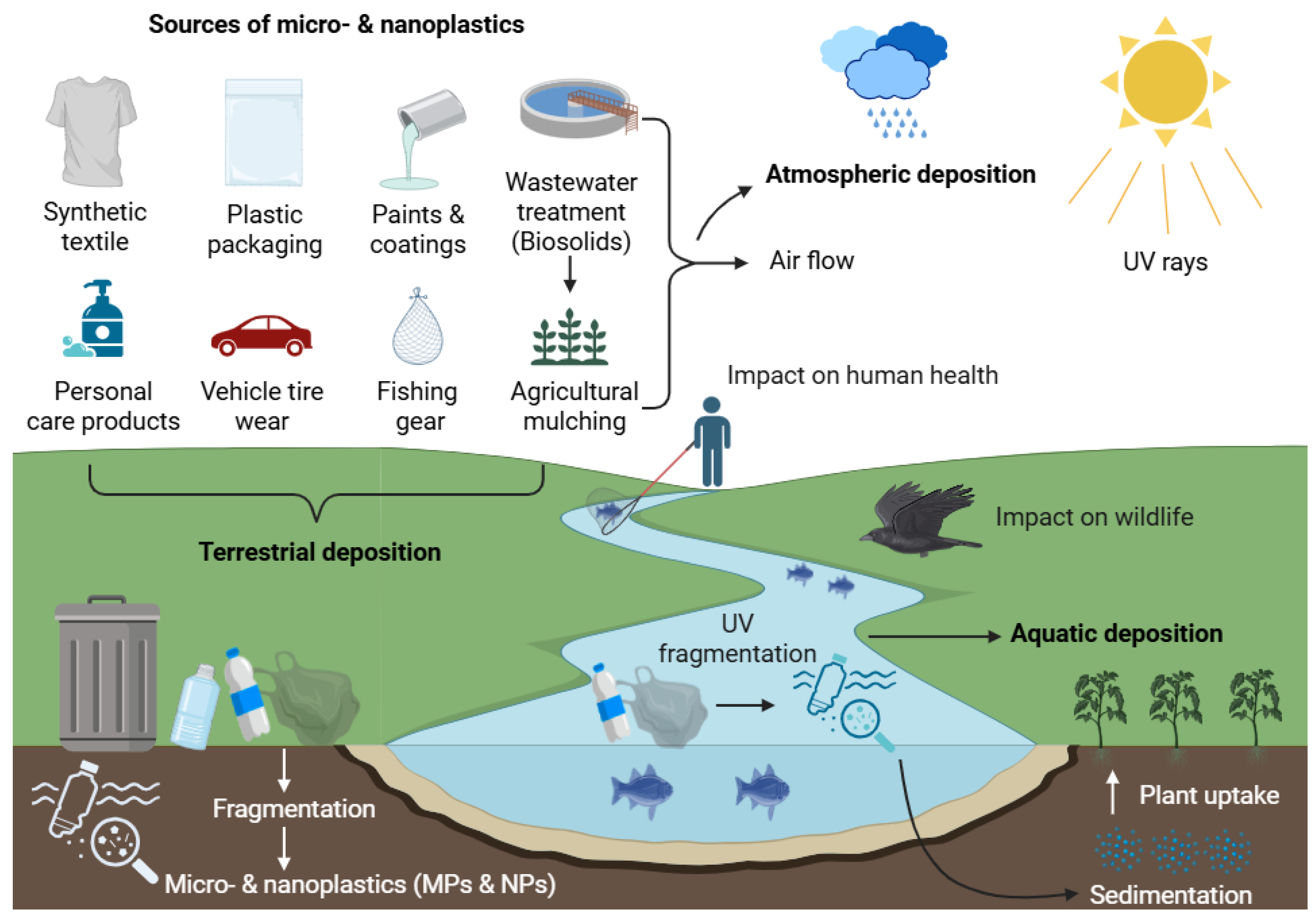
Figure 1. Sources, transport, and environmental distribution of MNPs.
Applsci 15 08813 g001
Figure 2. Overview of MNPs exposure pathways to humans and associated health impacts.
Applsci 15 08813 g002
Figure 3. Mechanistic map of MNP toxicity to human cells.
Applsci 15 08813 g003
Figure 4. Summary of the main adverse effects of MNPs on major human organ systems.
Applsci 15 08813 g004
Table 2. Human health effects of MNPs by physiological system.
Physiological SystemHuman Model/SampleMNPs Type and SizeExposureKey Health EffectsMechanistic PathwaysRef.
RespiratoryAlveolar epithelial cells (A549)PS-NPs (25 nm, 70 nm)1.14–25 μg/mL, 24–48 h↓ Cell viability; ↑ apoptosis; ↑ IL-8, TNF-α, and NF-κBCaspase activation, cytochrome c release, and pro-inflammatory signalling[113]
Lung epithelial cells (BEAS-2B)PS-MPs (1 μm)1–1000 μg/cm2, 24–48 hROS-induced cytotoxicity; ↓ transepithelial resistance; and ↑ COPD riskOxidative stress; barrier dysfunction[117,121]
Pulmonary alveolar cells (HPAEpIC)PS-NPs (40 nm)24 h↓ Cell viability; ↑ MMP-9; and surfactant protein A dysregulationRedox imbalance; apoptotic pathway activation[121]
DigestiveColon adenocarcinoma cells (Caco-2, HT29)PE-MPs (1–10 μm)21 mg/8 mL, 14 days↑ Pathobionts (e.g., Enterobacteriaceae); ↓ beneficial bacteria (e.g., Akkermansia)Gut dysbiosis, biofilm formation, and intestinal homeostasis loss[122]
Simulated gastrointestinal modelPET-MPsSingle dose (0.166 g), 72 hPET biotransformation; altered colonic microbiotaMicrobial adhesion, polymer degradation[123]
Colon cells + gut microbiotaPE-MPs100–1000 mg/L, 24–48 h↓ Gut diversity and metabolic disruptionROS-induced barrier dysfunction; immune activation[124]
CardiovascularEmbryonic stem cells (hiPSCs)PS-NPs (40 nm)1 × 10⁹/mL, 24 hImpaired heart valve development; ↓ LEFTY1/2; and ↑ CA4, OCLMDevelopmental gene dysregulation[125]
Red blood cells (RBCs)PS-NPs (50–250 nm)50–500 μg/mL, 1 hHemolysis (plasma-free medium)Membrane disruption[126]
Human umbilical vein endothelial cells (HUVECs)PS-NPs (100 nm, 500 nm)5–100 μg/mL↑ LDH release, membrane damage, and autophagyER stress; autophagosome formation[127]
NervousForebrain cortical spheroids (3D model)PS-MPs (1 μm, 10 μm)5–100 μg/mL, 4–30 days↓ Neuronal maturation; ↓ cortical layer VI markersAltered Nestin, PAX6, and HOXB4 expression[128]
ReproductivePlacenta/meconium (mother–infant pairs)PA, PU MPs (>20 μm)Clinical detectionMicrobiota dysbiosis (Proteobacteria ↑); inverse PE-microbiota correlationEpigenetic modulation, microbial translocation[129]
Testicular cells (NTE)PS-MPs (50 nm)200 μg/mL, 24 h↑ JNK1/2/3, TNF-α; dysregulated PI3K-AKT pathwayOxidative stress, pro-inflammatory signalling[114]
Note: ↑ indicates an increase or upregulation of the parameter; ↓ indicates a decrease or downregulation of the parameter.
Disclaimer/Publisher’s Note: The statements, opinions and data contained in all publications are solely those of the individual author(s) and contributor(s) and not of MDPI and/or the editor(s). MDPI and/or the editor(s) disclaim responsibility for any injury to people or property resulting from any ideas, methods, instructions or products referred to in the content.