Article

Effects of Different Titanium Anodized Surfaces on Peri-Implant Soft Tissue Healing Around Dental Abutments: In Vitro and Proteomic Study

by
Francisco Romero-Gavilán
1,
Andreia Cerqueira
1,
Carlos Arias-Mainer
1,
David Peñarrocha-Oltra
2,
Claudia Salavert-Martínez
2,
Juan Carlos Bernabeu-Mira
2,*,
Iñaki García-Arnáez
3,
Félix Elortza
4,
Mariló Gurruchaga
3,
Isabel Goñi
3 and
Julio Suay
1
1
Department of Industrial Systems Engineering and Design, Universitat Jaume I, Avda. Vicent Sos Baynat s/n, 12071 Castellón de la Plana, Spain
2
Oral Surgery Unit, Department of Stomatology, Faculty of Medicine and Dentistry, University of Valencia, C/Gascó Oliag 1, 46010 Valencia, Spain
3
Departament of Polymers and Advanced Materials: Physics, Chemistry and Technology, Universidad del País Vasco, P. M. de Lardizábal 3, 20018 San Sebastián, Spain
4
Proteomics Platform, CIC bioGUNE, Basque Research and Technology Alliance (BRTA), CIBERehd, Bizkaia Science and Technology Park, 48160 Derio, Spain
*
Author to whom correspondence should be addressed.
Appl. Sci. 2025, 15(13), 7349; https://doi.org/10.3390/app15137349
Submission received: 17 April 2025 / Revised: 23 May 2025 / Accepted: 28 May 2025 / Published: 30 June 2025
(This article belongs to the Special Issue Application of Advanced Therapies in Oral Health)

Abstract

Objectives: This study aimed to evaluate the effects of different titanium (Ti) anodized surfaces on soft tissue healing around dental implant abutments. Methods: Discs of machined (MC), pink anodized (PA) and yellow anodized (YA) surfaces were morphologically characterized and evaluated in vitro. Cell adhesion and collagen synthesis by human gingival fibroblasts (hGFs) were assessed to evaluate the regenerative potential of the surfaces under study. Their inflammatory potential was evaluated in THP-1 cell cultures by measuring cytokine secretion, and their proteomic adsorption patterns were characterized using nano-liquid chromatography mass spectrometry (nLC-MS/MS). Statistical significance was considered at 5%. In relation to proteomics, statistical differences were evaluated using the Student t-test with the Perseus application. Results: The anodization process resulted in a reduction in the surface roughness parameter (Ra) relative to the machined titanium (p < 0.05). No differences in hGF adhesion were found between the surfaces after one day. PA induced increased hGF collagen synthesis after 7 days (p < 0.05). The secretion of TNF-α was lower for anodized surfaces than for MC, and its concentration was lower for PA than for YA (p < 0.05). In turn, TGF-β was higher for PA and YA versus MC after one and three days of culture. A total of 176 distinct proteins were identified and 26 showed differences in adhesion between the anodized surfaces and MC. These differential proteins were related to coagulation, lipid metabolism, transport activity, plasminogen activation and a reduction in the immune response. Conclusions: Anodized Ti surfaces showed promising anti-inflammatory and regenerative potential for use in dental implant abutments. Anodization reduced surface roughness, increased collagen synthesis and lowered TNF-α secretion while increasing TGF-β levels compared to machined surfaces. Identified proteins related to coagulation and lipid metabolism supported these findings. Clinical relevance: Anodized surfaces could offer improved short-term peri-implant soft tissue healing over machined surfaces. The analysis of abutment surface, instead of implant surface, is a new approach that can provide valuable information.

1. Introduction

Soft tissue integration is a challenge in implant-prosthetic treatment, and attention has shifted to the portion between the peri-implant soft tissue and the transmucosal component [1,2]. Although titanium-based implants facilitate osseointegration with the host bone [3], the limited number of gingival fibroblasts around implants may reduce the healing and regeneration of the connective tissue, with the risk of bacterial exposure [4].
Only a small proportion of gingival fibroblasts (GFs) are found in the tissues around implants (3–5%), with significantly lower cell counts than around healthy periodontal connective tissue (approximately 15%) [5]. The limited GFs may reduce the healing and regeneration of the connective tissue (6–12 weeks are normally needed for peri-implant connective tissue maturation), which can result in bacterial exposure. In sum, soft tissue integration around Ti implants is mainly achieved through the physical “adaptation” of the peri-implant mucosa, rather than through biological “integration” around the natural tissue (such as teeth), which forms an effective barrier protecting the underlying structures [6]. The lack or weakness of transmucosal sealing can cause the rapid progression of peri-implant diseases; consequently, the application of surface treatments that enhance the “adaptation” of the soft tissues may represent an effective clinical strategy for the long-term prevention of peri-implant diseases [5].
Cellular functions can be enhanced and modulated by surface modifications on a macro-, micro- and nano-scale [7]. Electrochemical anodization to produce controlled TiO2 nanotubes/nanopores seems to be a popular technique for augmenting the bioactivity of Ti-based orthopedic and dental implants [8]. However, further research is needed to understand the effect of such surface treatments upon the soft tissues [9].
The biological response to an implanted device is determined by the interaction between the implant materials and the tissues in contact with them. This response is controlled by multiple regenerative pathways that condition the implantation outcome depending on the activation or/and development of immune, coagulation, angiogenic or collagen synthesis processes [10]. The contact of the implant with proteins from tissues and fluids leads to the adsorption of a protein layer on the implant surface [11]. The material surface parameters, such as wettability, roughness or chemistry, play a key role in this protein adsorption phenomenon [12]. Likewise, the activation of regenerative processes and the formation of the provisional extracellular matrix are determined by the protein layer, with the subsequent induction of cellular and tissue responses [13,14].
The aim of this study was to evaluate the potential of Ti anodized surfaces to improve soft tissue healing around dental implant abutments. For this purpose, the anodized surfaces were evaluated in vitro with both macrophages and fibroblasts to analyze their regenerative and inflammatory potentials. Additionally, to achieve a better understanding of how these dental abutment surfaces affect the initial soft tissue regenerative process, the proteomic adsorption patterns on the anodized Ti surfaces were characterized using nLC-MS/MS. The null hypothesis underlying this study is that the anodization of Ti surfaces does not produce any significant differences in biological responses, in particular, in cell behavior (inflammatory or regenerative) or in protein adsorption patterns, compared to non-anodized control surfaces.

2. Materials and Methods

2.1. Sample Preparation

Titanium grade 5 (Ti6Al4V) samples were machined into a disc shape (6 mm in diameter). The samples were then degreased and subsequently rinsed with purified water in an ultrasonic bath. Then, the machined discs were anodized under two different conditions, resulting in pink and yellow anodized surfaces (PA and YA, respectively) (Galimplant S.L.U, Sarria, Galicia, Spain). The anodizing process was carried out following the established procedures. Initially, cleaning of the surface was carried out, eliminating the natural layer of oxide and subsequently immersing the disc in an electrolytic cell containing a solution based on phosphoric acid at two given energy intensities, generating two different titanium oxide thicknesses, which, upon the reflection of light, cause the human eye to identify different colors (pink and yellow, respectively). At the end of the process, the samples were rinsed in deionized water for two minutes and washed with deionized water in an ultrasonic bath for 20 min. Machined (non-anodized) samples (MC) were used as controls.

2.2. Morphological Characterization

The morphology of the surface treatments was studied using a Leica-Zeiss LEO scanning electron microscope (SEM) (Leica, Wetzlar, Germany). Platinum sputtering was applied to increase the sample conductivity for SEM examination. The surface roughness was measured with an optical profilometer (PLm2300, Sensofar, Barcelona, Spain) operating in a confocal mode to obtain a 3D image. A 20× objective was used, providing a field of view of 240 × 180 µm. Three discs were evaluated for each surface treatment, performing three measurements for each disc. The results were displayed as the arithmetic average roughness (Ra).

2.3. In Vitro Experimentation

2.3.1. Cell Cultures

The peri-implant soft tissue regenerative potential of these anodized treatments was evaluated using human gingival fibroblasts (hGFs) (LGC Standards, Barcelona, Spain). The fibroblasts were maintained in an incubator at 37 °C (5% CO2, 95% H2O) in Fibroblast Basal Medium (ATCC, American Type Culture Collection; Manassas, VA, USA) supplemented with 5 ng mL−1 rhFGF b, 7.5 mM L-glutamine, 50 µg mL−1 ascorbic acid, 1 µg mL−1 hydrocortisone hemisuccinate, 5 µg mL−1 rh insulin, 2% FBS and 1% penicillin/streptomycin (ATCC). The inflammatory potential was assessed with human monocytes (THP-1) (ECACC, Public Health England, Porton Down, Salisbury, UK) cultured in RPMI-1640 medium (10% FBS and 1% penicillin/streptomycin; Merck, Waltham, MA, USA) and incubated at 37 °C (5% CO2, 95% H2O). The differentiation of THP-1 into macrophages was performed through phorbol 12-myristate 13-acetate (PMA; Merck). In both cases, the culture medium was replaced every two days. Cultures without samples and MC discs were used as controls.

2.3.2. Cell Adhesion

The capability of the studied surfaces to promote cell adhesion was evaluated by analyzing the hGF cytoskeleton arrangement. For this purpose, the cells were seeded onto the samples at a density of 10 × 103 cells cm−2 in 96-well plates (Corning Inc.; Somerville, MA, USA). After 1 and 3 days of culture, the cell adhesion assay was performed following the methodology previously described in [15]. Briefly, hGFs were washed with phosphate-buffered saline (PBS) solution and then fixed with 4% paraformaldehyde (Merck). Then, the cells were permeabilized with 0.1% Triton X-100, incubated with phalloidin (1:100; Abcam, Cambridge, UK) and diluted in 0.1% w/v bovine serum albumin (BSA)-PBS for one hour. Subsequently, the nuclei were stained with a mounting medium with DAPI (Abcam) for 5 min. Fluorescence was measured using a Leica TCS SP8 Confocal Laser Scanning Microscope (Leica). The images were obtained using LAS X software (Leica) and analyzed with Image J 2.0.0 software (National Institutes of Health; Bethesda, MD, USA). The experiment was performed in triplicate, and 30 photographs were taken for each condition to carry out the cell area measurements.

2.3.3. Collagen Synthesis

The amount of collagen secreted by hGF on the different surfaces was evaluated by Sirius Red (SR) staining in saturated picric acid (SR; Merck). Cells were seeded onto the samples in 96-well plates at a density of 20 × 103, 10 × 103 and 5 × 10³ cells cm−2 for 1, 3 and 7 days, respectively. At the corresponding timepoint, the cells were fixed with 4% paraformaldehyde (Merck) for 20 min. After washing with PBS, the samples were incubated with the SR solution for 24 h. The dye residues were washed with water and then the samples were treated with 1M NaOH (Merck) to extract the stain. Absorbance at 570 nm was measured in a Multiskan FC microplate reader (Thermo Fisher Scientific, Waltham, MA, USA). The experiment was carried out in triplicate.

2.3.4. Inflammatory Potential: Cytokine Secretion

The inflammatory potential of the anodized treatments was evaluated in vitro using THP-1 cells, which were cultured on the discs in 96-well plates at densities of 3 × 10⁵ and 1.5 × 10⁴ for one and three days, respectively. After these times, the culture media were collected for the measurement of both tumor necrosis factor α (TNF-α) and transforming growth factor β (TGF-β) cytokine concentrations. For this purpose, TNF-α and TGF-β ELISA kits (ref. 88-7346-88 and 88-8350-88, respectively; Invitrogen, Thermo Fisher Scientific) were used, following the instructions of the manufacturer. The assays were performed in quadruplicate.

2.4. Proteomics

2.4.1. Protein Layer Elution

Samples were incubated with 0.2 µL of human serum (from male AB plasma; Merck) for three hours (37 °C, 5% CO2) in 96-well plates. Non-adsorbed proteins were removed by rinsing the samples five times with ddH2O and once with 100 mM NaCl 50 mM Tris–HCl (pH 7.0) buffer. The adsorbed proteins were eluted using 2 M thiourea, 7 M urea, 4% CHAPS and 200 mM dithiothreitol solution. The experiment was performed in quadruplicate, and each replicate was obtained by pooling the protein layer eluates from four samples. All the reagents were purchased from Merck.

2.4.2. nLC-MS/MS Measurements and Bioinformatics Analyses

The eluted protein layers were analyzed using an Evosep ONE chromatograph (Evosep, Odense C, Denmark) coupled to a hybrid trapped ion mobility-quadrupole time of flight mass spectrometer (timsTOF Pro with PASEF; Bruker, Billerica, MA, USA). Each condition was analyzed in quadruplicate. Protein identification was performed with MaxQuant software (v2.0.3.0) (http://maxquant.org/). Differential analyses between the proteomic profiles identified for the anodized surfaces and the control (MC) were carried out by using the Perseus platform (https://www.maxquant.org/perseus/) (accessed on 8 February 2022). The functional analyses of differential proteins were carried out with PANTHER (http://www.pantherdb.org/) and the UniProt database (https://www.uniprot.org/). In addition, STRING v.11.5 (Search Tool for the Retrieval of Interacting Genes/Proteins; https://string-db.org/) (accessed on 16 December 2022) was used to generate protein–protein interaction networks, connecting the linked proteins to their associated gene ontology (GO) terms. The UniProt ID codes were utilized as protein names.

2.5. Statistical Analysis

GraphPad Prism® version 5.04 (GraphPad Software Inc., La Jolla, CA, USA) was used to analyze the effects of the anodizing treatments, using one-way analysis of variance (ANOVA) with the non-parametric Kruskal–Wallis test.
Statistical significance was considered for p < 0.05. In relation to proteomics, statistical differences were calculated using the Student t-test with the Perseus software (v1.6). Protein adsorption between conditions was considered differential when the difference proved statistically significant (p ≤ 0.05) and the ratio of abundance for these conditions was greater than 1.5 in either direction.

3. Results

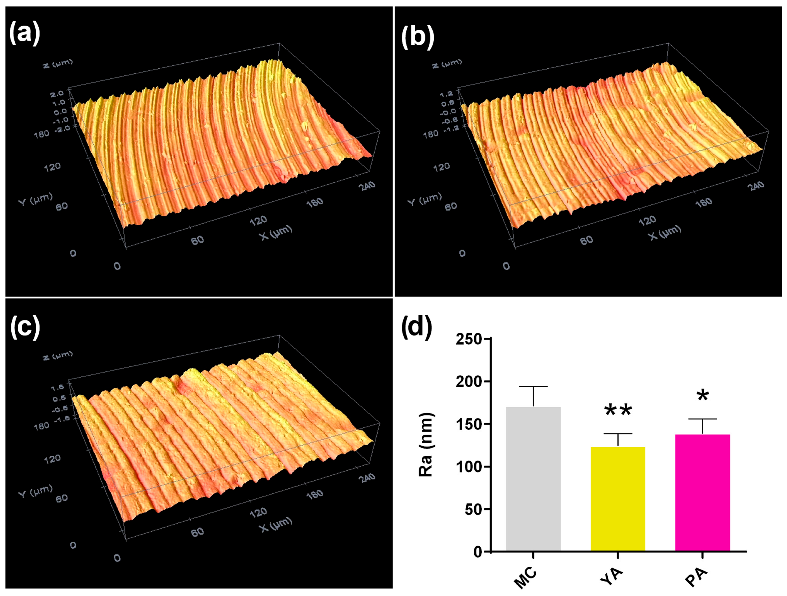
3.1. Morphological Characterization

Figure 1 shows the SEM micrographs of the initial Ti machined surface (Figure 1a), and this same surface after performing both types of anodizing (YA, Figure 1b; PA, Figure 1c). Grooves resulting from the machining process can be observed in the MC. On anodizing with both the YA and PA treatments, the formation of titanium oxide gives rise to the blurring of these initial grooves. In addition, with YA anodizing, this attenuation of the grooves on a micrometer scale is greater than in PA, which is likely associated with the greater formation of TiO2. On the nanometer scale (Figure 1a’–c’), the anodized surfaces are seen to be considerably smoother compared to MC.
In agreement with the SEM findings, the analysis of the surfaces with an optical profilometer showed the attenuation of the morphological irregularities, with a significant decrease in roughness after the anodizing of the machined Ti discs (Figure 2). In fact, the average Ra values measured for MC, YA and PA are 171, 125 and 139 nm, respectively.

3.2. In Vitro Experimentation

3.2.1. Cytoskeletal Arrangement and Collagen Secretion

The cytoskeleton organization of the hGF was evaluated after one and three days of culture (Figure 3a) for the anodized samples and MC. The cells cultured on the three surfaces had elongated shapes and protruding lamellipodia, showing good adhesion for all samples. In addition, fibroblasts were seen to be grown following the geometrical pattern which was present in all the samples. Fibroblast area measurements after one day of culture showed no differences in cell adhesion between the surfaces (Figure 3b). Regarding the collagen synthesized by hGF, increased collagen secretion was recorded for PA after 7 days of culture (Figure 3c).

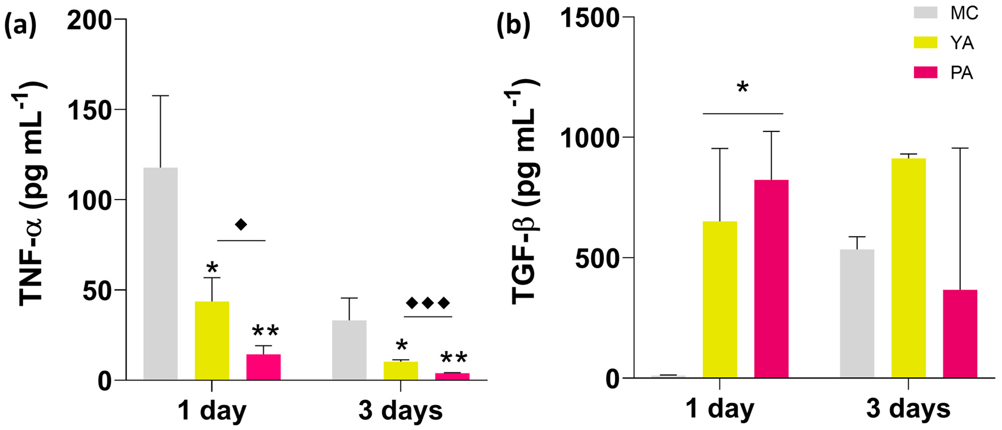
3.2.2. Inflammatory Potential: Cytokine Quantification by ELISA

To evaluate the effect of the anodization process upon the inflammatory response compared to the control (MC), the secretion of pro- and anti-inflammatory cytokines (TNF-α and TGF-β, respectively) by THP-1 macrophages was quantified at one and three days (Figure 4). TNF-α secretion showed a significant decrease in those cells cultured on anodized surfaces versus MC at one and three days. Additionally, the measured TNF-α concentration was lower for PA than YA. In the case of TGF-β, secretion increased for PA and YA versus MC after one and three days of culture, respectively.

3.3. Proteomic Analysis

The nLC–MS/MS analysis identified 176 distinct proteins adhered to the studied surfaces. Perseus comparative analyses were performed between the anodized treatments (YA and PA) with respect to the control surface (MC), revealing that 26 proteins differentially adhered onto the materials due to anodizing (Table 1). Ten proteins were found to be differentially more adsorbed onto YA versus MC, whereas another ten were less adsorbed. In PA, 13 proteins increased their affinity for this surface versus MC, and 10 reduced their affinity. Based on the Uniprot database, among the proteins differentially more adsorbed on the anodized surfaces, FA10, ZPI, HRG, KLKB1 and PLMN were associated with blood coagulation functions. In turn, APOF and APOL1 apolipoproteins, also found to be more adsorbed secondary to anodizing, were related to lipid metabolism and transport activity. Proteins related to the immune response were also found to be more adsorbed onto PA and YA. In this regard, immunoglobulin HV313, pentraxin SAMP and IC1 and C4BPB (with regulatory/inhibitory functions in the complement system) were differentially attached onto these surfaces. TENT, A2AP and IBP4 also increased their affinity for the anodized samples. TENT is an extracellular matrix protein that can promote plasminogen activation, whereas A2P2, a serine-type endopeptidase inhibitor, is related to the regulatory functions of this process. IBP4 in turn is an insulin growth factor (IGF)-binding protein that can promote the IGF and MAPK pathways. Regarding the proteins that reduced their affinity for the anodized samples, FA11, HABP2 and FA5 were associated with blood coagulation functions. Apolipoproteins APOM, APOC4 and APOE, and the high-density lipoprotein SAA4, were also found to be differentially less adhered to the PA and YA surfaces. The same applied to DSG1, with functions in cell-to-cell adhesion, and FHR5, S10A9, G3P and DCD, related to the immune response.
Bioinformatics tools were used to carry out the functional analysis of the proteins identified as differentially adsorbed between surfaces. In Figure 5, the Panther functional classification and the String analyses results are displayed. Panther revealed that proteins differentially more and less adsorbed onto anodized samples were related to biological regulation, localization, multicellular organismal processes and cellular and metabolic functions (Figure 5a,a’). Upregulated proteins were additionally associated with biological processes involved in interspecies interactions between organisms, responses to a stimulus, developmental processes and, in a small percentage, with biomineralization, signaling, growth and immune system functions. In turn, in the classification of proteins with less affinity for anodized surfaces, locomotion and biological adhesion functions were found. Regarding the Panther classification based on pathways, proteins more adsorbed onto anodized surfaces were linked to blood coagulation and the plasminogen activation cascades (Figure 5b). On the other hand, glycolysis and blood coagulation were the pathways related to the proteins with a greater affinity for MC (Figure 5b’). Two interaction networks were detected between the proteins most adsorbed onto PA and YA by the String analysis (Figure 5c). One was composed of two apolipoproteins and the other, larger network was composed of proteins with functions in blood coagulation, fibrinolysis and the complement system. The proteins with reduced affinity formed two interaction networks (Figure 5c’), one of them composed mainly of the proteins associated with blood coagulation.

4. Discussion

The insertion of a dental implant implies the introduction of a foreign body into a living host and leads to immediate tissue damage and cell disruption resulting from the surgical procedure [16]. Blood protein adsorption onto the surface of the implant initiates tissue healing by activating the immune response [17]. To promote proper tissue regeneration, suitable fibroblast activity without chronic inflammation is required. The collagen produced by fibroblasts is crucial to form a correct extracellular matrix (ECM), and acute proinflammatory environments adversely affect the ECM quality required for tissue regeneration [18]. The in vivo biocompatibility of the biomaterial and its tissue regeneration capacity depend on the modulation of immune cells, which are driven by complement system proteins [19]. In this sense, the composition, conformation and number of proteins bound onto the abutment surfaces can lead to distinct biological responses depending on the specific surface characteristics.
In this study, two distinct anodized Ti treatments (YA and PA) were studied. The anodization process led to morphological differences between the YA and PA surfaces versus the control (MC), as the oxidation attenuated the initial grooves typical of the machining process. This fact was supported by the roughness measurements, since a mild decrease in the Ra parameter was observed for the anodized surfaces versus MC. Yanagisawa et al. [20] found gingival connective tissue compatibility to be constant within the smooth-classified surface roughness category, as different Ti-treated surfaces with distinct Ra values (0.075, 0.217, 0.671 and 1.024 μm) showed no significant differences in human oral fibroblast attachment. In concordance with the present study, the measured decrease in Ra within this range did not impair hGF adhesion onto the Ti anodized surface.
The proteomic characterization of the protein layers adsorbed onto the anodized surfaces identified 26 proteins differentially attached onto these treated surfaces versus the control (MC). A total of 14 proteins increased their adsorption after anodization (UP), while another 12 reduced their adsorption (DOWN). It should be highlighted that the anodization of the Ti abutment implied a significant change in the adsorption of proteins associated with coagulation functions. In fact, the protein that showed the greatest increase in adsorption was coagulation factor X (FA10), which was the first factor of the common coagulation pathway [21]. During hemostasis, FA10 was converted into factor Xa (FA10a), which finally led to the conversion of prothrombin into thrombin, following the formation of fibrin and the blood clot [22]. FA10a stimulated fibroblast procollagen production, proliferation and calcium signaling [23]. In this sense, the in vitro evaluation of collagen synthesis by human fibroblasts cultured onto the anodized surfaces showed a significant increase for PA after 7 days—this being the treatment showing the highest affinity for FA10 versus MC (7.7-fold higher). The anodized surfaces, in addition to having a greater adsorption of FA10, showed an increased affinity for other coagulation-related proteins such as KLKB1, ZPI, HRG and PLMN, as well as a decrease for coagulation pathway factors FA5 and FA11. KLKB1 played a role in the activation of the intrinsic coagulation pathway through the surface-activating coagulation system [24]. On the other hand, HRG and ZPI were associated with a regulatory role during blood clot formation. HRG was related to both anticoagulant and antifibrinolytic functions, being able to regulate platelet function in vivo [25]. ZPI is a serpin family protein protease inhibitor with anticoagulant functions, which, in conjunction with its cofactor (protein Z), can inhibit prothrombinase-bound FA10a during prothrombin activation [26]. After clot formation, fibrinolysis causes fibrin degradation, leading proper tissue repair. PLMN plays a key role in fibrinolysis, and its activation to plasmin allows fibrin matrix degradation and remodeling to achieve wound healing [27]. In addition, TENT, which enhances plasminogen activation and is related to the wound tissue healing process [28], also shows increased affinity for anodized surfaces. These results are supported by the Panther analysis, in which proteins differentially more adsorbed onto anodized surfaces were related to blood coagulation and plasminogen activating cascades. In addition, an interaction network including molecules with functions in fibrinolysis and blood coagulation was identified among the UP-differential proteins by the STRING analysis. As has been demonstrated in previous works, the differential adsorption of blood coagulation-related proteins onto a biomaterial can condition clot formation around that surface [13]. The fibrin clot serves as a matrix for cytokines and growth factors that “jump start” wound healing, and provides a scaffold for the chemoattraction of inflammatory cells, the initiation of cell movements and connective tissue contraction [29].
Regarding the effect of anodizing treatments on the abutment immune response, the levels of TNF-α secreted by the THP-1 cells, related to the M1 proinflammatory macrophage phenotype, showed a significant decrease for anodized surfaces (especially PA) when compared to MC. At the same time, TGF-β, which exerts strong anti-inflammatory action, increased its secretion in those cells cultured on anodized surfaces. This cytokine is associated with M2 or tissue reparative macrophage phenotype polarization, being a key regulator of myofibroblast differentiation, wound contraction and remodeling [29]. Of note in the proteomics results was the great adsorption of SAMP, IC1 and C4BPB on PA. IC1 and C4BPB are known regulators/inhibitors of the complement system pathway and are associated with anti-inflammatory functions [30]. On the other hand, the role of pentraxin SAMP on the regenerative process around biomaterials is still unclear, but it has been associated with inflammation, tissue remodeling and coagulation [31]. Pilling and Gomer [32] found that SAMP reduced the accumulation of inflammatory macrophages and prevented fibrosis in vivo. This, together with the lower adsorption of proinflammatory proteins on the anodized surfaces, correlated with the in vitro findings. Moreover, insulin growth factor (IGF)-1 activity is regulated by the IGF binding proteins; here, IBP4, which inhibits the activity of IGF-1 and is related to antifibrotic activity [33,34], showed a greater adherence onto PA versus MC (5.4-fold higher). Renier et al. [20] demonstrated that IGF-I is a monocyte/macrophage activating factor that enhances production, so its IBP4-related inhibition might explain the significant decrease in the in vitro TNF-α release observed for PA. This favoring of anodized surfaces towards M2 macrophage phenotype polarization ensures the resolution of inflammation and the optimal transition to the proliferative phase of wound healing [29].

5. Conclusions

Within the limitations of this study, the morphological, cellular and proteomic analyses corresponding to the different Ti anodized surfaces of the dental implant abutments appeared to show a better short-term healing response of the peri-implant soft tissues than in the case of the machined Ti surface (MC). Pink anodized surfaces showed a lower immune response than the other treatments, accompanied by increased hGF collagen synthesis. Additional in vivo and clinical studies would be required to apply these findings.

Author Contributions

Conceptualization, M.G., I.G., J.S. and D.P.-O.; Data Curation, F.R.-G., A.C., C.A.-M., C.S.-M., J.C.B.-M. and I.G.-A.; Investigation, F.R.-G., A.C. and F.E.; Formal analysis, F.R.-G., C.A.-M., I.G.-A. and F.E.; Writing—Original Draft, F.R.-G.; Writing—Review & Editing, F.R.-G., C.S.-M. and J.C.B.-M.; Funding acquisition, D.P.-O., M.G., I.G. and J.S.; Project administration, D.P.-O., M.G., I.G. and J.S. All authors have read and agreed to the published version of the manuscript.

Funding

This work was supported by the Science and Innovation Ministry of Spain [PDC2021-120924-I00/AEI/10.13039/501100011033/Unión Europea NextGenerationEU/PRTR]; Universitat Jaume I [UJI-B2021-25]; and the Basque Government [MARSA21/07]. Andreia Cerqueira was supported by the Margarita Salas postdoctoral contract MGS/2022/10 (UP2022-024) financed by the European Union-NextGenerationEU.

Data Availability Statement

Data are contained within the article.

Acknowledgments

The authors would like to thank Raquel Oliver, José Ortega and Iraide Escobés for their valuable technical assistance.

Conflicts of Interest

The authors do not declare any conflicts of interest.

References

  1. Corvino, E.; Pesce, P.; Mura, R.; Marcano, E.; Canullo, L. Influence of Modified Titanium Abutment Surface on Peri-implant Soft Tissue Behavior: A Systematic Review of In Vitro Studies. Int. J. Oral Maxillofac. Implant. 2020, 35, 503–519. [Google Scholar] [CrossRef] [PubMed]
  2. Canullo, L.; Annunziata, M.; Pesce, P.; Tommasato, G.; Nastri, L.; Guida, L. Influence of abutment material and modifications on peri-implant soft-tissue attachment: A systematic review and meta- analysis of histological animal studies. J. Prosthet. Dent. 2021, 125, 426–436. [Google Scholar] [CrossRef] [PubMed]
  3. Williams, D.F. Titanium for Medical Applications. In Titanium in Medicine: Material Science, Surface Science, Engineering, Biological Responses and Medical Applications; Brunette, D.M., Tengvall, P., Textor, M., Thomsen, P., Eds.; Springer: Berlin, Germany, 2001; pp. 12–24. [Google Scholar]
  4. Kordbacheh Changi, K.; Finkelstein, J.; Papapanou, P.N. Peri-implantitis prevalence, incidence rate, and risk factors: A study of electronic health records at a U.S. dental school. Clin. Oral Implant. Res. 2019, 30, 306–314. [Google Scholar] [CrossRef] [PubMed]
  5. Al Rezk, F.; Trimpou, G.; Lauer, H.C.; Weigl, P.; Krockow, N. Response of soft tissue to different abutment materials with different surface topographies: A review of the literature. Gen. Dent. 2018, 66, 18–25. [Google Scholar]
  6. Amberg, R.; Elad, A.; Rothamel, D.; Fienitz, T.; Szakacs, G.; Heilmann, S.; Witte, F. Design of a migration assay for human gingival fibroblasts on biodegradable magnesium surfaces. Acta Biomater. 2018, 79, 158–167. [Google Scholar] [CrossRef]
  7. Guo, T.; Gulati, K.; Arora, H.; Han, P.; Fournier, B.; Ivanovski, S. Orchestrating soft tissue integration at the transmucosal region of titanium implants. Acta Biomater. 2021, 124, 33–49. [Google Scholar] [CrossRef]
  8. Gulati, K.; Kogawa, M.; Maher, S.; Atkins, G.; Findlay, D.; Losic, D. Titania Nanotubes for Local Drug Delivery from Implant Surfaces. In Electrochemically Engineered Nanoporous Materials: Methods, Properties and Applications; Losic, D., Santos, A., Eds.; Springer International Publishing: Cham, Switzerland, 2015; pp. 307–355. [Google Scholar]
  9. Cinquini, C.; Marchio, V.; Di Donna, E.; Alfonsi, F.; Derchi, G.; Nisi, M.; Barone, A. Histologic Evaluation of Soft Tissues around Dental Implant Abutments: A Narrative Review. Materials 2022, 15, 3811. [Google Scholar] [CrossRef]
  10. Lyu, Z.; Yu, Q.; Chen, H. Interactions of Biomaterials Surfaces with Proteins and Cells. In Polymeric Biomaterials for Tissue Regeneration: From Surface/Interface Design to 3D Constructs; Gao, C., Ed.; Springer: Singapore, 2016; pp. 103–122. [Google Scholar] [CrossRef]
  11. Othman, Z.; Cillero Pastor, B.; van Rijt, S.; Habibovic, P. Understanding interactions between biomaterials and biological systems using proteomics. Biomaterials 2018, 167, 191–204. [Google Scholar] [CrossRef]
  12. Romero-Gavilán, F.; Gomes, N.C.; Ródenas, J.; Sánchez, A.; Azkargorta, M.; Iloro, I.; Elortza, F.; Arnáez, I.G.; Gurruchaga, M.; Goñi, I.; et al. Proteome analysis of human serum proteins adsorbed onto different titanium surfaces used in dental implants. Biofouling 2017, 33, 98–111. [Google Scholar] [CrossRef]
  13. Romero-Gavilán, F.; Cerqueira, A.; Anitua, E.; Tejero, R.; García-Arnáez, I.; Martinez-Ramos, C.; Ozturan, S.; Izquierdo, R.; Azkargorta, M.; Elortza, F.; et al. Protein adsorption/desorption dynamics on Ca-enriched titanium surfaces: Biological implications. J. Biol. Inorg. Chem. 2021, 26, 715–726. [Google Scholar] [CrossRef]
  14. Romero-Gavilán, F.; Sanchez-Pérez, A.M.; Araújo-Gomes, N.; Azkargorta, M.; Iloro, I.; Elortza, F.; Gurruchaga, M.; Goñi, I.; Suay, J. Proteomic analysis of silica hybrid sol-gel coatings: A potential tool for predicting the biocompatibility of implants in vivo. Biofouling 2017, 33, 676–689. [Google Scholar] [CrossRef] [PubMed]
  15. Cerqueira, A.; Romero-Gavilán, F.; García-Arnáez, I.; Martinez-Ramos, C.; Ozturan, S.; Izquierdo, R.; Azkargorta, M.; Elortza, F.; Gurruchaga, M.; Suay, J.; et al. Characterization of magnesium doped sol-gel biomaterial for bone tissue regeneration: The effect of Mg ion in protein adsorption. Mater. Sci. Eng. Mater. Biol. Appl. C 2021, 125, 112114. [Google Scholar] [CrossRef] [PubMed]
  16. von Wilmowsky, C.; Moest, T.; Nkenke, E.; Stelzle, F.; Schlegel, K.A. Implants in bone: Part I. A current overview about tissue response, surface modifications and future perspectives. Oral Maxillofac. Surg. 2014, 18, 243–257. [Google Scholar] [CrossRef] [PubMed]
  17. Romero-Gavilán, F.; Araújo-Gomes, N.; Cerqueira, A.; García-Arnáez, I.; Martínez-Ramos, C.; Azkargorta, M.; Iloro, I.; Elortza, F.; Gurruchaga, M.; Suay, J.; et al. Proteomic analysis of calcium-enriched sol–gel biomaterials. J. Biol. Inorg. Chem. 2019, 24, 563–574. [Google Scholar] [CrossRef]
  18. Stunova, A.; Vistejnova, L. Dermal fibroblasts—A heterogeneous population with regulatory function in wound healing. Cytokine Growth Factor Rev. 2018, 39, 137–150. [Google Scholar] [CrossRef]
  19. Araújo-Gomes, N.; Romero-Gavilán, F.; Zhang, Y.; Martinez-Ramos, C.; Elortza, F.; Azkargorta, M.; de Llano, J.M.; Gurruchaga, M.; Goñi, I.; Beucken, J.v.D.; et al. Complement proteins regulating macrophage polarisation on biomaterials. Colloids Surf. B Biointerfaces 2019, 181, 125–133. [Google Scholar] [CrossRef]
  20. Renier, G.; Clément, I.; Desfaits, A.C.; Lambert, A. Direct stimulatory effect of insulin-like growth factor-I on monocyte and macrophage tumor necrosis factor-α production. Endocrinology 1996, 137, 4611–4618. [Google Scholar] [CrossRef] [PubMed]
  21. Johari, V.; Loke, C. Brief Overview of the Coagulation Cascade. Dis. Mon. 2012, 58, 421–423. [Google Scholar] [CrossRef]
  22. Brummel-Ziedins, K.; Mann, K.G. Molecular Basis of Blood Coagulation. In Hematology, 7th ed.; Elsevier Inc.: Amsterdam, The Netherlands, 2018; pp. 1885–1905.e8. [Google Scholar]
  23. Blanc-Brude, O.P.; Archer, F.; Leoni, P.; Derian, C.; Bolsover, S.; Laurent, G.J.; Chambers, R. Factor Xa stimulates fibroblast procollagen production, proliferation, and calcium signaling via PAR1 activation. Exp. Cell Res. 2005, 304, 16–27. [Google Scholar] [CrossRef]
  24. Schmaier, A.H.; McCrae, K.R. The plasma kallikrein-kinin system: Its evolution from contact activation. J. Thromb. Haemost. 2007, 5, 2323–2329. [Google Scholar] [CrossRef]
  25. Tsuchida-Straeten, N.; Ensslen, S.; Schäfer, C.; Wöltje, M.; Denecke, B.; Moser, M.; Gräber, S.; Wakabayashi, S.; Koide, T.; Jahnen-Dechent, W. Enhanced blood coagulation and fibrinolysis in mice lacking histidine-rich glycoprotein (HRG). J. Thromb. Haemost. 2007, 3, 865–872. [Google Scholar] [CrossRef] [PubMed]
  26. Huang, X.; Swanson, R.; Kroh, H.K.; Bock, P.E. Protein Z-dependent protease inhibitor (ZPI) is a physiologically significant inhibitor of prothrombinase function. J. Biol. Chem. 2019, 294, 7644–7657. [Google Scholar] [CrossRef] [PubMed]
  27. Miles, L.A.; Ny, L.; Wilczynska, M.; Shen, Y.; Ny, T.; Parmer, R.J. Plasminogen receptors and fibrinolysis. Int. J. Mol. Sci. 2021, 22, 1712. [Google Scholar] [CrossRef]
  28. Iba, K.; Hatakeyama, N.; Kojima, T.; Murata, M.; Matsumura, T.; Wewer, U.M.; Wada, T.; Sawada, N.; Yamashita, T. Impaired cutaneous wound healing in mice lacking tetranectin. Wound Repair Regen. 2009, 17, 108–112. [Google Scholar] [CrossRef]
  29. Opneja, A.; Kapoor, S.; Stavrou, E.X. Contribution of platelets, the coagulation and fibrinolytic systems to cutaneous wound healing. Thromb. Res. 2009, 179, 56–63. [Google Scholar] [CrossRef] [PubMed]
  30. Zipfel, P.F.; Skerka, C. Complement regulators and inhibitory proteins. Nat. Rev. Immunol. 2009, 9, 729–740. [Google Scholar] [CrossRef]
  31. Bottazzi, B.; Inforzato, A.; Messa, M.; Barbagallo, M.; Magrini, E.; Garlanda, C.; Mantovani, A. The pentraxins PTX3 and SAP in innate immunity, regulation of inflammation and tissue remodelling. J. Hepatol. 2016, 64, 1416–1427. [Google Scholar] [CrossRef]
  32. Pilling, D.; Gomer, R.H. Persistent lung inflammation and fibrosis in serum amyloid P component (Apcs-/-) knockout mice. PLoS ONE 2014, 9, 29–33. [Google Scholar] [CrossRef]
  33. Su, Y.Y.; Nishimoto, T.; Hoffman, S.; Nguyen, X.X.; Pilewski, J.M.; Feghali-Bostwick, C. Insulin-like growth factor binding protein-4 exerts antifibrotic activity by reducing levels of connective tissue growth factor and the C-X-C chemokine receptor 4. FASEB Bioadv. 2019, 1, 167–179. [Google Scholar] [CrossRef]
  34. Smith, Y.E.; Toomey, S.; Napoletano, S.; Kirwan, G.; Schadow, C.; Chubb, A.J.; Mikkelsen, J.H.; Oxvig, C.; Harmey, J.H. Recombinant PAPP-A resistant insulin-like growth factor binding protein 4 (dBP4) inhibits angiogenesis and metastasis in a murine model of breast cancer. BMC Cancer 2018, 18, 1016. [Google Scholar] [CrossRef]
Figure 1. Scanning electron micrograph of (a) MC, (b) YA and (c) PA surfaces. Scale bars, 10 µm (ac) and 1 µm (a’c’).
Applsci 15 07349 g001
Figure 2. Optical profilometer measurements for (a) MC, (b) YA and (c) PA. Arithmetic average of roughness results ((d); Ra). Bars indicate standard deviations. Statistical analysis was performed using one-way ANOVA with the Tukey post hoc test, showing significant differences for anodized samples versus MC (*, p < 0.05; **, p < 0.01).
Applsci 15 07349 g002
Figure 3. Confocal fluorescence images of the cytoskeletal arrangement of hGF on MC, YA and PA surfaces after one and three days of culture (a). Actin filaments were stained with phalloidin (green) and nuclei with DAPI (blue). Scale bar: 100 μm. Area measurements of the fibroblasts adhering to the different samples after one day (b). Collagen secreted by hGF cells after 1, 3 and 7 days of culture on the different surface treatments (c). Asterisks (p ≤ 0.05 (*)) indicate a statistically significant difference for PA versus MC, and diamonds indicate statistically significant differences between PA and YA (p ≤ 0.05 (♦)). Results are shown as mean ± SE.
Applsci 15 07349 g003
Figure 4. Cytokine quantification by ELISA in THP-1 at one and three days: (a) transforming growth factor beta (TGF-β) and (b) tumor necrosis α (TNF-α). The asterisks (p ≤ 0.05 (*) and p ≤ 0.01 (**)) indicate statistically significant differences between PA and YA versus MC, and the diamonds indicate statistically significant differences between PA and YA (p ≤ 0.05 (♦) and p ≤ 0.001 (♦♦♦)). Results are shown as mean ± SD.
Applsci 15 07349 g004
Figure 5. Panther classification of (a) biological functions and (b) pathways associated with the UP (a,b) and DOWN (a’,b’) proteins differentially adsorbed onto anodized surfaces versus MC. String interaction networks between proteins differentially more (c) and less (c’) adsorbed onto anodized surfaces versus MC. Line thickness indicates interaction strength and colors are related to the protein functional classification.
Applsci 15 07349 g005
Table 1. Perseus comparative analysis between proteins adsorbed onto the anodized surfaces (YA and PA) versus MC. Proteins with p ≤ 0.05 (yellow) and a ratio greater than 1.5 (in either direction) are considered as significantly different (boldface). Proteins differentially more adsorbed (UP) are marked in red, while those less adsorbed (DOWN) appear in green.
YA/MCPA/MC
AccessionDescriptionProtein NameUnique
Peptides
p ValueRatiop ValueRatio
P00742FA10Coagulation factor X97.7 × 10−44.81.4 × 10−37.7
P22692IBP4Insulin-like growth factor-
binding protein 4
26.7 × 10−23.92.5 × 10−25.4
Q9UK55ZPIProtein Z-dependent protease inhibitor79.6 × 10−34.26.7 × 10−35.1
P01766HV313Immunoglobulin heavy
variable 3-13
12.4 × 10−25.12.8 × 10−24.5
P05452TETNTetranectin91.2 × 10−42.37.1 × 10−62.8
P08697A2APAlpha-2-antiplasmin36.6 × 10−32.61.4 × 10−22.2
P04196HRGHistidine-rich glycoprotein151.8 × 10−31.91.2 × 10−31.9
P03952KLKB1Plasma kallikrein166.0 × 10−51.85.6 × 10−41.8
P00747PLMNPlasminogen369.4 × 10−61.93.9 × 1051.7
P20851C4BPBC4b-binding protein beta chain51.5 × 10−21.51.6 × 10−31.7
P02743SAMPSerum amyloid P-component104.6 × 10−41.57.3 × 1051.7
O14791APOL1Apolipoprotein L165.9 × 10−41.71.0 × 10−31.6
P05155IC1Plasma protease C1 inhibitor124.6 × 10−31.32.6 × 10−31.5
Q13790APOFApolipoprotein F31.4 × 10−21.69.8 × 10−11.0
O95445APOMApolipoprotein M48.3 × 10−30.67.8 × 10−10.9
P55056APOC4Apolipoprotein C-IV41.2 × 10−20.62.2 × 10−10.8
P03951FA11Coagulation factor XI319.5 × 10−11.01.2 × 10−20.7
Q9BXR6FHR5Complement factor H-related protein 551.7 × 10−20.59.0 × 10−20.6
P81605DCDDermcidin56.3 × 10−30.53.1 × 10−20.5
Q02413DSG1Desmoglein-1143.8 × 10−20.23.4 × 10−10.5
P02649APOEApolipoprotein E271.9 × 10−30.52.1 × 10−40.4
Q14520HABP2Hyaluronan-binding protein 2161.8 × 10−40.47.7 × 10−40.4
P35542SAA4Serum amyloid A-4 protein49.4 × 10−50.42.4 × 1050.4
P12259FA5Coagulation factor V62.7 × 10−20.38.5 × 1050.3
P06702S10A9Protein S100-A959.8 × 10−20.22.4 × 10−20.2
P04406G3PGlyceraldehyde-3-phosphate
dehydrogenase
54.0 × 10−30.39.6 × 10−40.2
Disclaimer/Publisher’s Note: The statements, opinions and data contained in all publications are solely those of the individual author(s) and contributor(s) and not of MDPI and/or the editor(s). MDPI and/or the editor(s) disclaim responsibility for any injury to people or property resulting from any ideas, methods, instructions or products referred to in the content.

