Abstract
In recent decades, there has been an increase in the development of non-invasive and non-destructive analytical techniques in the field of cultural heritage. The present study aims to characterize the frescoes in the hypogeum environment of the San Pietro a Corte complex in Salerno (Campania, Italy) through a multi-analytical approach that couples Infrared Reflectography with X-Ray Fluorescence Spectrometry. Thermographic and hygrometric measurements were also performed to evaluate their state of conservation in relation to environmental parameters such as relative humidity and temperature at the frescoed walls. Spectroscopic investigations revealed a predominant use of natural pigments—chiefly iron-rich earths—and uncovered details invisible to the naked eye that aid art historians in refining stylistic attributions. Hygrometric data showed that the central zones of the frescoes retain the highest moisture levels, underscoring the need for a carefully tailored conservation plan. Overall, this multi-analytical methodology provides important information that enables conservators and restorers to understand both the materials and the preservation requirements of these artworks from a scientific and conservation perspective.
1. Introduction and Historical Background
The San Pietro a Corte (SPC) Complex is located in the historical center of Salerno (Campania, Italy). Since its construction, in the eighth century AD, its chapel has had diverse uses (e.g., a place for graduation ceremonies of the Salerno Medical School). Six meters below the current street level, there are two different environments: one in the east side containing a Paleochristian Church, which previously was a temple of Priapus, having some wall frescoes, and another in the west consisting of the thermal structure of a frigidarium pool [1]. Figure 1 shows the location of the two better preserved frescoes inside the hypogeum environment of SPC: the fresco “Madonna in trono con bambino”, representing the Madonna on the throne with the Child on the right and S. Caterina d’Alessandria on the left, is highlighted in yellow; the “Teoria di Santi” fresco, depicting the Virgin seated on a throne with Jesus, S. Giacomo, S. Pietro, S. Caterina d’Alessandria, and an unidentified saint, is highlighted in green [2].
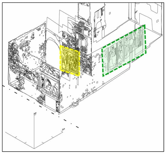
Figure 1.
Location of the frescoes inside the hypogeum (from left to right): “Madonna in trono con bambino” in yellow, and “Teoria di Santi” in green.
The SPC complex has been the subject of recent studies due to problems related to the conservation of these frescoes, exposed to phenomena of salt crystallization and efflorescence [2,3]. It is well known that the main factors in the deterioration of stone materials and wall paintings located in enclosed spaces are temperature excursions, water and humidity [4,5,6], soluble salts [7,8,9,10,11], and microorganisms [12,13,14]. High humidity values over long periods of time cause alternating cycles of hydration and dehydration, which, in the presence of soluble salts, lead to solubilization and recrystallisation phenomena, resulting in an increase in pressure within the material to the point of damaging part of its structure [15]. In the SPC hypogeum, saltpeter efflorescences of a biological origin were found, probably due to the infiltration of wastewater from nearby houses [2]. The high humidity of the air found in the complex and the presence of water infiltrations make the conservation of its works of art very difficult. In fact, the signs of degradation due to these phenomena are clearly evident: frescoes are completely covered by salt efflorescences as shown in Figure 2 (for comparison the red square highlights the area of the fresco that has been cleaned with a soft brush).
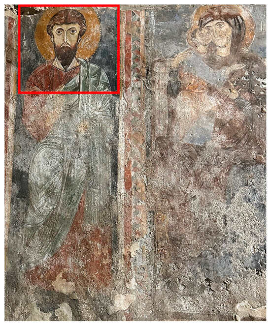
Figure 2.
Photo of a portion of “Teoria di Santi” fresco, where salt efflorescences are evident; the red square highlights the area of the fresco that has been cleaned with a soft brush.
From a stylistic viewpoint, these frescoes, dating back to the mid-twelfth to thirteenth century AD, have a pictorial matrix related to Byzantine culture. Although medieval artists generally had access to a wide palette, the hues employed in mural painting were necessarily more restricted: the alkaline wall matrix (usually slaked lime with sand or ground marble as fillers) is too aggressive toward most organic pigments, limiting painters to inorganic colorants [16]. Consequently, accurate identification of materials and techniques is critical for selecting appropriate restoration treatments and for devising preventive−conservation strategies [17,18,19]. Because the frescoes are already in a fragile state, the study relies on non-destructive analytical methods, now widely favored in cultural-heritage research precisely to avoid additional damage [20,21,22]. Rapid progress over the past decade has expanded the range and sensitivity of such techniques [23,24,25,26,27,28,29]. Portable instrumentation is particularly valuable: it allows for in situ characterization of raw materials and decay products, and pinpoints areas suitable for micro-sampling when laboratory study is indispensable [30,31].
The present work focuses on the characterization of the fresco paintings of the hypogeum environment of the SPC complex applying a multi-analytical protocol. Infrared Reflectography (IR-R) and X-ray Fluorescence (XRF) spectroscopy were used to map pigments and under-drawings, while thermographic and hygrometric surveys revealed moisture gradients and structural discontinuities along the painted surfaces. This integrated screening offers an initial picture of the frescoes’ conservation state [32], highlights zones that may warrant deeper diagnostics [33,34], and provides essential baseline data for future interventions.
2. Materials and Methods
Thermographic analyses were carried out inside the frigidarium environment, specifically surveying the walls at the two investigated frescoes. A FLIR T540 thermal imaging camera was used and the following parameters were set: Field of view: 25° × 19°; Minimum focusing distance: 0.40 m; Thermal sensitivity: 0.045 °C to + 30 °C; IR resolution: 320 × 240 pixels; Temperature range: −20 °C to +350 °C.
Hygrometric investigations were carried out with an MO257 moisture meter from EXTECH Instruments (Boston, MA, USA), which detects relative humidity in contact with the surface by means of a spherical probe avoiding surface damage. The measuring points identified on the surface of the fresco “Madonna in trono con bambino” are arranged along a vertical band measuring 20 × 200 cm, in correspondence with the figure of the Madonna on the right side of the fresco, measuring the humidity values every 20 cm. The values measured with the hygrometer were then processed with the software IC Capture 2.4 from the Imaging Source Europe Gmbh (Bremen, Germany), which made it possible to obtain a ‘mapping’ of the humidity distributed on the surface (hygrometric tomography).
Infrared Reflectography (IR-R) analyses were carried out with an IRIS DIGITAL BASIC instrument from Bresciani S.r.l. (Milano, Italy) consisting of an objective with interchangeable pass-band filter lenses, capable of recording responses in the infrared spectral range up to 1100 nm. The lenses are interchangeable and were used in sequence (715 nm; 850 nm; 1000 nm) to detect the presence of any retouches or pre-paintings carried out on the “Madonna in trono con bambino” fresco.
X-Ray Fluorescence (XRF) Spectrometry measurements for elemental pigment analysis on the two frescoes (Madonna in trono con Bambino” and “Teoria di Santi”) were performed using a Hitachi X-MET 8000 Expert GEO handheld XRF spectrometer (Hitachi High-Tech Analytical Science, Tokyo, Japan), equipped with a Rh anode X-ray tube. The following acquisition parameters were set:
- -
- Light element detection: voltage of 8–15 kV, current of 100–150 µA;
- -
- Detection of heavy elements: voltage of 15–45 kV, current of 100–33 µA;
- -
- Acquisition time equal to 30 s;
- -
- Spot size equal to 3 mm.
Figure 3 shows the measuring points for the two frescos.
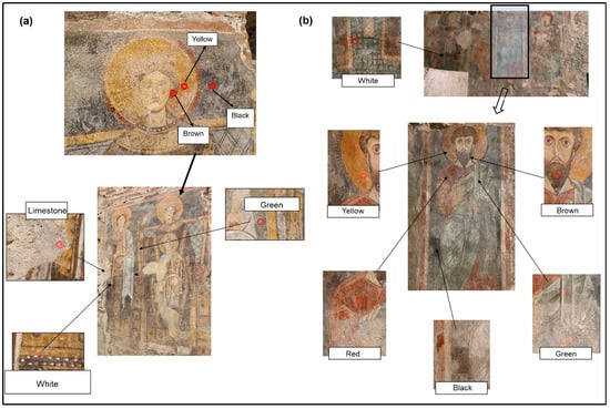
Figure 3.
Measuring points of XRF analysis on the frescoes: (a) “Madonna in trono con bambino” and (b) “Teoria di Santi”. Red circles indicate the exact point of measurement.
3. Results
3.1. Characterization of Temperature and Humidity on the Fresco Walls
Thermographic and hygrometric surveys were carried out inside the frigidarium, targeting the sections of wall that bear frescoes.
Because the chamber lies as much as 6 m below ground level and remains perennially humid, surface temperatures proved almost uniform, varying only from 18.9 up to 19.6 °C (ΔT = 0.7). This negligible gradient rendered infrared imaging uninformative, so no thermograms are presented.
In contrast, the in situ moisture readings collected with a contact hygrometer were post-processed to create false-color maps of water content (hygrometric tomograms) across each painted surface. Figure 4 shows the hygrometric tomography of the fresco “Madonna in trono con Bambino”.
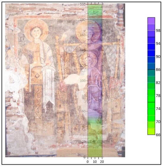
Figure 4.
Hygrometric tomography of the fresco “Madonna in trono con Bambino”.
As can be seen from the image shown in Figure 4 and the relative humidity scale in false colors, the investigated fresco is affected by a widespread phenomenon of surface humidity. The phenomenon is especially visible in the central and upper areas of the fresco (highlighted by the colors turning to blue−violet), which represents a state of maximum saturation. A similar trend had been observed for the fresco “Teoria di Santi”, with higher humidity values in the central part of the fresco.
3.2. Spectroscopic Investigations
IR-R was used on the fresco “Madonna in trono con Bambino” using different filters (715 nm–850 nm–1000 nm) to reveal possible hidden details, such as retouches, underdrawings and pentimenti, beneath the surface of the fresco. The main results of these measurement are reported in Figure 5, with images focused on the area where the Child Jesus is present (details of its face, the hand of the Madonna, their robes, and part of the throne where they are seated).
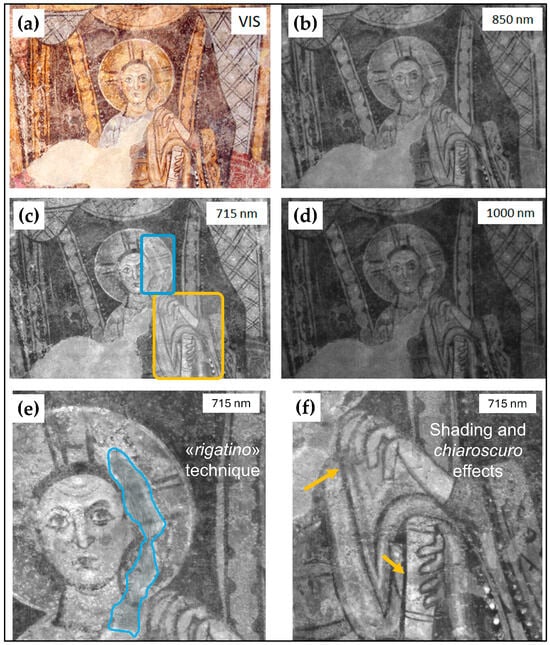
Figure 5.
IR-R images obtained with different filters: (a) Visible; (b) 850 nm; (c) 715 nm; (d) 1000 nm. The magnifications of areas marked in blue and yellow in the image (c) are also shown to highlight the details observed with IR-R: area marked in blue for rigatino technique (e) and yellow arrows for shading and chiaroscuro effect (f).
Because superficial salt efflorescences are largely transparent in the infrared, whereas the pigments absorb in that region, IR imaging yields a far clearer reading of the fresco—an essential step before any cleaning intervention. In the 715 nm band (Figure 5c), subtle surface modelling becomes evident, such as the nuanced shading of the Madonna’s hand resting on Jesus’ shoulder (contoured in Figure 5f with yellow arrows), revealing a more elaborate execution than can be discerned with the naked eye. The presence of thin shading details and chiaroscuro effects (Figure 5f) are visible only on this specific wavelength (715 nm), suggesting that the artist was involved in a more sophisticated and accurate compositional process. The detection of these nuances through imaging techniques reveals that the artist intentionally used light and shadow to enhance the realism and depth of the figures. IR-R technique exposes areas of past reintegration and retouching (e.g., the rigatino restoration technique for the halo of Jesus, contoured in blue in Figure 5c,e). Conversely, no pentimenti or preparatory under-drawings were detected at the longer wavelengths typically used to reveal them (850 nm, 1000 nm).
Elemental analysis by portable X-ray fluorescence (XRF) was carried out on the two most representative and best-preserved frescoes in the hypogeum—”Madonna in trono con bambino” and “Teoria di Santi”. The resulting spectra, presented in Figure 6 and Figure 7, include measurements on both the lime-based substrate and the principal color areas (white, yellow, red, brown, green, and black).
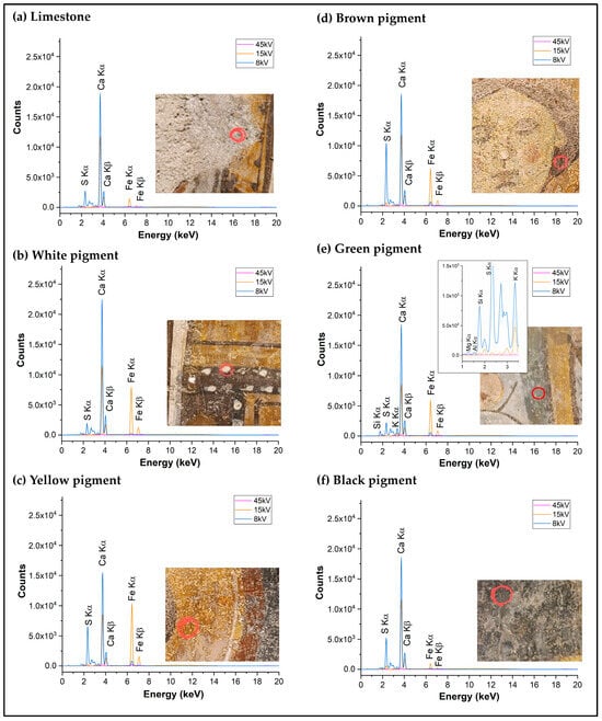
Figure 6.
XRF spectra of the fresco “Madonna in trono con bambino” measured at the following points: (a) limestone; (b) white pigment; (c) yellow pigment; (d) brown pigment; (e) green pigment with a magnification of the area between 1 and 3.5 keV; (f) black pigment. The bottom right of each spectrum shows the image of the measurement point: red circles indicate the exact point of measurement. The overlayed spectra (blue, orange, and magenta) are referred to the different voltages (8, 15, 45 kV) used for the detection of light and heavy elements.
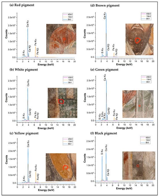
Figure 7.
XRF spectra of the fresco “Teoria di Santi” measured at the following points: (a) red pigment; (b) white pigment; (c) yellow pigment; (d) brown pigment; (e) green pigment; (f) black pigment. The bottom right of each spectrum shows the image of the measurement point: red circles indicate the exact point of measurement. The overlayed spectra (blue, orange, and magenta) are referred to the different voltages (8, 15, and 45 kV) used for the detection of light and heavy elements.
By comparing the data obtained with the XRF database for pigments [35], the various pigments were assigned as shown in Table 1. From the analysis of the lime substrate (Figure 6a), in addition to calcium, iron, and sulfur were also found, the former probably due to the use of small quantities of iron-based earths mixed with the lime matrix, and the latter due to the presence of both sulfate-based salt efflorescence and gypsum. For this reason, a contribution of these elements was always observed in the subsequent pigment analyses.

Table 1.
Result of XRF analysis: elemental composition of the pigment from “Madonna in trono con bambino” and “Teoria di Santi”.
The XRF survey on the white pigments of both frescoes shows calcium as the dominant element, with only minor iron and sulfur. Because no lead, tin, barium, zinc, or titanium were detected, the use of lead white, tin white, barium-based “fixed” white, zinc white, or titanium white can be ruled out. The white layer is therefore consistent with chalk, i.e., calcium carbonate (CaCO3). Yellow, brown, and red areas all display markedly higher iron than the lime substrate, plus variable calcium and sulfur that originate from the plaster and salt efflorescences rather than the pigments themselves. The absence of cadmium and silicon excludes cadmium yellow (CdS) and enamel yellows (SiO2 + CdS). The iron abundance instead points to iron-oxide earths: yellow = yellow ochre (limonite, FeO(OH)), brown = burnt sienna (limonite with hematite), and red = red ochre (hematite, Fe2O3·nH2O). The green strokes exhibit the same iron signature but with detectable Si, K, Mg, and Al. The presence of low amounts of silicon and potassium and traces of magnesium and aluminum (see the magnification in Figure 6e) can be attributed to the use of Green Earth (silico-aluminates of Fe2+/Fe3+). The black pigment, on the other hand, is the only organic pigment detected. Calcium, sulfur, and traces of strontium are the only elements detected; as carbon itself is invisible to XRF, this indicates the possible presence of charcoal black.
4. Discussion
The multi-analytical protocol adopted here—relying exclusively on non-destructive techniques—yields critical information on both the pigment palette and the conservation state of the hypogeum frescoes in the San Pietro a Corte complex (Salerno, Italy), thereby giving conservators a sound scientific basis for future interventions.
Thermographic and hygrometric surveys reveal a pervasive surface-moisture problem: relative-humidity values approach 100% in the central zones of the paintings. Such readings are consistent with the microclimatic conditions of this subterranean space, where high ambient humidity is readily absorbed by the porous plaster.
Infrared reflectography revealed interesting technical and iconographic details of the representation not directly perceptible to the naked eye [36] (mainly due to high occurrence of saline efflorescences on fresco surface): the presence of a shading (attributable to a more elaborate execution) and signs of painting integration and restoration on the “Madonna in trono con bambino fresco” can be observed. The improved legibility of the figure of the Madonna could allow for comparative studies with other contemporary examples in southern Italy [17].
The results of the XRF analysis, carried out on the main colors of the representation (white, red, yellow, brown, green, and black), are in line with those reported in the literature for other medieval frescoes discovered in southern Italy [7,17,37,38]: the palette is dominated by naturally occurring earth pigments—long prized for availability, chromatic range, and compatibility with the alkaline fresco technique [37]. Calcium (from the lime plaster) and sulfur (from both sulphate efflorescences and sulfation of paint layers) are ubiquitous. The white pigment shows a composition similar to that of the substrate; therefore, it has been identified as chalk, in line with data reported in similar literature studies [17,37,38,39]. The only organic pigment found in this study is the black one that can be attributed to the use of charcoal black. The detection of iron, associated with its oxides and hydroxides, as the main element for the red, yellow, brown, and green colors, suggest the use of red and yellow ochres, burnt sienna and green earth [37,40,41]. Such earthy pigments (red, yellow, brown, and green) were usually employed thanks to their complete compatibility for the fresco technique [42,43].
As observed in other hypogeum contexts [7,17], the inherent durability of these inorganic pigments has preserved the pictorial layers despite harsh environmental conditions. The greatest conservation threat remains the elevated humidity—particularly in the frescoes’ central areas, where it coincides with extensive salt efflorescence. The data obtained suggest that the areas most at risk are in the central portions of the frescoes. Taking action to decrease the moisture content within the site is not advisable, as this would facilitate the crystallization of currently dissolved salts (such as sulphates, which are more dangerous) causing further plaster detachments. Likely, the site is currently in a state of equilibrium, which requires a careful and delicate conservation plan in order to best protect it.
5. Conclusions
In this study, the fresco paintings of the hypogeum environment of the SPC complex (Salerno, Italy) were characterized using non-invasive analytical techniques: IR-R and portable XRF provided detailed mapping of the pictorial layers, while thermographic and hygrometric surveys documented temperature and moisture distribution across the walls.
XRF results indicate a palette dominated by natural earth pigments—red and yellow ochres, burnt sienna, and green earth—alongside chalk (calcite) for white passages and charcoal black as the sole organic pigment. IR-R exposed iconographic and technical details invisible to the naked eye, enriching stylistic comparisons with other regional works. Hygrometric mapping pinpointed the central zones of the murals as the most at-risk areas owing to elevated humidity, likely caused by infiltration, suggesting a careful and delicate conservation plan in order to best protect these frescoes.
Overall, the multi-analytical dataset offers conservators a robust scientific foundation for developing targeted, minimally invasive interventions, and long-term preventive strategies.
Author Contributions
Conceptualization, A.P., M.R., C.G. and E.C.; methodology, C.G.; software, C.G. and A.F.; validation, O.M., M.R. and C.N.; formal analysis, C.G.; investigation, C.G.; resources, R.F., M.R. and O.M.; data curation, C.G., A.F. and M.R.; writing—original draft preparation, M.R. and C.G.; writing—review and editing, M.R. and O.M.; visualization, R.F., C.N., E.C., O.M. and M.R.; supervision, E.C., O.M. and A.P.; project administration, E.C. and A.P.; funding acquisition, M.R. All authors have read and agreed to the published version of the manuscript.
Funding
This research was funded by the University of Salerno (FARB 2024: ORSA243323).
Institutional Review Board Statement
Not applicable.
Informed Consent Statement
Not applicable.
Data Availability Statement
Data available upon request.
Acknowledgments
The authors gratefully acknowledged Luciana Santimone for technical support.
Conflicts of Interest
Authors C.G., C.N. and E.C. were employed by the company Istemi S.r.l. The remaining authors declare that the research was conducted in the absence of any commercial or financial relationships that could be construed as a potential conflict of interest.
Abbreviations
The following abbreviations are used in this manuscript:
| IR-R | Infrared Reflectography |
| XRF | X-Ray Fluorescence Spectrometry |
| SPC | San Pietro a Corte |
References
- Complesso Monumentale Di San Pietro a Corte. Available online: https://cultura.comune.salerno.it/web/itinerari.aspx?url=Complesso-di-S-Pietro-a-Corte (accessed on 30 May 2025).
- Ricciardi, M.; Pironti, C.; Motta, O.; Fiorillo, R.; Camin, F.; Faggiano, A.; Proto, A. Investigations on Historical Monuments’ Deterioration through Chemical and Isotopic Analyses: An Italian Case Study. Environ. Sci. Pollut. Res. 2022, 29, 29409–29418. [Google Scholar] [CrossRef]
- Pironti, C.; Ricciardi, M.; Proto, A.; Cucciniello, R.; Fiorentino, A.; Fiorillo, R.; Motta, O. New Analytical Approach to Monitoring Air Quality in Historical Monuments through the Isotopic Ratio of CO2. Environ. Sci. Pollut. Res. 2022, 29, 29385–29390. [Google Scholar] [CrossRef]
- Frasca, F.; Verticchio, E.; Caratelli, A.; Bertolin, C.; Camuffo, D.; Siani, A.M. A Comprehensive Study of the Microclimate-Induced Conservation Risks in Hypogeal Sites: The Mithraeum of the Baths of Caracalla (Rome). Sensors 2020, 20, 3310. [Google Scholar] [CrossRef]
- Brunet, J.; Vouvé, J.; Malaurent, P. Conservation of Subterranean Historic and Prehistoric Monuments: The Importance of the Environment and Microclimate; Conservation of Ancient Sites on the Silk Road; Getty Conservation Institute: Los Angeles, CA, USA, 1997. [Google Scholar]
- Gómez-Laserna, O.; Prieto-Taboada, N.; Morillas, H.; Arrizabalaga, I.; Olazabal, M.Á.; Arana, G.; Madariaga, J.M. Analytical Study to Evaluate the Origin and Severity of Damage Caused by Salt Weathering in a Historical Palace House: The Attack of Infiltration Water. Anal. Methods 2015, 7, 4608–4615. [Google Scholar] [CrossRef]
- Zicarelli, M.A.; La Russa, M.F.; Alberghina, M.F.; Schiavone, S.; Greca, R.; Pogliani, P.; Ricca, M.; Ruffolo, S.A. A Multianalytical Investigation to Preserve Wall Paintings: A Case Study in a Hypogeum Environment. Materials 2023, 16, 1380. [Google Scholar] [CrossRef] [PubMed]
- Germinario, L.; Oguchi, C.T. Underground Salt Weathering of Heritage Stone: Lithological and Environmental Constraints on the Formation of Sulfate Efflorescences and Crusts. J. Cult. Herit. 2021, 49, 85–93. [Google Scholar] [CrossRef]
- Arnold, A.; Zehnder, K. Monitoring Wall Paintings Affected by Soluble Salts; The Conservation of Wall Paintings; Getty Conservation Institute: Los Angeles, CA, USA, 1991; pp. 103–136. [Google Scholar]
- Comite, V.; Bergomi, A.; Lombardi, C.A.; Borelli, M.; Fermo, P. Characterization of Soluble Salts on the Frescoes by Saturnino Gatti in the Church of San Panfilo in Villagrande Di Tornimparte (L’Aquila). Appl. Sci. 2023, 13, 6623. [Google Scholar] [CrossRef]
- Pironti, C.; Ricciardi, M.; Motta, O.; Venier, M.; Faggiano, A.; Cucciniello, R.; Proto, A. Sulphurous Air Pollutants and Exposure Events of Workers in Thermal-Mineral Springs: A Case Study of Contursi Terme (Salerno, Italy). Environ. Sci. Pollut. Res. 2023, 30, 3112–3120. [Google Scholar] [CrossRef]
- Madariaga, J.M. Analytical Chemistry in the Field of Cultural Heritage. Anal. Methods 2015, 7, 4848–4876. [Google Scholar] [CrossRef]
- Padovnik, A.; Bosiljkov, V.B.; Ropret, P.; Kosel, J. Influence of Methyl Cellulose in Injection Grout on Mould Growth on Mural Paintings—Preliminary Results. In Proceedings of the 6th Historic Mortars Conference (HMC), Ljubljana, Slovenia, 21–23 September 2022; Bokan Bosiljkov, V., Padovnik, A., Turk, T., Eds.; Conservation and Restoration of Historic Mortars and Masonry Structures. Springer Nature: Cham, Switzerland, 2023; pp. 608–620. [Google Scholar]
- Ljaljević Grbić, M.; Dimkić, I.; Janakiev, T.; Kosel, J.; Tavzes, Č.; Popović, S.; Knežević, A.; Legan, L.; Retko, K.; Ropret, P.; et al. Uncovering the Role of Autochthonous Deteriogenic Biofilm Community: Rožanec Mithraeum Monument (Slovenia). Microb. Ecol. 2024, 87, 87. [Google Scholar] [CrossRef]
- Flatt, R.J. Salt Damage in Porous Materials: How High Supersaturations Are Generated. J. Cryst. Growth 2002, 242, 435–454. [Google Scholar] [CrossRef]
- Murat, Z. Wall Paintings through the Ages: The Medieval Period (Italy, Twelfth to Fifteenth Century). Archaeol. Anthropol. Sci. 2021, 13, 191. [Google Scholar] [CrossRef]
- Ricca, M.; Alberghina, M.F.; Houreh, N.D.; Koca, A.S.; Schiavone, S.; La Russa, M.F.; Randazzo, L.; Ruffolo, S.A. Preliminary Study of the Mural Paintings of Sotterra Church in Paola (Cosenza, Italy). Materials 2022, 15, 3411. [Google Scholar] [CrossRef] [PubMed]
- Piovesan, R.; Siddall, R.; Mazzoli, C.; Nodari, L. The Temple of Venus (Pompeii): A Study of the Pigments and Painting Techniques. J. Archaeol. Sci. 2011, 38, 2633–2643. [Google Scholar] [CrossRef]
- D’Amico, S.; Comite, V.; Paladini, G.; Ricca, M.; Colica, E.; Galone, L.; Guido, S.; Mantella, G.; Crupi, V.; Majolino, D.; et al. Multitechnique Diagnostic Analysis and 3D Surveying Prior to the Restoration of St. Michael Defeating Evil Painting by Mattia Preti. Environ. Sci. Pollut. Res. 2022, 29, 29478–29497. [Google Scholar] [CrossRef]
- Amadori, M.L.; Barcelli, S.; Poldi, G.; Ferrucci, F.; Andreotti, A.; Baraldi, P.; Colombini, M.P. Invasive and Non-Invasive Analyses for Knowledge and Conservation of Roman Wall Paintings of the Villa of the Papyri in Herculaneum. Microchem. J. 2015, 118, 183–192. [Google Scholar] [CrossRef]
- Germinario, C.; Francesco, I.; Mercurio, M.; Langella, A.; Sali, D.; Kakoulli, I.; De Bonis, A.; Grifa, C. Multi-Analytical and Non-Invasive Characterization of the Polychromy of Wall Paintings at the Domus of Octavius Quartio in Pompeii. Eur. Phys. J. Plus 2018, 133, 359. [Google Scholar] [CrossRef]
- Gebremariam, K.F.; Kvittingen, L.; Banica, F.-G. Application of a Portable XRF Analyzer to Investigate the Medieval Wall Paintings of Yemrehanna Krestos Church, Ethiopia. X-Ray Spectrom. 2013, 42, 462–469. [Google Scholar] [CrossRef]
- Janssens, K.; Grieken, R.V. Non-Destructive Micro Analysis of Cultural Heritage Materials; Elsevier: Amsterdam, The Netherlands, 2004; ISBN 978-0-08-045442-9. [Google Scholar]
- Cheilakou, E.; Troullinos, M.; Koui, M. Identification of Pigments on Byzantine Wall Paintings from Crete (14th Century AD) Using Non-Invasive Fiber Optics Diffuse Reflectance Spectroscopy (FORS). J. Archaeol. Sci. 2014, 41, 541–555. [Google Scholar] [CrossRef]
- Crupi, V.; Fazio, B.; Fiocco, G.; Galli, G.; La Russa, M.F.; Licchelli, M.; Majolino, D.; Malagodi, M.; Ricca, M.; Ruffolo, S.A.; et al. Multi-Analytical Study of Roman Frescoes from Villa Dei Quintili (Rome, Italy). J. Archaeol. Sci. Rep. 2018, 21, 422–432. [Google Scholar] [CrossRef]
- Pitarch, À.; Ruiz, J.F.; de Vallejuelo, S.F.-O.; Hernanz, A.; Maguregui, M.; Madariaga, J.M. In Situ Characterization by Raman and X-Ray Fluorescence Spectroscopy of Post-Paleolithic Blackish Pictographs Exposed to the Open Air in Los Chaparros Shelter (Albalate Del Arzobispo, Teruel, Spain). Anal. Methods 2014, 6, 6641–6650. [Google Scholar] [CrossRef]
- Fermo, P.; Lombardi, C.A.; D’Amato, A.; Guglielmi, V.; Giudici, B.; Tomaino, A.; Pozzi, M.; Comite, V.; Bergomi, A.; Guardiano, L.; et al. Disclosing Colors and Pigments on Archaeological Objects from the Aga Khan Necropolis (West Aswan Egypt) through On-Site Analytical Methods: Preliminary Results. Heritage 2024, 7, 4980–4996. [Google Scholar] [CrossRef]
- Guglielmi, V.; Lombardi, C.A.; Fiocco, G.; Comite, V.; Bergomi, A.; Borelli, M.; Azzarone, M.; Malagodi, M.; Colella, M.; Fermo, P. Multi-Analytical Investigation on a Renaissance Polychrome Earthenware Attributed to Giovanni Antonio Amadeo. Appl. Sci. 2023, 13, 3924. [Google Scholar] [CrossRef]
- Guglielmi, V.; Comite, V.; Andreoli, M.; Demartin, F.; Lombardi, C.A.; Fermo, P. Pigments on Roman Wall Painting and Stucco Fragments from the Monte d’Oro Area (Rome): A Multi-Technique Approach. Appl. Sci. 2020, 10, 7121. [Google Scholar] [CrossRef]
- Costantini, I.; Castro, K.; Madariaga, J.M. Portable and Laboratory Analytical Instruments for the Study of Materials, Techniques and Environmental Impacts in Mediaeval Mural Paintings. Anal. Methods 2018, 10, 4854–4870. [Google Scholar] [CrossRef]
- Volpi, F.; Vagnini, M.; Vivani, R.; Malagodi, M.; Fiocco, G. Non-Invasive Identification of Red and Yellow Oxide and Sulfide Pigments in Wall-Paintings with Portable ER-FTIR Spectroscopy. J. Cult. Herit. 2023, 63, 158–168. [Google Scholar] [CrossRef]
- Caliano, E.; Messuti, N.; Napoli, C. The Diagnostic of Cultural Heritage: Active and Historical Researchdiscipline and Guardianship. The Case of the “Cenacolo” by Ghirlandaio of the Badia of Passignano in Florence; Associazione Italiana Prove Non Distruttive (AIPND): Milano, Italy, 2017. [Google Scholar]
- Caliano, E.; Gallo, C.; Messuti, N.; Napoli, C. Colors, Materials, and Techniques in Historical Buildings in Rome: Diagnostic Investigations and Case Studies. Stud. Conserv. 2023, 68, 365–379. [Google Scholar] [CrossRef]
- Gallo, C.; D’Agostino, L.; Messuti, N.; Napoli, C.; Quarta, M.; Valentini, D.; Caliano, E. Structural and Diagnostic Investigations on Materials and Plasters of the Biblioteca Umanistica of the University of Florence. In Proceedings of the International Conference Florence Heri-Tech: The Future of Heritage Science and Technologies, Florence, Italy, 16–18 May 2022; Furferi, R., Giorgi, R., Seymour, K., Pelagotti, A., Eds.; Springer International Publishing: Cham, Switzerland, 2022; pp. 183–195. [Google Scholar]
- Larsen, R.; Coluzzi, N.; Cosentino, A. Free XRF Spectroscopy Database of Pigments Checker. Int. J. Conserv. Sci. 2016, 7, 659–668. [Google Scholar]
- Daffara, C.; Fontana, R. Multispectral Infrared Reflectography to Differentiate Features in Paintings. Microsc. Microanal. 2011, 17, 691–695. [Google Scholar] [CrossRef]
- Pagano, S.; Germinario, C.; Alberghina, M.F.; Covolan, M.; Mercurio, M.; Musmeci, D.; Piovesan, R.; Santoriello, A.; Schiavone, S.; Grifa, C. Multilayer Technology of Decorated Plasters from the Domus of Marcus Vipsanus Primigenius at Abellinum (Campania Region, Southern Italy): An Analytical Approach. Minerals 2022, 12, 1487. [Google Scholar] [CrossRef]
- Salatino, G.; Zicarelli, M.A.; Ricca, M.; Macchia, A.; Randazzo, L.; Pogliani, P.; Arcudi, A.; Ruffolo, S.A.; La Russa, M.F. A Multi-Analytical Diagnostic on an Outdoor Wall Painting: The Study on the Déesis of St. Maria Annunziata’s Church, Motta San Giovanni (Reggio Calabria, Italy). Appl. Sci. 2023, 13, 10439. [Google Scholar] [CrossRef]
- Tsiapali, M.; Vivdenko, S.; Tsangalidis, H.; Konstanta, A.; Mitsos, D.; Mantzana, E.; Vasileiadou, A.; Zacharias, N. Archaeometric Investigation of Pigments of the Iconostasis from Saint Georgios Church of Sohos. J. Archaeol. Sci. Rep. 2023, 52, 104235. [Google Scholar] [CrossRef]
- Guglielmi, V.; Andreoli, M.; Comite, V.; Baroni, A.; Fermo, P. The Combined Use of SEM-EDX, Raman, ATR-FTIR and Visible Reflectance Techniques for the Characterisation of Roman Wall Painting Pigments from Monte d’Oro Area (Rome): An Insight into Red, Yellow and Pink Shades. Environ. Sci. Pollut. Res. 2022, 29, 29419–29437. [Google Scholar] [CrossRef] [PubMed]
- Hradil, D.; Grygar, T.; Hradilová, J.; Bezdička, P. Clay and Iron Oxide Pigments in the History of Painting. Appl. Clay Sci. 2003, 22, 223–236. [Google Scholar] [CrossRef]
- Vasco, G.; Serra, A.; Manno, D.; Buccolieri, G.; Calcagnile, L.; Buccolieri, A. Investigations of Byzantine Wall Paintings in the Abbey of Santa Maria Di Cerrate (Italy) in View of Their Restoration. Spectrochim. Acta Part A Mol. Biomol. Spectrosc. 2020, 239, 118557. [Google Scholar] [CrossRef]
- Mastrotheodoros, G.P.; Beltsios, K.G. Pigments—Iron-Based Red, Yellow, and Brown Ochres. Archaeol. Anthropol. Sci. 2022, 14, 35. [Google Scholar] [CrossRef]
Disclaimer/Publisher’s Note: The statements, opinions and data contained in all publications are solely those of the individual author(s) and contributor(s) and not of MDPI and/or the editor(s). MDPI and/or the editor(s) disclaim responsibility for any injury to people or property resulting from any ideas, methods, instructions or products referred to in the content. |
© 2025 by the authors. Licensee MDPI, Basel, Switzerland. This article is an open access article distributed under the terms and conditions of the Creative Commons Attribution (CC BY) license (https://creativecommons.org/licenses/by/4.0/).