Article

Effect of Contralateral Cervical Glide on the Suprascapular Nerve: An In Vitro and In Vivo Study

by
Marta Montané-Blanchart
1,†,
Maribel Miguel-Pérez
1,*,†,
Lourdes Rodero-de-Lamo
2,
Pasqual Navarro-Cano
3 and
Albert Pérez-Bellmunt
4,5,*,†
1
Unit of Human Anatomy and Embryology, Unity of Histology, Department of Pathology and Experimental Therapeutics, Faculty of Medicine and Health Sciences (Bellvitge Campus), University of Barcelona, 08907 Barcelona, Spain
2
Department of Statistics and Operations Research, Universitat Politècnica de Catalunya, 08028 Barcelona, Spain
3
Fisiolab Center, Clínica de Fisioteràpia, Av Cubelles, 31 Vilanova i la Geltrú, 08800 Barcelona, Spain
4
Basic Sciences Department, Universitat Internacional de Catalunya, 08017 Barcelona, Spain
5
ACTIUM Functional Anatomy Group, 08195 Barcelona, Spain
*
Authors to whom correspondence should be addressed.
These authors contributed equally to this work.
Appl. Sci. 2025, 15(13), 6987; https://doi.org/10.3390/app15136987
Submission received: 13 March 2025 / Revised: 23 May 2025 / Accepted: 13 June 2025 / Published: 20 June 2025
(This article belongs to the Special Issue Radiology and Biomedical Imaging in Musculoskeletal Research)

Abstract

Background: Suprascapular neuropathy is a known cause of shoulder pain. Although neurodynamic techniques are widely used to treat peripheral neuropathies, the mechanical behavior of the suprascapular nerve in the shoulder region remains poorly understood. Objectives: This study aimed to analyze the mechanical behavior of the suprascapular nerve during a contralateral cervical glide and an infraspinatus muscle contraction. Methods: The study was conducted in two phases. First, nerve movement was analyzed in 12 cryopreserved cadaveric shoulders using anatomical dissection. Second, suprascapular nerve displacement was assessed in 34 shoulders from 17 healthy volunteers using ultrasound imaging. Results: In cadaveric dissections, the contralateral cervical glide produced a proximal nerve displacement of 1.85 mm at the suprascapular notch. In the ultrasound study, this maneuver resulted in horizontal and vertical displacements of 1.18 mm and 0.39 mm, respectively. In contrast, infraspinatus muscle contraction caused a distal displacement of 3.21 mm in the cadaveric study, and ultrasound imaging showed horizontal and vertical displacements of 1.34 mm and 0.75 mm, respectively. All reported displacements were statistically significant (p < 0.05). Conclusions: The findings of both phases of the study contribute to a better understanding of suprascapular nerve biomechanics and may inform clinical neurodynamic interventions.

1. Introduction

Shoulder pain is a common musculoskeletal condition, accounting for approximately 1.3% of all musculoskeletal consultations [1,2,3,4]. Its prognosis can be poor, with 45% of patients continuing to experience symptoms 6 to 12 months after onset [5,6,7]. One possible and often underrecognized cause of shoulder pain is suprascapular neuropathy or compression [8,9], which may also be associated with worse clinical outcomes [10]. The suprascapular nerve (SNe) innervates the supraspinatus and infraspinatus muscles and provides approximately 70% of the sensory supply to the glenohumeral and acromioclavicular joints [11,12,13] and the suprascapular notch (SNo) is the most frequent site of compression or injury of this nerve [14,15,16,17,18].
Growing evidence indicates that reduced neural excursion is linked to peripheral neuropathies [19,20,21,22,23,24]. Neurodynamic tests involve sequences of movements designed to apply mechanical stress on the neural tissue to assess its physiological and mechanical integrity [19,25,26,27,28]. In addition to tension tests, neurodynamic techniques include neural glides—passive or active joint movements that promote nerve mobility relative to surrounding tissues [29,30,31,32]. Nerve gliding techniques are commonly employed in clinical rehabilitation due to their potential neuromechanical effects, including peripheral nerve excursion and intraneural tension changes [20,28]. At the cervical level, both contralateral cervical tilt and rotation have been proven to increase the proximal neural load on the SNe. In a similar way, scapular depression, retraction, posterior tilt, inferior rotation and protraction boost the distal neural load [18,23]. There is additional research on the lateral cervical glide, a movement which has been proven effective for the treatment of upper limb neuropathies [20,33,34]. In particular, the contralateral cervical glide (CLCG) has been proposed to increase neural loading through the upper limb [35,36], and adding active external rotation during SNe tension tests appears to further enhance neural stress on the SNe [36].
Although recent studies have clarified some of the joint movements that can be used to assess the SNe through neurodynamic testing [18,23], little is known about which movements function as effective neural glides. In neuropathies such as carpal tunnel syndrome, recognizing the joint motions that could create neural glides on the median nerve has been shown to be beneficial for enhancing treatment results [20,24,37,38,39].
Previous studies have shown that performing neurodynamic tests on cadavers—particularly involving nerves such as the median and sciatic nerves—enhances the understanding of these assessments, as well as the biomechanics and behavior of the nerves under examination [23,37,38,39,40,41]. In contrast, ultrasound imaging provides a practical, noninvasive method for investigating the nervous system and offers a more consistent means of evaluating nerve behavior in vivo [21,42,43,44].
Understanding the typical movement of each nerve is crucial prior to examining its alterations. Without comprehending its behavior in physiological conditions, detecting when it is dysfunctional becomes impossible. When examining the movement of the nerve relative to a specific area, the neural glide becomes more evident if the region being assessed remains as motionless as possible. The influence of the CLCG on the SNe remains unexamined, and similar to the isometric contraction of the infraspinatus muscle, it barely alters both the scapular position on the thorax and the SNo behavior.
Given the limited understanding of which joint movements facilitate gliding of the suprascapular nerve, this exploratory study aimed to examine the mechanical effects of a contralateral cervical glide and an isometric contraction of the infraspinatus muscle around the suprascapular notch, specifically focusing on either action resulted in detectable nerve gliding and the direction of that gliding.

2. Materials and Methods

This study was conducted in two phases, each employing different models and approaches. The first phase was based on an anatomical study using cadaveric specimens to analyze the mechanical behavior of the SNe in response to cervical movement and simulated infraspinatus muscle contraction. The second phase consisted of an in vivo ultrasound study to assess SNe displacement during a CLCG and an active isometric contraction of the infraspinatus muscle.

2.1. In Vitro Study (Anatomical Study)

2.1.1. Sample

Twelve shoulders from six cryopreserved specimens (three women, three men) were anatomically studied by dissection. The anatomical samples were stored under refrigerated conditions (−20 °C) and thawed to ambient temperature 36 h before the study to ensure normal tissue characteristics. None of these samples had evidence of traumatic injury or surgical scars in the area. Samples that presented interventions, prostheses, or surgical material were excluded. The average age of the deceased donors was 83 years (range 68–91 years). The sample was obtained from the local body donor program. There were no statistically significant differences between gender or side (all p-values were above the significance level α = 0.05), thus, the final sample consisted of twelve shoulders.

2.1.2. Procedure

The anatomical study involved the dissection of each specimen along the course of the SNe and its motor branches. Two silicone marker rings were placed around the nerve: the superior ring was located proximal to the SNo and the inferior ring was located distal to the spinoglenoid notch (SGN).
Two different maneuvers were studied: a CLCG focused on C5-C6 and a movement where the attachment of the infraspinatus muscle was brought together (this movement is intended to simulate the mechanical effect on the SNe that occurs during muscle contraction, see a detailed explanation in Appendix A). Three different paired measurements were performed with a Vernier caliper (Mitutoyo ABSOLUTE Solar Caliper Series 500 with ABSOLUTE technology, Aurora, IL, USA). Each pair of measurements was repeated five times. The paired measurements performed (represented in Figure 1) were:
  • The distance between the superior ring and the superior transverse scapular ligament (STSL) before and after a CLCG.
  • The distance between the inferior ring and the SGN before and after a CLCG.
  • The distance between the superior ring and the STSL before and after mimicking an infraspinatus muscle contraction.
  • The difference between each pair of measurements was calculated to obtain the estimated displacement in millimeters of the silicone rings placed on the SNe.

2.1.3. Data Analysis

It was first determined whether the individual’s laterality influenced the movement using the non-parametric Mann–Whitney U test, due to the lack of normality and the small sample size. In all cases, p-values were greater than the significance level (α = 0.05), indicating that it could not be demonstrated that the laterality influenced nerve movement. Then, since nerve movement was clearly observed in the anatomical samples, non-parametric confidence intervals were estimated using bootstrapping methods. This approach was chosen because the assumption of normality was not validated (p-values below the significance level α = 0.05), and the sample size was not large enough to rely on parametric methods. Bootstrapping allowed us to estimate a more robust measure of the observed nerve displacement. All statistical analyses were performed using Minitab statistical software (version 21, Minitab LLC, State College, PA, USA).

2.2. In Vivo Study

2.2.1. Sample

Eighteen healthy volunteers (10 women, 8 men) with an average age of 40 years (range 21–68 years) participated in the in vivo study. None of them had been diagnosed with a systemic pathology or presented symptoms of cervical or shoulder pathology in the last 6 months.
The inclusion criteria used were scores lower than 10 on the Neck Disability Index and scores lower than 15 on the “QuickDASH” questionnaire. The cervical and shoulder girdle joint range of motion had to be complete and painless [45]. The exclusion criteria were scores higher than 10 on the Neck Disability Index, scores higher than 15 on the “QuickDASH” questionnaire, or the presence of any pain during range of motion and provocation tests. After an initial exploration, one participant was excluded from the study.

2.2.2. Procedure

Both upper extremities of each volunteer were studied by ultrasound, with the CHISON EBit 30 Digital Color Doppler Ultrasound System, using the L12-E linear array transducer (12.0 MHz). For each arm, three videos were obtained involving two different maneuvers. The recording process is presented in Figure 2:
-
For Video S1, the transducer was placed on the SNe as close as possible to the SNo. A CLCG was performed (see Figure 2A and the detailed explanation on Appendix B).
-
For Video S2, the transducer was placed in the same position as for Video S1. An isometric external rotation contraction was performed (see Figure 2B and the detailed explanation in Appendix B).
-
For Video S3, the transducer was placed along the inferior border of the spine of the scapula to scan the SNe distal to the SGN. A CLCG was performed (see Figure 2C and the detailed explanation in Appendix B).

2.2.3. Data Processing

The movement of the SNe was analyzed using the trajectory tracking function of the KINOVEA (Kinovea-2023.1.2) program, a reliable free software tool for linear measurements that digitalizes the x- and y-axis coordinates [46]. This program has shown usefulness in previous studies [41]. Three tracking points were selected for each analyzed video: two on the SNe (movement points) and one outside the nerve (control point). Before deeming the chosen points valid, the whole video was examined by two researchers to ensure there were no major tracking mistakes. If a monitored point shifted beyond the chosen structure, the selection procedure was repeated until suitable points were obtained. A total of 240 position records were obtained for each of the three points studied on the horizontal and vertical axis. The difference in mm of each position relative to its initial position was calculated. As it was not possible to completely align the ultrasound head longitudinally on the SNe course, small positional fluctuations situated consistently within the nerve outline were indeed detected. Possessing 240 position records for each point was deemed adequate to minimize the effects of these tracking errors. Also, to estimate the movement, a trimmed mean excluding the 5% of extreme values of all the displacements registered for each point was calculated.

2.2.4. Data Analysis

Three video movements were analyzed in 17 individuals, examining both arms (34 arms in total). Before performing a statistical test on nerve displacement, it was first determined whether the individual’s laterality influenced the movement using the non-parametric Mann–Whitney U test, due to the lack of normality and the small sample size. In all cases, p-values were greater than the significance level (α = 0.05), indicating that it could not be demonstrated that the laterality influenced nerve movement. Consequently, both arms were considered independent data points and the total sample size for analysis was set at 34 arms.
The analysis focused on the differential movement between the movement point and the control point. Normality of the data was tested using the Anderson–Darling test, which indicated a deviation from normality of data. Given this result and the limited sample size, the assumptions required for parametric tests could not be safely met. Therefore, the non-parametric alternative was selected. To determine whether the maneuver did not produce significant movement (H0: η = 0) versus whether it did (H1: η > 0), a non-parametric Wilcoxon signed-rank test was performed. This test compared the displacement of each movement point against the control point within each video. All statistical analyses were performed using Minitab statistical software (version 21, Minitab LLC, State College, PA, USA).

3. Results

3.1. Anatomical Study

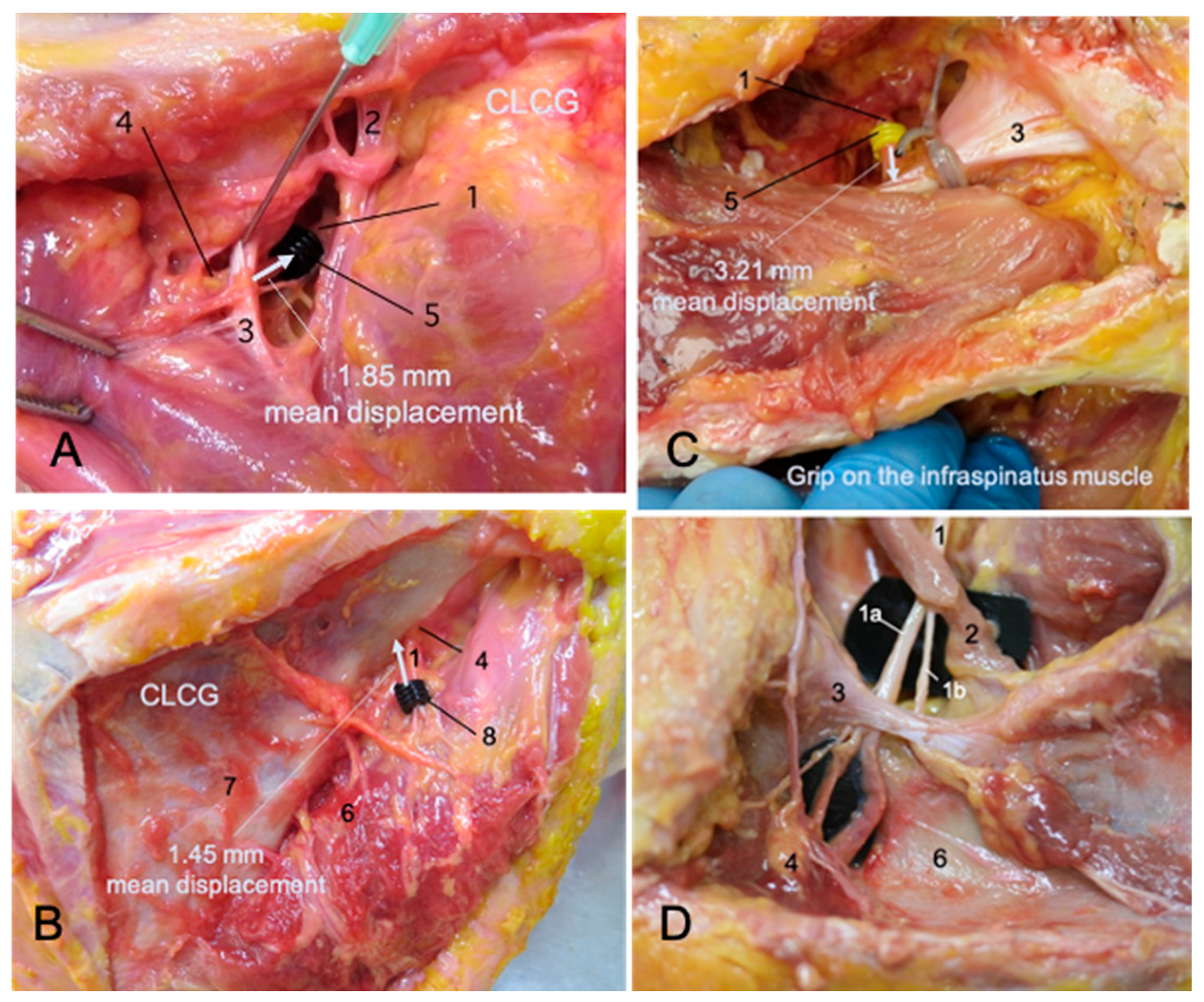
The CLCG resulted in a proximal glide of the SNe toward the spine measured proximal to the SNo (see Figure 3A) and distal to SGN (see Figure 3B). The Sne’s proximal average movement was 1.85 mm at the SNo level and 1.45 mm at the SNG level (95% CI). Summarized mean displacements are presented in Table 1.
Similarly, pulling the infraspinatus muscle resulted in a distal glide of the SNe toward the infraspinatus fossa (see Figure 3C), with an average movement of 3.21 mm at SNo level (95% CI). The movement at the SNo was particularly prominent when pinching the infraspinatus muscle, showing that this maneuver pulled the SNe toward the muscle.
During dissection, an anatomical variation was observed in four of the twelve shoulders (see Figure 3D), where the SNe was divided into its motor branches for the supraspinatus and infraspinatus muscles before entering the SNo. While this variation is clinically relevant, it did not affect the movement of the nerve.

3.2. In Vivo Study

In all the videos analyzed, a significant difference was recorded between the displacements of the movement point and the control point, which confirmed that nerve movement was indeed detected. All movement values were provided with 95% non-parametric confidence intervals using bootstrapping methods. This approach was chosen because the assumption of normality was not validated (p-values below the significance level α = 0.05), and the sample size was not large enough to rely on parametric methods. Bootstrapping allowed us to estimate a more robust measure of the observed nerve displacement. All statistical analyses were performed using Minitab statistical software (version 21, Minitab LLC, State College, PA, USA).
The presence of a vertical displacement value suggested that aligning the ultrasound head longitudinally with the nerve was not feasible. Considering the anatomy and course of the SNe, it was not possible to rectify this error. Therefore, it was decided to analyze and supply information on both horizontal and vertical displacements, as both motion components were recorded.
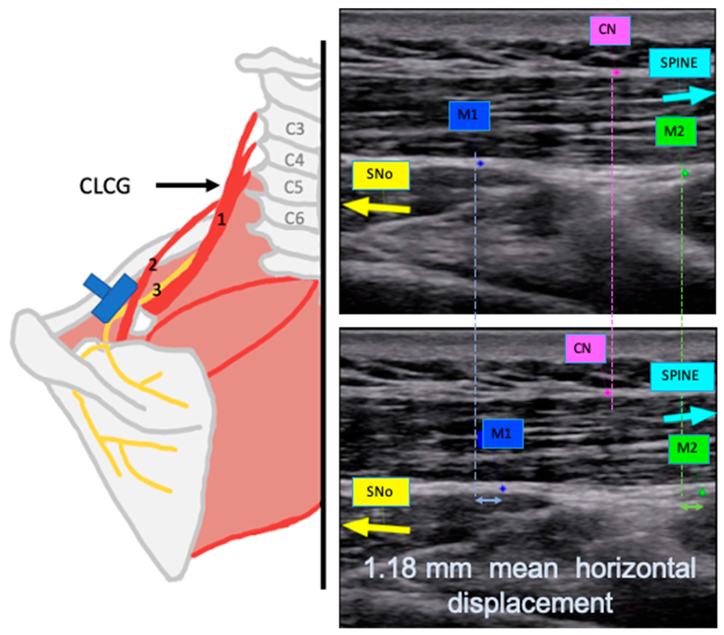
The CLCG resulted in a proximal glide of the SNe toward the spine, both at SNo level and at SGN level. In Video S1, recorded at SNo level, a CLCG resulted in a horizontal medial movement of 1.18 mm of the SNe toward the spine and a vertical cranial movement of 0.39 mm (see Figure 4 and detailed values in Table 1). In Video S3, the same maneuver moved the SNe horizontally and laterally toward the SGN by 1.65 mm, and vertically and cranially by 0.34 mm at the infraspinous fossa level (see Figure 5 and detailed values in Table 1).
The isometric infraspinatus muscle contraction resulted in a distal glide of the SNe toward the infraspinatus fossa, registered at the SNo level. In Video S2, an isometric external rotation contraction resulted in a horizontal lateral movement of 1.34 mm of the SNe and a vertical caudal movement of 0.75 mm (see Figure 6 and detailed values in Table 1).
Ultrasound imaging did not confirm the presence of the anatomical variation detected during dissection.

4. Discussion

This study investigated the mechanical behavior of the suprascapular nerve (SNe) using both cadaveric dissection and in vivo ultrasound imaging. Previous MRI-based simulations have shown that the SNe shifts from contacting the superior transverse scapular ligament (STSL) in the neutral arm position to the inferior portion of the suprascapular notch (SNo) during 90° abduction [17], although these findings were based on a single shoulder. This research showcased improved measurement accuracy through MRI, enabling precise investigation of the SNe location in relation to its bony structure. While a downward vertical shift of the SNe was demonstrated during shoulder abduction, no estimates were provided regarding its extent. Considering the SNe course, shoulder abduction might be viewed as a distal glide. This research demonstrated that the SNe distal glide during isometric contraction of the infraspinatus muscle, analyzed via ultrasound, also exhibited a caudal vertical component. In addition, this study confirmed the SNe glide by ultrasound in a larger sample, offering new insights into nerve mobility at the SNo.
Comparing the findings of the current study with other dissection studies was challenging. The cadaveric study performed to investigate the joint movement combinations that increase the strain on the SNe, while validating the movement elements applicable both proximally and distally, did not provide data on the possible SNe glide or its direction [23]. Additionally, the current dissection phase had considerable limitations. The primary constraint was found in the examination of distal gliding since it is unfeasible to reproduce genuine muscle tone or contraction in cadavers. Furthermore, the sample size is limited, and it was essential to expose almost the entire nerve course for its examination. However, the existence and the direction of SNe proximal and distal glide were fully confirmed.
To analyze whether the magnitude of movement recorded on the CLCG maneuver by dissection could be used as a guide, it was compared with those obtained in other studies of cervical movement in dissection. In this study, a contralateral lateral cervical glide (CLCG) produced greater SNe displacement than that reported for the median nerve during a similar cervical movement (2.25 mm vs. 0.71 mm) [39]. This difference may be due to variations in nerve length, anatomical path, and dissection methods. Unlike previous studies on the median nerve, our approach required full exposure of the SNe from the omohyoid level to the infraspinatus, potentially affecting its natural constraints. While the extent of movement noted in this study during its dissection phase must be interpreted with care, particularly the information gathered from the maneuver of approximating the origin and insertion of the infraspinatus muscle, the observation of the nerve gliding and its direction present new research opportunities.
In the ultrasound phase, both maneuvers—CLCG and isometric infraspinatus contraction—elicited measurable SNe displacement, although of smaller magnitude compared to the dissection. This reduction likely reflects the influence of fascial constraints present in living tissues. Moreover, the existence of a vertical motion vector clearly indicated that the ultrasound head could not be completely aligned with the nerve The tortuous course of the SNe greatly influenced this measurement error. Obtaining two motion vectors (horizontal and vertical), complicated direct comparisons with both the in vitro results and previous ultrasound studies on different nerves. In another study carried out on healthy volunteers, it was observed a 2.5 mm longitudinal median nerve displacement reported during lateral cervical glides [21]. The median nerve had sections where the ultrasound head could be longitudinally aligned, providing much more specific and reliable motion data. However, the SNe lacked a longitudinal section long enough to record a single longitudinal motion vector. Further research on this nerve would require both improvement of the ultrasound tracking system and consideration of expressing its movement values in two vectors. Nonetheless, the presence and direction of nerve glide can be reliably confirmed.
Importantly, this is the first study to assess the effects of CLCG and muscle contraction on the SNe. No previous work has evaluated these maneuvers in relation to SNe gliding. Although one study suggested the effect of active contraction on SNe mechanosensitivity [36], it has not been replicated in clinical trials. Neurodynamic testing is widely used for diagnosis and treatment of neuropathic conditions, including shoulder pain [19,20,26,47]. Tension-related dysfunctions can affect conduction and be identified via tension tests. However, gliding dysfunctions may present only with pain during specific glide maneuvers [19,48]. The ability to assess SNe gliding via ultrasound could enhance diagnostic precision in persistent shoulder pain.
Furthermore, different neurodynamic maneuvers have varied mechanical effects depending on movement sequence and location [37,38,40,42,49]. Despite the SNe being a key contributor to shoulder symptoms [11,12,13,23,50,51,52], little is known about how neurodynamic techniques influence its behavior. Future research should focus on establishing standardized neurodynamic protocols targeting the SNe. Emerging tools such as elastography could also help evaluate nerve biomechanics and tissue changes more effectively [42,43,44], supporting better clinical assessment and treatment strategies.
This study has some important limitations. First, although the analysis confirmed relative motion between the SNe and surrounding tissues, further research is needed to precisely quantify displacement magnitude. The intricate anatomy of the SNe made it truly challenging to precisely evidence the nerve behavior. Still, this represents an important step toward highlighting the limitations that the SNe presents for its study and to provide some preliminary information that could guide more precise research. Second, while the research through dissection provided valuable insights into the gliding capability of the SNe, these results could not be generalized to in vivo exploration since the nerve needed to be fully exposed, which altered its functionality, and muscle contraction could not be replicated in a cadaver. Another important limitation is the ultrasound analysis of nerve sliding since the disposition of the nerve during the in vivo and ultrasound study has limited its analysis. Finally, as this is the first study investigating neurodynamic gliding techniques applied to the SNe, comparative data or reference values are currently unavailable. To address this information gap, future research should be conducted to validate sonographic neural assessment with free support programs, making them more accessible, and compare the preliminary results obtained with a larger sample including volunteers who present shoulder pathology.

5. Conclusions

The study of the mechanical behavior of the suprascapular nerve through dissection and ultrasound suggested that the SNe exhibits distinct mechanical displacement patterns depending on the applied maneuver, whereby a CLCG caused a proximal glide of the nerve, and the contraction of the infraspinatus muscle generated a distal glide. These initial findings lay the groundwork for future research, particularly those obtained by ultrasound, which include both validating the analysis system by studying other nerves with available reference values and broadening the sample to include individuals with shoulder dysfunction.

Supplementary Materials

The following supporting information can be downloaded at: https://www.mdpi.com/article/10.3390/app15136987/s1, Maneuver Videos S1–S3.

Author Contributions

Conceptualization, M.M.-B., M.M.-P. and A.P.-B.; methodology, M.M.-B., A.P.-B. and M.M.-P.; validation, M.M.-B., M.M.-P. and L.R.-d.-L.; formal analysis L.R.-d.-L.; investigation M.M.-B., P.N.-C. and M.M.-P.; data curation M.M.-B., M.M.-P. and P.N.-C.; writing—original draft preparation, M.M.-B. and A.P.-B.; writing—review and editing, M.M.-B., M.M.-P. and A.P.-B.; supervision, M.M.-P. and A.P.-B.; project administration, M.M.-B. and A.P.-B. All authors have read and agreed to the published version of the manuscript.

Funding

This research received no external funding.

Institutional Review Board Statement

The study was conducted in accordance with the Declaration of Helsinki and approved by the Ethic Committee of the University of Barcelona (IRB00003099).

Informed Consent Statement

Written informed consent was obtained voluntarily from the deceased donors in life. Written informed consent was obtained from all volunteers for the sonographic study.

Data Availability Statement

Data are available upon request from the corresponding author.

Acknowledgments

The authors sincerely thank those who donated their bodies to science so that anatomical research could be performed. Many thanks to all the volunteers who participated in the study.

Conflicts of Interest

The authors declare no conflicts of interest.

Abbreviations

The following abbreviations are used in this manuscript:
SNeSuprascapular Nerve
SNoSuprascapular Notch
STSLSuperior Transverse Scapular Ligament
CLCGContralateral Cervical Glide
SGNSpinoglenoid Notch
ULNTTUpper Limb Neural Tension Test
CIConfidence Interval

Appendix A

Detailed Analomical Study Procedure

The torso specimen was placed in an upright position with its hands leaning in front on the table to stabilize it. Two researchers oversaw the measurement procedure. One of them, situated contralateral to the side studied, stabilized the specimen and executed the maneuver. With one hand, the scapula was stabilized on the side to be mobilized, and the ulnar edge of the other hand was used to grip the lateral surface of C5-C6, stabilizing the head of the specimen between the arm and the forearm. The second researcher was on the homolateral side of the torso when performing the different measurements.
The researcher who executed the maneuver was situated on the left side of the torso specimen, pressing it toward them to stabilize it. With the right hand supporting the trunk and maintaining scapular stability, the cubital side of the researcher’s left hand was placed on the lateral side of C5-C6. The contralateral cervical glide was performed by the researcher gradually pressing the ulnar edge of their left hand horizontally toward themselves until an articular end-feel was obtained.
Two different maneuvers were studied:
  • A contralateral cervical glide (CLCG) focusing on C5-C6 was performed. The maneuver was performed by the researcher gradually pressing the ulnar edge of the hand on the lateral surface of C5-C6 until an articular end-feel was obtained before returning to neutral position. The torso on the side contralateral to the measurement was stabilized, with the scapula being kept stable on the side that was being measured by the researcher who was performing the maneuver.
  • The infraspinatus muscle fibers were gripped to bring the origin and insertion closer together to mimic a muscle contraction.
Three different paired measurements were performed with a Vernier caliper (Mitutoyo ABSOLUTE Solar Caliper Series 500 with ABSOLUTE technology, Aurora, IL, USA). Each pair of measurements was repeated five times. The paired measurements were:
  • The distance between the superior ring and the STSL before and after a CLCG.
  • The distance between the inferior ring and the SGN before and after a CLCG.
  • The distance between the superior ring and the STSL before and after mimicking an infraspinatus contraction.

Appendix B

The subject was seated with their arms relaxed at each side of their body and the back was supported against a chair. Both arms were studied. To ensure accurate ultrasound recording, each maneuver was repeated two to three times. The recording process was:
  • For Video S1, the transducer was placed in the horizontal plane on the anterolateral aspect of the neck to image the roots of C5-C6-C7 and was moved toward the acromion to locate the SNe as close as possible to the SNo. A CLCG was performed (see Maneuver Video S1), by the researcher gradually pressing the ulnar edge of their hand on the lateral surface of C5-C6 until an articular end-feel was obtained. The torso on the side contralateral to the measurement was stabilized, ensuring that the scapula was stable on the side being measured.
  • For Video S2, the transducer was placed in the same position as for Video S1. An isometric external rotation contraction was performed (see Maneuver Video S2). The elbow was stabilized against the side of the trunk and a resistance was applied on the dorsal side of the wrist. An isometric contraction was chosen to prevent the ultrasound head from moving. A preliminary observation study was undertaken to determine the appropriate contraction intensity. Movement could be observed at the beginning of the contraction. No additional movement was observed when the contraction intensity increased. The SNe returned to its resting position when the contraction was released. Thus, it was decided that a mild isometric contraction would be used.
  • For Video S3, the transducer was placed along the inferior border of the spine of the scapula to scan the SNe distal to the SGN. A CLCG was performed (see Maneuver Video S3).
Maneuver Video S1: A contralateral cervical glide focusing on C5-C6 was performed. The maneuver was performed by a researcher gradually pressing the ulnar edge of their hand on the lateral surface of C5-C6 until an articular end-feel was obtained before returning to a neutral position. The torso on the side contralateral to the measurement was stabilized, ensuring that the scapula was stable on the side being measured. The transducer was placed in the horizontal plane on the anterolateral aspect of the neck to image the roots of C5-C6-C7 and was moved toward the acromion to locate the SNe as close as possible to the SNo. Two to three repetitions of the maneuver were performed.
Maneuver Video S2: An active isometric contraction of the infraspinatus muscle with mild intensity was performed. The elbow was stabilized against the side of the trunk and a resistance was applied on the dorsal side of the wrist. The transducer was placed in the same position as that for Video S1. Two to three repetitions of the isometric external rotation contraction were performed.
Maneuver Video S3: A contralateral cervical glide focusing on C5-C6 was performed. The maneuver was performed by a researcher gradually pressing the ulnar edge of their hand on the lateral surface of C5-C6 until an articular end-feel was obtained before returning to a neutral position. The torso on the side contralateral to the measurement was stabilized, ensuring that the scapula was stable on the side being measured. The transducer was placed along the inferior border of the spine of the scapula to scan the SNe distal to the SNo. Two to three repetitions of the maneuver were performed.

References

  1. Khosravi, F.; Amiri, Z.; Masouleh, N.A.; Kashfi, P.; Panjizadeh, F.; Hajilo, Z.; Shanayii, S.; Khodakarim, S.; Rahnama, L. Shoulder pain prevalence and risk factors in middle-aged women: A cross-sectional study. J. Bodyw. Mov. Ther. 2019, 23, 752–757. [Google Scholar] [CrossRef] [PubMed]
  2. Thoomes-de Graaf, M.; Scholten-Peeters, G.G.M.; Duijn, E.; Karel, Y.H.J.M.; van den Borne, M.P.J.; Beumer, A.; Ottenheijm, R.P.G.; Dinant, G.J.; Tetteroo, E.; Lucas, C.; et al. Inter-professional agreement of ultrasound-based diagnoses in patients with shoulder pain between physical therapists and radiologists in the Netherlands. Man. Ther. 2014, 19, 478–483. [Google Scholar] [CrossRef]
  3. Crookes, T.; Wall, C.; Byrnes, J.; Johnson, T.; Gill, D. Chronic shoulder pain. Aust. J. Gen. Pract. 2023, 52, 753–758. [Google Scholar] [CrossRef] [PubMed]
  4. Karel, Y.H.J.M.; Scholten-Peeters, G.G.M.; Thoomes-de Graaf, M.; Duijn, E.; van Broekhoven, J.B.; Koes, B.W.; Verhagen, A.P. Physiotherapy for patients with shoulder pain in primary care: A descriptive study of diagnostic- and therapeutic management. Physiotherapy 2017, 103, 369–378. [Google Scholar] [CrossRef] [PubMed]
  5. Uhl, T.L.; Smith-Forbes, E.V.; Nitz, A.J. Factors influencing final outcomes in patients with shoulder pain: A retrospective review. J. Hand Ther. 2017, 30, 200–207. [Google Scholar] [CrossRef]
  6. Kooijman, M.; Swinkels, I.; Van Dijk, C.; De Bakker, D.; Veenhof, C. Patients with shoulder syndromes in general and physiotherapy practice: An observational study. BMC Musculoskelet. Disord. 2013, 14, 128. [Google Scholar] [CrossRef]
  7. Lucas, J.; van Doorn, P.; Hegedus, E.; Lewis, J.; van der Windt, D. A systematic review of the global prevalence and incidence of shoulder pain. BMC Musculoskelet. Disord. 2022, 23, 1073. [Google Scholar] [CrossRef]
  8. Memon, M.; Kay, J.; Ginsberg, L.; Simunovic, N.; Bak, K.; Lapner, P.; Ayeni, O.R. Arthroscopic management of suprascapular neuropathy of the shoulder improves pain and functional outcomes with minimal complication rates. Knee Surg. Sport. Traumatol. Arthrosc. 2018, 26, 240–266. [Google Scholar] [CrossRef]
  9. Ohanisian, L.; Brown, N.; White, S.D.; Rubay, D.; Schwartz, P.M. Persistent Shoulder Pain Due to a Suprascapular Nerve Injury in the Setting of Trauma. Cureus 2019, 11, e4224. [Google Scholar] [CrossRef]
  10. Bachasson, D.; Singh, A.; Shah, S.; Lane, J.G.; Ward, S.R. The Role of the Peripheral and Central Neurvous Systems inRotator Cuff Disease. J. Shoulder Elb. Surg. 2015, 24, 1322–1335. [Google Scholar] [CrossRef]
  11. Laumonerie, P.; Dalmas, Y.; Tibbo, M.E.; Robert, S.; Faruch, M.; Chaynes, P.; Bonnevialle, N.; Mansat, P. Sensory innervation of the human shoulder joint: The three bridges to break. J. Shoulder Elb. Surg. 2020, 29, e499–e507. [Google Scholar] [CrossRef] [PubMed]
  12. Polguj, M.; Rożniecki, J.; Sibiński, M.; Grzegorzewski, A.; Majos, A.; Topol, M. The variable morphology of suprascapular nerve and vessels at suprascapular notch: A proposal for classification and its potential clinical implications. Knee Surg. Sport. Traumatol. Arthrosc. 2015, 23, 1542–1548. [Google Scholar] [CrossRef] [PubMed]
  13. Lee, S.H.; Choi, H.H.; Lee, D.G. Effectiveness of new nerve blocks method on the articular branches of the suprascapular and subscapular nerves to treat shoulder pain. Medicine 2020, 99, e22050. [Google Scholar] [CrossRef] [PubMed]
  14. Jezierski, H.; Podgórski, M.; Wysiadecki, G.; Olewnik, Ł.; De Caro, R.; Macchi, V.; Polguj, M. Morphological Aspects in Ultrasound Visualisation of the Suprascapular Notch Region: A Study Based on a New Four-Step Protocol. J. Clin. Med. 2018, 7, 491. [Google Scholar] [CrossRef]
  15. Polguj, M.; Synder, M.; Borowski, A.; Wojciechowski, M.; Wysiadecki, G.; Topol, M. Anterior Coracoscapular Ligament as a Factor Predisposing to or Protective for Suprascapular Neuropathy. Biomed Res. Int. 2016, 2016, 4134280. [Google Scholar] [CrossRef]
  16. Tasaki, A.; Nimura, A.; Mochizuki, T.; Yamaguchi, K.; Kato, R.; Sugaya, H.; Akita, K. Anatomic observation of the running space of the suprascapular nerve at the suprascapular notch in the same direction as the nerve. Knee Surg. Sport. Traumatol. Arthrosc. 2015, 23, 2667–2673. [Google Scholar] [CrossRef]
  17. Yoo, Y.S.; Jang, S.W.; Kim, Y.S.; Choi, J.A.; Oh, J.H.; Jeong, J.Y. Does the Suprascapular Nerve Move within the Suprascapular Notch? Biomechanical Perspective Using the Finite Element Method. Yonsei Med. J. 2022, 63, 657–664. [Google Scholar] [CrossRef]
  18. Kostretzis, L.; Theodoroudis, I.; Boutsiadis, A.; Papadakis, N.; Papadopoulos, P. Suprascapular Nerve Pathology: A Review of the Literature. Open Orthop. J. 2017, 11, 140–153. [Google Scholar] [CrossRef]
  19. Shacklock, M. Neurodynamics. Physiotherapy 1995, 81, 9–16. [Google Scholar] [CrossRef]
  20. Basson, C.A.; Stewart, A.; Mudzi, W.; Musenge, E. Effect of neural mobilization on nerve-related neck and arm pain: A randomized controlled trial. Physiother. Can. 2020, 72, 408–419. [Google Scholar] [CrossRef]
  21. Brochwicz, P.; von Piekartz, H.; Zalpour, C. Sonography assessment of the median nerve during cervical lateral glide and lateral flexion. Is there a difference in neurodynamics of asymptomatic people? Man. Ther. 2013, 18, 216–219. [Google Scholar] [CrossRef] [PubMed]
  22. Paquette, P.; El Khamlichi, Y.; Lamontagne, M.; Higgins, J.; Gagnon, D.H. Comparison of longitudinal excursion of a nerve-phantom model using quantitative ultrasound imaging and motion analysis system methods: A convergent validity study. Ultrasound 2017, 25, 143–149. [Google Scholar] [CrossRef] [PubMed]
  23. Bellot, N.; Ridehalgh, C.; Brismée, J.-M.; Crawford, M.; St-Pierre, M.-O.; Effatparvar, M.R.; Lavoie, F.-A.; Sobczak, S. Neurodynamic testing of the suprascapular nerve: An observational cadaveric study. Clin. Biomech. 2025, 121, 106380. [Google Scholar] [CrossRef] [PubMed]
  24. Beddaa, H.; Kably, B.; Marzouk, B.; Mouhi, I.; Marfak, A.; Azemmour, Y.; Alaoui, I.B.; Birouk, N. The effectiveness of the median nerve neurodynamic mobilisation techniques in women with mild or moderate bilateral carpal tunnel syndrome: A single-blind clinical randomised trial. S. Afr. J. Physiother. 2022, 78, 1823. [Google Scholar] [CrossRef]
  25. Joshi, K.C.; Eapen, C.; Kumar, S.P. Normal sensory and range of motion (ROM) responses during Thoracic Slump Test (ST) in asymptomatic subjects. J. Man. Manip. Ther. 2013, 21, 24–32. [Google Scholar] [CrossRef]
  26. Shacklock, M. Clinical Neurodynamics in Sports Injuries. Sport. Rehabil. 2014, 3, 50–56. [Google Scholar]
  27. Coppieters, M.W.; Alshami, A.M.; Babri, A.S.; Souvlis, T.; Kippers, V.; Hodges, P.W. Strain and excursion of the sciatic, tibial and plantar nerves during a modified straight leg raising test. J. Orthop. Res. 2006, 24, 1883–1889. [Google Scholar] [CrossRef]
  28. Coppieters, M.W.; Butler, D.S. Do “sliders” slide and “tensioners” tension? An analysis of neurodynamic techniques and considerations regarding their application. Man Ther. 2008, 13, 213–221. [Google Scholar] [CrossRef]
  29. Ellis, R.; Carta, G.; Andrade, R.J.; Coppieters, M.W. Neurodynamics: Is tension contentious? J. Man. Manip. Ther. 2022, 30, 3–12. [Google Scholar] [CrossRef]
  30. Carta, G.; Gambarotta, G.; Fornasari, B.E.; Muratori, L.; El Soury, M.; Geuna, S.; Raimondo, S.; Fregnan, F. The neurodynamic treatment induces biological changes in sensory and motor neurons in vitro. Sci. Rep. 2021, 11, 13277. [Google Scholar] [CrossRef]
  31. Cuenca-Martínez, F.; La Touche, R.; Varangot-Reille, C.; Sardinoux, M.; Bahier, J.; Suso-Martí, L.; Fernández-Carnero, J. Effects of Neural Mobilization on Pain Intensity, Disability, and Mechanosensitivity: An Umbrella Review With Meta-Meta-Analysis. Phys. Ther. 2022, 102, pzac040. [Google Scholar] [CrossRef] [PubMed]
  32. Baptista, F.M.; Nery, E.; Cruz, E.B.; Afreixo, V.; Silva, A.G. Effectiveness of Neural Mobilisation on Pain Intensity, Functional Status, and Physical Performance in Adults with Musculoskeletal Pain—A Systematic Review with Meta-Analysis. Clin. Rehabil. 2024, 38, 145–183. [Google Scholar] [CrossRef] [PubMed]
  33. Shou, M.K.; Study, C.; Lateral, C.; Sliders, N.; Syndrome, P. Comparative Study of Contralateral Cervical Lateral Glide and Neural Sliders in Athletes with Cervicobrachial Pain Syndrome. Int. J. Progress. Res. Sci. Eng. 2022, 3, 18–28. [Google Scholar]
  34. Rodríguez-Sanz, D.; Calvo-Lobo, C.; Unda-Solano, F.; Sanz-Corbalán, I.; Romero-Morales, C.; López-López, D. Cervical lateral glide neural mobilization is effective in treating Cervicobrachial pain: A randomized waiting list controlled clinical trial. Pain Med. 2017, 18, 2492–2503. [Google Scholar] [CrossRef]
  35. Shacklock, M.; Giménez Donoso, C.; Lucha López, M. Hacia un enfoque clínico- científico en el diagnóstico con test neurodinámicos (tensión neural). Fisioterapia 2017, 29, 288–297. [Google Scholar] [CrossRef]
  36. Zamorano, E. Capitulo 10: Manual de pruebas de provocación neural. In Movilización Neuromeníngea: Tratamiento de los Trastornos Mecanosensitivos del Sistema Nervioso; Editorial Médica Panamericana S.A.: Madrid, Spain, 2013; pp. 203–223. ISBN 8479039701. [Google Scholar]
  37. Boudier-Revéret, M.; Gilbert, K.K.; Allégue, D.R.; Moussadyk, M.; Brismée, J.M.; Sizer, P.S.; Feipel, V.; Dugailly, P.M.; Sobczak, S. Effect of neurodynamic mobilization on fluid dispersion in median nerve at the level of the carpal tunnel: A cadaveric study. Musculoskelet. Sci. Pract. 2017, 31, 45–51. [Google Scholar] [CrossRef]
  38. Ibrahim, D.; Ahbouch, A.; Qadah, R.M.; Kim, M.; Alrawaili, S.M.; Moustafa, I.M. Impact of the Order of Movement on the Median Nerve Root Function: A Neurophysiological Study with Implications for Neurodynamic Exercise Sequencing. J. Clin. Med. 2024, 13, 913. [Google Scholar] [CrossRef]
  39. Bueno-Gracia, E.; Pérez-Bellmunt, A.; Estébanez-de-Miguel, E.; López-de-Celis, C.; Caudevilla-Polo, S.; Shacklock, M.; González-Rueda, V. Effect of cervical contralateral lateral flexion on displacement and strain in the median nerve and flexor digitorum superficialis at the wrist during the ULNT1—Cadaveric study. Musculoskelet. Sci. Pract. 2020, 50, 102244. [Google Scholar] [CrossRef]
  40. Bueno-Gracia, E.; Fanlo-Mazas, P.; Malo-Urriés, M.; Rodriguez-Mena, D.; Montaner-Cuello, A.; Ciuffreda, G.; Shacklock, M.; Estébanez-de-Miguel, E. Diagnostic accuracy of the upper limb neurodynamic test 1 using neurodynamic sequencing in diagnosis of carpal tunnel syndrome. Musculoskelet. Sci. Pract. 2024, 69, 102897. [Google Scholar] [CrossRef]
  41. Pérez-Bellmunt, A.; López-de-Celis, C.; Estébanez-de-Miguel, E.; Pérez-Rey, J.; Shacklock, M.; Ortiz-Miguel, S.; Bueno-Gracia, E. Effect of cervical contralateral lateral flexion on the median nerve and fascia at the wrist—Cadaveric study. Musculoskelet. Sci. Pract. 2024, 73, 103146. [Google Scholar] [CrossRef]
  42. Ciuffreda, G.; Bueno-Gracia, E.; Albarova-Corral, I.; Montaner-Cuello, A.; Pérez-Rey, J.; Pardos-Aguilella, P.; Malo-Urriés, M.; Estébanez-de-Miguel, E. In Vivo Effects of Joint Movement on Nerve Mechanical Properties Assessed with Shear-Wave Elastography: A Systematic Review and Meta-Analysis. Diagnostics 2024, 14, 343. [Google Scholar] [CrossRef] [PubMed]
  43. Santilli, G.; Ciccarelli, A.; Martino, M.; Pacini, P.; Agostini, F.; Bernetti, A.; Giuliani, L.; Del Gaudio, G.; Mangone, M.; Colonna, V.; et al. Pain, Function, and Elastosonographic Assessment After Shockwave Therapy in Non-Calcific Supraspinatus Tendinopathy: A Retrospective Observational Study. J. Funct. Morphol. Kinesiol. 2025, 10, 39. [Google Scholar] [CrossRef] [PubMed]
  44. Snoj, Ž.; Wu, C.H.; Taljanovic, M.S.; Dumić-Čule, I.; Drakonaki, E.E.; Klauser, A.S. Ultrasound Elastography in Musculoskeletal Radiology: Past, Present, and Future. Semin. Musculoskelet. Radiol. 2020, 24, 156–166. [Google Scholar] [CrossRef] [PubMed]
  45. McKay, M.J.; Baldwin, J.N.; Ferreira, P.; Simic, M.; Vanicek, N.; Burns, J.; Nightingale, E.; Pourkazemi, F.; Sman, A.; Hiller, C.; et al. Normative reference values for strength and flexibility of 1000 children and adults. Neurology 2017, 88, 36–43. [Google Scholar] [CrossRef]
  46. Puig-Diví, A.; Escalona-Marfil, C.; Padullés-Riu, J.M.; Busquets, A.; Padullés-Chando, X.; Marcos-Ruiz, D. Validity and reliability of the Kinovea program in obtaining angles and distances using coordinates in 4 perspectives. PLoS ONE 2019, 14, e0216448. [Google Scholar] [CrossRef]
  47. Basson, A.; Olivier, B.; Ellis, R.; Coppieters, M.; Stewart, A.; Mudzi, W. The effectiveness of neural mobilization for neuromusculoskeletal conditions: A systematic review and meta-Analysis. J. Orthop. Sports Phys. Ther. 2017, 47, 593–615. [Google Scholar] [CrossRef]
  48. Topp, K.S.; Boyd, B.S. Structure and biomechanics of peripheral nerves: Nerve responses to physical stresses and implications for physical therapist practice. Phys. Ther. 2006, 86, 92–109. [Google Scholar] [CrossRef]
  49. Marks, M.; Schöttker-Königer, T.; Probst, A. Efficacy of cervical spine mobilization versus peripheral nerve slider techniques in cervicobrachial pain syndrome—A randomized clinical trial. J. Phys. Ther. 2011, 4, 9–17. [Google Scholar]
  50. Davis, F.B.; Katsuura, Y.; Dorizas, J.A. A retrospective review of 112 patients undergoing arthroscopic suprascapular nerve decompression. J. Orthop. 2020, 19, 31–35. [Google Scholar] [CrossRef]
  51. Fernandes, M.R.; Barbosa, M.A.; Sousa, A.L.L.; Ramos, G.C. Bloqueio do Nervo Supraescapular: Procedimento Importante na Prática Clínica. Rev. Bras. Anestesiol. 2012, 62, 96–104. [Google Scholar] [CrossRef]
  52. Leider, J.D.; Derise, O.C.; Bourdreaux, K.A.; Dierks, G.J.; Lee, C.; Varrassi, G.; Sherman, W.F.; Kaye, A.D. Treatment of suprascapular nerve entrapment syndrome. Orthop. Rev. 2021, 13, 25554. [Google Scholar] [CrossRef]
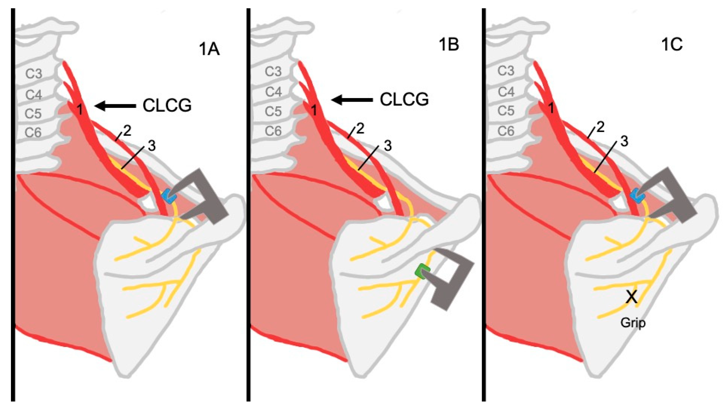
Figure 1. Maneuvers and measurement procedures in the cadaveric study. 1: scalene muscles; 2: omohyoid muscle; 3: suprascapular nerve; CLCG: contralateral cervical glide. (A) The distance between the superior ring (blue dot) and the STSL was measured before and after a CLCG. (B) The distance between the inferior ring (green dot) and the SGN was measured before and after a CLCG. (C) The distance between the superior ring (blue dot) and the STSL was measured before and after gripping the infraspinatus muscle.
Applsci 15 06987 g001
Figure 2. Videos and maneuvers recorded during the in vivo study. 1: scalene muscles; 2: omohyoid muscle; 3: suprascapular nerve; blue dot: fixation; blue arrow: direction of the maneuver; green arrow: direction of an isometric contraction. (A) Recording of Video S1. The transducer was placed on the anterolateral region of the neck, longitudinal to the Sne, as close as possible to the SNo. A CLCG was repeated two to three times while recording the video. (B) Recording of Video S2. The transducer was placed on the anterolateral region of the neck longitudinal to the SNe as close as possible to the SNo. An isometric external rotation contraction was repeated 2–3 times while recording the video. (C) Recording of Video S3. The transducer was placed on the infraspinatus fossa, longitudinal to the Sne, as close as possible to the SGN. A CLCG was repeated two to three times while recording.
Applsci 15 06987 g002
Figure 3. Dissection images of the anatomical results. Anatomical structures are marked as follows: 1: suprascapular nerve (SNe); 2: omohyoid muscle; 3: superior transverse scapular ligament (STSL); 4: suprascapular notch (SNo); 5: superior ring (a silicone ring situated around the SNe before the STSL); 6: infraspinatus muscle; 7: scapula; 8: inferior ring (a silicone ring situated around the SNe motor branch); 1a: motor branch into the infraspinatus muscle; 1b: motor branch into the supraspinatus muscle; contralateral cervical glide (CLCG). (A) A CLCG observed at the SNo. (B) A CLCG observed at the infrascapular fossa. (C) A simulated infraspinatus muscle contraction observed at the SNo. (D) Anatomical variation of the SNe.
Applsci 15 06987 g003
Figure 4. A CLCG observed at SNo by ultrasound. 1: scalene muscles; 2: omohyoid muscle; 3: suprascapular nerve; CLCG: contralateral cervical glide. The image shows two full screenshots of the ultrasound, the image on top was taken in the rest position, while the image on the bottom was captured at the end of the CLCG. This example of Video S1 was recorded on a left shoulder. The directions of the spine and the SNo are indicated by a light blue and yellow arrow, respectively. Three points were tracked: the blue and green points on the SNe and the pink point on the tissues surrounding the nerve. Vertical lines are provided from the top image to show the displacement of the points between captures. Specifically, in this example, the control point not only showed a lower magnitude of movement than the nerve points, but it also moved in the opposite direction.
Applsci 15 06987 g004
Figure 5. A CLCG observed at the infraspinous fossa by ultrasound. 1: scalene muscles; 2: omohyoid muscle; 3: suprascapular nerve; CLCG: contralateral cervical glide. The image shows two full screenshots of the ultrasound, the image on top was taken in the rest position, while the image on the bottom was captured at the end of the CLCG. This example of Video S3 was recorded on a left shoulder. The directions of the infraspinous muscle and the SGN are indicated by red and purple, respectively. Three points were tracked: the blue and green points on the SNe and the pink point on the tissues surrounding the nerve. The image on top shows the rest position, and the image on the bottom shows the end of the maneuver position. Vertical lines are provided from the top image to show the displacement of the points between captures. The control point showed a lower magnitude of movement than at least one of the nerve points toward the SGN.
Applsci 15 06987 g005
Figure 6. An isometric infraspinous contraction observed at the SNo by ultrasound. 1: scalene muscles; 2: omohyoid muscle; 3: suprascapular nerve. The image shows two full screenshots of the ultrasound; the image on top was taken in the rest position, while the image on the bottom was captured at the end of the infraspinatus muscle contraction. This example from Video S2 was recorded on a left shoulder. The directions of the spine and the SNo are indicated by a light blue and yellow arrow, respectively. Three points were tracked: the blue and green points on the SNe and the pink point on the tissues surrounding the nerve. The image on top shows the rest position, and the image on the bottom shows the end of the maneuver position. Vertical lines are provided from the top image to show the displacement of the points between captures. The control point showed a lower magnitude of movement than the nerve points, while the nerve points showed a mean horizontal displacement of 1.34 mm toward the spine.
Applsci 15 06987 g006
Table 1. Suprascapular nerve displacement.
Cadaveric StudyIn Vivo Study
Contralateral cervical glide observed at the suprascapular notch.1.85 (1.73, 1.98) mmHorizontal1.18 (0.79, 1.31) mm
Vertical0.39 (0.23, 0.54) mm
Infraspinatus muscle contraction observed at the suprascapular notch (simulated in the dissection study).3.21 (2.93, 3.46) mmHorizontal1.34 (1.05, 1.75) mm
Vertical0.75 (0.47, 1.02) mm
Contralateral cervical glide observed at the spinoglenoid notch.1.45 (1.35, 1.56) mmHorizontal1.65 (1.08, 2.00) mm
Vertical0.34 (0.22, 0.54) mm
This table summarizes the average estimated movement of the suprascapular nerve. For the cadaveric study, the mean movement is presented along with its 95% confidence interval. For the in vivo study, a trimmed mean—excluding 5% of extreme values—is shown, also accompanied by its 95% confidence interval. All values reported correspond to the mean or trimmed mean with their respective 95% confidence intervals.
Disclaimer/Publisher’s Note: The statements, opinions and data contained in all publications are solely those of the individual author(s) and contributor(s) and not of MDPI and/or the editor(s). MDPI and/or the editor(s) disclaim responsibility for any injury to people or property resulting from any ideas, methods, instructions or products referred to in the content.