


Chemical Analysis and Antimicrobial Potential Assessment of Wild Laurel from the National Park Skadar Lake, Montenegro
 , , and
Abstract
1. Introduction
2. Materials and Methods
2.1. Plant Material and Chemicals
2.2. Microorganisms
2.3. Chemical Analysis of Plant Material
2.4. Isolation of the Essential Oil
2.5. Chemical Analysis of Essential Oils
2.5.1. Gas Chromatography—GC
2.5.2. Gas Chromatography–Mass Spectrometry (GC–MS)
2.6. Preparation of Essential Oils for Antimicrobial Experiments
2.7. Antibacterial Activity
2.8. Antifungal Activity
2.9. Statistical Analysis
3. Results
3.1. Chemical Analysis
3.2. Antibacterial Activity
3.3. Antifungal Activity
4. Discussion
4.1. Chemical Analysis
4.2. Antibacterial Activity
5. Conclusions
Author Contributions
Funding
Institutional Review Board Statement
Informed Consent Statement
Data Availability Statement
Conflicts of Interest
Abbreviations
| EOL | leaves essential oil |
| EOF | fruits essential oil |
| EOs | essential oils |
| WHO | World Health Organization |
| ATCC | American-type culture collection |
| NCIMB | National Collection of Industrial, Food, and Marine Bacteria |
| MIC | minimal inhibitory concentration |
| TP | total phenolic |
| GA | gallic acid |
| SD | standard deviation |
| TT | total tannins |
| PVPP | polyvinylpolypyrrolidone |
| TF | total flavonoid |
| GC | gas chromatography |
| CLSI | Clinical and Laboratory Standards Institute |
| MHB | Müller–Hinton broth |
| RI | retention indices |
| SFE | supercritical fluid extraction |
| FID | flame ionization detector |
References
- Ng, W.J.; Shit, C.S.; Ee, K.Y.; Chai, T.T. Plant natural products for mitigation of antibiotic resistance. In Sustainable Agriculture Reviews 49. Sustainable Agriculture Reviews; Panwar, H., Sharma, C., Lichtfouse, E., Eds.; Springer: Cham, Switzerland, 2021; Volume 49, pp. 57–91. [Google Scholar]
- Angelini, P. Plant-derived antimicrobials and their crucial role in combating antimicrobial resistance. Antibiotics 2024, 13, 746. [Google Scholar] [CrossRef] [PubMed]
- Khameneh, B.; Iranshahy, M.; Soheili, V.; Fazly Bazzaz, B.S. Review on plant antimicrobials: A mechanistic viewpoint. Antimicrob. Resist. Infect. Control 2019, 8, 118. [Google Scholar] [CrossRef] [PubMed]
- Yap, P.S.X.; Yiap, B.C.; Ping, H.C.; Lim, S.H.E. Essential oils, a new horizon in combating bacterial antibiotic resistance. Open Microbiol. J. 2014, 8, 6. [Google Scholar] [CrossRef] [PubMed]
- Sırıken, B.; Yavuz, C.; Güler, A. Antibacterial Activity of Laurus nobilis: A review of literature. Med. Sci. Discov. 2018, 5, 374–379. [Google Scholar] [CrossRef]
- Bakkali, F.; Averbeck, S.; Averbeck, D.; Idaomar, M. Biological effects of essential oils—A review. Food. Chem. Toxicol. 2008, 46, 446–475. [Google Scholar] [CrossRef]
- El, S.N.; Karagozlu, N.; Karakaya, S.; Sahın, S. Antioxidant and antimicrobial activities of essential oils extracted from Laurus nobilis L. leaves by using solvent-free microwave and hydrodistillation. Food Nutr. Sci. 2014, 5, 97–106. [Google Scholar] [CrossRef]
- Zeković, Z.P.; Lepojević, Ž.D.; Mujić, I.O. Laurel extracts obtained by steam distillation, supercritical fluid and solvent extraction. J. Nat. Prod. 2009, 2, 104–109. [Google Scholar]
- Muñiz-Márquez, D.B.; Martínez-Ávila, G.C.; Wong-Paz, J.E.; Belmares-Cerda, R.; Rodríguez-Herrera, R.; Aguilar, C.N. Ultrasound-assisted extraction of phenolic compounds from Laurus nobilis L. and their antioxidant activity. Ultrason. Sonochem. 2013, 20, 1149–1154. [Google Scholar] [CrossRef]
- Moghtader, M.; Farahmand, A. Evaluation of the antibacterial effects of essential oil from the leaves of Laurus nobilis L. in Kerman Province. J. Microbiol. Antimicrob. 2013, 5, 13–17. [Google Scholar] [CrossRef]
- Caputo, L.; Nazzaro, F.; Souza, L.F.; Aliberti, L.; De Martino, L.; Fratianni, F.; Coppola, R.; De Feo, V. Laurus nobilis: Composition of essential oil and its biological activities. Molecules 2017, 22, 930. [Google Scholar] [CrossRef]
- Jemâa, J.M.B.; Tersim, N.; Toudert, K.T.; Khouja, M.L. Insecticidal activities of essential oils from leaves of Laurus nobilis L. from Tunisia, Algeria and Morocco, and comparative chemical composition. J. Stored Prod. Res. 2012, 48, 97–104. [Google Scholar] [CrossRef]
- Fernandez, C.M.M.; da Rosa, M.F.; Fernandez, A.C.A.M.; Lorenzetti, F.B.; Raimundo, K.F.; Cortez, D.A.G.; Gonçalves, J.E.; Simões, M.R.; Colauto, N.B.; Lobo, V.D.S.; et al. Larvicidal activity against Aedes aegypti of essential oil of Laurus nobilis leaves obtained at different seasons. J. Essent. Oil Res. 2018, 30, 379–387. [Google Scholar] [CrossRef]
- Abu-Dahab, R.; Kasabri, V.; Afifi, F.U. Evaluation of the Volatile Oil Composition and Antiproliferative Activity of Laurus nobilis L. (Lauraceae) on Breast Cancer Cell Line Models. Rec. Nat. Prod. 2014, 8, 136–147. [Google Scholar]
- Fidan, H.; Stefanova, G.; Kostova, I.; Stankov, S.; Damyanova, S.; Stoyanova, A.; Zheljazkov, V.D. Chemical composition and antimicrobial activity of Laurus nobilis L. essential oils from Bulgaria. Molecules 2019, 24, 804. [Google Scholar] [CrossRef]
- Peña-Bautista, C.; Baquero, M.; Vento, M.; Cháfer-Pericás, C. Free radicals in Alzheimer’s disease: Lipid peroxidation biomarkers. Clin. Chim. Acta 2019, 491, 85–90. [Google Scholar] [CrossRef]
- Charles, D.J. Antioxidant Properties of Spices, Herbs and Other Sources; Springer Science & Business Media: Berlin/Heidelberg, Germany, 2012; pp. 181–187. [Google Scholar]
- Al-Kalaldeh, J.Z.; Abu-Dahab, R.; Afifi, F.U. Volatile oil composition and antiproliferative activity of Laurus nobilis, Origanum syriacum, Origanum vulgare, and Salvia triloba against human breast adenocarcinoma cells. Nutr. Res. 2010, 30, 271–278. [Google Scholar] [CrossRef]
- Leyel, C.F. A Modern Herbal; Grieve, M., Ed.; Penguin Books: Harmondsworth, UK, 1984; pp. 154–196. [Google Scholar]
- Demirbas, A.; Demirbas, M.F. Algae Energy: Algae as a New Source of Biodiesel; Springer Science & Business Media: Berlin/Heidelberg, Germany, 2010; pp. 171–178. [Google Scholar]
- Ilić, Z.S.; Stanojević, L.; Milenković, L.; Šunić, L.; Milenković, A.; Stanojević, J.; Cvetković, D. Chemical Profiling of Essential Oils from Main Culinary Plants—Bay (Laurus nobilis L.) and Rosemary (Rosmarinus officinalis L.) from Montenegro. Horticulturae 2024, 10, 1249. [Google Scholar] [CrossRef]
- Kovacevic, N.N.; Simic, M.D.; Ristic, M.S. Essential oil of Laurus nobilis from Montenegro. Chem. Nat. Compd. 2007, 43, 408–411. [Google Scholar] [CrossRef]
- Simić, A.; Soković, M.D.; Ristić, M.; Grujić-Jovanović, S.; Vukojević, J.; Marin, P.D. The chemical composition of some Lauraceae essential oils and their antifungal activities. Phytother. Res. 2004, 18, 713–717. [Google Scholar] [CrossRef]
- Singleton, V.L.; Rossi, J.A. Colorimetry of total phenolics with phosphomolybdic-phosphotungstic acid reagents. Am. J. Enol. Vitic. 1965, 16, 144–158. [Google Scholar] [CrossRef]
- Council of Europe. European Pharmacopeia, 11th ed.; Council of Europe: Strasbourg, France, 2024. [Google Scholar]
- Jugoslovenska Farmakopeja IV SFRJ [Pharmacopoea Jugoslavica], 4th ed.; Savezni Zavod za Zdravstvenu Zaštitu: Belgrade, Serbia, 1984. (In Serbian)
- Tadić, V.M.; Žugić, A.; Martinović, M.; Stanković, M.; Maksimović, S.; Frank, A.; Nešić, I. Enhanced skin performance of emulgel vs. cream as systems for topical delivery of herbal actives (immortelle extract and hemp oil). Pharmaceutics 2021, 13, 1919. [Google Scholar] [CrossRef] [PubMed]
- Adams, R.P. Identification of Essential Oil Components by Gas Chromatography/Mass Spectrometry, 4th ed.; Allured Publishing Corporation: Carol Stream, IL, USA, 2007. [Google Scholar]
- Michielin, E.M.; Salvador, A.A.; Riehl, C.A.; Smânia, A., Jr.; Smânia, E.F.; Ferreira, S.R. Chemical composition and antibacterial activity of Cordia verbenacea extracts obtained by different methods. Bioresour. Technol. 2009, 100, 6615–6623. [Google Scholar] [CrossRef] [PubMed]
- Stefanova, G.; Girova, T.; Gochev, V.; Stoyanova, M.; Petkova, Z.; Stoyanova, A.; Zheljazkov, V.D. Comparative study on the chemical composition of laurel (Laurus nobilis L.) leaves from Greece and Georgia and the antibacterial activity of their essential oil. Heliyon 2020, 6, e05491. [Google Scholar] [CrossRef]
- Awada, F.; Hamade, K.; Kassir, M.; Hammoud, Z.; Mesnard, F.; Rammal, H.; Fliniaux, O. Laurus nobilis leaves and fruits: A review of metabolite composition and interest in human health. Appl. Sci. 2023, 13, 4606. [Google Scholar] [CrossRef]
- Petkova, Z.; Stefanova, G.; Girova, T.; Antova, G.; Stoyanova, M.; Damianova, S.; Gochev, V.; Stoyanova, A.; Zheljazkov, V.D. Phytochemical investigations of laurel fruits (Laurus nobilis). Nat. Prod. Commun. 2019, 14, 1934578X19868876. [Google Scholar] [CrossRef]
- Türkmen, M.; Koçer, O. Variation of components in laurel (Laurus nobilis L.) fixed oil extracted by different methods. Int. J. Chem. Technol. 2021, 5, 167–171. [Google Scholar] [CrossRef]
- Castilho, P.C.; Costa, M.D.C.; Rodrigues, A.; Partidário, A. Characterization of laurel fruit oil from Madeira Island, Portugal. J. Am. Oil Chem. Soc. 2005, 82, 863–868. [Google Scholar] [CrossRef]
- Nurbas, M.; Bal, Y. Recovery of fixed and volatile oils from Laurus nobilis L. fruit and leaves by solvent extraction method. Eng. Arch. Fac. EskişehirOsmangazi Univ. 2005, 18, 15–24. [Google Scholar]
- Butturini, E.; Cavalieri, E.; Carcereri de Prati, A.; Darra, E.; Rigo, A.; Shoji, K.; Murayama, N.; Yamazaki, H.; Watanabe, Y.; Suzuki, H.; et al. Two naturally occurring terpenes, dehydrocostuslactone and costunolide, decrease intracellular GSH content and inhibit STAT3 activation. PLoS ONE 2011, 6, e20174. [Google Scholar] [CrossRef]
- Wang, J.; Yu, Z.; Wang, C.; Tian, X.; Huo, X.; Wang, Y.; Sun, C.; Feng, L.; Ma, J.; Zhang, B.; et al. Dehydrocostus lactone, a natural sesquiterpene lactone, suppresses the biological characteristics of glioma, through inhibition of the NF-κB/COX-2 signaling pathway by targeting IKKβ. Am. J. Cancer Res. 2017, 7, 1270. [Google Scholar] [PubMed]
- Boucher, H.; Miller, L.G.; Razonable, R.R. Serious Infections Caused by Methicillin-Resistant Staphylococcus aureus. Clin. Infect. Dis. 2010, 51, S183–S197. [Google Scholar] [CrossRef]
- Tilahun, B.; Faust, A.C.; McCorstin, P.; Ortegon, A. Nasal colonization and lower respiratory tract infections with methicillin-resistant Staphylococcus aureus. Am. J. Crit. Care. 2015, 24, 8–12. [Google Scholar] [CrossRef]
- Atef, N.M.; Shanab, S.M.; Negm, S.I.; Abbas, Y.A. Evaluation of Antimicrobial Activity of Some Plant Extracts against Antibiotic Susceptible and Resistant Bacterial Strains Causing Wound Infection. Bull. Natl. Res. Cent. 2019, 43, 144. [Google Scholar] [CrossRef]
- Tsonis, I.; Karamani, L.; Xaplanteri, P.; Kolonitsiou, F.; Zampakis, P.; Gatzounis, G.; Marangos, M.; Assimakopoulos, S.F. Spontaneous cerebral abscess due to Bacillus subtilis in an immunocompetent male patient: A case report and review of literature. World J. Clin. Cases. 2018, 6, 1169. [Google Scholar] [CrossRef]
- Dahl, A.; Iversen, K.; Tonder, N.; Hoest, N.; Arpi, M.; Dalsgaard, M.; Chehri, M.; Soerensen, L.L.; Fanoe, S.; Junge, S.; et al. Prevalence of infective endocarditis in Enterococcus faecalis bacteremia. J. Am. Coll. Cardiol. 2019, 74, 193–201. [Google Scholar] [CrossRef]
- Choi, U.; Kim, E.J.; Lyu, D.H.; Park, B.H.; Chung, H.; Han, C.H.; Bae, S. Ureteral stent induced urinary tract infection and microbial inconsistency between bladder and renal pelvis. Urogenit. Tract Infect. 2021, 16, 61–66. [Google Scholar] [CrossRef]
- Arias, C.A.; Murray, B.E. The rise of the Enterococcus: Beyond vancomycin resistance. Nat. Rev. Microbiol. 2012, 10, 266–278. [Google Scholar] [CrossRef]
- Zida, A.; Bamba, S.; Yacouba, A.; Ouedraogo-Traore, R.; Guiguemdé, R.T. Anti-Candida albicans Natural Products, Sources of New Antifungal Drugs: A Review. J. Mycol. Méd. 2017, 27, 1–19. [Google Scholar] [CrossRef]
- Juergens, L.J.; Worth, H.; Juergens, U.R. New perspectives for mucolytic, anti-inflammatory and adjunctive therapy with 1, 8-cineole in COPD and asthma: Review on the new therapeutic approach. Adv. Ther. 2020, 37, 1737–1753. [Google Scholar] [CrossRef]
- Hoch, C.C.; Petry, J.; Griesbaum, L.; Weiser, T.; Werner, K.; Ploch, M.; Verschoor, A.; Multhoff, G.; Dezfouli, A.B.; Wollenberg, B. 1,8-cineole (eucalyptol): A versatile phytochemical with therapeutic applications across multiple diseases. Biomed. Pharmacother. 2023, 167, 115467. [Google Scholar] [CrossRef] [PubMed]
- Guo, F.; Chen, Q.; Liang, Q.; Zhang, M.; Chen, W.; Chen, H.; Yun, Y.; Zhong, Q.; Chen, W. Antimicrobial Activity and Proposed Action Mechanism of Linalool Against Pseudomonas fluorescens. Front. Microbiol. 2021, 12, 562094. [Google Scholar] [CrossRef] [PubMed]
- Cao, Y.; Zhang, H.; Liu, H.; Liu, W.; Zhang, R.; Xian, M.; Liu, H. Biosynthesis and production of sabinene: Current state and perspectives. Appl. Microbiol. Biotechnol. 2018, 102, 1535–1544. [Google Scholar] [CrossRef] [PubMed]
- Salehi, B.; Upadhyay, S.; Erdogan Orhan, I.; Kumar Jugran, A.L.D.; Jayaweera, S.A.; Dias, D.; Sharopov, F.; Taheri, Y.; Martins, N.; Baghalpour, N.; et al. Therapeutic Potential of α- and β-Pinene: A Miracle Gift of Nature. Biomolecules 2019, 9, 738. [Google Scholar] [CrossRef]


| No. | Compound | KIa | L. nobilis leaf EOL (%) | L. nobilis fruit EOF (%) | L. nobilis fruit fatty oil (%) |
|---|---|---|---|---|---|
| 1. | pentanol | 762 | 0.1 | ||
| 2. | hexanal | 801 | 0.9 | 0.3 | |
| 3. | isopropyl-2-methyl butanoate | 880 | 2.9 | ||
| 4. | n-nonane | 900 | 0.5 | ||
| 5. | heptanal | 901 | 0.1 | ||
| 6. | tricyclene | 921 | 0.1 | ||
| 7. | α-thujene | 924 | 0.2 | 0.3 | |
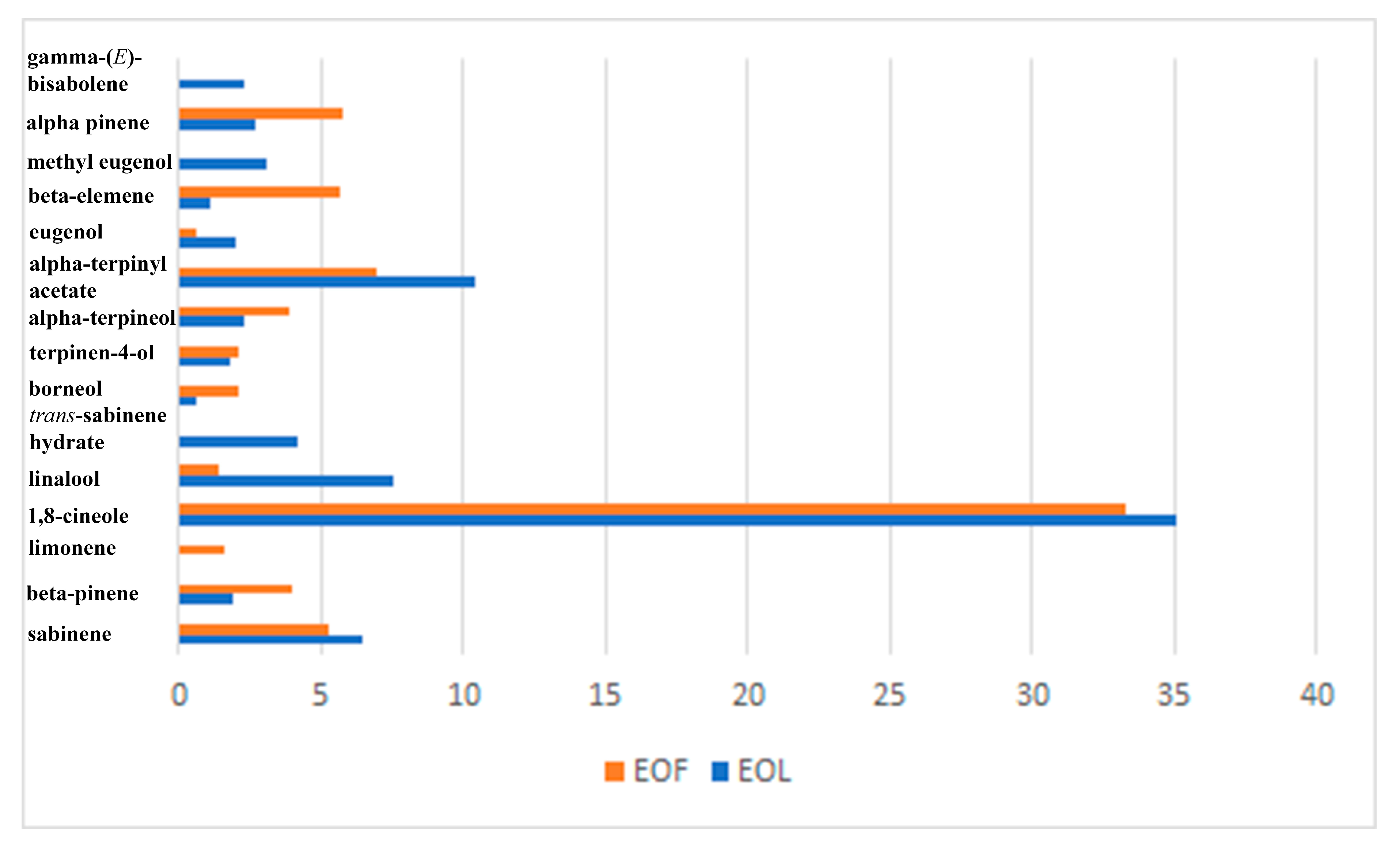
| 8. | α-pinene | 932 | 2.7 | 5.8 | 0.6 |
| 9. | camphene | 946 | 0.3 | 2.7 | 0.2 |
| 10. | hexanoic acid | 967 | 0.2 | ||
| 11. | sabinene | 969 | 6.5 | 5.3 | 0.3 |
| 12. | β-pinene | 974 | 1.9 | 4.0 | 0.4 |
| 13. | 1,8-dehydrocineole | 988 | 0.1 | 0.1 | |
| 14. | myrcene | 988 | 0.7 | 0.2 | |
| 15. | n-octanal | 998 | 0.2 | 0.1 | |
| 16. | α-phellandrene | 1002 | - | 0.9 | |
| 17. | δ-3-carene | 1008 | - | 0.2 | |
| 18. | α-terpinene | 1014 | 0.2 | 0.3 | |
| 19. | p-cymene | 1020 | 0.5 | ||
| 20. | limonene | 1024 | 1.6 | 0.1 | |
| 21. | 1,8-cineole | 1026 | 35.1 | 33.3 | |
| 22. | (E)-β-ocimene | 1044 | 0.1 | ||
| 23. | (2E)-octen-1-al | 1049 | 0.1 | ||
| 24. | γ-terpinene | 1054 | 0.5 | 0.5 | 0.1 |
| 25. | cis-sabinene hydrate | 1065 | 0.4 | 0.1 | |
| 26. | cis-linalool oxide (furanoid) | 1067 | 0.1 | ||
| 27. | terpinolene | 1086 | 0.2 | 0.2 | |
| 28. | 2-nonanone | 1087 | 0.1 | ||
| 29. | p-cymenene | 1095 | 0.1 | ||
| 30. | linalool | 1095 | 7.6 | 1.4 | |
| 31. | trans-sabinene hydrate | 1098 | 4.2 | ||
| 32. | n-nonanal | 1100 | 0.3 | ||
| 33. | isopentyl 2-methyl butanoate | 1100 | 0.1 | ||
| 34. | cis-p-menth-2-en-1-ol | 1118 | 0.1 | ||
| 35. | trans-p-menth-2-en-1-ol | 1136 | 0.1 | ||
| 36. | camphor | 1141 | 0.1 | ||
| 37. | (2E)-nonen-1-al | 1157 | 0.1 | ||
| 38. | δ-terpineol | 1162 | 0.3 | 0.1 | |
| 39. | borneol | 1165 | 0.6 | 2.1 | |
| 40. | terpinene-4-ol | 1174 | 1.8 | 2.1 | 0.1 |
| 41. | α–terpineol | 1186 | 2.3 | 3.9 | 0.2 |
| 42. | n-decanal | 1201 | 0.1 | ||
| 43. | octanol acetate | 1211 | 0.1 | ||
| 44. | linalool formate | 1214 | 0.1 | ||
| 45. | neral | 1235 | 0.1 | ||
| 46. | linalool acetate | 1254 | 0.7 | 0.1 | |
| 47. | (2E)-decanal | 1260 | 0.2 | 0.1 | |
| 48. | geranial | 1264 | 0.1 | ||
| 49. | nonanoic acid | 1267 | 0.1 | ||
| 50. | verbenyl acetate | 1280 | 0.1 | ||
| 51. | bornyl acetate | 1287 | 0.3 | 1.2 | 0.1 |
| 52. | trans-sabinyl acetate | 1289 | 0.2 | ||
| 53. | (2E,4Z)-decadienal | 1292 | 0.2 | t | |
| 54. | 2-undecanone | 1293 | 0.6 | ||
| 55. | (2E, 4E)-decadienal | 1315 | 0.5 | t | |
| 56. | δ-terpinyl acetate | 1316 | 0.5 | 0.3 | |
| 57. | trans-p-menth-6-en-2,8-diol | 1324 | 1.3 | ||
| 58. | trans-carvyl acetate | 1339 | 0.1 | 0.2 | |
| 59. | α-cubebene | 1345 | 0.1 | 0.1 | |
| 60. | α-terpinyl acetate | 1346 | 10.4 | 7.0 | 1.0 |
| 61. | eugenol | 1356 | 2.0 | 0.6 | 0.7 |
| 62. | hydroxycinnamyl acetate | 1366 | 0.1 | ||
| 63. | α-ylangene | 1373 | 0.2 | t | |
| 64. | α-copaene | 1374 | - | 0.2 | t |
| 65. | β-bourbonene | 1387 | 0.2 | t | |
| 66. | β-elemene | 1389 | 1.1 | 5.7 | 2.8 |
| 67. | cyperone | 1398 | 0.1 | ||
| 68. | methyl eugenol | 1403 | 3.1 | ||
| 69. | α-gurjunene | 1409 | 0.1 | ||
| 70. | trans-(E)-caryophyllene | 1417 | 1.4 | 0.9 | 0.5 |
| 71. | α-guaiene | 1437 | 0.1 | 0.4 | 0.2 |
| 72. | 6,9-guaiadinen | 1442 | 0.2 | ||
| 73. | cinnamyl acetate | 1443 | 0.1 | ||
| 74. | (E)-isoeugenol | 1448 | 0.2 | ||
| 75. | cis-muurola-3,5-diene | 1448 | 0.2 | t | |
| 76. | α-humulene | 1452 | 0.9 | 0.4 | 0.2 |
| 77. | allo-aromadendrene | 1458 | 0.2 | t | |
| 78. | 9-epi-(E)-caryophyllene | 1464 | 0.2 | 0.1 | |
| 79. | χ-muurolene | 1478 | 0.3 | 0.2 | 0.1 |
| 80. | γ-curcumene | 1481 | 0.1 | t | |
| 81. | germacrene D | 1484 | 0.3 | 0.4 | 0.3 |
| 82. | β-selinene | 1489 | 0.1 | 0.4 | 0.4 |
| 83. | aciphyllene | 1499 | 0.2 | t | |
| 84. | bicyclogermacrene | 1500 | 1.0 | 1.0 | 0.5 |
| 85. | β-himachalene | 1500 | 0.3 | ||
| 86. | germacrene A | 1508 | 0.2 | ||
| 87. | γ-cadinene | 1513 | t | 0.3 | 0.3 |
| 88. | (Z)-γ-bisabolene | 1514 | 0.5 | t | |
| 89. | trans-cubebol | 1514 | 0.1 | ||
| 90. | δ-cadinene | 1522 | 0.5 | 0.7 | 0.5 |
| 91. | zonarene | 1528 | 0.1 | 0.6 | |
| 92. | γ-(E)-bisabolene | 1529 | 2.3 | ||
| 93. | trans-cadina-1,4-diene | 1533 | 0.1 | 0.7 | |
| 94. | α-cadinene | 1537 | 0.1 | ||
| 95. | silphiperfol-5-en-3-ol A | 1557 | 0.1 | ||
| 96. | β-calacorene | 1564 | 0.1 | ||
| 97. | dodecanoic acid/lauric acid | 1565 | 1.4 | 13.8 | |
| 98. | (2E)-tridecanol | 1568 | 0.1 | ||
| 99. | germacrene-D-4-ol | 1574 | 0.2 | 0.1 | |
| 100. | fokienol | 1576 | 0.9 | ||
| 101. | spathulenol | 1577 | 0.9 | 0.3 | 1.1 |
| 102. | pygmaein | 1581 | 0.4 | ||
| 103. | caryophyllene oxide | 1582 | 0.5 | ||
| 104. | globulol | 1590 | 0.2 | 0.2 | 0.3 |
| 105. | viridiflorol | 1592 | 0.2 | ||
| 106. | ethyl dodecanoate | 1594 | 0.1 | ||
| 107. | isoaromadendrene epoxide | 1594 | 0.5 | ||
| 108. | ledol | 1602 | 0.1 | 0.2 | 0.2 |
| 109. | humulene epoxide | 1608 | 0.1 | ||
| 110. | 1,10-diepicubenol | 1618 | 0.1 | ||
| 111. | 1-epi-cubenol | 1627 | 0.1 | 0.2 | |
| 112. | τ-cadinol | 1640 | 0.2 | 0.1 | 0.5 |
| 113. | τ-muurolol | 1640 | t | 0.2 | |
| 114. | cubenol | 1645 | 0.1 | t | |
| 115. | β-eudesmol | 1649 | 0.6 | ||
| 116. | α-cadinol | 1653 | 0.9 | 0.2 | 0.6 |
| 117. | 14-hydroxy-(Z)-caryophyllene | 1666 | 2.5 | ||
| 118. | cedr-8-en-13-ol | 1668 | 0.2 | 0.6 | |
| 119. | guaia-3,10(14)-dien-11-ol | 1676 | t | 0.1 | |
| 120. | shyobunol | 1688 | 0.7 | ||
| 121. | amorpha-4,9-dien-2-ol | 1700 | 0.4 | ||
| 122. | zerumbone | 1732 | 0.1 | ||
| 123. | amorpha-4,9-dien-7,14-anhydro | 1735 | 0.1 | ||
| 124. | (E)-β-santalol | 1739 | 0.3 | ||
| 125. | (E)-2-hexyl-cinnamaldehyde | 1749 | 1.7 | 5.0 | |
| 126. | (E)-nuciferol | 1754 | 0.2 | ||
| 127. | α-sinensal | 1755 | 0.4 | ||
| 128. | (Z)-2-hexyl-cinnamaldehyde | 1773 | 1.0 | ||
| 129. | n-pentadecanol | 1773 | 0.4 | ||
| 130. | (Z)-nerolidol isobutyrate | 1783 | 0.4 | ||
| 131. | β-costol | 1785 | 0.7 | ||
| 132. | 8α-acetoxyelemol | 1792 | 0.2 | 1.4 | |
| 133. | isopropyl tetradecanoate/myristic acid | 1812 | 0.1 | 1.0 | |
| 134. | 2α-acetoxy-11-metoxy-amorpha-4,7-diene | 1861 | 0.8 | ||
| 135. | flourensadiol | 1864 | 1.5 | ||
| 136. | (E)-β-santalol acetate | 1867 | 0.9 | ||
| 137. | (Z)-valerenyl acetate | 1897 | 0.7 | ||
| 138. | costunolide | 1897 | 0.5 | ||
| 139. | dihydrocostunolide | 1898 | 0.9 | ||
| 140. | farnesyl acetone | 1927 | 0.1 | ||
| 141. | hexadecanoic acid/palmitic acid | 1956 | 8.0 | ||
| 142. | dehydrocostunolide | 1963 | 0.7 | ||
| 143. | β-cyclocostunolide | 1983 | 0.7 | ||
| 144. | dehydrocostuslactone MW 230 | 2006 | 21.0 | ||
| 145. | eremanthin | 2018 | 1.7 | ||
| 146. | methyl linoleate | 2095 | 2.2 | ||
| 147. | linoleic acid | 2139 | 3.1 | ||
| 148. | oleic acid | 2141 | 5.6 | ||
| 149. | linolenic acid | 2143 | 0.4 | ||
| 150. | ethyl octadecanoate/stearic acid | 2193 | 0.8 | ||
| 151. | docosane | 2200 | 0.3 | ||
| 152. | arachidonic caid | 2324 | 0.2 | ||
| 153. | pentacosane | 2500 | 0.6 | ||
| 154. | bechenic acid | 2567 | 1.1 | ||
| 155. | triacontane | 3000 | 1.0 | ||
| 156. | tetratriacontane | 3400 | 0.5 | ||
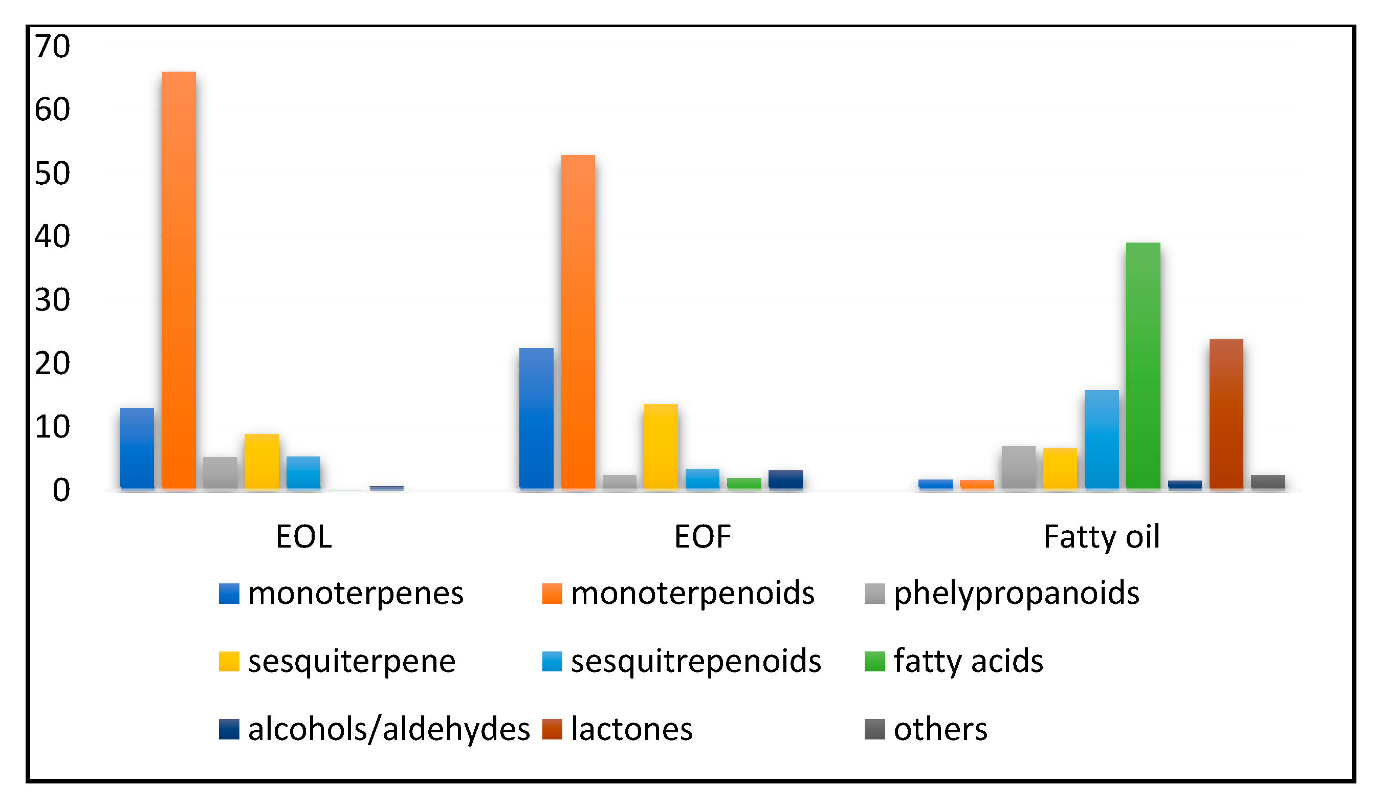
| Monoterpenes | 13.0 | 22.4 | 1.7 | ||
| Monoterpenoids | 66.0 | 52.9 | 1.6 | ||
| Phelypropanoids | 5.2 | 2.4 | 6.9 | ||
| Sesquiterpene | 8.9 | 13.6 | 6.6 | ||
| Sesquitrepenoids | 5.3 | 3.3 | 15.8 | ||
| Fatty acids | 0.1 | 1.9 | 39.1 | ||
| Alcohols/aldehydes | 0.6 | 3.1 | 1.5 | ||
| Lactones | 23.8 | ||||
| Others | 2.4 | ||||
| The percentage of the total chemical compounds | 99.1 | 99.6 | 99.4 | ||
| Test Microorganisms | EOL MIC * (μg/mL) | EOF MIC * (μg/mL) |
|---|---|---|
| Staphylococcus aureus ATCC25923 | 500 | 500 |
| Enterococcus faecalis ATCC29212 | 250 | 250 |
| Bacillus subtilis ATCC6633 | 500 | 250 |
| Escherichia coli ATCC25922 | 1000 | 1000 |
| Klebsiella pneumoniae ATCC13083 | 1000 | 1000 |
| Salmonella Abony NCTC6017 | 1000 | 500 |
| Pseudomonas aeruginosa ATCC27853 | 1000 | 1000 |
Disclaimer/Publisher’s Note: The statements, opinions and data contained in all publications are solely those of the individual author(s) and contributor(s) and not of MDPI and/or the editor(s). MDPI and/or the editor(s) disclaim responsibility for any injury to people or property resulting from any ideas, methods, instructions or products referred to in the content. |
© 2025 by the authors. Licensee MDPI, Basel, Switzerland. This article is an open access article distributed under the terms and conditions of the Creative Commons Attribution (CC BY) license (https://creativecommons.org/licenses/by/4.0/).
Share and Cite
Bojović, D.; Šoškić, M.; Žugić, A.; Milenković, M.T.; Ljumović, I.; Tadić, V.M. Chemical Analysis and Antimicrobial Potential Assessment of Wild Laurel from the National Park Skadar Lake, Montenegro. Appl. Sci. 2025, 15, 6741. https://doi.org/10.3390/app15126741
Bojović D, Šoškić M, Žugić A, Milenković MT, Ljumović I, Tadić VM. Chemical Analysis and Antimicrobial Potential Assessment of Wild Laurel from the National Park Skadar Lake, Montenegro. Applied Sciences. 2025; 15(12):6741. https://doi.org/10.3390/app15126741
Chicago/Turabian StyleBojović, Dragica, Miomir Šoškić, Ana Žugić, Marina T. Milenković, Iva Ljumović, and Vanja M. Tadić. 2025. "Chemical Analysis and Antimicrobial Potential Assessment of Wild Laurel from the National Park Skadar Lake, Montenegro" Applied Sciences 15, no. 12: 6741. https://doi.org/10.3390/app15126741
APA StyleBojović, D., Šoškić, M., Žugić, A., Milenković, M. T., Ljumović, I., & Tadić, V. M. (2025). Chemical Analysis and Antimicrobial Potential Assessment of Wild Laurel from the National Park Skadar Lake, Montenegro. Applied Sciences, 15(12), 6741. https://doi.org/10.3390/app15126741