Article

Enhancing Doxorubicin Efficacy in Hepatocellular Carcinoma: The Multi-Target Role of Muscari comosum Extract

by
Alessandro Pistone
,
Ilenia Matera
,
Vittorio Abruzzese
,
Maria Antonietta Castiglione Morelli
,
Martina Rosa
and
Angela Ostuni
*
Department of Basic and Applied Sciences, University of Basilicata, Via dell’ Ateneo Lucano 10, 85100 Potenza, Italy
*
Author to whom correspondence should be addressed.
Appl. Sci. 2025, 15(12), 6509; https://doi.org/10.3390/app15126509
Submission received: 30 April 2025 / Revised: 3 June 2025 / Accepted: 5 June 2025 / Published: 10 June 2025

Abstract

Hepatocellular carcinoma (HCC) is still a leading cause of cancer-related mortality worldwide, characterized by poor prognosis and limited therapeutic efficacy of conventional chemotherapeutics such as doxorubicin. Phytochemicals are promising adjuvants in cancer therapy due to their multi-targeted effects. In this in vitro study, we investigated the impact of a methanol–water extract (70:30 v/v, MET70) from Muscari comosum bulbs, rich in polyphenols and flavonoids, on doxorubicin-treated HepG2 human hepatoma cells. Co-treatment with MET70 increased intracellular reactive oxygen species (ROS) associated with downregulation of Nrf2 signaling, suppression of antioxidant enzymes (SOD2, GPX-1) and decreased mitochondrial UCP2 expression. MET70 modulated the inflammatory response induced by doxorubicin by decreasing TNF-α and increasing IL-6 expression. MET70 also promoted protein homeostasis through PDIA2 upregulation without exacerbating endoplasmic reticulum stress and inhibited autophagy by reducing Beclin-1 levels, contributing to increased chemosensitivity. Moreover, MET70 downregulated ABCC1 expression, suggesting a role in overcoming multidrug resistance. All these findings demonstrate that Muscari comosum extract enhances doxorubicin efficacy by targeting redox balance, inflammatory signaling, autophagy, and drug resistance, offering a promising redox-based strategy for improving HCC therapy. However, further studies should be performed in vivo.

1. Introduction

According to WHO/IARC GLOBOCAN 2022 estimates, the global cancer burden is rising rapidly driven by aging populations, evolving risk factors, and regional disparities. Liver cancer ranks sixth in global incidence and third in mortality and stands out as one of the deadliest malignancies, despite its strong association with largely preventable risk factors (chronic hepatitis B and C, alcohol consumption, and aflatoxin exposure), particularly affecting low-and middle-income countries [1]. Despite significant advancements in therapeutic strategies, including radiotherapy, surgery, immunotherapy and emerging gene therapies, chemotherapy remains a mainstay of cancer treatment, particularly for metastatic or inoperable cases [2]. However, the success of chemotherapy is often hampered by multidrug resistance (MDR), which accounts for over 90% of cancer-related deaths. MDR arises from multiple factors, including tumor-specific mechanisms, host influences and tumor–host interactions [3]. A critical contributor to MDR is the overexpression of ATP-binding cassette (ABC) transporters, which actively remove chemotherapeutic agents from cells, thereby reducing their intracellular concentration and diminishing their therapeutic efficacy [4,5]. Notable ABC transporters involved in drug resistance include P-glycoprotein (P-gp/ABCB1), MRP1 (ABCC1), MRP7 (ABCC10), and BCRP (ABCG2) [6].
Doxorubicin, a widely used anthracycline antibiotic derived from Streptomyces peucetius, is used either as a single agent or in combination regimens to treat a variety of solid tumors, including hepatocellular carcinoma (HCC) [7,8]; its cytotoxic effects are mediated through DNA intercalation, topoisomerase II inhibition, and the generation of reactive oxygen species (ROS). ROS production induces oxidative stress, resulting in lipid peroxidation, DNA damage, and protein dysfunction, which trigger apoptosis in cancer cells [9]. However, oxidative stress also harms healthy tissues, particularly the heart, and is a major contributor to dose-limiting cardiotoxicity [10,11]. Moreover, the development of MDR mechanisms, such as ABC transporter overexpression, significantly compromises doxorubicin’s therapeutic potential [12]. In recent years, various strategies have been investigated to overcome MDR, including the use of MDR modulators, nanoparticle-based drug delivery systems, metallic nanoparticles, and RNA interference therapies [13,14,15].
Increasing attention has also been given to phytochemicals as adjunctive agents in chemotherapy [16]. These naturally derived compounds have shown promise in enhancing drug accumulation in tumor cells, modulating key signaling pathways and reducing oxidative damage to healthy tissues. Among the most studied classes of phytochemicals are flavonoids, alkaloids, stilbenes (e.g., resveratrol), and curcuminoids (e.g., curcumin), which have demonstrated chemosensitizing properties [17,18]. Polyphenols and flavonoids exert their effects by modulating key signaling pathways such as PI3K/AKT/mTOR, which regulates cell metabolism, and MAPK/ERK1/2, involved in tumor proliferation. Additionally, these compounds inhibit the NF-κB pathway, thereby reducing pro-inflammatory cytokines production and promoting apoptosis in cancer cells [19].
Leopoldia comosa (LC), commonly known as Muscari comosum, is an herbaceous plant belonging to the Liliaceae family, native to the Mediterranean region. Its bulbs are a valuable source of bioactive compounds, including phenolic acids, flavonoids, phytosterols, glycosides [20]. Phytosterols exhibit anticancer effects by inhibiting tumor growth, inflammation, and oxidative stress by modulating NF-κB and MAPK/ERK pathways. They also enhance antioxidant defenses and protect mitochondrial function [21]. Glycosides play a key role in chemosensitization across different tumor types via multiple mechanisms. Their cytotoxic and antiproliferative effects involve the regulation of the cell cycle and the induction of apoptosis through increased caspase expression [22]. In Muscari comosum bulbs the presence of homoisoflavanones was identified, particularly 3-benzyl-4-chromanones, which interact with estrogen receptors and suggest potential therapeutic applications in hormone-dependent cancers [23]. There are few studies investigating the biological effects of Muscari comosum extracts on cellular models. In vitro studies on MCF-7 breast cancer cells revealed a dose-dependent antiproliferative effect of raw extracts [24]. In HepG2 cells, Muscari comosum bulb extract influenced cell viability by regulating antioxidant-related genes; the effects were correlated to the concentration of polyphenols and flavonoids, especially in the methanol–water extract [25].
Given the interplay between ROS production and doxorubicin’s cytotoxicity, this study aimed to investigate whether a methanol–water extract of Muscari comosum bulbs (70:30 v/v, MET70) enhances doxorubicin efficacy in HepG2 cells. Strategically, we started by evaluating cell viability and consequently we verified whether the observed reduction in viability correlates with changes in biomarkers involved in redox balance, inflammation, the unfolded protein response (UPR), the ER stress, autophagy, and MDR-related transporters. The overall goal is to assess the potential of MET70 to improve therapeutic outcomes in hepatocellular carcinoma.

2. Materials and Methods

2.1. Reagents

The following products were purchased from Sigma–Aldrich, S.p.A. (Milan, Italy): Dimethyl sulfoxide (DMSO, CAS: 67-68-5), methanol (CAS: 67-56-1), isopropanol (CAS: 67-63-0), Triton X-100 (CAS: 9002-93-1), Doxorubicin hydrochloride (CAS: 25316-40-9), MTT reagent (3-(4,5-dimethylthiazol-2-yl)-2,5-diphenyltetrazolium bromide, CAS: 298-93-1), DCFH-DA (2′,7′-dichlorodihydrofluorescein diacetate, CAS: 4091-99-0), SDS (CAS: 151-21-3), NP-40 (CAS: 9016-45-9), sodium deoxycholate (CAS: 302-95-4), Tris-HCl (CAS: 1185-53-1), glycerol (CAS: 56-81-5), β-mercaptoethanol (CAS: 60-24-2), bromophenol blue (CAS: 115-39-9), Protease Inhibitor Cocktail (CAS 30827-99-7), radioimmunoprecipitation assay (RIPA) buffer (CAS number 127087-87-0) (0.1% sodium dodecyl sulfate, 1% NP-40, 0.5% sodium deoxycholate in PBS, pH 7.4), Bradford solution (CAS number 67-56-1). The following reagents were purchased from Euroclone (Milan, Italy): fetal bovine serum (FBS, CAS number 9014-81-7), trypsin-ethylenediaminetetraacetic acid solution (CAS number 9002-07-7), glutamine (CAS number 56-85-9), penicillin–streptomycin and phosphate-buffered solution (PBS). Reagents used for qRT-PCR were purchased: Quick-RNA MiniPrep Kit from Zymo Research, Irvine, CA, USA; the High-Capacity cDNA Reverse Transcription Kit from Applied Biosystems, Foster City, CA, USA; iTaq™ Universal SYBR Green Supermix from Bio-Rad; primers were purchased from Eurofins Genomics Europe Shared Services GmbH, Ebersberg, Germany. The reagent used for Western blot: nitrocellulose membranes from Amersham Bioscience (Buckinghamshire, UK), ECL™ Western blot Detection Reagents from GE Healthcare (Chicago, IL, USA) and the SuperSignal™ West Pico PLUS Chemiluminescent Substrate from Thermo Scientific (Waltham, MA, USA).

2.2. Cell Culture and Treatments

The human hepatocellular carcinoma cell lines (HepG2) purchased from ATCC (American Type Culture Collection) were cultured in Dulbecco’s Modified Eagle’s Medium (DMEM) containing 4.5 g/L glucose, 10% fetal bovine serum (FBS), 2 mM L-glutamine, 100 U/mL penicillin and 100 µg/mL streptomycin, at 37 °C, in a humidified atmosphere with 5% CO₂. Doxorubicin stock solution (3.68 mM) was prepared in sterile water. Methanol and aqueous extracts (as previously described [25]) were dissolved at 2 mg/mL of sterile water containing 0.2% (v/v) dimethyl sulfoxide (DMSO). Control cells were treated with equivalent concentrations of DMSO. All stock solutions were diluted in DMEM to the required concentrations immediately before use.

2.3. Viability Assay

Cell viability was assessed using the MTT assay (3-(4,5-dimethylthiazol-2-yl)-2,5-diphenyltetrazolium bromide). HepG2 cells (1.5 × 10⁴ cells/well) were seeded in 96-well plates and treated after 24 h with doxorubicin (0.3–100 μM), either alone or in combination with 100 μg/mL of Muscari comosum methanol/water (70:30 v/v) extract (MET70) for 72 h. Following treatment, cells were incubated with medium containing 0.75 mg/mL MTT for 4 h at 37 °C. The medium was then removed and formazan crystals were solubilized with a 1:1 DMSO:isopropanol solution containing 1% Triton X-100. Absorbance was measured at 570 nm, with background subtraction at 630 nm, using a microplate reader (Multiskan™ GO Microplate Spectrophotometer, Thermo Scientific, Waltham, MA, USA). The half-maximal inhibitory concentration (IC50) of the extract was determined by non-linear curve fitting using GraphPad Prism 6 (GraphPad Software, San Diego, CA, USA). Cell viability was expressed as a percentage of control cells treated with vehicle only. All experiments were performed in triplicate and repeated three times.

2.4. Intracellular Reactive Oxygen Species (ROS) Assay

Intracellular reactive oxygen species (ROS) levels were quantified using the 2′,7′-dichlorodihydrofluorescein diacetate (DCFH-DA) assay. HepG2 cells (1.5 × 10⁴ cells/well) were seeded in a 96-well black polystyrene plate with a clear bottom. After 24 h, cells were treated with DMSO (0.02%), MET70 (100 µg/mL), doxorubicin (0.5 µM), or a combination of doxorubicin and MET70 for 72 h. Following treatment, cells were incubated with DCFH-DA (10 µM in PBS) for 30 min at 37 °C. Fluorescence intensity was measured using a GloMax™ Multi-Detection System (Promega) with a blue filter (excitation: 490 nm; emission: 510–570 nm). To account for signal interference from doxorubicin, which emits fluorescence in the same range, the signal from wells containing both the probe and the drug was adjusted by subtracting the doxorubicin-specific signal. Fluorescence values were normalized to protein content measured using the Bradford assay. Results are presented as a percentage of the negative control (DMSO-treated cells). Each treatment was performed in triplicate.

2.5. Real-Time Reverse Transcription PCR (qRT-PCR)

Total RNA was extracted from cells using the Quick-RNA MiniPrep Kit and reverse transcribed into cDNA using random primers and the High-Capacity cDNA Reverse Transcription Kit. Quantitative real-time PCR (qRT-PCR) was performed on the 7500 Fast Real-Time PCR System (Applied Biosystems) with iTaq™ Universal SYBR Green Supermix. PCR specificity was confirmed by melting curve analysis. Relative mRNA expression levels were calculated using the 2−ΔCt method, with β-actin as the endogenous reference gene. Primers were designed to span exon–exon junctions to avoid amplification of genomic DNA (Table 1).

2.6. Western Blot Analysis

Samples were prepared as previously reported [26]. Briefly, cells were lysed in RIPA buffer (0.1% sodium dodecyl sulfate, 1% NP-40, 0.5% sodium deoxycholate in PBS, pH 7.4) supplemented with a protease and phosphatase inhibitor cocktail, using sonication. The lysates were centrifuged at 13.000 rpm for 10 min at 4 °C and protein concentration was measured using the Bradford solution. Proteins (40 μg) were mixed with Laemmli sample buffer (60 mM Tris-HCl, pH 6.8, 10% glycerol, 2% SDS, 1% β-mercaptoethanol and 0.002% bromophenol blue) and separated by SDS-PAGE. After electrophoresis, proteins were transferred onto nitrocellulose membranes. Membranes were blocked in 5% non-fat milk prepared in PBS with 0.05% Tween-20 (PBST) for 2 h at room temperature and incubated overnight at 4 °C with specific primary antibodies: 1:400 anti-β-actin monoclonal antibody (A5441; Sigma-Aldrich, Merck KGaA, Darmstadt, Germany); 1:1000 monoclonal anti-α -tubulin (T9026; Sigma-Aldrich, Merck KGaA); 1:500 MRP1 Polyclonal Antibody (PA5-30594, Invitrogen, Thermo Fisher Scientific, Waltham, MA, USA); 1:20 Anti-BCRP/ABCG2 antibody (ab24115, Abcam, Cambridge, UK); 1:1000 Anti-MDR1/ABCB1 Antibody (E1Y7B, Cell Signaling, Danvers, MA, USA); 1:400 GPX-1/2 antibody (sc-133160, Santa Cruz, Dallas, TX, USA); 1:1000 purified anti-ATF4 antibody (W16016A, BioLegend, San Diego, CA, USA); 1:1000 purified anti-ATF6 antibody (W17028A, BioLegend). The membrane was washed with PBST and incubated with an HRP-conjugated secondary antibody at room temperature for 1 h. Signal was visualized using ECL™ Western blotting Detection Reagents or the SuperSignal™ West Pico PLUS Chemiluminescent Substrate on a Chemidoc™ XRS detection system (Bio-Rad Laboratories, Hercules, CA, USA) equipped with Image Lab 5.1 software. Densitometric analysis was performed using GelAnalizer 19.1 (Istvan Lazar, www.gelanalyzer.com (accessed on 1 January 2023). Protein expression levels were normalized to the control sample, set at 100. Each experiment was performed in triplicate.

2.7. Statistical Analysis

All assays were performed independently at least three times. Data are expressed as the mean ± standard error of the mean (SEM). Statistical analysis was conducted using one-way ANOVA in GraphPad Prism 8 (GraphPad Software, San Diego, CA, USA), followed by Holm–Sidak’s test for multiple comparisons. A significance level of α = 0.05 was used to determine statistical significance (p < 0.05, * p < 0.01, ** p < 0.001).

3. Results

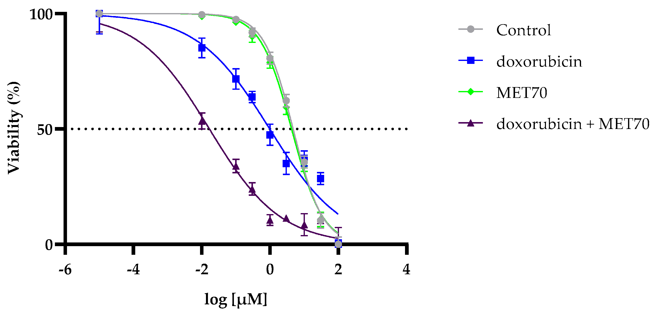
3.1. Enhanced Reduction in Hepg2 Cell Viability by Combined Treatment with Doxorubicin and Muscari comosum Extract (MET70)

Chemical characterization of the bulb of Muscari comosum revealed a diverse range of phytochemicals, including phenolic acids, fatty acids, flavonoids, triterpenes, phytosterols, and homoisoflavones [20,23]. Our previous study demonstrated that the 70% methanol extract (MET70) had the highest total phenolic content (58.72 ± 1.11 mg gallic acid equivalents/g extract) and one of the highest flavonoid contents (20.37 ± 1.24 mg quercetin equivalents/g extract) among the tested extracts. Additionally, MET70 exhibited strong antioxidant activity in vitro and reduced HepG2 cell viability by approximately 30% at a concentration of 100 µg/mL after 72 h of exposure [25].
To assess whether the bulb extract could enhance the effect of doxorubicin on cell viability, HepG2 cells were treated with increasing doses of doxorubicin, both alone and in combination with MET70 (100 µg/mL). The MTT assay (Figure 1) revealed that doxorubicin reduced cell viability in a dose-dependent manner after 72 h, with an IC50 of 0.98 µM (95% confidence interval, 0.645–1.461 µM). Notably, the combination of doxorubicin and MET70 significantly reduced cell viability, with an IC50 of 0.01675 µM (95% confidence interval, 0.0106–0.02514 µM). Treatment with the MET70 extract alone resulted in only a slight reduction in cell viability, whereas the untreated control group maintained consistently high viability values, supporting the robustness of the assay. These results indicate that, while MET70 exhibits limited cytotoxicity by itself, it significantly enhances the susceptibility of HepG2 cells to doxorubicin.
Based on these results, subsequent experiments were conducted using a sublethal dose of 0.5 µM doxorubicin in combination with 100 µg/mL MET70.

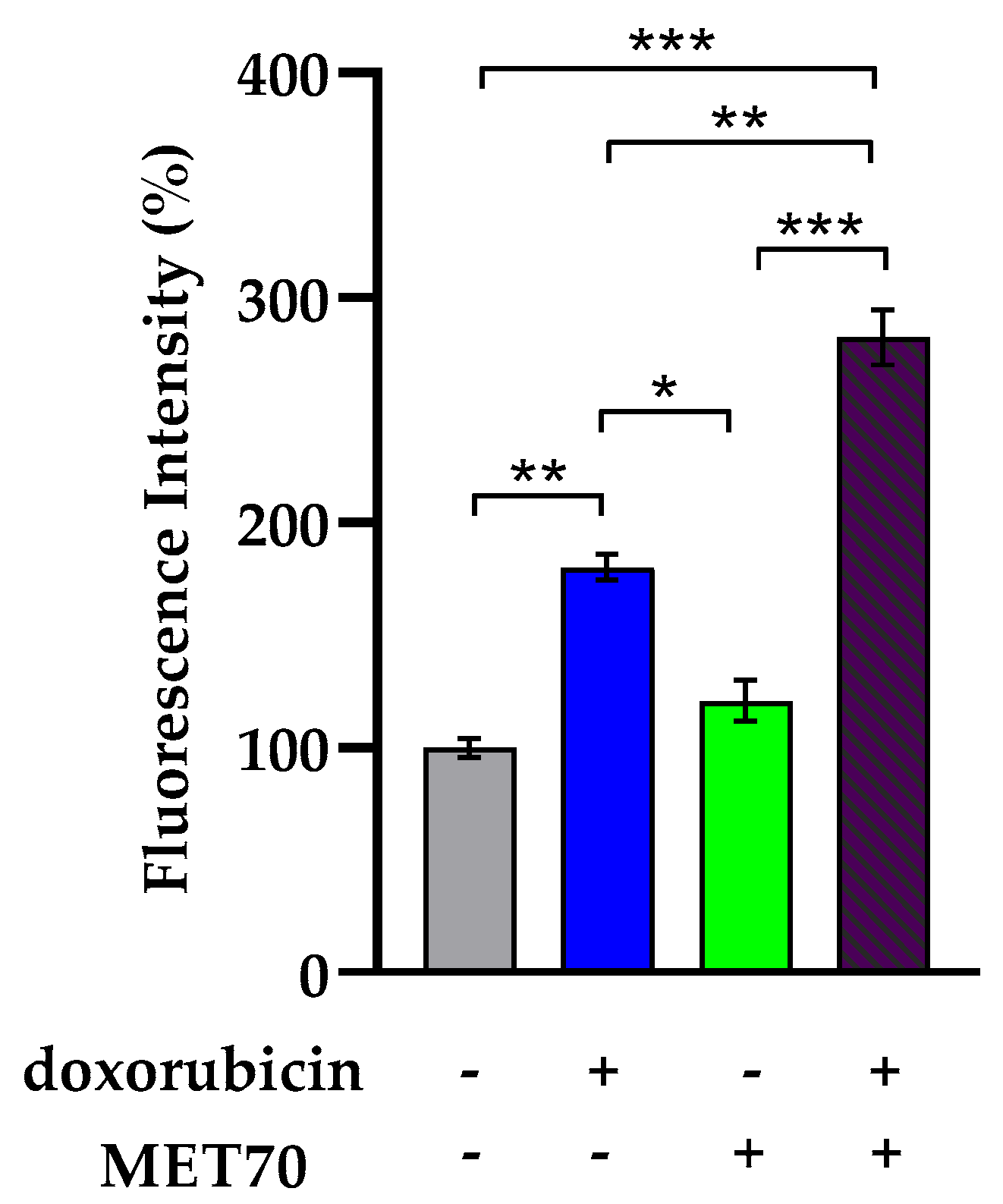
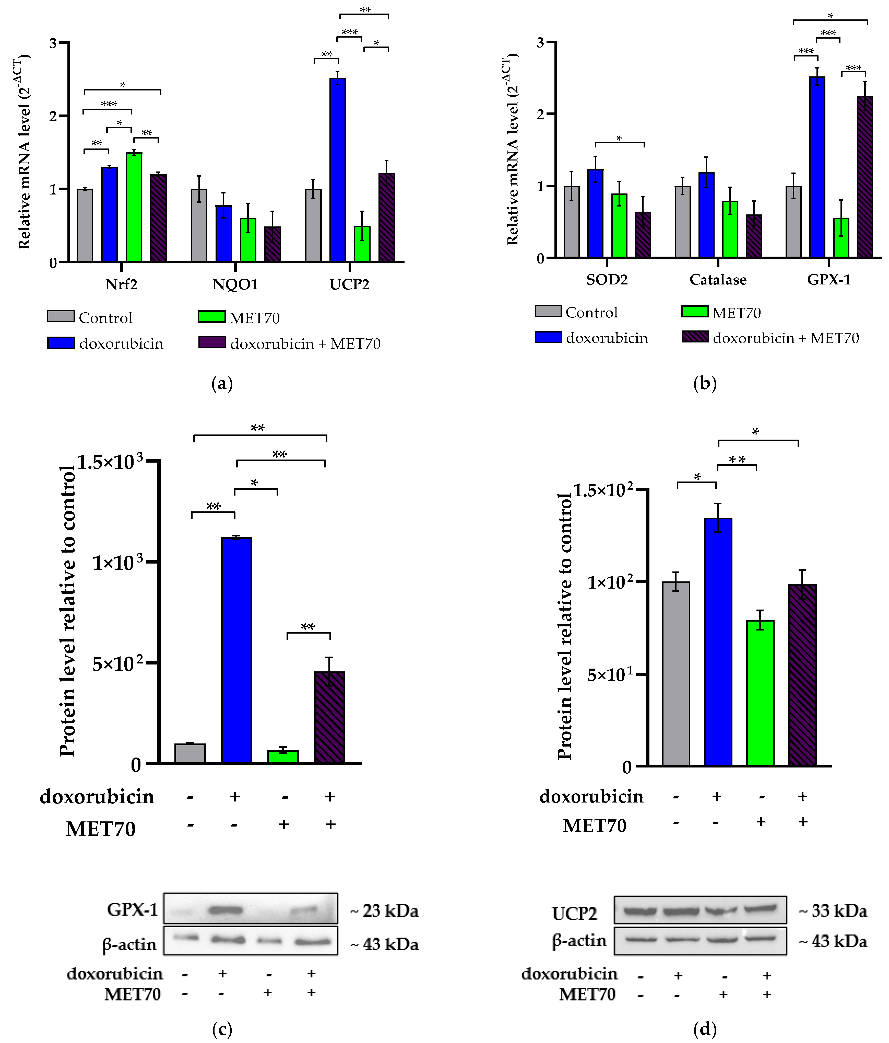
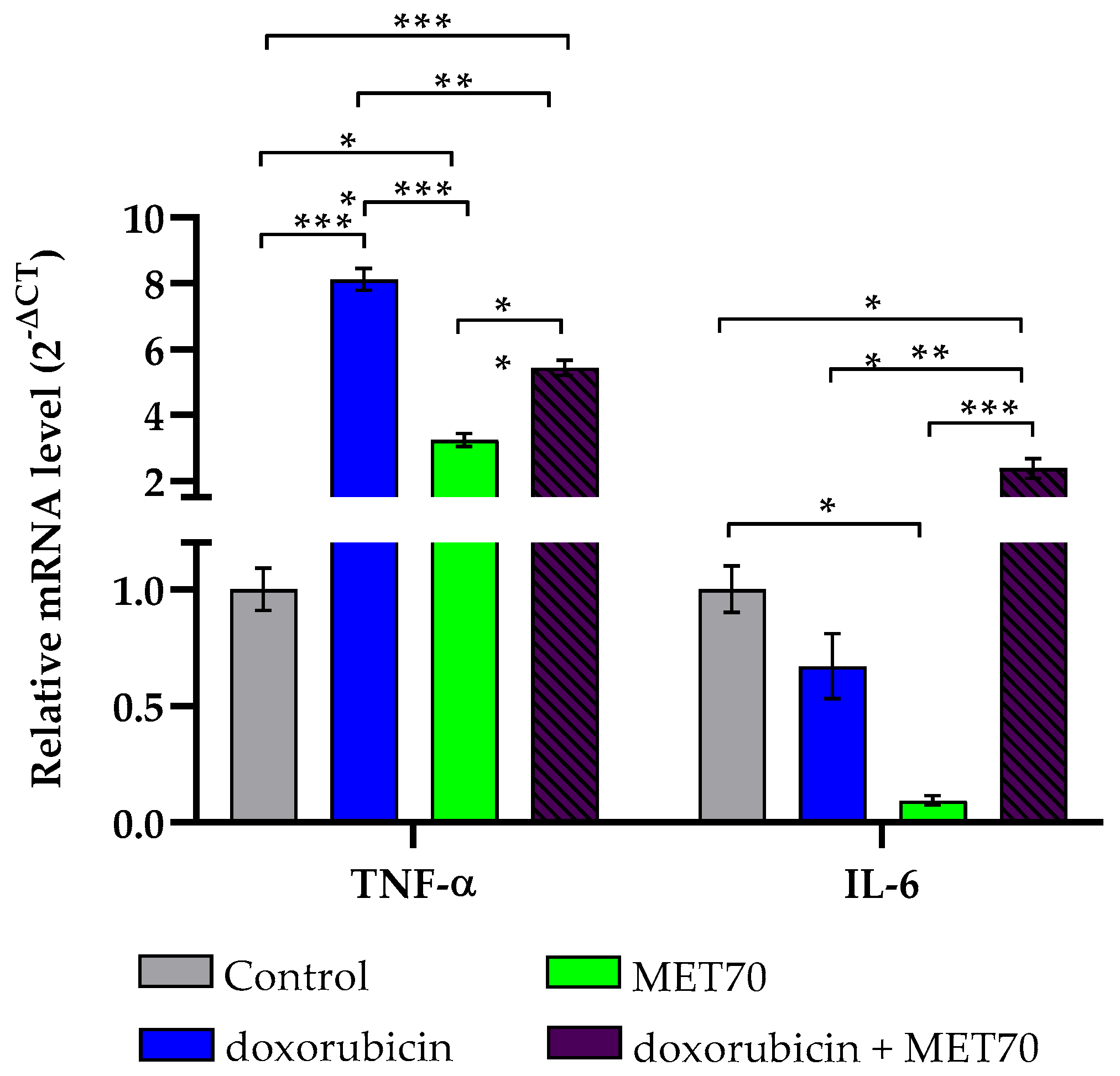
3.2. Impact of Combined Doxorubicin and Muscari comosum Extract (MET70) Treatment on Intracellular ROS Levels and Redox/Inflammatory Gene Expression

To evaluate the effect of MET70 treatment in combination with doxorubicin on intracellular reactive oxygen species (ROS) accumulation, we measured the oxidation of a non-fluorescent probe (2′,7′-dichlorodihydrofluorescein diacetate, DCFH-DA) to its fluorescent form (2′,7′-dichlorofluorescein, DCF). As shown in Figure 2, after 72 h, the combination of the drug and extract resulted in a significant increase in intracellular ROS levels, enhancing the effect of doxorubicin. Furthermore, treatment with MET70 alone did not alter the level of cellular ROS.
We analyzed the expression of genes involved in the regulation of redox balance. The expression level of Nrf2, a key transcription factor that regulates the production of ROS-scavenging enzymes, increased after treatment with doxorubicin alone but did not change significantly in combination with MET70. MET70 itself increased the expression level of Nrf2 (Figure 3a). The combined treatment had no effect on the catalase and NAD(P)H Quinone Dehydrogenase 1 (NQO1) expression; on the contrary, it decreased both superoxide dismutase 2 (SOD2) and Glutathione peroxidase 1 (GPX-1) expression levels (Figure 3b,c). The expression level of mitochondrial uncoupling protein 2 (UCP2), a mitochondrial protein involved in the resolution of oxidative stress, decreased following the combined treatment (Figure 3b,d). Ultimately, the reduced expression of SOD2 and, in particular, of both GPX-1 and UCP2 could be responsible for the greater oxidative stress and therefore the lower viability of cells treated with doxorubicin combined with MET70.
The expression level of tumor necrosis factor alpha (TNF-α) increased significantly in the presence of doxorubicin. However, the effect was reduced when doxorubicin was combined with MET70. Doxorubicin in combination with MET70 induced an increase in the expression of interleukin 6 (IL-6) (Figure 4).

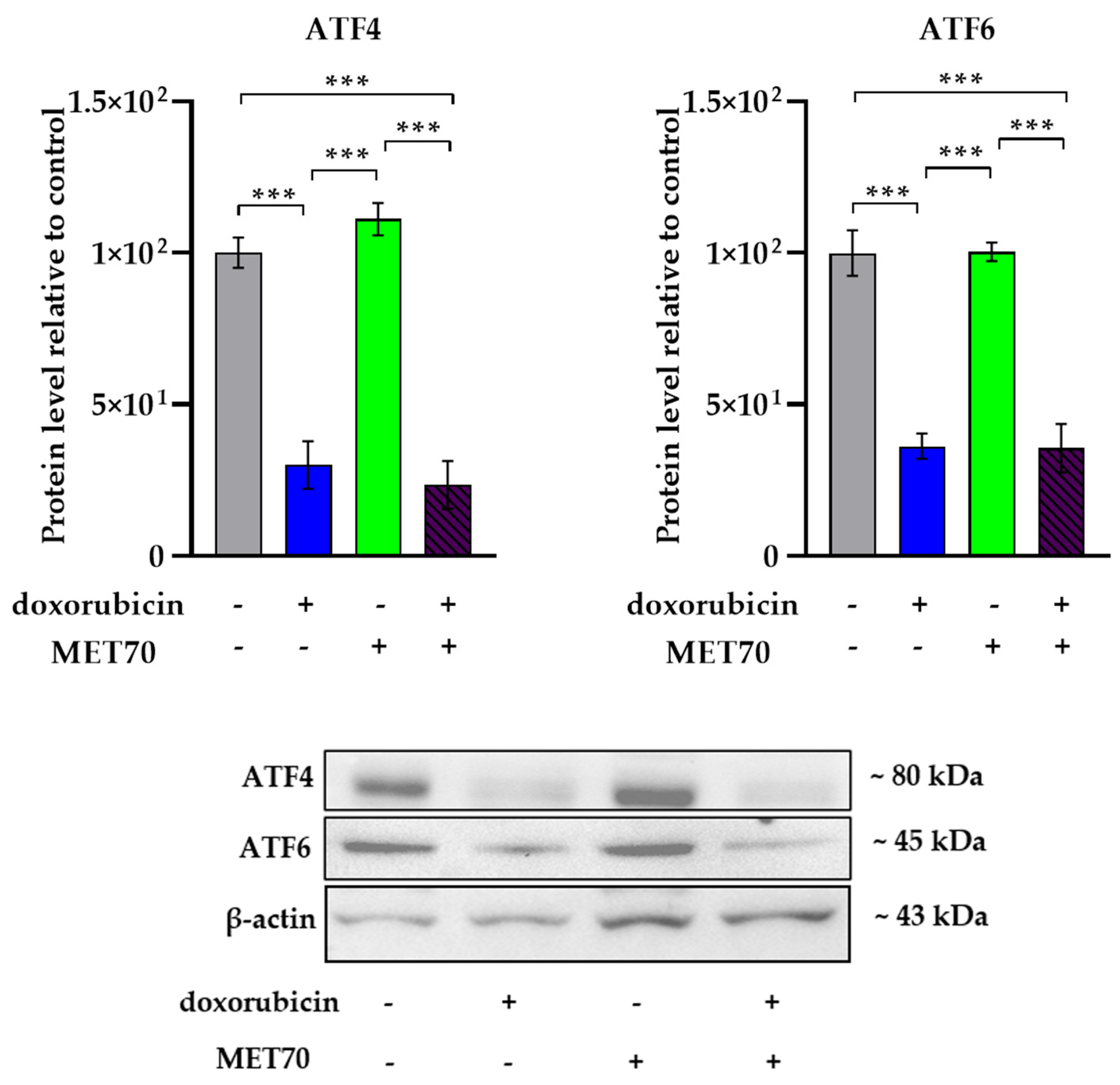
3.3. Effect of Doxorubicin-Muscari comosum Extract Treatment on the UPR Pathway

To investigate the involvement of endoplasmic reticulum (ER) stress, we analyzed the expression levels of key ER stress markers in HepG2 cells treated with doxorubicin alone or in combination with MET70.
RT-qPCR analysis revealed that doxorubicin treatment decreased the expression of the chaperone BiP and the disulfide isomerase PDIA2. However, co-treatment with MET70 did not alter BiP expression and significantly increased PDIA2 expression compared to doxorubicin alone. Doxorubicin also increased the expression of the ER stress sensor DNA damage-inducible transcript 3 (CHOP), while the combination with MET70 did not significantly modify this effect (Figure 5).
Moreover, the expression levels of activating transcription factors 4 (ATF4) and 6 (ATF6), two proteins acting downstream in the unfolded protein response (UPR), were similarly decreased by doxorubicin alone and by the combination treatment, without evidence of a synergistic effect (Figure 6).
Overall, these results suggest that the addition of MET70 to doxorubicin does not significantly exacerbate or mitigate the ER stress response induced by doxorubicin in HepG2 cells.

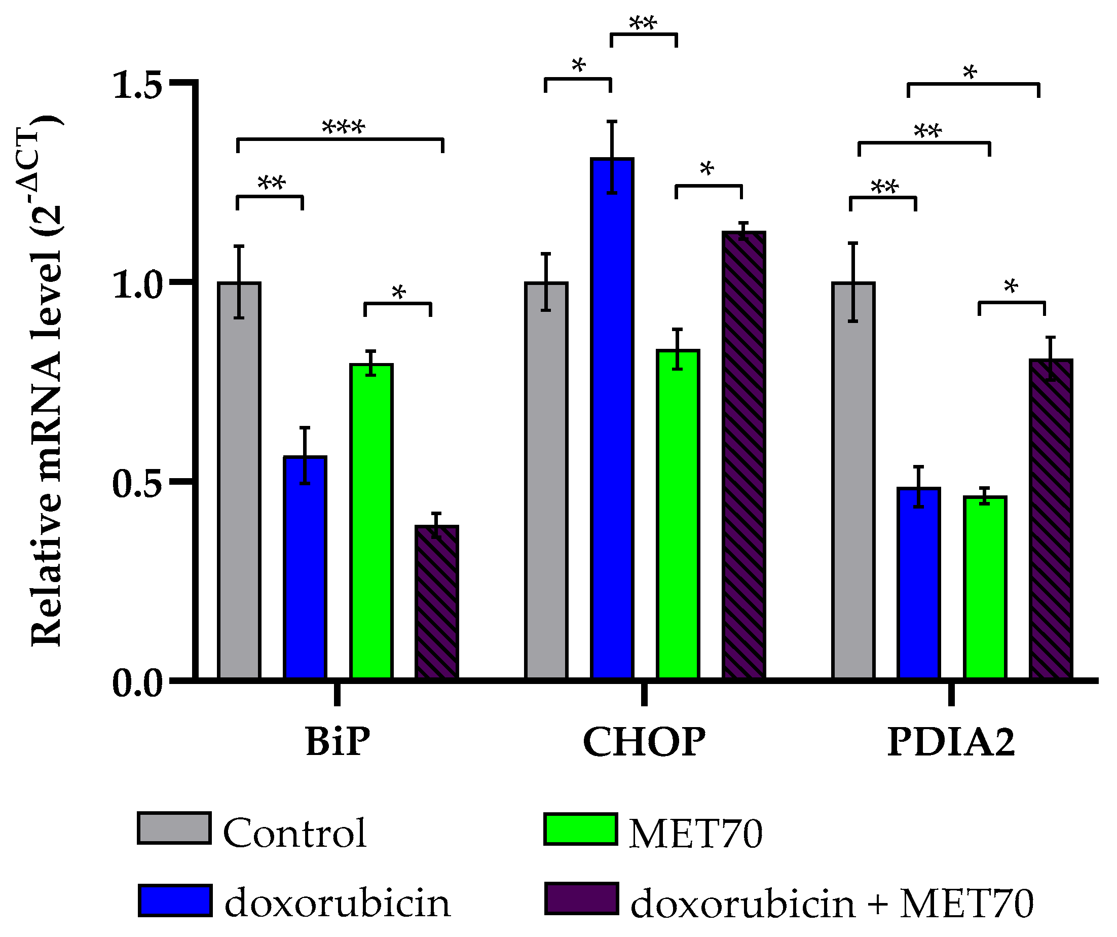
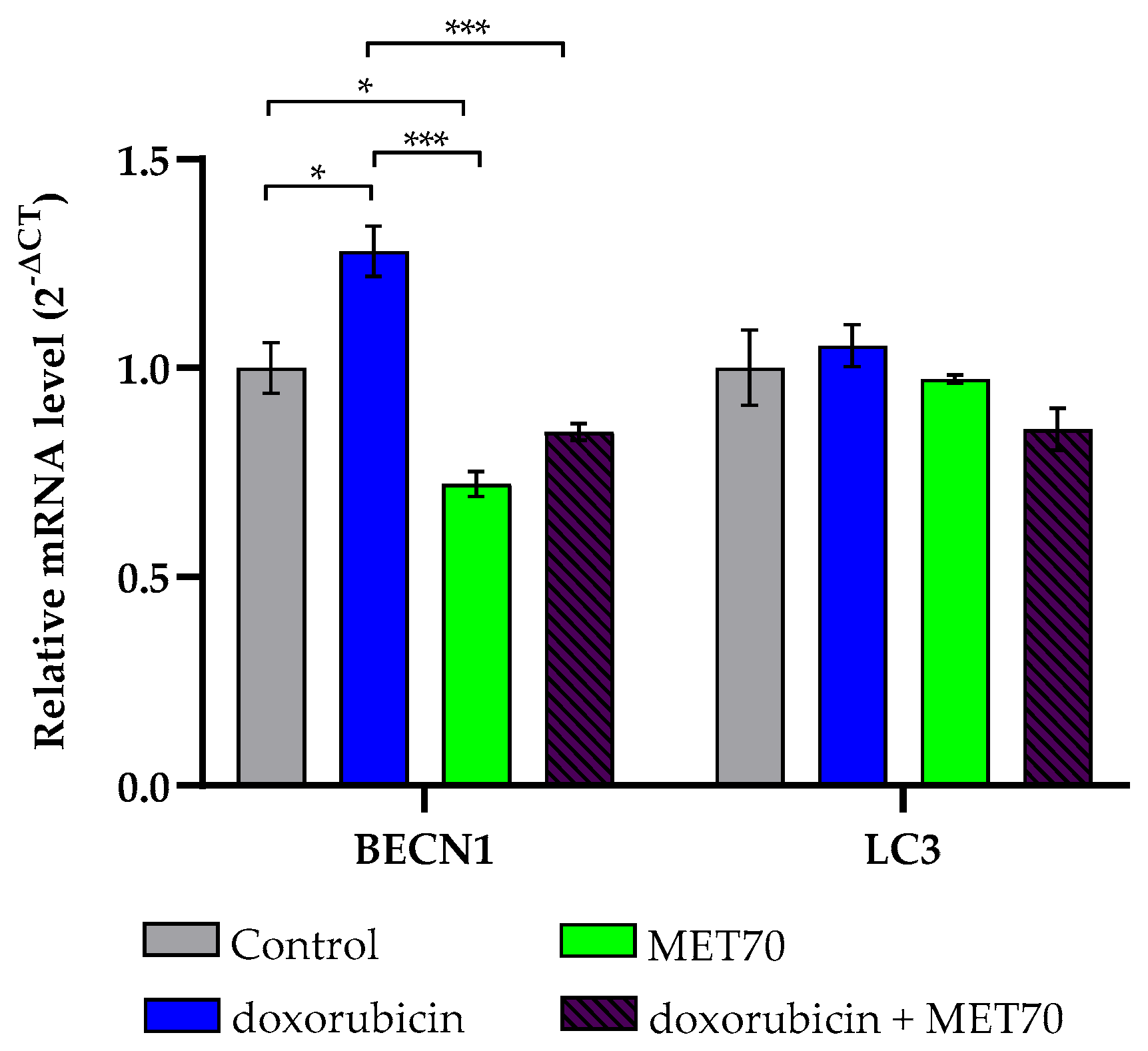
3.4. Modulation of Endoplasmic Reticulum Stress Markers by Combined Doxorubicin and Muscari comosum Extract (MET70) Treatment

Cancer cells often exploit autophagy as a survival mechanism under conditions of excessive stress and in response to cytotoxic agents. To investigate the impact of the combined doxorubicin and Muscari comosum extract (MET70) treatment on autophagy, we evaluated the expression levels of two key autophagy-related genes, LC3 and Beclin-1.
No significant changes in the expression of microtubule-associated protein 1A/1B-light chain 3 (LC3) were observed following treatment with doxorubicin alone or in combination with MET70 (Figure 7). However, while doxorubicin alone increased the expression of Beclin-1 (BECN1), the combination treatment attenuated this effect, suggesting that inhibition of autophagy may contribute to the enhanced cytotoxicity of doxorubicin.

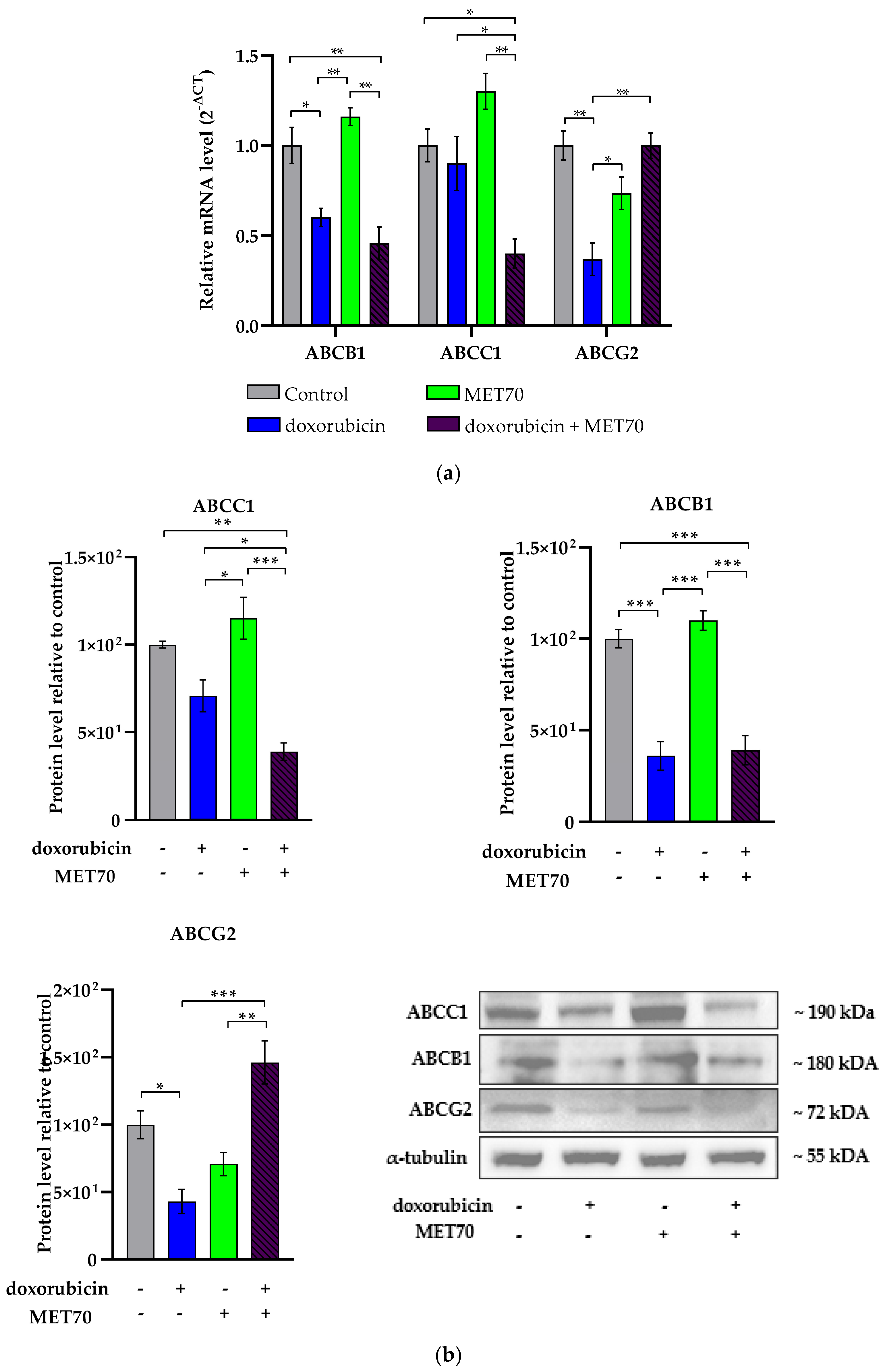
3.5. Modulation of ABC Transporters Expression by Combined Doxorubicin and Muscari comosum Extract (MET70) Treatment

Multidrug resistance (MDR) remains a major obstacle in cancer therapy, often mediated by the efflux of chemotherapeutic agents through ATP-binding cassette (ABC) transporters. Among the most relevant transporters are ABCB1, ABCC1, and ABCG2. In this study, we investigated the modulatory effects of the combined treatment with doxorubicin and Muscari comosum extract (MET70) on the expression of these transporters.
RT-qPCR and Western blot analyses demonstrated that doxorubicin treatment alone significantly reduced both mRNA and protein levels of ABCB1 and ABCG2 compared to control cells, while ABCC1 expression remained unchanged (Figure 8a,b). Interestingly, compared to doxorubicin alone, the combination with MET70 did not further alter ABCB1 expression, but significantly decreased ABCC1 levels and increased ABCG2 expression.
These findings suggest that MET70 may differentially modulate ABC transporter expression, potentially contributing to the sensitization of HepG2 cells to doxorubicin.

4. Discussion

Hepatocellular carcinoma (HCC), the most common form of liver cancer, accounts for approximately 75–85% of all liver cancer cases. Despite advancements in treatment strategies, HCC remains a major clinical challenge, with a five-year survival rate of only 8.37% [27,28]. A promising strategy for improving cancer therapy involves the use of phytochemicals as adjuvants to conventional chemotherapy, due to their demonstrated anti-tumor, anti-inflammatory, and anti-angiogenic properties [16,17,18,19,21,22].
In this study, we evaluated for the first time the potential of a methanol–water extract of Muscari comosum (MET70) to enhance the efficacy of doxorubicin in HepG2 liver cancer cells. The combination treatment significantly reduced cell viability compared to either agent alone, indicating a synergistic cytotoxic effect and the possibility of using lower doses of doxorubicin to mitigate its systemic toxicity. Although limited studies have evaluated M. comosum in cancer models [24,25], previous reports have highlighted its high polyphenol and flavonoid content, which are known to be associated with anticancer effects [17,19,21,22].
Doxorubicin exerts its cytotoxic effects through multiple mechanisms, including the induction of reactive oxygen species (ROS) [7,8,9,10,11,29]. Interestingly, MET70 alone did not significantly alter ROS levels in our model, but when combined with doxorubicin, a substantial increase in ROS production was observed. This synergistic effect may occur due to the downregulation of antioxidant defenses, including glutathione peroxidase-1 (GPX-1) and superoxide dismutase (SOD2), as well as the potential interference with the nuclear factor erythroid 2–related factor 2 (Nrf2) signaling pathway, as well as reduced levels of mitochondrial uncoupling protein 2 (UCP2). This enhanced ROS production likely contributed to the increased cytotoxicity observed following the combination treatment [30,31,32,33]. Although MET70 alone upregulated Nrf2 and did not significantly increase ROS, the combination treatment with doxorubicin suppressed antioxidant defenses (e.g., GPX-1, SOD2), potentially explaining the observed ROS synergy and reduced viability. While cancer cells can tolerate moderate ROS through adaptive mechanisms, excessive ROS surpasses antioxidant capacity, resulting in oxidative damage and cell death. Thus, MET70 may potentiate doxorubicin efficacy by disrupting redox homeostasis beyond a tolerable threshold.
It is well established that elevated levels of pro-inflammatory cytokines can further promote ROS generation, creating a feedback loop that exacerbates oxidative stress [33]. Oxidative stress and inflammation are closely linked with ROS promoting the release of pro-inflammatory cytokines. Doxorubicin activates NF-κB and increases IL-6 and TNF-α levels [10,34,35]. We found that co-treatment with MET70 increased IL-6 expression, suggesting that MET70 may modulate inflammatory responses during chemotherapy. Similar behavior has been noted for polyphenol-rich extracts that both exacerbate or attenuate cytokine responses, depending on the context [18]. Although IL-6 can promote tumorigenesis in chronic inflammation, its acute elevation in response to high ROS levels may facilitate cancer cell death.
We also investigated markers of the unfolded protein response (UPR), which is activated during ER stress, particularly under conditions of proteotoxicity induced by chemotherapy. As expected, doxorubicin modulated several ER stress markers and reduced the expression of the chaperone BiP and protein disulfide isomerase PDIA2 while increasing the expression of the pro-apoptotic ER stress sensor CHOP. Additionally, doxorubicin decreased the expression of the downstream UPR mediators ATF4 and ATF6. Given the cytotoxic synergy observed between MET70 and doxorubicin, it was initially hypothesized that MET70 would exacerbate ER stress responses. As a crude plant extract, MET70 contains a complex mixture of bioactive compounds capable of simultaneously targeting multiple cellular pathways. Contrary to our expectations, the data reveal a selective modulation of the ER stress response. Co-treatment with MET70 did not further alter BiP, CHOP, ATF4, or ATF6 expression but significantly upregulated PDIA2 compared to doxorubicin alone. These findings suggest that MET70 does not amplify doxorubicin-induced ER stress but may instead enhance protein folding capacity via PDIA2 upregulation [36,37]. This modulatory effect could support ER homeostasis under chemotherapeutic stress, indicating that MET70 enhances cytotoxicity primarily by promoting redox imbalance rather than intensifying UPR-mediated cell death.
We also observed that MET70 downregulated Beclin-1, a key initiator of autophagy that interacts with the PI3KC3 complex to promote autophagosome formation and activate the autophagic pathway. Beclin-1 functions as a tumor suppressor during early cancer development. However, in established tumors, autophagy is often activated as a protective mechanism in response to chemotherapy-induced ER stress and ROS generation. Therefore, MET70-mediated suppression of Beclin-1 may impair this survival pathway, thereby enhancing doxorubicin cytotoxicity [38].
Finally, we evaluated the expression of multidrug resistance (MDR)-related ABC transporters. Doxorubicin alone downregulated ABCB1 and ABCG2, while MET70 treatment selectively reduced ABCC1 expression in combination. These transporters are major contributors to drug efflux and resistance in cancer therapy [39,40,41]. Notably, while ABCG2 was upregulated in the co-treatment condition, the downregulation of ABCC1 and ABCB1 may still result in a net increase in doxorubicin accumulation within the cell.

5. Conclusions

Taken together, our findings support the potential of Muscari comosum extract as a promising chemosensitizer for hepatocellular carcinoma by enhancing doxorubicin efficacy through multiple mechanisms such as increased oxidative stress, modulation of inflammatory signaling, autophagy inhibition, ER stress attenuation, and downregulation of MDR-related transporters. These effects contribute to greater cytotoxicity at potentially lower doses of doxorubicin, suggesting a strategy to reduce its systemic toxicity. However, it is important to emphasize that this study was conducted exclusively in vitro. Further in vivo studies are essential to evaluate the therapeutic relevance, safety profile, and pharmacokinetics of MET70. Future work should also focus on using biocompatible extraction methods, such as aqueous ethanol and on isolating and characterizing the active constituents responsible for the observed effects.

Author Contributions

Conceptualization, A.O.; formal analysis, A.P., I.M. and V.A.; investigation, A.P. and M.R.; writing—original draft preparation, A.O. and V.A.; writing—review and editing, A.O. and M.A.C.M.; visualization, I.M.; supervision, A.O.; project administration, A.O. All authors have read and agreed to the published version of the manuscript.

Funding

This research received no external funding.

Institutional Review Board Statement

Not applicable.

Informed Consent Statement

Not applicable.

Data Availability Statement

The original contributions presented in this study are included in the article. Further inquiries may be addressed to the corresponding author.

Acknowledgments

The authors thank Rocco Rossano of the Department of Basic and Applied Sciences (University of Basilicata, Potenza-Italy) for donating Muscari comosum bulbs.

Conflicts of Interest

The authors declare no conflicts of interest.

References

  1. Bray, F.; Laversanne, M.; Sung, H.; Ferlay, J.; Siegel, R.L.; Soerjomataram, I.; Jemal, A. Global cancer statistics 2022: GLOBOCAN estimates of incidence and mortality worldwide for 36 cancers in 185 countries. CA Cancer J. Clin. 2024, 74, 229–263. [Google Scholar] [CrossRef] [PubMed]
  2. Anand, U.; Dey, A.; Chandel, A.K.S.; Sanyal, R.; Mishra, A.; Pandey, D.K.; De Falco, V.; Upadhyay, A.; Kandimalla, R.; Chaudhary, A.; et al. Cancer Chemotherapy and beyond: Current Status, Drug Candidates, Associated Risks and Progress in Targeted Therapeutics. Genes Dis. 2022, 10, 1367–1401. [Google Scholar] [CrossRef]
  3. Bukowski, K.; Kciuk, M.; Kontek, R. Mechanisms of Multidrug Resistance in Cancer Chemotherapy. Int. J. Mol. Sci. 2020, 21, 3233. [Google Scholar] [CrossRef]
  4. Sajid, A.; Rahman, H.; Ambudkar, S.V. Advances in the Structure, Mechanism and Targeting of Chemoresistance-Linked ABC Transporters. Nat. Rev. Cancer 2023, 23, 762–779. [Google Scholar] [CrossRef]
  5. Xiao, H.; Zheng, Y.; Ma, L.; Tian, L.; Sun, Q. Clinically-Relevant ABC Transporter for Anti-Cancer Drug Resistance. Front. Pharmacol. 2021, 12, 705. [Google Scholar] [CrossRef] [PubMed]
  6. Pote, M.S.; Gacche, R.N. ATP-Binding Cassette Efflux Transporters and MDR in Cancer. Drug Discov. Today 2023, 28, 103537. [Google Scholar] [CrossRef]
  7. Mattioli, R.; Ilari, A.; Colotti, B.; Mosca, L.; Fazi, F.; Colotti, G. Doxorubicin and Other Anthracyclines in Cancers: Activity, Chemoresistance and Its Overcoming. Mol. Aspects Med. 2023, 93, 101205. [Google Scholar] [CrossRef] [PubMed]
  8. Sohail, M.; Sun, Z.; Li, Y.; Gu, X.; Xu, H. Research Progress in Strategies to Improve the Efficacy and Safety of Doxorubicin for Cancer Chemotherapy. Expert Rev. Anticancer Ther. 2021, 21, 1385–1398. [Google Scholar] [CrossRef] [PubMed]
  9. Kciuk, M.; Gielecińska, A.; Mujwar, S.; Kołat, D.; Kałuzińska-Kołat, Ż.; Celik, I.; Kontek, R. Doxorubicin-An Agent with Multiple Mechanisms of Anticancer Activity. Cells 2023, 12, 659. [Google Scholar] [CrossRef]
  10. Vitale, R.; Marzocco, S.; Popolo, A. Role of Oxidative Stress and Inflammation in Doxorubicin-Induced Cardiotoxicity: A Brief Account. Int. J. Mol. Sci. 2024, 25, 7477. [Google Scholar] [CrossRef]
  11. Linders, A.N.; Dias, I.B.; López Fernández, T.; Tocchetti, C.G.; Bomer, N.; Van der Meer, P. A Review of the Pathophysiological Mechanisms of Doxorubicin-Induced Cardiotoxicity and Aging. NPJ Aging 2024, 10, 9. [Google Scholar] [CrossRef] [PubMed]
  12. Limeres, M.J.; Moretton, M.A.; Bernabeu, E.; Chiappetta, D.A.; Cuestas, M.L. Thinking Small, Doing Big: Current Success and Future Trends in Drug Delivery Systems for Improving Cancer Therapy with Special Focus on Liver Cancer. Mater. Sci. Eng. C 2019, 95, 328–341. [Google Scholar] [CrossRef] [PubMed]
  13. Duan, C.; Yu, M.; Xu, J.; Li, B.-Y.; Zhao, Y.; Kankala, R.K. Overcoming Cancer Multi-Drug Resistance (MDR): Reasons, Mechanisms, Nanotherapeutic Solutions, and Challenges. Biomed. Pharmacother. 2023, 162, 114643. [Google Scholar] [CrossRef]
  14. Koirala, M.; DiPaola, M. Overcoming Cancer Resistance: Strategies and Modalities for Effective Treatment. Biomedicines 2024, 12, 1801. [Google Scholar] [CrossRef]
  15. Rodríguez-Félix, F.; Graciano-Verdugo, A.Z.; Moreno-Vásquez, M.J.; Lagarda-Díaz, I.; Barreras-Urbina, C.G.; Armenta-Villegas, L.; Olguín-Moreno, A.; Tapia-Hernández, J.A. Trends in Sustainable Green Synthesis of Silver Nanoparticles Using Agri-Food Waste Extracts and Their Applications in Health. J. Nanomater. 2022, 8874003. [Google Scholar] [CrossRef]
  16. Kangra, K.; Kakkar, S.; Mittal, V.; Kumar, V.; Aggarwal, N.; Chopra, H.; Malik, T.; Garg, V. Incredible use of plant-derived bioactives as anticancer agents. RSC Adv. 2025, 15, 1721–1746. [Google Scholar] [CrossRef] [PubMed]
  17. Almilaibary, A. Phyto-therapeutics as anti-cancer agents in breast cancer: Pathway targeting and mechanistic elucidation. Saudi J. Biol. Sci. 2024, 31, 103935. [Google Scholar] [CrossRef]
  18. Pandey, P.; Lakhanpal, S.; Mahmood, D.; Kang, H.N.; Kim, B.; Kang, S.; Choi, J.; Choi, M.; Pandey, S.; Bhat, M.; et al. An updated review summarizing the anticancer potential of flavonoids via targeting NFkB pathway. Front. Pharmacol. 2025, 15, 1513422. [Google Scholar] [CrossRef]
  19. Hon, K.W.; Naidu, R. Synergistic Mechanisms of Selected Polyphenols in Overcoming Chemoresistance and Enhancing Chemosensitivity in Colorectal Cancer. Antioxidants 2024, 13, 815. [Google Scholar] [CrossRef]
  20. Jaiswal, V.; Lee, H.-J. The Bioactivity and Phytochemicals of Muscari comosum (Leopoldia comosa), a Plant of Multiple Pharmacological Activities. Int. J. Mol. Sci. 2024, 25, 2592. [Google Scholar] [CrossRef]
  21. Shen, M.; Yuan, L.; Zhang, J.; Wang, X.; Zhang, M.; Li, H.; Jing, Y.; Zeng, F.; Xie, J. Phytosterols: Physiological Functions and Potential Application. Foods 2024, 13, 1754. [Google Scholar] [CrossRef] [PubMed]
  22. Matsuo, Y.; Mimaki, Y. Search for new steroidal glycosides with anti-cancer potential from natural resources. J. Nat. Med. 2024, 78, 807–827. [Google Scholar] [CrossRef] [PubMed]
  23. Grande, F.; Rizzuti, B.; Occhiuzzi, M.A.; Ioele, G.; Casacchia, T.; Gelmini, F.; Guzzi, R.; Garofalo, A.; Statti, G. Identification by Molecular Docking of Homoisoflavones from Leopoldia Comosa as Ligands of Estrogen Receptors. Molecules 2018, 23, 894. [Google Scholar] [CrossRef] [PubMed]
  24. Casacchia, T.; Sofo, A.; Casaburi, I.; Marrelli, M.; Conforti, F.; Statti, G.A. Antioxidant, Enzyme-Inhibitory and Antitumor Activity of the Wild Dietary Plant Muscari comosum (L.) Mill. Int. J. Plant Biol. 2017, 8, 6895. [Google Scholar] [CrossRef]
  25. Giglio, F.; Castiglione Morelli, M.A.; Matera, I.; Sinisgalli, C.; Rossano, R.; Ostuni, A. Muscari comosum L. Bulb Extracts Modulate Oxidative Stress and Redox Signaling in Hepg2 Cells. Molecules 2021, 26, 416. [Google Scholar] [CrossRef]
  26. Matera, I.; Miglionico, R.; Abruzzese, V.; Marchese, G.; Ventola, G.M.; Castiglione Morelli, M.A.; Bisaccia, F.; Ostuni, A. A Regulator Role for the ATP-Binding Cassette Subfamily C Member 6 Transporter in HepG2 Cells: Effect on the Dynamics of Cell-Cell and Cell-Matrix Interactions. Int. J. Mol. Sci. 2023, 24, 16391. [Google Scholar] [CrossRef]
  27. Sagnelli, E.; Macera, M.; Russo, A.; Coppola, N.; Sagnelli, C. Epidemiological and Etiological Variations in Hepatocellular Carcinoma. Infection 2020, 48, 7–17. [Google Scholar] [CrossRef]
  28. Sarveazad, A.; Agah, S.; Babahajian, A.; Amini, N.; Bahardoust, M. Predictors of 5 Year Survival Rate in Hepatocellular Carcinoma Patients. J. Res. Med. Sci. 2019, 24, 86. [Google Scholar] [CrossRef]
  29. Prasanna, P.L.; Renu, K.; Valsala Gopalakrishnan, A. New Molecular and Biochemical Insights of Doxorubicin-Induced Hepatotoxicity. Life Sci. 2020, 250, 117599. [Google Scholar] [CrossRef]
  30. Mirzaei, S.; Zarrabi, A.; Hashemi, F.; Zabolian, A.; Saleki, H.; Azami, N.; Hamzehlou, S.; Farahani, M.V.; Hushmandi, K.; Ashrafizadeh, M.; et al. Nrf2 Signaling Pathway in Chemoprotection and Doxorubicin Resistance: Potential Application in Drug Discovery. Antioxidants 2021, 10, 349. [Google Scholar] [CrossRef]
  31. Luby, A.; Alves-Guerra, M.-C. UCP2 as a Cancer Target through Energy Metabolism and Oxidative Stress Control. Int. J. Mol. Sci. 2022, 23, 15077. [Google Scholar] [CrossRef] [PubMed]
  32. Pons, D.G.; Nadal-Serrano, M.; Torrens-Mas, M.; Valle, A.; Oliver, J.; Roca, P. UCP2 Inhibition Sensitizes Breast Cancer Cells to Therapeutic Agents by Increasing Oxidative Stress. Free Radic. Biol. Med. 2015, 86, 67–77. [Google Scholar] [CrossRef] [PubMed]
  33. Checa, J.; Aran, J.M. Reactive Oxygen Species: Drivers of Physiological and Pathological Processes. J. Inflamm. Res. 2020, 13, 1057–1073. [Google Scholar] [CrossRef] [PubMed]
  34. Cabillic, F.; Corlu, A. Regulation of Transdifferentiation and Retrodifferentiation by Inflammatory Cytokines in Hepatocellular Carcinoma. Gastroenterology 2016, 151, 607–615. [Google Scholar] [CrossRef]
  35. Yu, L.-X.; Ling, Y.; Wang, H.-Y. Role of Nonresolving Inflammation in Hepatocellular Carcinoma Development and Progression. NPJ Precis Oncol. 2018, 2, 6. [Google Scholar] [CrossRef]
  36. Ye, Z.; Zhang, J.; Aslam, M.; Blumental-Perry, A.; Tew, K.D.; Townsend, D.M. Protein Disulfide Isomerase Family Mediated Redox Regulation in Cancer. Adv. Cancer Res. 2023, 160, 83–106. [Google Scholar] [CrossRef]
  37. Tao, J.; Yin, L.; Wu, A.; Zhang, J.; Zhang, J.; Shi, H.; Liu, S.; Niu, L.; Xu, L.; Feng, Y.; et al. PDIA2 Bridges Endoplasmic Reticulum Stress and Metabolic Reprogramming During Malignant Transformation of Chronic Colitis. Front. Oncol. 2022, 12, 836087. [Google Scholar] [CrossRef]
  38. Hashemi, M.; Nadafzadeh, N.; Imani, M.H.; Rajabi, R.; Ziaolhagh, S.; Bayanzadeh, S.D.; Norouzi, R.; Rafiei, R.; Koohpar, Z.K.; Raei, B.; et al. Targeting and Regulation of Autophagy in Hepatocellular Carcinoma: Revisiting the Molecular Interactions and Mechanisms for New Therapy Approaches. Cell Commun. Signal. 2023, 21, 32. [Google Scholar] [CrossRef]
  39. Kumar, A.; Jaitak, V. Natural Products as Multidrug Resistance Modulators in Cancer. Eur. J. Med. Chem. 2019, 176, 268–291. [Google Scholar] [CrossRef]
  40. Ganesan, M.; Kanimozhi, G.; Pradhapsingh, B.; Khan, H.A.; Alhomida, A.S.; Ekhzaimy, A.; Brindha, G.R.; Prasad, N.R. Phytochemicals Reverse P-Glycoprotein Mediated Multidrug Resistance via Signal Transduction Pathways. Biomed. Pharmacother. 2021, 139, 111632. [Google Scholar] [CrossRef]
  41. Muriithi, W.; Macharia, L.W.; Heming, C.P.; Echevarria, J.L.; Nyachieo, A.; Filho, P.N.; Neto, V.M. ABC Transporters and the Hallmarks of Cancer: Roles in Cancer Aggressiveness beyond Multidrug Resistance. Cancer Biol. Med. 2020, 17, 253. [Google Scholar] [CrossRef] [PubMed]
Figure 1. Dose–response curves of doxorubicin, MET70 and their combination on HepG2 cells. HepG2 cells were cultured for 72 h in the presence of increasing concentrations of doxorubicin, either alone or in combination with the Muscari comosum extract (MET70). MET70 alone and untreated control groups were also included. Cell viability was evaluated using the MTT assay and the concentration required to reduce cell viability by 50% (IC50) for each condition was calculated using GraphPad Prism. Each data point represents the mean of at least three independent experiments, with vertical bars indicating the standard deviation.
Applsci 15 06509 g001
Figure 2. Effect of doxorubicin and doxorubicin combined with MET70 on intracellular ROS levels in HepG2 cells. Cells were treated for 72 h with doxorubicin (0.5 μM), MET70 (100 µg/mL), or their combination. Data are presented as mean fluorescence ± SEM from three independent experiments, each performed in triplicate. Statistical significance was assessed using one-way ANOVA followed by Holm–Sidak correction for multiple comparisons (* p < 0.05; ** p < 0.01; *** p < 0.001).
Applsci 15 06509 g002
Figure 3. Effect of doxorubicin–MET70 combined treatment on mRNA and protein expression of redox balance regulators in HepG2 cells. Cells were treated with doxorubicin (0.5 μM) and MET70 (100 μg/mL), either alone or in combination, for 72 h. DMSO (0.02%) was used as a vehicle control. (a,b) Relative mRNA expression levels of SOD2, Catalase, GPX-1, Nrf2, NQO1 and UCP2, analyzed by qRT-PCR were calculated using the 2ΔCt method, with β-actin as the endogenous reference gene. Data are presented as mean ± SEM from three independent experiments. (c,d) Protein expression levels of GPX-1 and UCP2, assessed by Western blot analysis. Representative blots and densitometric quantifications are shown, with protein levels normalized to β-actin and expressed relative to control (set to 100%). Data are presented as mean ± SEM from at least three independent experiments. Statistical significance was determined by one-way ANOVA followed by a Holm–Sidak correction for multiple comparisons (* p < 0.05; ** p < 0.01; *** p < 0.001).
Applsci 15 06509 g003
Figure 4. Effect of doxorubicin–MET70 combined treatment on the mRNA expression of genes encoding pro-inflammatory cytokines in HepG2 cells. Cells were treated with doxorubicin (0.5 μM) and MET70 (100 μg/mL), either alone or in combination, for 72 h. DMSO (0.02%) was used as the vehicle control. Relative mRNA expression levels of TNF-α and IL-6 analyzed by qRT-PCR were calculated using the 2ΔCt method, with β-actin as the endogenous reference gene. Data are presented as mean ± SEM from three independent experiments. Statistical significance was determined using one-way ANOVA followed by Holm–Sidak correction for multiple comparisons (* p < 0.05; ** p < 0.01; *** p < 0.001).
Applsci 15 06509 g004
Figure 5. Effect of doxorubicin–MET70 combined treatment on the mRNA expression of genes involved in endoplasmic reticulum stress in HepG2 cells. Cells were treated with doxorubicin (0.5 μM) and MET70 (100 μg/mL), either alone or in combination, for 72 h. DMSO (0.02%) was used as the vehicle control. Relative mRNA expression levels of BiP, CHOP and PDIA2 analyzed by qRT-PCR were calculated using the 2ΔCt method, with β-actin as the endogenous reference gene. Data are expressed as mean ± SEM from at least three independent experiments. Statistical significance was assessed using one-way ANOVA followed by Holm–Sidak correction for multiple comparisons (* p < 0.05; ** p < 0.01; *** p < 0.001).
Applsci 15 06509 g005
Figure 6. Effect of doxorubicin–MET70 combined treatment on the protein expression of ATF4 and ATF6 in HepG2 cells. Cells were treated with doxorubicin (0.5 μM) and MET70 (100 μg/mL), either alone or in combination, for 72 h. DMSO (0.02%) was used as the vehicle control. Protein expression levels were assessed by Western blot analysis. Representative blot and densitometric quantification are shown, with protein levels normalized to β-actin and expressed relative to control (set to 100%). Data are presented as mean ± SEM from at least three independent experiments. Statistical significance was determined using one-way ANOVA followed by Holm–Sidak correction for multiple comparisons (*** p < 0.001).
Applsci 15 06509 g006
Figure 7. Effect of doxorubicin–MET70 combined treatment on the mRNA expression of genes involved in autophagy in HepG2 cells. Cells were treated with doxorubicin (0.5 μM) and MET70 (100 μg/mL), either alone or in combination, for 72 h. DMSO (0.02%) was used as the vehicle control. Relative mRNA expression levels of BECN1 and LC3 analyzed by qRT-PCR were calculated using the 2−ΔCt method, with β-actin as the endogenous reference gene. Data are presented as mean ± SEM from at least three independent experiments. Statistical significance was determined using one-way ANOVA followed by Holm–Sidak correction for multiple comparisons (* p < 0.05; *** p < 0.001).
Applsci 15 06509 g007
Figure 8. Effect of doxorubicin–MET70 combined treatment on the mRNA expression (a) and protein levels (b) of key ABC transporters involved in multidrug resistance in HepG2 cells. Cells were treated with doxorubicin (0.5 μM) and MET70 (100 μg/mL), either alone or in combination, for 72 h. DMSO (0.02%) was used as the vehicle control. (a) Relative mRNA expression levels of ABCB1, ABCC1 and ABCG2, analyzed by qRT-PCR were calculated using the 2−ΔCt method, with β-actin as the endogenous reference gene. Data are presented as mean ± SEM from at least three independent experiments. (b) Protein expression levels of ABC transporters assessed by Western blot analysis. Representative blots and densitometric quantifications are shown, with protein levels normalized to β-actin and expressed relative to control (set to 100%). Data are presented as mean ± SEM from at least three independent experiments. Statistical significance was determined by one-way ANOVA followed by Holm–Sidak correction for multiple comparisons (* p < 0.05; ** p < 0.01; *** p < 0.001).
Applsci 15 06509 g008
Table 1. List of primers used in this study.
GeneAccession NumberForward PrimerReverse Primer
β-actinNM_001101.35′-CCTGGCACCCAGCACAAT-3′5′-GCCGATCCACACGGAGTACT-3′
ABCB1NM_000927.55′-CCTTCAGGGTTTCACATTTGG-3′5′-ACTCACATCCTGTCTGAGCA-3′
ABCC1NM_004996.45′-GATCATGCTCACTTTCTGGC-3′5′-TGGGCATCCTCTTTTAAGGC-3′
ABCG2NM_004827.25′-ATCACTGATCCTTCCATCTTG-3′5′-GCTTAGACATCCTTTTCAGG-3′
BECN1NM_003766.55′-AGCTGCCGTTATACTGTTCTG-3′5′-ACTGCCTCCTGTGTCTTCAATCTT -3′
BiPNM_005347.55′-GAATCGCCTGACACCTGAAGA-3′5′-GTTTGCTGATAATTGGTTGAACA-3′
CATNM_001752.45′-GAATCGCCTGACACCTGAAGA-3′5′-GTTGAATCTCCGCACTTCTCCAG-3′
CHOPNM_001195053.15′-GTACCTATGTTTCACCTCCTG-3′5′-TCTCCTTCATGCGCTGCTTTC-3′
GPX-1NM_000581.45‘-CAGTCGGTGTATGCCTTCTCG-3′5′-CTCGTTCATCTGGGTGTAGTCC-3′
IL-6NM_000600.55′-GGATTCAATGAG GAGACTTG-3′5′-CTACTCTCAAATCTGTTCTGG-3′
LC3NM_032514.45′-GAGAGCAGCATCCAACCAAAA-3′5′- CCGTTCACCAACAGGAAGAAGG-3′
NQO1NM_0009035′-GGTGGTGGAGTCGGACCTCTA-3′5′-AGGGTCCTTCAGTTTACCTGTGAT-3′
NRF2NM_00114541.35′-AACTACTCCCAGGTTGCCCA-3′5′-CATTGTCATCTACAAACGGGAA-3′
PDIA2NM_006849.45′-ACGGAGTACCCTACGCTCAAGTT-3′5′-CGTCCCGTGGTCCTGTGTA-3′
SOD2NM_000636.45′-CCGACCTGCCCTACGACTAC-3′5′-AACGCCTCCTGGTACTTCTCC-3′
TNF-αNM_000594.45′-GCAGTCAGATCATCTTCTCG-3′5′-TGAAGAGGACCTGGCAGTAG-3′
UCP2NM_001381943.15′-GCTGGAGGTGGTCGGAGATA-3′5′-TTACGAGCAACATTGGGAGAG-3′
β-actin, Beta Actin; ABCB1, ATP Binding Cassette Subfamily B Member 1; ABCC1, ATP Binding Cassette Subfamily C Member 1; ABCG2, ATP binding cassette subfamily G member 2; BECN1, Beclin-1; CAT, Catalase; CHOP, DNA damage-inducible transcript 3, (DDIT3); GPX-1, Glutathione peroxidase 1; BiP, Endoplasmic reticulum chaperone (GRP78); IL6, interleukin 6; LCR, Microtubule-Associated Protein 1A/1B-Light Chain 3 (MAP1LC3A); NRF2, Nuclear factor erythroid 2-related factor 2 (NFE2L2); NQO1, NAD(P)H Quinone Dehydrogenase 1; PDIA2, Protein Disulfide Isomerase Family A Member 2; SOD2, superoxide dismutase 2; TNF-α, Tumor necrosis factor alpha; UCP2, Mitochondrial uncoupling protein 2.
Disclaimer/Publisher’s Note: The statements, opinions and data contained in all publications are solely those of the individual author(s) and contributor(s) and not of MDPI and/or the editor(s). MDPI and/or the editor(s) disclaim responsibility for any injury to people or property resulting from any ideas, methods, instructions or products referred to in the content.

