Article

Musculoskeletal Pain and Compensatory Mechanisms in Posture and Adaptation to Sport in Players from the Polish Men’s Goalball National Team—Cross Sectional Study

by
Barbara Rosołek
1,
Dan Iulian Alexe
2,
Diana Celebańska
1 and
Anna Zwierzchowska
1,*
1
Institute of Sport Sciences, Academy of Physical Education in Katowice, 40-065 Katowice, Poland
2
Department of Physical and Occupational Therapy, “Vasile Alecsandri” University of Bacău, 600115 Bacău, Romania
*
Author to whom correspondence should be addressed.
Appl. Sci. 2025, 15(11), 6363; https://doi.org/10.3390/app15116363
Submission received: 25 March 2025 / Revised: 30 May 2025 / Accepted: 3 June 2025 / Published: 5 June 2025
(This article belongs to the Special Issue Physiology and Biomechanical Monitoring in Sport)

Abstract

The aim of the study was to verify the relationship between musculoskeletal pain of elite Polish goalball players and selected physique and posture characteristics. We examined 12 players. The mean age was 21.8 ± 6.0 years, and a mean training experience of 6.3 ± 3.4 years. Physique (body mass, body height, waist circumference, fat tissue, fat-free soft tissue) and posture (thoracic kyphosis and lumbar lordosis) and range of motion (in the thoracic and lumbar regions) were assessed. The incidences and locations of musculoskeletal pain were identified using the Nordic Musculoskeletal Questionnaire, covering the period from the last seven days (NMQ-7) and six months (NMQ-6). Due to the small group size, non-parametric tests (Spearman’s rank correlation) were used. The significance level was set at p < 0.05. Players were more likely to report musculoskeletal pain in the last six months than in the previous week. Pain reported in both NMQ6 and NMQ7 was most common in the wrists/hands and lower back, and, in NMQ6, also in the shoulders and ankles/feet. There were significant negative correlations of total NMQ7 with lumbar lordosis angle in the habitual standing position (R = −0.6; p = 0.04), trunk flexion (R = −0.8, p = 0.002), and trunk extension (R = −0.6; p = 0.03), and a positive correlation with thoracic kyphosis angle in trunk flexion (R = 0.8, p = 0.005). There was a statistically significant, inversely proportional relationship of thoracic kyphosis angle values in the habitual position (R = −0.58; p = 0.049) and thoracic kyphosis angle THA in trunk flexion (R = −0.6; p = 0.038) with time of disability. Relationships between some body posture parameters and musculoskeletal pain in the studied athletes were also noted.

1. Introduction

Nowadays, we observe the continuous development of professional sports, including sports for people with disabilities [1]. More and more people with disabilities are involved in professional sports. Sports associations are being established, and sponsors are increasingly willing to support athletes with disabilities. Professionalization in Paralympic sports is associated with the athletes’ desire to maximize results, and thus, with increased training loads, there is an increase in injuries and traumas, consequently contributing to musculoskeletal pain in athletes.
Previous studies have focused mainly on the frequency and location of musculoskeletal pain in athletes [2,3,4,5,6,7]. However, there is a paucity of research explaining their etiopathogenesis in athletes with disabilities. The etiopathogenesis of pain in this specific group, differentiated by type, degree, and location of disability, is multifactorial. Musculoskeletal pain may be a consequence of adaptation to sport, which triggers the development of an external compensatory mechanism, which was defined by Zwierzchowska et al. [6] as the mechanism responsible for the adaptation of the athlete’s body to specific movements resulting from a given sport [7].
On the other hand, the intrinsic compensatory mechanism is defined as self-activating changes in the musculoskeletal system that are related to the individual anatomical structure [6]. This mechanism has been identified and defined in the group of people with disabilities of the musculoskeletal system. In the case of visual impairment, the internal mechanism will not be related to anatomical structures, but only to physiological and sensory functions, i.e., the vestibular system (exteroreceptors, interoreceptors, proprioceptors, vestibular organ, kinesthetic sensation, sound perception).
Therefore, it is important to identify and define compensatory mechanisms within the group of sensory impairments (vision) by stratifying the compensatory senses. This hypothesis forms the basis for the development of a theoretical model of sensory hierarchy in response to the deprivation of a sense in the control of postural stability (Figure 1).
Deprivation of vision disrupts the neurocontrol of posture and its stability, which reduces overall well-being. Concurrently, the implementation of specialized unilateral training, often referred to as adaptation training, has been observed to trigger pain-related symptoms in some individuals with vision loss or impairment. It is confirmed by authors who indicate that visual impairments are associated with neck, shoulder, and back pain [8]. In addition, musculoskeletal pain in athletes may be related to adaptation to the training load and the specificity of the sport, i.e., external compensatory mechanisms, as already demonstrated in a group of sitting volleyball players [3,7,9]. To the best of the authors’ knowledge, there is a lack of research in this aspect in visually impaired athletes.
The loss or impairment of vision causes the activation of internal compensatory mechanisms, thus adversely affecting postural stability [10], static balance [11], children’s posture (higher thoracic kyphosis, lower lumbar lordosis, shoulder deviation in frontal plane, lateral deviation of the spine) [12], and locomotion [13,14]. In childhood, postural abnormalities are observed in blind people and are perpetuated, resulting in disturbed biomechanics of habitual posture (especially sway in the sagittal plane) and motor skills. Specific adaptive changes in the aspect of motor skills and the activation of internal compensatory mechanisms were observed in blind children in coordination skills such as kinesthetic differentiation, speed of reaction to sound, and motor adaptation. Above all, the authors point to limitations in free and voluntary physical activity, which may consequently lead to an increase in musculoskeletal complaints [15,16]. Such differences in the motor development of blind children undoubtedly affect the morphofunctional state of the blind adult, including his or her body posture, movement skills, and motor abilities. This may be particularly evident when the blind are subjected to specialized sports training, which always affects biomechanical adaptation in body posture, as has been shown among both able-bodied and disabled athletes.
Physical activity, including sport for the visually impaired, is an antidote to the sedentary lifestyle imposed by disability and the lack of naturally acquired skills to support their development. Scientific studies have shown a low level of physical activity among people with visual impairments, which is related to internal barriers (fear and reluctance to exercise) and external barriers, including architectural and urban barriers. As already mentioned, a specific group consists of people with visual impairments who practice sports. A particular challenge for this disability group is practicing team sports, where the essence is competing against an opponent and working together as a team while overcoming individual limitations. For many years, the only game for the blind and visually impaired was goalball. It is a Paralympic sport in which players compete in teams of six (three on field, three substitutes) and their goal is to hit a 1.25 kg rubber ball with metal bells embedded inside it into the opponent’s goal [17,18]. During the attack, players usually take a few steps of run-up or turn, followed by a throw. In defense, players assume a low position, moving to a fall on the side with arms stretched above the head and legs straight. The variability of body position during the game and its dynamics are a challenge for players and can cause injuries that generate musculoskeletal pain. Authors argue that goalball is a high-injury risk sport [19,20]. Willick et al. [21] showed that during the London Paralympic Games (2012), goalball was the sport with the third highest injury rate (injury rating level 19.5), thus generating musculoskeletal pain.
While there have been studies on visually impaired athletes, there are no studies that have considered the influence of both disability and sports training as variables influencing body posture and musculoskeletal pain. Given the gap in research assessing both the variability in posture due to blindness (intrinsic compensatory mechanism) and goalball practice (adaptation to the sport and induction of an extrinsic compensatory mechanism), the study aimed to examine the relationship between musculoskeletal pain in elite Polish goalball players and selected physique and postural characteristics. Therefore, the operational objectives of our study of elite Polish goalball players were: (1) determination of the incidence and locations of musculoskeletal pain; (2) assessment of body structure and composition; and (3) identification of the presence of biomechanical changes in posture (amount of anteroposterior curvature of the spine, pelvic alignment).
It was assumed that adaptation to sport is a factor that generates biomechanical compensations in posture and body physique, which may vary due to visual impairment and affect habitual posture and, consequently, the occurrence of musculoskeletal pain.

2. Materials and Methods

2.1. Participants

The Polish men’s national goalball team was invited to participate in the study. Inclusion criteria were: (1) being a national goalball team player, (2) consent to participate in research. Exclusion criteria were the lack of consent of the subject. We studied twelve players (mean age 21.8 ± 6.0 years) with an average time of disability of 20 ± 6.9 years (seven players were born with disabilities) and an average training experience of 6.3 ± 3.4 years. According to the current IBSA (International Blind Sports Federation) regulations [18], one athlete was classified in sport class B1, four in sport class B2, and seven in sport class B3. B1 are players who are completely blind and have a sense of light, but do not recognize objects or their outlines regardless of direction or distance. Class B2 are players who have the ability to recognize objects or their outlines, visual acuity of 2/60, and/or a field of vision limitation of 5 degrees. Class B3 are players with visual acuity from over 2/60 to 6/60 and/or a field of vision limitation of 5 to 20 degrees.

2.2. Protocol of the Study

Data were collected in January 2022 after the detraining following the league season in Poland and immediately before the first national team preparation camp before international competitions in Lithuania and Germany (IBSA—International Blind Sports Federation ranking tournaments). The research was part of a lifestyle project and the risk of lifestyle diseases of adults with disabilities.
All measurements were carried out at the Laboratory of Densitometry and Diagnostics of Body Structure, Composition, and Posture (Institute of Healthy Living, Academy of Physical Education in Katowice, Poland). Participants were informed about the advantages and disadvantages of the study and provided written informed consent. Participants were allowed to withdraw from the study at any moment. The research protocol was approved by the Bioethics Committee at the Academy of Physical Education in Katowice, Poland (9/2012 with addendum KB/13/2022) and met the ethical standards of the Declaration of Helsinki, 2013. The patients’ body composition was assessed on an empty stomach. The spine and its range of motion were examined without prior warm-up.

2.3. Measurements

Body structure and posture were assessed using a direct observation method with the support of guides for blind people or volunteers. The frequency and location of musculoskeletal pain was assessed using a questionnaire method.
Prevalence of musculoskeletal pain was measured using the Nordic Musculoskeletal Questionnaire from the last seven days (NMQ-7) and six months (NMQ-6) [22]. The questionnaire includes the following nine body parts: the neck, shoulders, upper back, elbows, wrists, lower back, hips/thighs, knees, and ankles/feet. The players completed the questionnaire with the assistance of the researchers or trained volunteers who read the questions and wrote down the answers.
The following physique characteristics were measured: body weight (BM) (using a Tanita MC-780MA S, Tanita, Poland), body height (BH) (using a Charder HM-200P stadiometer), waist circumference (WC) (using a tape measure, at the midpoint between the lower edge of the last palpable rib and the apex of the iliac crest, at the end of the expiratory phase), and hip circumference (HC) (using a tape measure, placed parallel to the ground taking into account the largest gluteal muscle circumference) was also measured [23]. Body Mass Index (BMI) was calculated by using the formula: BMI = BM (kg)/BH2 (m). Body Adiposity Index (BAI) was calculated using the formula: BAI = (HC (cm)/(BH1.5 (m))) − 18) [24].
Body composition was assessed using dual-energy X-ray absorptiometry (Lunar iDXA, seria Lunar Prodigy Advance, GE HealthCare, USA), which is the gold standard but also represents the most effective method for assessing body composition in people with disabilities, especially in the case of individuals with limb loss, muscular dystrophy, enforced sedentariness, or limitations in natural physical activity, as is the case among blind people.
Spinal curvatures (thoracic kyphosis and lumbar lordosis) and range of motion in the thoracic and lumbar regions were measured using a non-invasive device Medi Mouse (Idiag M360). Measurements were taken in three positions: habitual standing position, trunk flexion, and trunk extension. The measurements started by putting the Medi Mouse at the C7 level. Next, the device was moved at a constant speed up to the S5 level. With regard to the range of motion of the thoracic and lumbar spine, the Medi Mouse system indicates with a minus sign the forward projection of the individual spinal segments. All measurements were automatically recorded on a computer with Idiag M360pro.Ink 7.7.0 software, which indicates the values from anteroposterior spinal curvatures, the type of sagittal spinal deviation (thoracic hypo/hyperkyphosis, lumbar hypo/hyperlordosis), and range of motion.

2.4. Statistical Analysis

Minimum and maximum values were indicated for all quantitative variables (BM, BH, WC, HC, BMI, BAI, FAT, FfST), and mean values ( x ¯ ) and standard deviations (SD) were calculated. A qualitative assessment of body posture was made by calculating the percentage of normal results, thoracic hypo/hyperkyphosis, lumbar hypo/hyperlordosis, and anterior/posterior pelvic tilt (the values of kyphosis and lordosis were referred to the Medi Mouse norms (Idiag M360), as the “value reference range” for each subject). The prevalence and location of musculoskeletal pain in each body area were determined as a percentage. The correlation between physique and posture variables and the frequency and location of musculoskeletal pain was verified. The relationship (correlation) between the sum of the areas in which the subjects reported musculoskeletal complaints (NMQ 6 and NMQ 7) and the value of the curvatures (TKA and LLA) was verified. In addition, the relationship (correlation) of time to disability with TKA and LLA was verified. Moreover, the relationship (correlation) of time to disability with TKA and LLA was verified. Due to the small group size, non-parametric tests (Spearman’s rank correlation) were used. The significance level was set at p < 0.05.

3. Results

The characterization of the physical characteristics of the participants is presented in Table 1. A large dispersion of the minimum and maximum values was observed for the measured somatic parameters and indicators. Five players were overweight (according to BMI).
Elite goalball players were more likely to report musculoskeletal pain in the last six months (38 times) than in the last week (24 times). The pain reported in both NMQ6 and NMQ7 was most common in the wrists/hands and lower back, and, in NMQ6, also in the shoulders and ankles/feet (Table 2). Four players reported no pain in both the last seven days and the last six months. The maximum number of areas reported by players as being affected by musculoskeletal complaints was five.
There were statistically significant negative correlations of total NMQ7 with lumbar lordosis angle (LLA) in the habitual standing position (R = −0.6; p = 0.04), trunk flexion (R = −0.8, p = 0.002), and trunk extension (R = −0.6; p = 0.03), and a positive correlation with thoracic kyphosis angle (TKA) in trunk flexion (R = 0.8, p = 0.005).
There was a statistically significant, inversely proportional correlation of TKA values in the habitual position (R = −0.58; p = 0.049) and TKA in trunk flexion (R = −0.6; p = 0.038) with time of disability. The qualitative assessment showed that four athletes were characterized by normal posture, i.e., normal values of TKA, LLA, and pelvic alignment. Furthermore, 75% of the participants had a normal thoracic kyphosis angle and 50% had a normal lumbar lordosis angle (Table 3).
Table 4 shows the range of motion for each athlete and the average of all participants. In terms of the range of motion of the thoracic and lumbar spine, the Medi Mouse indicated, with a minus sign (‘−’), the forward direction of projection of the individual spinal segments, thus showing that the athletes studied were characterized by a flattening of the kyphosis and a deepening of the lordosis during trunk extension. In most of the players, normal values of the range of motion in flexion were observed, and values outside the norm indicated a decrease in mobility in both the thoracic and lumbar spine. In the case of range of motion in extension, the results were not as clear-cut. Some players showed an increase, while others showed a decrease in mobility in the lumbar spine.

4. Discussion

Both postural abnormalities and injuries can contribute to musculoskeletal pain [25]. The aim of the present study was to verify the relationship between the musculoskeletal pain of elite Polish goalball players and selected physique (body structure and composition) and posture characteristics (anteroposterior curvature of the spine, pelvic alignment).
In the case of blind and visually impaired people, an important variable that can influence the incidence of musculoskeletal pain is body posture, particularly its anatomical anterior and posterior projections, which develops under altered biomechanical conditions (no visuomotor information and integration in the formation of the pattern of habitual upright posture). Posture development is a multidimensional process. It is influenced by internal factors, including the type and degree of disability, generating the emergence of internal compensatory mechanisms, and external factors, including lifestyle, responsible for the emergence of external compensatory mechanisms [9]. Their interdependence is not clear, at least in terms of the time of disability, and type of disability (amputation, monoplegia, paraplegia), nor has the strength of the impact of these factors on posture been demonstrated. Our research shows a relationship between the shape of lumbar lordosis and the incidence of musculoskeletal pain (in the habitual position, in trunk flexion and trunk extension).
Another important variable analyzed in our study was the time of disability, as we assumed that it has an effect on the development of the habitual posture in line with the anatomical (biomechanical) pattern. In the group of Polish goalball players studied, the average age of the athletes and the time of disability were similar. Furthermore, the average training experience oscillated around 6 years; thus, the development of the players’ posture most often took place in the absence of vision or impaired vision and was related to the time of disability, which was confirmed by the correlation of the variable of time of disability with THA in standing and trunk flexion.
Pereira et al. [26], in a study of visually impaired athletes, showed an overall rightward inclination of the body, a forward positioning of the head, and a forwardly inclined pelvis, followed by a flexed knee and a valgus ankle aligned in dorsiflexion position, which corresponds with our qualitative analysis of the posture of elite goalball athletes, indicating the presence of an internal compensatory mechanism that is independent of sports training and is due to the specificity of the disability. This thesis was confirmed by the research of Soo Hoo et al. [27], which concludes that wheelchair use is a sufficient risk factor for shoulder pain regardless of sport, as the incidence was comparable to the control group. This hypothesis confirms our hypothesis about the existence of an internal compensatory mechanism in the posture of people with visual impairments, which is related to the specificity of the disability.
It should also be noted that among the goalball players studied, lumbar abnormalities (hyper and hypolordosis) were the most common, and hyperkyphosis co-occurred with hyperlordosis each time, which may reflect the adaptation of body posture to the specifics of goalball training. This hypothesis corresponds with the theses of a systematic review and meta-analysis among non-disabled athletes, where the authors showed that the specificity of sports training is the most common variable that influences variability in spinal curvature and posture [6]. Most studies among able-bodied athletes have revealed a directly proportional relationship between spinal curvature abnormalities or disorders and sport-specific training [28,29,30,31]. At the same time, there are no studies in the available literature on the assessment of spinal curvature in the sagittal plane among blind athletes/goalball players. Therefore, it can be estimated that the practice of sport by people with disabilities, the associated training loads, and the sport-specific movements/exercises induce biomechanical adaptations in posture and postural control [32], as has been shown in other disability groups [3]. The authors indicate that poorer postural control is associated with an increased risk of lesions, especially in the ankle joint [33]. According to Magno e Silva et al. [34,35], 80–87% of the musculoskeletal injuries reported in visually impaired athletes (swimmers and blind footballers) occur in the lower extremities. Furthermore, according to Muñoz-Jiménez et al. [36], the majority of injuries in five-a-side football players occurred in the upper body, specifically the trunk (69.4%). This is similar to the observations of Zwierzchowska et al. [37], which indicates that 92% of injuries in goalball players involved the upper limb. The research presented above focuses primarily on injuries that generate musculoskeletal pain, while it does not take into account the physical and biomechanical conditions of the participants’ posture, which causes difficulties in interpreting our observations among elite goalball athletes. Against the background of the research results presented, physical conditions and, in particular, physique and body composition should not be ignored, as these are the variables that determine the formation and evolution of posture in ontogenesis. Although our results failed to confirm this phenomenon, we suppose that this was caused by the low number of players studied and the homogeneity due to the somatic status resulting from sport.
Our data and their analyses do not lead to the unequivocal conclusion that there is an external mechanism in the study group of goalball players as an effect of the adaptation of the body posture to the specific sports training of the goalball players, which may contribute to musculoskeletal pain. We hypothesize that the occurrence of musculoskeletal pains is more a consequence of the goalball player’s posture, which is influenced by the alignment of the anatomical curvature of the spine and the absence of vision (anatomical and biomechanical integration). The pattern of posture developed under altered biomechanical conditions is primarily generated by an internal compensatory mechanism, and this contributes to the musculoskeletal complaints indicated by athletes. In view of this thesis, compensatory motor training in static and dynamic conditions should be recommended for goalball athletes. Therefore, training aimed at integrating proprioceptive sensory perception and body awareness, especially in the habitual standing position, is recommended. Training should also include the integration of vestibular system functions (sound perception, kinesthetic, proprioceptive, vestibular organ) with the biomechanics of maintaining upright posture. Regardless of these recommendations, as in any professional sport, strength and conditioning are essential to compensate for the adaptations of the musculoskeletal and postural system to the specific goalball training, which should support the physical and biomechanical potential of the blind athlete
A limitation of our study is the small group size and lack of a control group. However, the analyses were not only quantitative but also qualitative, which in our opinion reinforces the value of the presented results and conclusions. Furthermore, the group of goalball players surveyed was comprised of elite athletes from the Polish national team who qualified for division A last year, and the team has been significantly rejuvenated. Therefore, including Polish league players in the analysis would result in heterogeneity of the study group, as Polish goalball teams have players over 50 years of age whose body posture is evolving as a result of ontogenetic involutionary changes. We would also like to point out that taking into account the measurement of physical activity of the respondents could provide additional information in the interpretation of musculoskeletal pain in goalball players. The direction of further research should include the assessment of body posture in the frontal plane, which will probably allow for the assessment of the impact of goalball as an asymmetric sport.

5. Conclusions

The results of the study indicate that musculoskeletal pain was a common complaint among the Polish national goalball team. The pain reported in both NMQ6 and NMQ7 was most common in the wrists/hands and lower back, and, in NMQ6, also in the shoulders and ankles/feet. Moreover, it was reported that 75% of the participants had a normal thoracic kyphosis angle and 50% had a normal lumbar lordosis angle. Relationships between some body posture parameters and musculoskeletal pain in the studied athletes were also noted.

Author Contributions

Conceptualization, A.Z. and B.R.; methodology, A.Z. and B.R.; validation, B.R., D.C. and D.I.A.; formal analysis, B.R.; investigation, D.C. and B.R.; resources, A.Z. and B.R.; data curation, B.R.; writing—original draft preparation, A.Z., D.I.A. and B.R.; writing—review and editing, D.C. and D.I.A.; visualization, B.R. and A.Z.; supervision, A.Z., D.I.A. and D.C.; project administration, A.Z.; funding acquisition, A.Z. All authors have read and agreed to the published version of the manuscript.

Funding

This research was funded by Academy of Physical Education in Katowice-Institute of Sport Sciences, Grant Number AWF/INS/ZB2.

Institutional Review Board Statement

The study was conducted in accordance with the Declaration of Helsinki, and approved by the Bioethics Committee at the Academy of Physical Education in Katowice, Poland (number 9/2012 8 March 2012 with addendum KB/13/2022).

Informed Consent Statement

Informed consent was obtained from all subjects involved in the study.

Data Availability Statement

The raw data supporting the conclusions of this article will be made available by the authors on request.

Acknowledgments

We would like to thank the players and coaching staff of the Polish Men’s Goalball National Team, especially coach Piotr Szymala.

Conflicts of Interest

The authors declare no conflicts of interest.

References

  1. Fadian, U.F.L.; Hidayatullah, M.F.; Riyadi, S.; Umar, F. Development of Paralympic Sport 2008-2022 through Systematic Literature Review. KnE Soc. Sci. 2024, 9, 533–544. [Google Scholar] [CrossRef]
  2. Zwierzchowska, A.; Gaweł, E.; Rosołek, B. Determinants of the prevalence and location of musculoskeletal pain in elite Para athletes. Medicine 2022, 101, e31268. [Google Scholar] [CrossRef] [PubMed]
  3. Zwierzchowska, A.; Gawel, E.; Celebanska, D.; Rosolek, B. Musculoskeletal pain as the effect of internal compensatory mechanisms on structural and functional changes in body build and posture in elite Polish sitting volleyball players. BMC Sports Sci. Med. Rehabil. 2022, 14, 49. [Google Scholar] [CrossRef] [PubMed]
  4. Zwierzchowska, A.; Gaweł, E.; Gómez, M.A.; Żebrowska, A. Prediction of injuries, traumas and musculoskeletal pain in elite Olympic and Paralympic volleyball players. Sci. Rep. 2023, 13, 11064. [Google Scholar] [CrossRef] [PubMed]
  5. Moura, L.R.D.S.; Gomes, J.F.M.; Belache, F.A.T.C.; Meziat Filho, N.A.M.; Vigário, P.D.S. Musculoskeletal Pain, Mood, and Sports Injury in Wheelchair Power Soccer Players. J. Sport. Rehabil. 2024, 33, 161–165. [Google Scholar] [CrossRef]
  6. Zwierzchowska, A.; Gaweł, E.; Maszczyk, A.; Roczniok, R. The importance of extrinsic and intrinsic compensatory mechanisms to body posture of competitive athletes a systematic review and meta-analysis. Sci. Rep. 2022, 12, 8808. [Google Scholar] [CrossRef]
  7. Gaweł, E.; Zwierzchowska, A. Effect of Compensatory Mechanisms on Postural Disturbances and Musculoskeletal Pain in Elite Sitting Volleyball Players: Preparation of a Compensatory Intervention. Int. J. Environ. Res. Public Health 2021, 18, 10105. [Google Scholar] [CrossRef]
  8. Thorud, H.S.; Mork, R.; Bjørset, C.O.; Gilson, S.J.; Hagen, L.A.; Langaas, T.; Pedersen, H.R.; Svarverud, E.; Vikesdal, G.H.; Baraas, R.C. Laboured reading and musculoskeletal pain in school children—The role of lifestyle behaviour and eye wear: A cross-sectional study. BMC Pediatr. 2022, 22, 416. [Google Scholar] [CrossRef]
  9. Zwierzchowska, A.; Gawel, E.; Karpinski, J.; Maszczyk, A.; Zebrowska, A. The effect of swimming on the body posture, range of motion and musculoskeletal pain in elite para and able-bodied swimmers. BMC Sports Sci. Med. Rehabil. 2023, 15, 122. [Google Scholar] [CrossRef]
  10. Ray, C.T.; Horvat, M.; Croce, R.; Mason, R.C.; Wolf, S.L. The impact of vision loss on postural stability and balance strategies in individuals with profound vision loss. Gait Posture 2008, 28, 58–61. [Google Scholar] [CrossRef]
  11. da Silva, E.S.; Fischer, G.; da Rosa, R.G.; Schons, P.; Teixeira, L.; Hoogkamer, W.; Peyré-Tartaruga, L.A. Gait and functionality of individuals with visual impairment who participate in sports. Gait Posture 2018, 62, 355–358. [Google Scholar] [CrossRef] [PubMed]
  12. de Pádua, M.; Sauer, J.F.; João, S. Quantitative Postural Analysis of Children with Congenital Visual Impairment. J. Manip. Physiol. Ther. 2018, 41, 62–70. [Google Scholar] [CrossRef] [PubMed]
  13. Hallemans, A.; Ortibus, E.; Meire, F.; Aerts, P. Low vision affects dynamic stability of gait. Gait Posture 2010, 32, 547–551. [Google Scholar] [CrossRef] [PubMed]
  14. Bair, W.N.; Barela, J.A.; Whitall, J.; Jeka, J.J.; Clark, J.E. Children with developmental coordination disorder benefit from using vision in combination with touch information for quiet standing. Gait Posture 2011, 34, 183–190. [Google Scholar] [CrossRef]
  15. Barbosa Porcellis da Silva, R.; Marques, A.C.; Reichert, F.F. Objectively measured physical activity in Brazilians with visual impairment: Description and associated factors. Disabil. Rehabil. 2018, 40, 2131–2137. [Google Scholar] [CrossRef]
  16. Cai, Y.; Schrack, J.A.; Wang, H.; E, J.Y.; Wanigatunga, A.A.; Agrawal, Y.; Urbanek, J.K.; Simonsick, E.M.; Ferrucci, L.; Swenor, B.K. Visual Impairment and Objectively Measured Physical Activity in Middle-Aged and Older Adults. J. Gerontol. A Biol. Sci. Med. Sci. 2021, 76, 2194–2203. [Google Scholar] [CrossRef]
  17. International Paralympic Committee. Available online: https://www.paralympic.org/goalball (accessed on 30 July 2024).
  18. International Blind Sports Federation. Goalball Rules and Regulations 2022–2024. Available online: https://goalball.sport/about-goalball/rules-and-downloads/ (accessed on 6 August 2024).
  19. Hirschmüller, A.; Kosel, J.; Südkamp, N.P.; Kubosch, E.J. Epidemiology of Injuries and Illnesses in Handicapped Athletes Competing at the Paralympic Games. Ger. J. Sports Med. 2005, 66, 294–300. [Google Scholar] [CrossRef]
  20. Ottesen, T.; Mashkovskiy, E.; Gentry, M.; Jensen, D.; Webborn, N.; Tuakli-Wosornu, Y. Acute and chronic musculoskeletal injury in para-sport: A systematic review. Phys. Med. Rehabil. Clin. N. Am. 2018, 29, 205–243. [Google Scholar] [CrossRef]
  21. Willick, S.E.; Webborn, N.; Emery, C.; Blauwet, C.A.; Pit-Grosheide, P.; Stomphorst, J.; Van de Vliet, P.; Patino Marques, N.A.; Martinez-Ferrer, J.O.; Jordaan, E.; et al. The epidemiology of injuries at the London 2012 Paralympic Games. Br. J. Sports Med. 2013, 47, 426–432. [Google Scholar] [CrossRef]
  22. Kuorinka, I.; Jonsson, B.; Kilbom, A.; Vinterberg, H.; Biering-Sørensen, F.; Andersson, G.; Jørgensen, K. Standardised Nordic questionnaires for the analysis of musculoskeletal symptoms. Appl. Ergon. 1987, 18, 233–237. [Google Scholar] [CrossRef]
  23. WHO. Waist Circumference and Waist-Hip Ratio: Report of a WHO Expert Consultation; WHO: Geneva, Switzerland, 2008. [Google Scholar]
  24. Bergman, R.N.; Stefanovski, D.; Buchanan, T.A.; Sumner, A.E.; Reynolds, J.C.; Sebring, N.G.; Xiang, A.H.; Watanabe, R.M. A better index of body adiposity. Obesity 2011, 19, 1083–1089. [Google Scholar] [CrossRef]
  25. Ozdemir, S.; Gencbas, D.; Tosun, B.; Bebis, H.; Sinan, O. Musculoskeletal Pain, Related Factors, and Posture Profiles Among Adolescents: A Cross-Sectional Study from Turkey. Pain. Manag. Nurs. 2021, 22, 522–530. [Google Scholar] [CrossRef] [PubMed]
  26. Pereira, R.C.M.; Vigário, P.S.; Mainenti, M.R.M.; Silva, D.T.R.; Lima, T.R.L.; Lemos, T. Computerized photogrammetric assessment of postural alignment in visually impaired athletes. J. Bodyw. Mov. Ther. 2019, 23, 142–147. [Google Scholar] [CrossRef] [PubMed]
  27. Soo Hoo, J.A.; Kim, H.; Fram, J.; Lin, Y.S.; Page, C.; Easthausen, I.; Jayabalan, P. Shoulder pain and ultrasound findings: A comparison study of wheelchair athletes, nonathletic wheelchair users, and nonwheelchair users. PM&R 2022, 14, 551–560. [Google Scholar] [CrossRef]
  28. Grabara, M. A comparison of the posture between young female handball players and non-training peers. J. Back. Musculoskelet. Rehabil. 2014, 27, 85–92. [Google Scholar] [CrossRef] [PubMed]
  29. Sanz-Mengibar, J.M.; Sainz-de-Baranda, P.; Santonja-Medina, F. Training intensity and sagittal curvature of the spine in male and female artistic gymnasts. J. Sports Med. Phys. Fit. 2018, 58, 465–471. [Google Scholar] [CrossRef]
  30. Ginés-Díaz, A.; Martínez-Romero, M.T.; Cejudo, A.; Aparicio-Sarmiento, A.; Sainz de Baranda, P. Sagittal Spinal Morphotype Assessment in Dressage and Show Jumping Riders. J. Sport. Rehabil. 2019, 29, 533–540. [Google Scholar] [CrossRef]
  31. Sainz de Baranda, P.; Cejudo, A.; Moreno-Alcaraz, V.J.; Martinez-Romero, M.T.; Aparicio-Sarmiento, A.; Santonja-Medina, F. Sagittal spinal morphotype assessment in 8 to 15 years old Inline Hockey players. PeerJ 2020, 8, e8229. [Google Scholar] [CrossRef]
  32. Santos, C.D.N.; Carvalho, T.L.D.; Felício, L.R.; Mainenti, M.R.M.; Vigário, P.D.S. Postural control in athletes with different degrees of visual impairment. J. Phys. Educ. 2018, 29, e2936. [Google Scholar] [CrossRef]
  33. Hrysomallis, C. Relationship between balance ability, training and sports injury risk. Sports Med. 2007, 37, 547–556. [Google Scholar] [CrossRef]
  34. Magno e Silva, M.; Bilzon, J.; Duarte, E.; Gorla, J.; Vital, R. Sport injuries in elite paralympic swimmers with visual impairment. J. Athl. Train. 2013, 48, 493–498. [Google Scholar] [CrossRef] [PubMed]
  35. Magno e Silva, M.; Morato, M.P.; Bilzon, J.L.; Duarte, E. Sports injuries in brazilian blind footballers. Int. J. Sports Med. 2013, 34, 239–243. [Google Scholar] [CrossRef] [PubMed]
  36. Muñoz-Jiménez, J.; Gámez-Calvo, L.; Rojas-Valverde, D.; León, K.; Gamonales, J.M. Analysis of Injuries and Wellness in Blind Athletes during an International Football Competition. Int. J. Environ. Res. Public Health 2022, 19, 8827. [Google Scholar] [CrossRef] [PubMed]
  37. Zwierzchowska, A.; Rosołek, B.; Celebańska, D.; Gawlik, K.; Wójcik, M. The Prevalence of Injuries and Traumas in Elite Goalball Players. Int. J. Environ. Res. Public Health 2020, 17, 2496. [Google Scholar] [CrossRef]
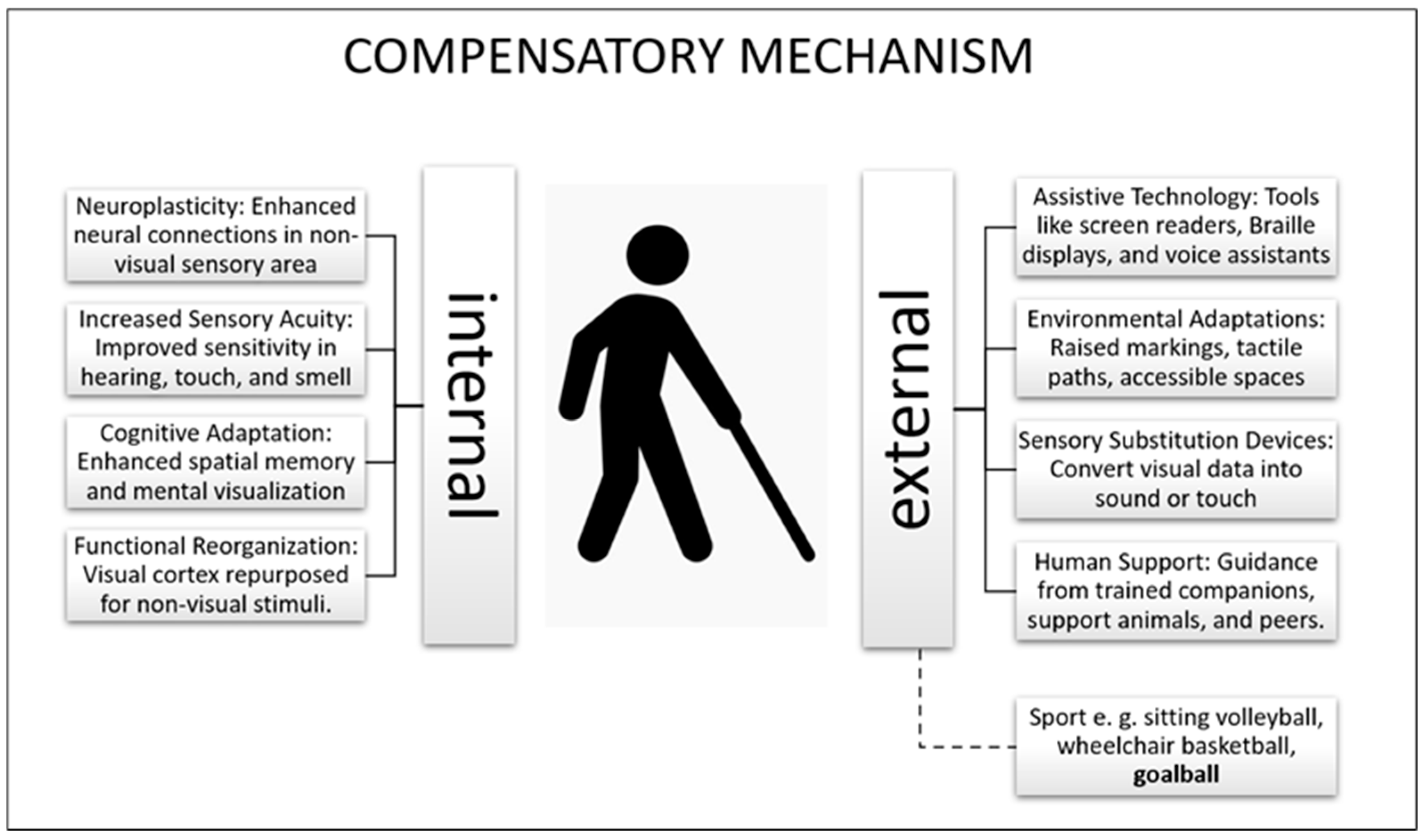
Figure 1. Theoretical model of compensatory mechanism (own source).
Applsci 15 06363 g001
Table 1. Characteristics of respondents’ physical status (N = 12).
CharacteristicsMean ± SDMin-MaxQualitative Assessment
BM (kg)81.4 ± 14.157.2–108.7-
BH (cm)177,0; 17170.0–195.3-
WC (cm)86.9 ± 9.970.5–106.0<90 n = 9; >90 ** n = 3
HC (cm)102.5 ± 8.187.0–119.0-
BMI (kg/m2)24.6 ± 3.119.1–29.9<25 n = 7; ≥25 and <30 ** n = 5
BAI23.9 ± 2.819.9–28.3<21 n = 3; >21 ** n = 9
FAT (%) *21.8 ± 7.112.3–37.7-
FfST (%) *74.2 ± 6.859.2–83.4-
BM—body mass; BH—body height; WC—waist circumference; HC—hip circumference; BMI—body mass index; BAI—body adiposity index; FAT—fat tissue; FfST—Fat-free soft tissue; *—n = 11; **—above normal; italics—variables with non-normal distribution for which the median and interquartile range were given.
Table 2. Prevalence of musculoskeletal pain by body area in the last 7 days (NMQ7) and 6 months (NMQ6).
Body PartsNMQ7
n/%
NMQ6
n/%
neck/nape of the neck1/8.33/25
arms4/33.35/41.7
upper back1/8.34/33.3
elbows04/33.3
wrists/hands6/506/50
lower back5/41.75/41.7
hips/thighs2/16.73/25
knees2/16.73/25
ankles/feet3/255/41.7
n—number of participants.
Table 3. Assessment of spinal curvature and pelvic alignment of athletes (Medi Mouse).
Player NumberTKA,
HP (°)
Value
Reference Range
LLA,
HP (°)
Value
Reference Range
Pelvic Alignment
in HSP
Value
Reference Range
Qualitative Assessment
of Body Posture
Player 153 ↑
28–48
42 ↑
18–30
23 ↑
8–16
Hyperkyphosis,
hyperlordosis,
anterior pelvic tilt
Player 252 •
39–55
29 •
23–31
9 ↓
10–16
Normal kyphosis,
normal lordosis,
posterior pelvic tilt
Player 347 •
34–52
33 •
21–35
20 ↑
8–18
Normal kyphosis,
normal lordosis,
anterior pelvic tilt
Player 457 ↑
28–48
41 ↑
18–30
23 ↑
8–16
Hyperkyphosis,
hyperlordosis,
anterior pelvic tilt
Player 546 •
35–55
27 •
17–31
11 •
6–16
Normal kyphosis,
normal lordosis,
pelvis aligned correctly
Player 642 •
39–55
19 ↓
23–31
9 ↓
10–16
Normal kyphosis,
hypolordosis,
posterior pelvic tilt
Player 754 ↑
34–52
42 ↑
21–35
20 ↑
8–18
Hyperkyphosis,
hyperlordosis,
anterior pelvic tilt
Player 836 •
34–52
29 •
21–35
18 •
8–18
Normal kyphosis,
normal lordosis,
pelvis aligned correctly
Player 947 •
34–52
19 ↓
21–35
2 ↓
8–18
Normal kyphosis,
hypolordosis,
posterior pelvic tilt
Player 1041 •
34–52
24 •
21–35
8 •
8–18
Normal kyphosis,
normal lordosis,
pelvis aligned correctly
Player 1151 •
34–52
32 •
21–35
15 •
8–18
Normal kyphosis,
normal lordosis,
pelvis aligned correctly
Player 1248 •
28–48
10 ↓
18–30
5 ↓
8–16
Normal kyphosis,
hypolordosis,
posterior pelvic tilt
All players
x ¯ ± SD
(min–max)
47.8 ± 6.0
36.0–57.0
28.9 ± 9.9
10.0–42.0
13.6 ± 7.2
2.0–23.0
-
TKA—thoracic kyphosis angle, HP—habitual position, LLA—lumbar lordosis angle, HSP—habitual standing position, ↑—above-normal value; ↓—below-normal value; •—within reference limits, x ¯ —mean value, SD—standard deviation.
Table 4. Range of motion of THA and LLA during trunk flexion, and trunk extension (MediMouse).
Player NumberRange of Motion (Flexion)Range of Motion (Extension)
TK
Value
Reference Range
LL
Value
Reference Range
TK
Value
Reference Range
LL
Value
Reference Range
Player 131 •
11–33
60 •
60–76
1 •
−17–5
−14 •
−27–(−9)
Player 26 •
2–24
63 •
55–69
−12 •
−23–(−3)
−15 •
−24–(−8)
Player 317 •
6–28
67 •
56–74
14 ↑
−22–6
−20 •
−28–(−10)
Player 47 ↓
11–33
75 •
60–76
1 •
−17–5
−7 ↓
−27–(−9)
Player 515 •
9–25
60 •
45–61
14 ↑
−23–(−7)
−11 •
−20–4
Player 62 •
2–24
69 •
55–69
−13 •
−23–(3)
−3 ↓
−24–(−8)
Player 711 •
6–28
70 •
56–74
−9 •
−22–6
−13 ↓
−28–(−20)
Player 822 •
6–28
70 •
56–74
−8 •
−22–6
1 ↓
−28–(−10)
Player 97 •
6–28
67 •
56–74
13 ↑
−22–6
−20 •
−28–(−10)
Player 104 ↓
6–28
76 •
56–74
−5 •
−22–6
−3 ↓
−28–(−10)
Player 1114 •
6–28
51 ↓
56–74
0 •
−22–6
−10 •
−28–(−10)
Player 1210 ↓
11–33
58 ↓
60–76
−7 •
−17–5
−28 ↑
−27–(−9)
All players
x ¯ ± SD
(min–max)
12.2 ± 8.3
(2–31)
65.6 ± 7.3
(51–76)
−0.9 ± 9.9
(−13–14)
−11.9 ± 8.3
(−28–1)
TK—thoracic kyphosis, LL—lumbar lordosis, x ¯ —mean value, SD—standard deviation, ↑—above-normal value; ↓—below-normal value; •—within reference limits.
Disclaimer/Publisher’s Note: The statements, opinions and data contained in all publications are solely those of the individual author(s) and contributor(s) and not of MDPI and/or the editor(s). MDPI and/or the editor(s) disclaim responsibility for any injury to people or property resulting from any ideas, methods, instructions or products referred to in the content.