Article

Comparison of Condylar Guidance in Opening and Protrusion Using Electronic Axiography and CBCT: An Observational Cross-Sectional Study

1
Department of Conservative Dentistry, Faculty of Medicine, Medical University of Gdańsk, 80-210 Gdańsk, Poland
2
Department of Prosthetic Dentistry, Faculty of Medicine, Medical University of Gdańsk, 80-210 Gdańsk, Poland
3
Department of Prosthodontics, College of Medicine and Dentistry, Ulster University, Birmingham B5 5JE, UK
*
Author to whom correspondence should be addressed.
Appl. Sci. 2025, 15(11), 5993; https://doi.org/10.3390/app15115993
Submission received: 31 March 2025 / Revised: 1 May 2025 / Accepted: 19 May 2025 / Published: 26 May 2025

Abstract

:
This observational cross-sectional study evaluated the differences in condylar guidance angles during mandibular protrusion and mouth opening using digital axiography and CBCT. A total of 30 healthy subjects (60 TMJs) aged 20–24 years were examined. Mandibular movements were recorded using the Modjaw system, and fossa height was assessed from CBCT images. A statistically significant correlation was observed between condylar angles measured during protrusion and opening (r = 0.57, p < 0.001). Protrusive measurements were more consistent, while opening exhibited greater variability. The results suggest that protrusive movements are more reliable for articulator programming. Digital tools such as Modjaw can enhance precision and workflow efficiency in TMJ diagnostics.

1. Introduction

The temporomandibular joint (TMJ) occupies a central anatomical position in the craniofacial structure, connecting the temporal bone, cranial base, and mandibular condyle [1,2]. The stomatognathic system includes the TMJ, teeth, maxilla, mandible, muscles, ligaments, nerves, and vasculature [3]. The TMJ enables mandibular mobility, essential for chewing and speaking through coordinated masticatory and accessory muscle action [1]. Analogue and digital articulators simulate these dynamics to optimize prosthetic treatment by adjusting occlusion and joint movement [2,4,5,6,7,8]. Their use improves restoration predictability and alignment, enhancing prosthodontic outcomes [8,9,10]. Digital workflows have spurred the development of virtual articulators (VAs), with research focused on their accuracy and clinical utility [9,11,12,13]. VAs offer superior occlusal analysis and jaw movement simulation compared to mechanical systems. Concurrently, electronic axiography provides precise recordings of condylar path movements, aiding biomechanical assessments and prosthodontic planning [14,15,16,17]. Calibrating virtual or mechanical articulators requires two posterior determinants: sagittal condylar inclination (SCI) and transverse condylar inclination (TCI, or Bennett angle) [18,19]. SCI measures the condylar path angle relative to sagittal and horizontal planes. TCI quantifies the non-working condyle’s horizontal deviation during mediotrusive movement (Figure 1). Mean SCI values range between 20 and 33 degrees [20], while the Bennett angle averages 15 degrees, reflecting lateral mandibular displacement [13,21,22]. These parameters ensure functional harmony in prosthetic reconstructions. The Modjaw device (MODJAW, Lyon, France) streamlines full-mouth rehabilitation by replacing traditional tools like facebows and mechanical articulators [13,23]. Expanding on prior research linking condylar path anatomy to anterior guidance during protrusion [24], this study addresses a critical gap: whether SCI calibration should occur during protrusive movements or mandibular opening. Clarifying this distinction will refine articulator programming and prosthetic rehabilitation accuracy, advancing diagnostic and clinical applications.

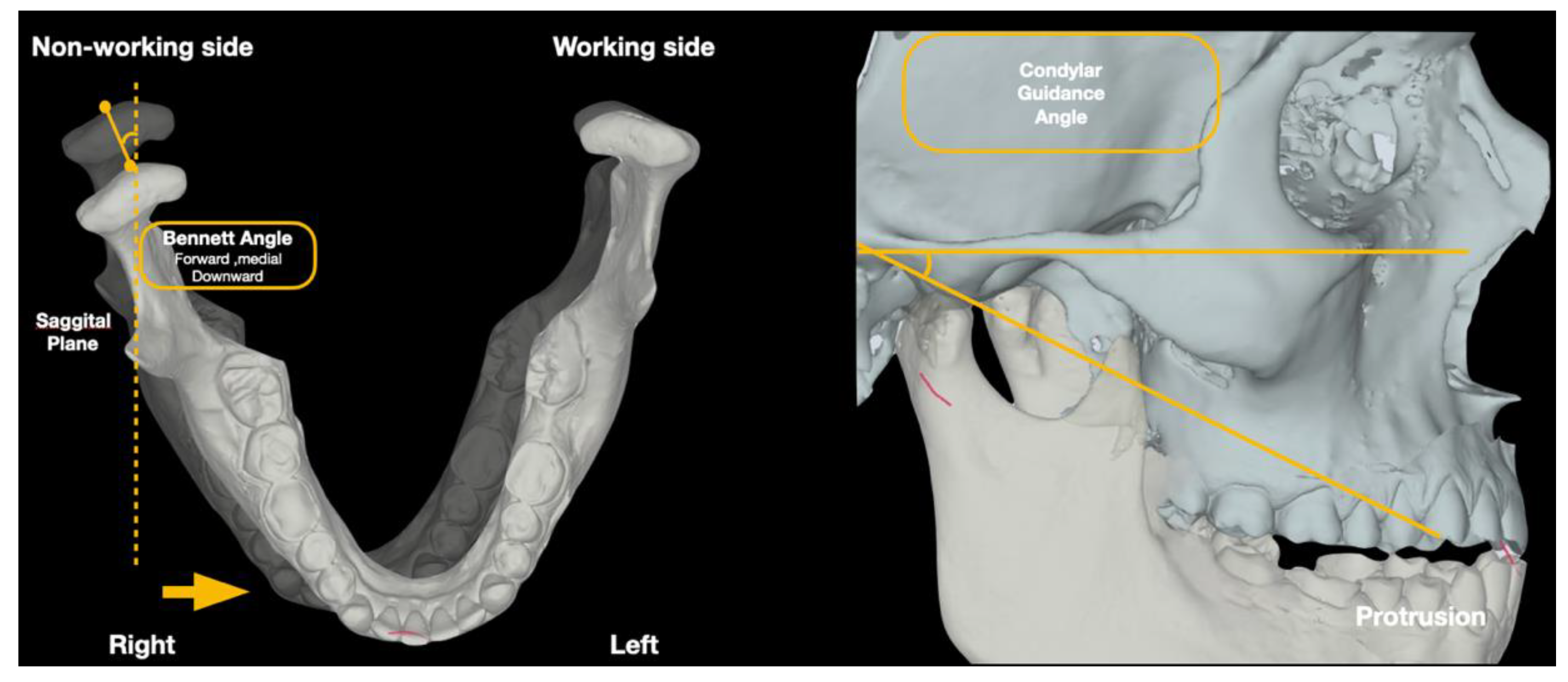
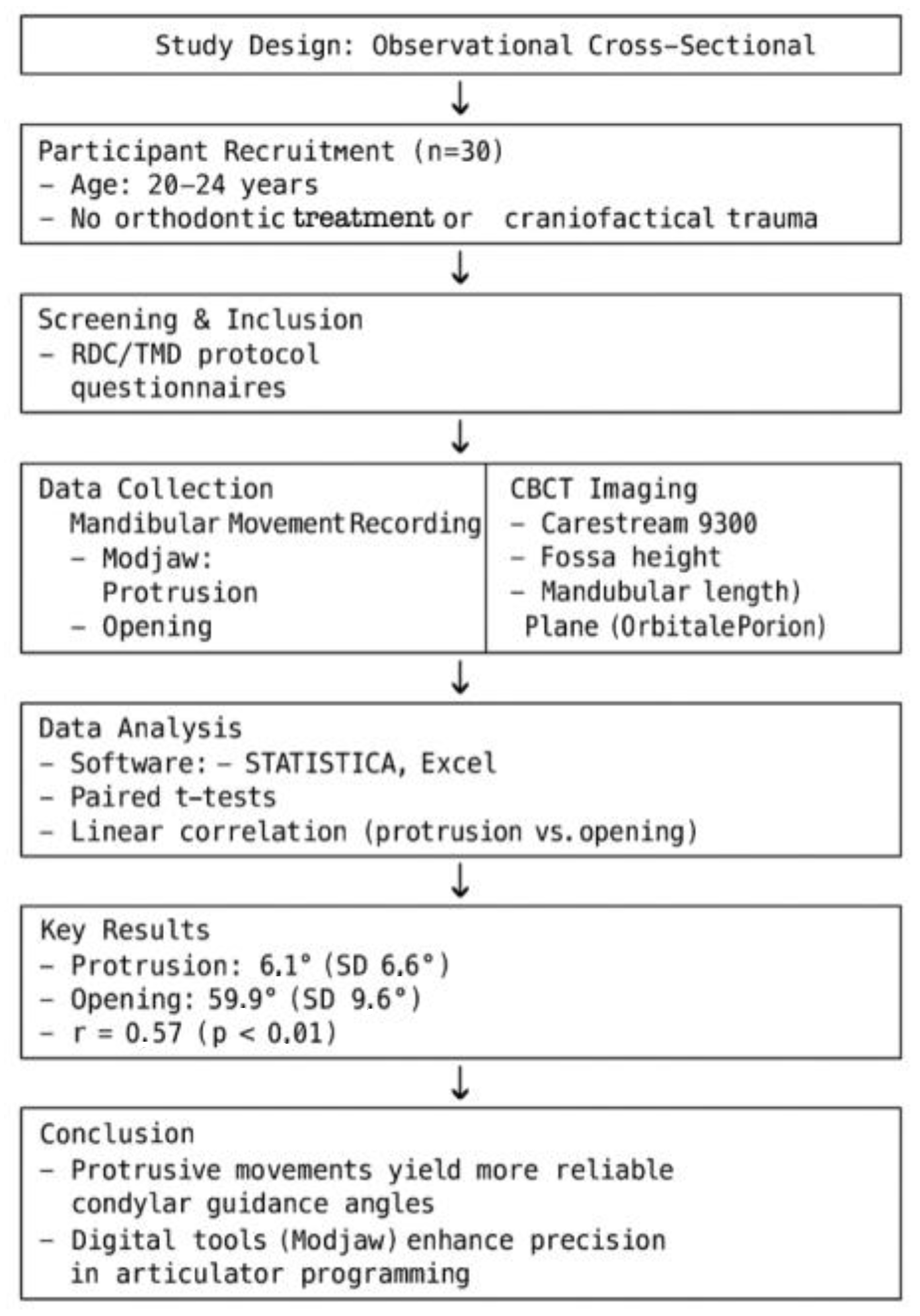
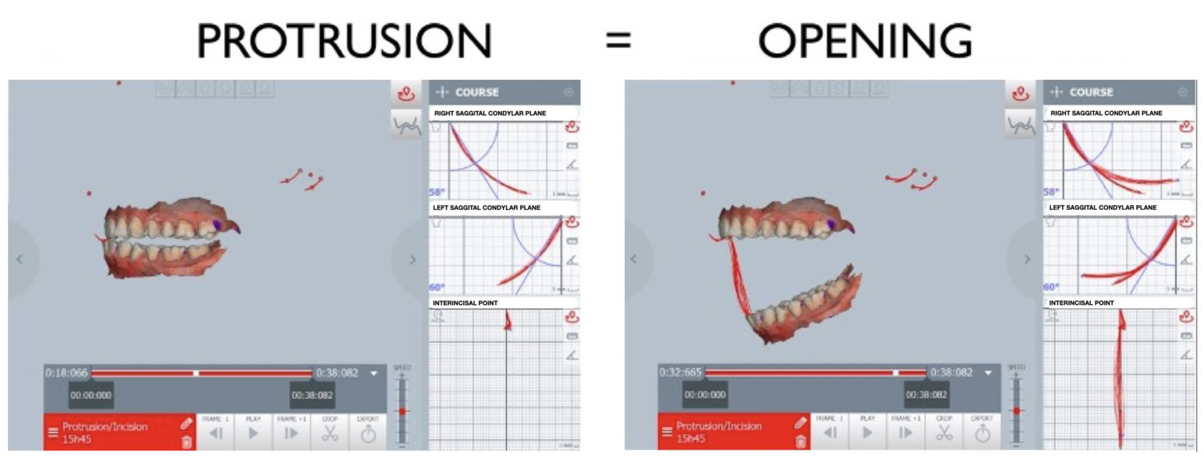
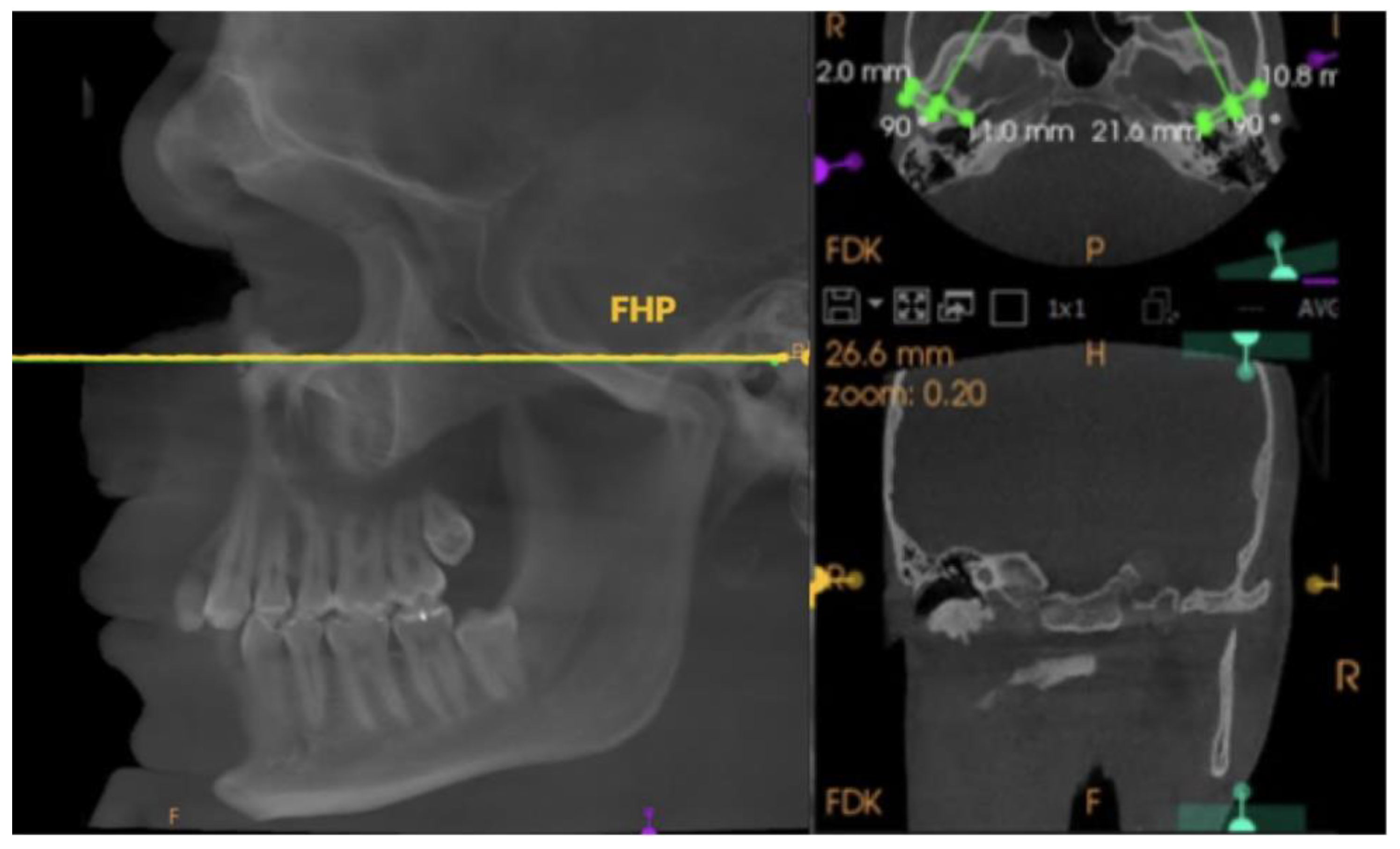
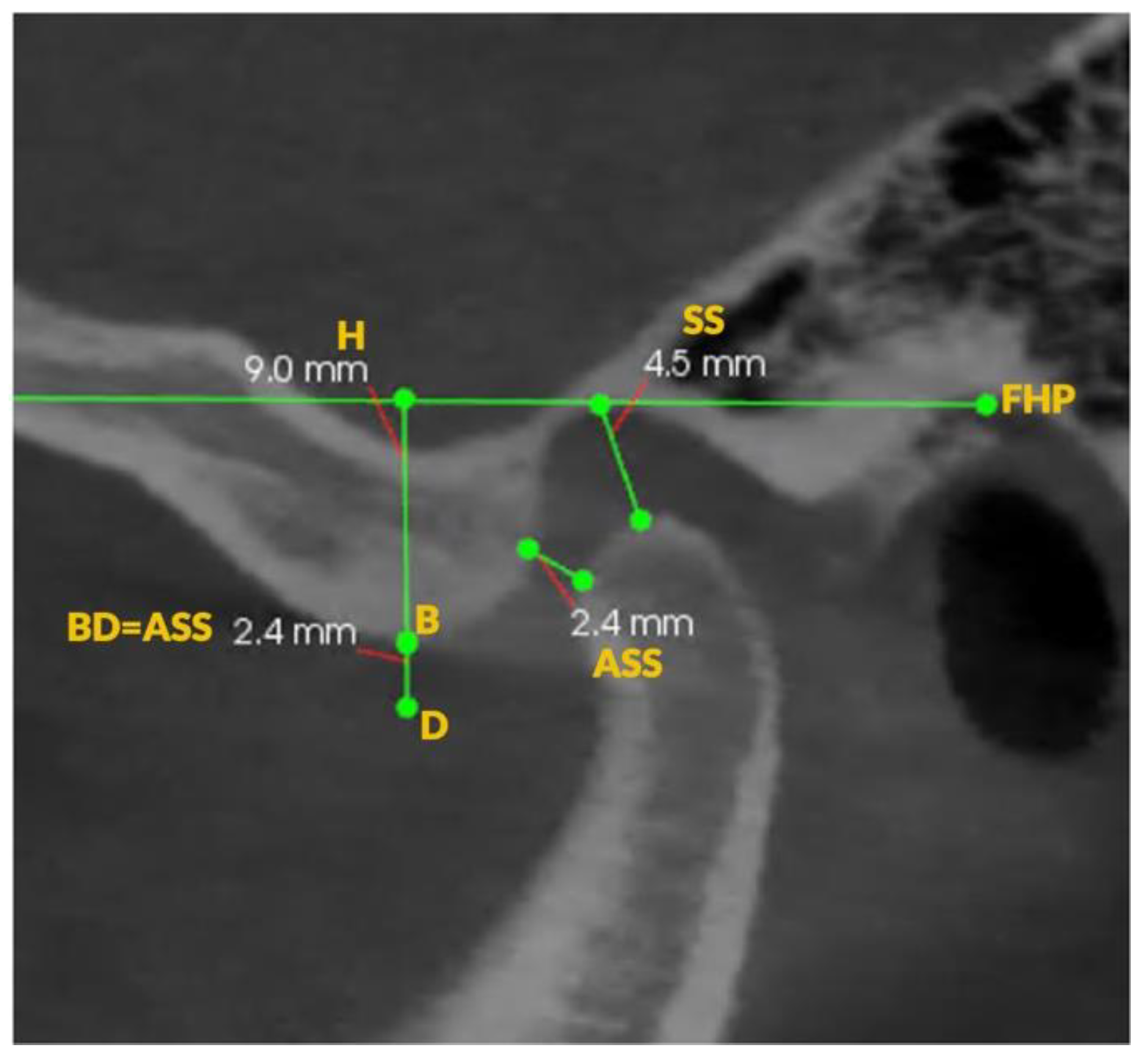
2. Materials and Methods

This study was conducted at the Medical University of Gdańsk (Gdańsk, Poland). Thirty healthy individuals (aged 20–24 years) without a history of temporomandibular disorders (TMD), orthodontic treatment, or craniofacial trauma were recruited from among university volunteers. Subjects were screened via a questionnaire and the Research Diagnostic Criteria/Temporomandibular Disorders (RDC/TMD) [25] protocol. Flow chart Figure 2 detailing study design process. Examinations were performed using the Modjaw device (MODJAW, Lyon, France) to capture real-time mandibular movement and cone beam computed tomogram (CBCT) imaging (Carestream 9300) to measure glenoid fossa height/anatomy.
Mandibular movement recordings included the following (Figure 3):
-
Repetitive mouth opening and closing;
-
Protrusive movements from maximum intercuspation (MIP);
-
Full mouth opening from MIP.
The Modjaw system (MODJAW, Lyon, France) optically tracked mandibular motion in real time. Each subject performed multiple cycles of the above movements. The Frankfort Horizontal Plane (FHP) was used as the reference, constructed by connecting the left Orbitale and Porion landmarks bilaterally (Figure 4) [26]. In the sagittal view, a cross-sectional plane perpendicular to a line passing through the innermost and outermost points of the condyle in the transverse section was selected individually for each TMJ, ensuring consistent orientation for measurements (Figure 5). CBCT imaging was performed by a skilled radiology technician using the Carestream 9300 system (Carestream Dental, Atlanta, GA, USA) under standardized settings: 120 kV, 3.20 mA, 40 s exposure, and a delivered dose of 1698.19 mGy/cm2. Each patient was positioned in a standing posture with the mandible in maximum intercuspation position (MIP) to ensure consistency. Image analysis was conducted using CS Imaging 8.0.5 software to measure the vertical dimension of the glenoid fossa. For each movement, Modjaw TWIM version software was used to calculate the sagittal condylar guidance angle (SCI, in degrees) for the right and left TMJs.
The temporomandibular joint (TMJ) space was evaluated by identifying the antero-superior space (ASS) and superior space (SS). The ASS corresponds to the thickness of the intermediate disc band, while the SS reflects the thickness of the posterior disc band. In addition, the vertical height of the fossa (H) was measured (Figure 6). All measurements from the CBCT images were performed by two independent examiners; the vertical fossa height values obtained showed negligible differences (inter-observer variation < 0.2 mm, indicating high consistency).
The research protocol was approved by the Independent Bioethics Committee for Scientific Research at the Medical University of Gdańsk (approval number: NKBBN/1043/2021-2022) and retrospectively registered at ClinicalTrials.gov (NCT05637372). All participants were provided with oral and written information about the study and gave informed consent to participate.
Statistical Analysis: All calculations were performed using Microsoft Excel 2019 and STATISTICA 13.6 (TIBCO Software Inc., Palo Alto, CA, USA; 2020) Data Science Workbench, version 14. Descriptive statistics (mean, median, standard deviation, and range) were calculated for all quantitative variables. The Shapiro–Wilk test was used to assess the normality of distributions. Paired comparisons (e.g., right vs. left, protrusion vs. opening) were made using paired t-tests for normally distributed data or Wilcoxon signed-rank tests if non-normal. Pearson’s correlation coefficient was used to evaluate relationships between variables when both were approximately normally distributed (Spearman’s rank correlation otherwise). A multiple linear regression analysis was explored to identify potential predictors of the condylar guidance angle, but the final analysis relied on the simpler correlation models given the sample size. Statistical significance was set at p < 0.05. The choice of statistical methods was reviewed in consultation with a biostatistician (Dr. Tomasz Bandurski—Department of Radiological Informatics and Statistics at the Medical University of Gdańsk.).

3. Results

The measurements of the parameters for all 30 patients (60 TMJ) were collated and subjected to statistical analysis. The aim was to obtain values of the guidance angles during protrusive movement, mouth opening, and the Bennett angle, as determined by digital axiography (Table 1), and test the correlation between the averaged values from both joints during protrusion and mouth opening, as well as the correlation between the Bennett angle and the height of the articular fossa (Table 2), which are presented in the tables below.
The sagittal condylar guidance angles recorded via electronic axiography showed distinct profiles for protrusive movements and mouth opening. During mandibular protrusion, the condylar guidance angles averaged approximately 56–57° (mean of ~56.6° when combining both joints), with a relatively narrow dispersion (SD ~6–7° per side). Most individual protrusive angles clustered in the 50–65° range, indicating a fairly consistent distribution across the 30 subjects. The median protrusive angle was around 56–58°, closely matching the mean, suggesting a roughly symmetrical (normal) distribution without strong skew. In contrast, during full mouth opening, condylar guidance angles tended to be slightly steeper on average (mean ~59.9° for the averaged bilateral values). The distribution was broader: values ranged from the mid-40s up to nearly 90° (see Table 1). The group mean for opening angles was about 3–4 degrees higher than for protrusion, and the standard deviation was larger (approximately 9–11° for individual joint measurements, versus ~6–7° in protrusion), indicating greater variability. The median opening angle (~59–61° depending on side) was slightly lower than the mean, reflecting the influence of a few high-angle measurements on the average. Overall, the protrusive condylar paths showed more uniform angles, whereas the opening movements exhibited a wider spread of values.
These data reveal a trend where protrusive excursions produce more consistent condylar guidance angles, while maximal opening can yield both moderate and very steep condylar paths in different individuals. Notably, the majority of opening angle measurements still fell within a moderate range (roughly 50–70° for most participants), but the presence of several higher values increased the overall mean. All recorded sagittal guidance angles in this young adult sample were well above the textbook “average” articulator setting (typically ~30°), which highlights that the method of measurement (electronic axiography with Frankfort horizontal reference) and the full range of motion captured can result in larger numerical angles. This underscores the importance of using individualized measurements, as group mean values alone may not represent any given patient’s condylar inclination accurately (Figure 7).

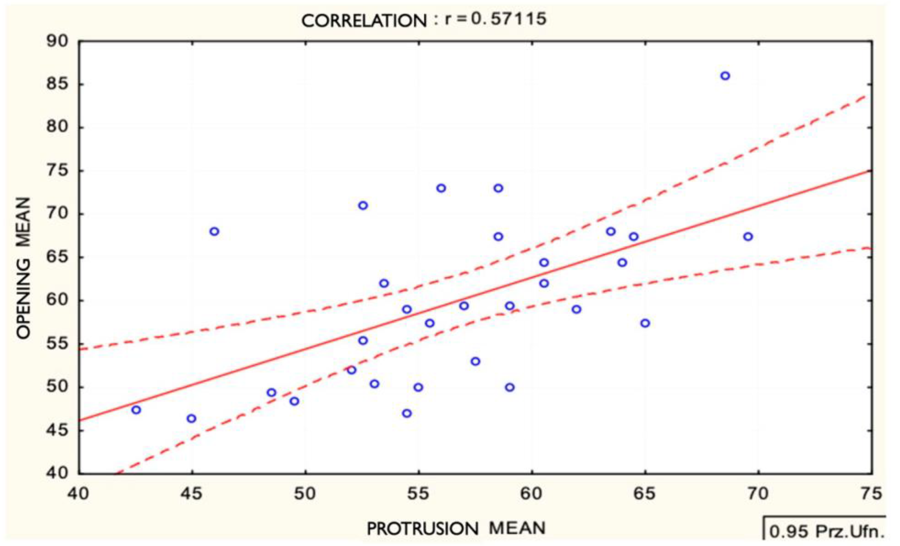
Correlation Between Protrusive and Opening Guidance Angles

Statistical analysis (Table 2) revealed a moderate positive correlation between the condylar guidance angles measured in protrusion and those measured during mouth opening. Using each subject’s mean bilateral angle for the two movements, the Pearson correlation coefficient was r = 0.57 (p < 0.001). This indicates that individuals with a relatively steep condylar guidance in one type of movement tended to also show a steep guidance angle in the other movement. In practical terms, a subject who exhibited a high protrusive inclination (for example, well above the average) often also had an above-average inclination during full opening, and vice versa for lower-angle cases. However, the correlation was far from perfect. The coefficient of determination was r2 ≈ 0.33, meaning only about 33% of the variance in opening angle can be statistically explained by the protrusive angle. The remaining ~67% of variability is due to factors other than a simple linear relationship—in other words, there is substantial individual variation in how the condyle translates during opening versus protrusion. Figure 6 (scatterplot of protrusive vs. opening angles) illustrated this trend: while there is an upward slope to the data points (confirming a general positive association), the points are fairly widely scattered rather than falling tightly along a line. Some subjects with moderate protrusive angles had much steeper opening angles than expected, and a few with high protrusive angles still showed only moderate opening values.
The clinical implication of this finding is that protrusive and opening condylar guidance angles, though related, are not interchangeable. A moderate correlation suggests that a clinician who calibrates an articulator using a protrusive record can expect the opening pathway inclination to be in a similar ballpark for many patients, but not with high precision. The statistically significant relationship supports the idea that the steepness of the condylar path is partly a characteristic of the individual’s joint anatomy (and thus influences all movements to some extent). Yet, the differences in movement patterns (protrusion being a mostly linear forward translation vs. opening involving rotation plus translation along a different condylar trajectory) result in divergence between the two measurements. From the study’s primary objective standpoint, this result is notable: it confirms that there is a correlation between the two methods of determining sagittal condylar inclination, but also underscores that protrusive movement recordings are more reliable (given their lower variability) for articulator programming. The data demonstrated that protrusive measurements had tighter consistency (as described above), which, combined with the moderate correlation, suggests that articulators set using protrusive records would approximate the patient’s functional movements more predictably than those set using maximal opening records. In summary, while a shared trend exists between the protrusive and opening guidance angles, the degree of scatter in the data reinforces the need to measure protrusive inclination directly for the most accurate result, rather than relying on mouth-opening values as a proxy.

4. Discussion

This study highlights the importance of mandibular movement type when measuring condylar guidance angles. Protrusive movements provided more stable values, supporting their use in articulator programming. These findings align with the literature suggesting protrusion as the most reliable reference for sagittal condylar inclination. The use of digital axiography provides enhanced diagnostic capabilities in prosthodontics, reducing variability and supporting individualized treatment planning.
This study investigated the measurement of condylar guidance angles during protrusion and mouth opening using digital axiography. The findings offer valuable insights into the variations and potential reliability of these measurements, providing a basis for refining clinical approaches to temporomandibular joint (TMJ) analysis.
A significant correlation was observed between the condylar guidance angles measured during protrusion and mouth opening. Notably, the guidance angles obtained during mouth opening exhibited greater deviations from normative values compared to those measured during protrusion. This observation underscores that the type of mandibular movement assessed plays an important role in the precision and reproducibility of condylar guidance angle measurements. Specifically, protrusive movements yielded more stable and less variable results, supporting their potential as a preferred standard for these evaluations.

4.1. Correlation Between Protrusion and Mouth Opening

The results revealed a significant correlation between the condylar guidance angles measured during protrusion and mouth opening. However, measurements during mouth opening demonstrated greater variability, reflecting deviations from normative values. These findings are consistent with earlier studies [15,17] that highlight distinct condylar path patterns associated with different mandibular movements. They further emphasize the superior stability and reproducibility of protrusive measurements, underscoring their value in ensuring accurate articulator programming. The increased variability during mouth opening is likely attributable to the sinusoidal trajectories [19] of the condylar path, influenced by complex anatomical and functional dynamics. This research provides a significant contribution to the field, as it is among the first to simultaneously assess these parameters in dental patients using advanced digital axiography, thereby paving the way for improved diagnostic and prosthetic methodologies.

4.2. Clinical Implications of Movement Type

The findings underscore the importance of selecting appropriate mandibular movements for condylar guidance measurements. Protrusive movements, characterized by their consistency and reliability, are reaffirmed as the gold standard for articulator calibration. Moreover, CGA plays a pivotal role in determining occlusal morphology, as variations in CGA directly influence the morphology of posterior teeth. Studies [27,28,29] have shown that changes in CGA can alter cusp angles and occlusal relationships, affecting both the functionality and esthetics of restorations. Digital tools, such as the Modjaw device, offer a transformative approach to condylar guidance assessment by enabling real-time tracking of mandibular dynamics, thereby enhancing workflow efficiency and accuracy in prosthodontic treatments. These tools not only streamline clinical processes but also ensure the accurate reproduction of functional dental morphology tailored to individual patients.

4.3. Methodological Considerations

The selection of horizontal reference planes plays a pivotal role in the accuracy of sagittal condylar inclination (SCI) measurements. This study employed the axis–orbital plane (AOP), a widely accepted standard in the literature due to its reproducibility and clinical applicability. Previous research [19,23] has highlighted significant discrepancies in sagittal condylar inclination (SCI) values depending on the reference plane used. Notably, measurements taken with the axis–orbital plane (AOP) tend to produce higher SCI values compared to the Frankfort horizontal or Camper planes, with differences reported to reach up to 10 degrees. These findings emphasize the critical need for standardizing reference planes, especially when integrating digital workflows into articulator programming and prosthodontic rehabilitation.
While the AOP offers reliable and consistent measurements, comparisons with other methodologies, such as intraoral and extraoral recordings, reveal inherent variability introduced by differing techniques. Digital axiography, particularly when paired with the AOP, has proven to provide superior consistency and accuracy. These insights reinforce the AOP’s utility as a reference standard in both clinical and research contexts, paving the way for enhanced precision in condylar guidance assessments.

5. Limitations

This study has limitations. The small sample size (n = 30) and narrow age range (20–24 years) limit statistical power and generalizability to older or clinical populations. Participants were asymptomatic and lacked orthodontic history, introducing selection bias. These factors highlight the need for future studies with broader cohorts and advanced anatomical assessments.

6. Conclusions

Digital axiography confirmed that condylar guidance angles measured during protrusion are more reproducible than those measured during mouth opening. These findings emphasize the utility of protrusive movements in articulator programming and the potential of digital tools like Modjaw in modern prosthodontic diagnostics and rehabilitation.

Author Contributions

Conceptualization, Ł.L., M.T.-P. and R.Y.; Methodology, Ł.L.; Resources, Ł.L.; Data curation, Ł.L.; Writing—original draft, Ł.L., M.T.-P. and R.Y.; Writing—review & editing, M.T.-P. and R.Y.; Supervision, A.Ż.; Project administration, A.Ż. All authors have read and agreed to the published version of the manuscript.

Funding

The authors did not receive any external funding for the completion of this work.

Institutional Review Board Statement

The study was approved by the Independent Bioethics Committee for Scientific Research at the Medical University of Gdańsk (protocol number NKBBN/1043/2021-2022, 25 January 2022).

Informed Consent Statement

Informed consent was obtained from all subjects involved in the study.

Data Availability Statement

The original contributions presented in this study are included in the article. Further inquiries can be directed to the corresponding author.

Conflicts of Interest

The authors declare no conflicts of interest.

References

  1. Iturriaga, V.; Bornhardt, T.; Velasquez, N. Temporomandibular Joint: Review of Anatomy and Clinical Implications. Dent. Clin. N. Am. 2023, 67, 199–209. [Google Scholar] [CrossRef] [PubMed]
  2. Woodford, S.C.; Robinson, D.L.; Mehl, A.; Lee, P.V.S.; Ackland, D.C. Measurement of normal and pathological mandibular and temporomandibular joint kinematics: A systematic review. J. Biomech. 2020, 111, 109994. [Google Scholar] [CrossRef] [PubMed]
  3. Hatcher, D.C. Anatomy of the Mandible, Temporomandibular Joint, and Dentition. Neuroimaging Clin. N. Am. 2022, 32, 749–761. [Google Scholar] [CrossRef]
  4. Starcke, E.N.; Engelmeier, R.L. The history of articulators: The wonderful world of “Grinders”. Part I. J. Prosthodont. 2006, 15, 129–140. [Google Scholar] [CrossRef]
  5. Kranjčić, J.; Hunt, D.; Kiršić, S.P.; Kovačić, I.; Vukšić, J.; Vojvodić, D. Articular Eminence Morphology of American Historic and Contemporary Populations. Acta Stomatol. Croat. 2021, 55, 397–405. [Google Scholar] [CrossRef]
  6. Sharpling, B. Semi-adjustable articulators. In Practical Procedures in Aesthetic Dentistry; John Wiley & Sons, Ltd.: Hoboken, NJ, USA, 2017; pp. 60–66. [Google Scholar]
  7. Goldstein, G.; Goodacre, C. Selecting a Virtual Articulator: An Analysis of the Factors Available with Mechanical Articulators and their Potential Need for Inclusion with Virtual Articulators. J. Prosthodont. 2023, 32, 10–17. [Google Scholar] [CrossRef]
  8. Cabot, L.B. Using articulators to enhance clinical practice. Br. Dent. J. 1998, 184, 272–276. [Google Scholar] [CrossRef]
  9. Doshi, K.N.; Sathe, S.; Dubey, S.A.; Bhoyar, A.; Dhamande, M.; Jaiswal, T. A Comprehensive Review on Virtual Articulators. Cureus 2024, 16, e52554. [Google Scholar] [CrossRef]
  10. Hobo, S.; Shillingburg, H.T.; Whitsett, L.D. Articulator selection for restorative dentistry. J. Prosthet. Dent. 1976, 36, 35–43. [Google Scholar] [CrossRef]
  11. Lepidi, L.; Galli, M.; Mastrangelo, F.; Venezia, P.; Joda, T.; Wang, H.L.; Li, J. Virtual Articulators and Virtual Mounting Procedures: Where Do We Stand? J. Prosthodont. 2021, 30, 24–35. [Google Scholar] [CrossRef]
  12. Lepidi, L.; Suriano, C.; Wang, H.L.; Granata, S.; Joda, T.; Li, J. Digital fixed complete-arch rehabilitation: From virtual articulator mounting to clinical delivery. J. Prosthet. Dent. 2022, 127, 398–403. [Google Scholar] [CrossRef] [PubMed]
  13. Manziuc, M.M.; Dîrzu, A.; Almăşan, O.; Leucuţa, D.C.; Tăut, M.; Ifrim, C.; Berindean, D.; Kui, A.; Negucioiu, M.; Buduru, S. Cadiax Compact 2 and MODJAW comparative analysis of condylar inclination: Innovative digital approaches in dentistry. J. Prosthet. Dent. 2024, 133, 482–488. [Google Scholar] [CrossRef] [PubMed]
  14. Tuppy, F.; Celar, R.M.; Celar, A.G.; Piehslinger, E.; Jäger, W. The reproducibility of condylar hinge axis positions in patients, by different operators, using the electronic mandibular position indicator. J. Orofac. Pain 1994, 8, 315–320. [Google Scholar] [PubMed]
  15. Hue, O. The Sagittal Condylar Paths in Edentulous Patients: Analysis with Computerized Axiography. Int. J. Prosthodont. 2016, 29, 11–16. [Google Scholar] [CrossRef]
  16. Gsellmann, B.; Schmid-Schwap, M.; Piehslinger, E.; Slavicek, R. Lengths of condylar pathways measured with computerized axiography (CADIAX) and occlusal index in patients and volunteers. J. Oral Rehabil. 1998, 25, 146–152. [Google Scholar] [CrossRef]
  17. Hüe, O. Condylar paths during protrusion in edentulous patients: Analysis with electronic axiography. J. Prosthodont. 2011, 20, 294–298. [Google Scholar] [CrossRef]
  18. Lewandowska, A.; Mańka-Malara, K.; Kostrzewa-Janicka, J. Sagittal Condylar Inclination and Transversal Condylar Inclination in Different Skeletal Classes. J. Clin. Med. 2022, 11, 2664. [Google Scholar] [CrossRef]
  19. Bapelle, M.; Dubromez, J.; Savoldelli, C.; Tillier, Y.; Ehrmann, E. Update on the parameters influencing the adjustment of the sagittal and transversal condylar inclination of dental articulators. Quintessence Int. 2022, 53, 78–88. [Google Scholar]
  20. Caro, A.J.; Peraire, M.; Martinez-Gomis, J.; Anglada, J.M.; Samsó, J. Reproducibility of lateral excursive tooth contact in a semi-adjustable articulator depending on the type of lateral guidance. J. Oral Rehabil. 2005, 32, 174–179. [Google Scholar] [CrossRef]
  21. Amarnath, G.S.; Kumar, U.; Hilal, M.; Muddugangadhar, B.C.; Anshuraj, K.; Shruthi, C.S. Comparison of Cone Beam Computed Tomography, Orthopantomography with Direct Ridge Mapping for Pre-Surgical Planning to Place Implants in Cadaveric Mandibles: An Ex-Vivo Study. J. Int. Oral Health 2015, 7 (Suppl. S1), 38. [Google Scholar]
  22. Aljohani, A.O.; Sghaireen, M.G.; Abbas, M.; Alzarea, B.K.; Srivastava, K.C.; Shrivastava, D.; Issrani, R.; Mathew, M.; Alsharari, A.H.L.; Alsharari, M.A.D.; et al. Comparative Evaluation of Condylar Guidance Angles Measured Using Arcon and Non-Arcon Articulators and Panoramic Radiographs—A Systematic Review and Meta-Analysis. Life 2023, 13, 1352. [Google Scholar] [CrossRef] [PubMed]
  23. Bapelle, M.; Dubromez, J.; Savoldelli, C.; Tillier, Y.; Ehrmann, E. Modjaw® device: Analysis of mandibular kinematics recorded for a group of asymptomatic subjects. Cranio J. Craniomandib. Sleep Pract. 2024, 42, 483–489. [Google Scholar] [CrossRef] [PubMed]
  24. Lassmann, Ł.; Nowak, Z.; Orthlieb, J.D.; Żółtowska, A. Complicated Relationships between Anterior and Condylar Guidance and Their Clinical Implications—Comparison by Cone Beam Computed Tomography and Electronic Axiography—An Observational Cohort Cross-Sectional Study. Life 2023, 13, 335. [Google Scholar] [CrossRef]
  25. Schiffman, E.; Ohrbach, R.; Truelove, E.; Look, J.; Anderson, G.; Goulet, J.P.; List, T.; Svensson, P.; Gonzalez, Y.; Lobbezoo, F.; et al. Diagnostic Criteria for Temporomandibular Disorders (DC/TMD) for Clinical and Research Applications: Recommendations of the International RDC/TMD Consortium Network and Orofacial Pain Special Interest Group. J. Oral. Facial Pain Headache 2014, 28, 6–27. [Google Scholar] [CrossRef]
  26. Chae, J.M.; Park, J.H.; Tai, K.; Mizutani, K.; Uzuka, S.; Miyashita, W.; Seo, H.Y. Evaluation of condyle-fossa relationships in adolescents with various skeletal patterns using cone-beam computed tomography. Angle Orthod. 2020, 90, 224–232. [Google Scholar] [CrossRef]
  27. Oancea, L.; Stegaroiu, R.; Cristache, C.M. The influence of temporomandibular joint movement parameters on dental morphology. Ann. Anat.-Anat. Anz. 2018, 218, 49–58. [Google Scholar] [CrossRef]
  28. Price, R.B.; Kolling, J.N.; Clayton, J.A. Effects of changes in articulator settings on generated occlusal tracings Part I: Condylar inclination progressive side shift settings. J. Prosthet. Dent. 1991, 65, 237–243. [Google Scholar] [CrossRef]
  29. Price, R.B.; Kolling, J.N.; Clayton, J.A. Effects of changes in articulator settings on generated occlusal tracings Part II: Immediate side shift intercondylar distance rear top wall settings. J. Prosthet. Dent. 1991, 65, 377–382. [Google Scholar] [CrossRef]
Figure 1. Aiding visualization of the TCI and CGA.
Applsci 15 05993 g001
Figure 2. Flowchart detailing the study design process.
Applsci 15 05993 g002
Figure 3. Illustrates recordings obtained using the Modjaw device, providing a detailed comparison between mandibular protrusion and mouth opening movements in sagittal views. The tracings clearly depict the trajectories of each movement and the calculation of CGA.
Applsci 15 05993 g003
Figure 4. FHP connecting the left Orbitale and Porion landmarks.
Applsci 15 05993 g004
Figure 5. Transverse view highlighting FHP.
Applsci 15 05993 g005
Figure 6. Vertical height of the fossa and the corresponding measurements of the joint space.
Applsci 15 05993 g006
Figure 7. Correlation between the condylar guidance angle during opening and protrusion.
Applsci 15 05993 g007
Table 1. Values of the guidance angles during protrusive movement, mouth opening determined using digital axiography.
PatientProtrusionOpening
RightLeftRightLeft
153535051
263666471
348704951
471668389
553596977
668567048
764646564
859596158
959586878
1057526157
1164636571
1270696966
1359626465
1448514651
1565656253
1653445643
1749586064
1857576059
1947457264
2057524945
2160615767
2253625452
2359587065
2443425144
2555507171
2656556154
2751545655
2854565347
2948425142
3055495450
Table 2. Correlation between the averaged values from both joints during protrusion and mouth opening.
Variables X and YNumber of CasesMeanStandard Deviationr(X,Y)r2p
Protrusion Mean3056.60006.62990.5711470.3262090.000979
Opening Mean59.88339.5923
Disclaimer/Publisher’s Note: The statements, opinions and data contained in all publications are solely those of the individual author(s) and contributor(s) and not of MDPI and/or the editor(s). MDPI and/or the editor(s) disclaim responsibility for any injury to people or property resulting from any ideas, methods, instructions or products referred to in the content.

