Review

Non-Invasive Food Authentication Using Vibrational Spectroscopy Techniques for Low-Resolution Food Fingerprinting

Shandong Key Laboratory of Applied Technology for Protein and Peptide Drugs, School of Pharmaceutical Sciences and Food Engineering, Liaocheng University, Liaocheng 252000, China
*
Authors to whom correspondence should be addressed.
Appl. Sci. 2025, 15(11), 5948; https://doi.org/10.3390/app15115948
Submission received: 10 April 2025 / Revised: 15 May 2025 / Accepted: 23 May 2025 / Published: 25 May 2025

Abstract

To address issues of food authenticity, such as fraud and origin tracing, it is essential to employ methods in food fingerprinting that are efficient, economical, and easy to use. This review highlights the capabilities of vibrational spectroscopy techniques, including mid-infrared (MIR), near-infrared (NIR), and Raman spectroscopy, as non-invasive tools for food authentication. These methods offer rapid, cost-effective, and environmentally friendly analysis across diverse food matrices. This review further discusses recent advances such as hyperspectral imaging, portable devices, and data fusion strategies that integrate chemometrics and artificial intelligence. Despite their promise, challenges remain, including limited sensitivity for certain compounds, spectral overlaps, fluorescence interference in Raman spectroscopy, and the need for standardized validation protocols. Looking forward, trends such as the miniaturization of devices, real-time monitoring, and AI-enhanced spectral interpretation are expected to significantly advance the field of food authentication.

1. Introduction

With the advancement of the Food 4.0 framework, the food industry has widely adopted the systematic verification of raw materials, finished products, and manufacturing workflows. This shift reflects a growing emphasis on integrating digital technologies to enable full-spectrum monitoring and validation across the production lifecycle [1,2]. This approach emphasizes the integration of advanced digital technologies, including the Internet of Things (IoT), artificial intelligence (AI), and diverse sensing systems, into the food industry to address critical challenges such as safety and security [3,4,5]. Building effective traceability frameworks and authenticity measures requires a comprehensive and methodical grasp of food-related processes, logistics, and storage conditions throughout the supply chain and value network. Moreover, beyond the core focus on traceability and authenticity, the adoption of analytical processing techniques plays a pivotal role in shaping a forward-thinking, sustainable, and adaptive food manufacturing sector [6].
Food fingerprinting has emerged as a critical approach for verifying authenticity, ensuring consistency, and maintaining the traceability of food products. This technique significantly contributes to fostering transparency and supporting sustainable practices within the food sector, ultimately benefiting consumers [7,8,9]. For example, Zheng’s group developed high-performance MoO3 nanosheet gas sensors for triethylamine detection to assess fish freshness [10]. Moreover, machine learning models were applied to classify and estimate non-refrigeration time with different quantities of variables to ensure the quality and safety of hydrobiological species for human consumption [11]. Importantly, the implementation of food safety, security, authentication, and traceability systems requires the involvement of multiple stakeholders. Typically, these efforts are supported by a collaborative network that includes food producers, regulatory bodies, technology developers, and academic researchers. Such cooperation, which is rooted in multidisciplinary engagement, facilitates the creation and refinement of innovative applications for the industry [12,13].
The food manufacturing sector faces numerous challenges and risks concerning food authenticity, including cross-contamination, mislabeling, unintentional ingredient errors, and deliberate economic food fraud. These issues are prevalent across various stages of the food supply and value chains, posing substantial disruptions to the industry [14,15]. To address these concerns, there is a growing need for early problem detection and the continuous advancement of real-time monitoring and verification methods to ensure food authenticity from production to consumption.
To verify the authenticity of food products, numerous analytical strategies have been employed, ranging from conventional proximate methods used to quantify constituents like moisture, proteins, and fats to sophisticated instrumental platforms. These include techniques such as gas and liquid chromatography (GC and LC), mass spectrometry (MS), high-performance liquid chromatography (HPLC), integrated or hyphenated analytical methods, and molecular approaches based on DNA profiling [8,16,17,18,19]. While these sophisticated methods provide high-resolution insights capable of identifying specific molecules and compounds, they are often criticized for their drawbacks, including high costs, lengthy processing times, and operational inefficiency. These limitations can hinder their practicality within the fast-paced, dynamic frameworks required by modern food supply and value chain systems for effective traceability and fingerprinting.
Techniques based on vibrational spectroscopy, including mid-infrared (MIR), near-infrared (NIR), and Raman spectroscopy, have gained recognition as reliable tools for assessing food authenticity in both research settings and industrial or commercial operations. These methods offer significant advantages, such as the ability to analyze diverse samples without requiring extensive preparation, enabling nondestructive and efficient measurements. Additionally, they are characterized by their rapid testing capabilities, cost-effectiveness, and environmentally sustainable nature. Such attributes make these techniques highly versatile, supporting applications across a wide spectrum of activities, from on-farm evaluations and real-time in-line monitoring to traceability during transportation and storage, as well as quality checks at the retail or consumer level [20,21,22,23]. These methods are now considered viable candidates for addressing key limitations of conventional techniques in food fingerprinting applications.
Motivated by these evolving needs and developments, this survey aims to critically examine the state-of-the-art applications of vibrational spectroscopy in food authenticity and traceability (the literature search methodology is detailed in the Supplementary File). Specifically, we seek to address the following key questions: What are the current analytical challenges in food authentication and how can vibrational spectroscopy address them? What types of food matrices and adulteration scenarios have been explored using MIR, NIR, and Raman spectroscopy? How are these techniques integrated with chemometric and machine learning models to enhance detection capabilities? What are the current limitations and future opportunities for implementing these techniques in real-time, industrial food environments?
By answering these questions, we aim to provide a comprehensive overview of vibrational spectroscopy-based food fingerprinting approaches, highlight their practical relevance, and identify future research directions. As depicted in Figure 1, selecting an appropriate technique fundamentally depends on the primary goal of the analysis. Addressing this requires a well-defined understanding of food fingerprinting and its objectives. Furthermore, developing a robust food authenticity protocol involves the careful consideration of multiple factors. These include the selection of suitable methods or techniques, the design of experiments aligned with the specific research question, the formulation of an effective sampling strategy, the establishment of clear criteria to define “authentic” food, and the practical application of the resulting data (Figure 2). Therefore, in this review, we introduced the fundamental principles and instrumentation of vibrational spectroscopy techniques used in food analysis; reviewed recent applications of MIR, NIR, FTIR, and Raman spectroscopy in food authenticity and traceability; discussed the integration of chemometric and machine learning approaches to enhance analytical performance; and concluded current challenges, limitations, and future perspectives in applying these techniques to real-world food systems.

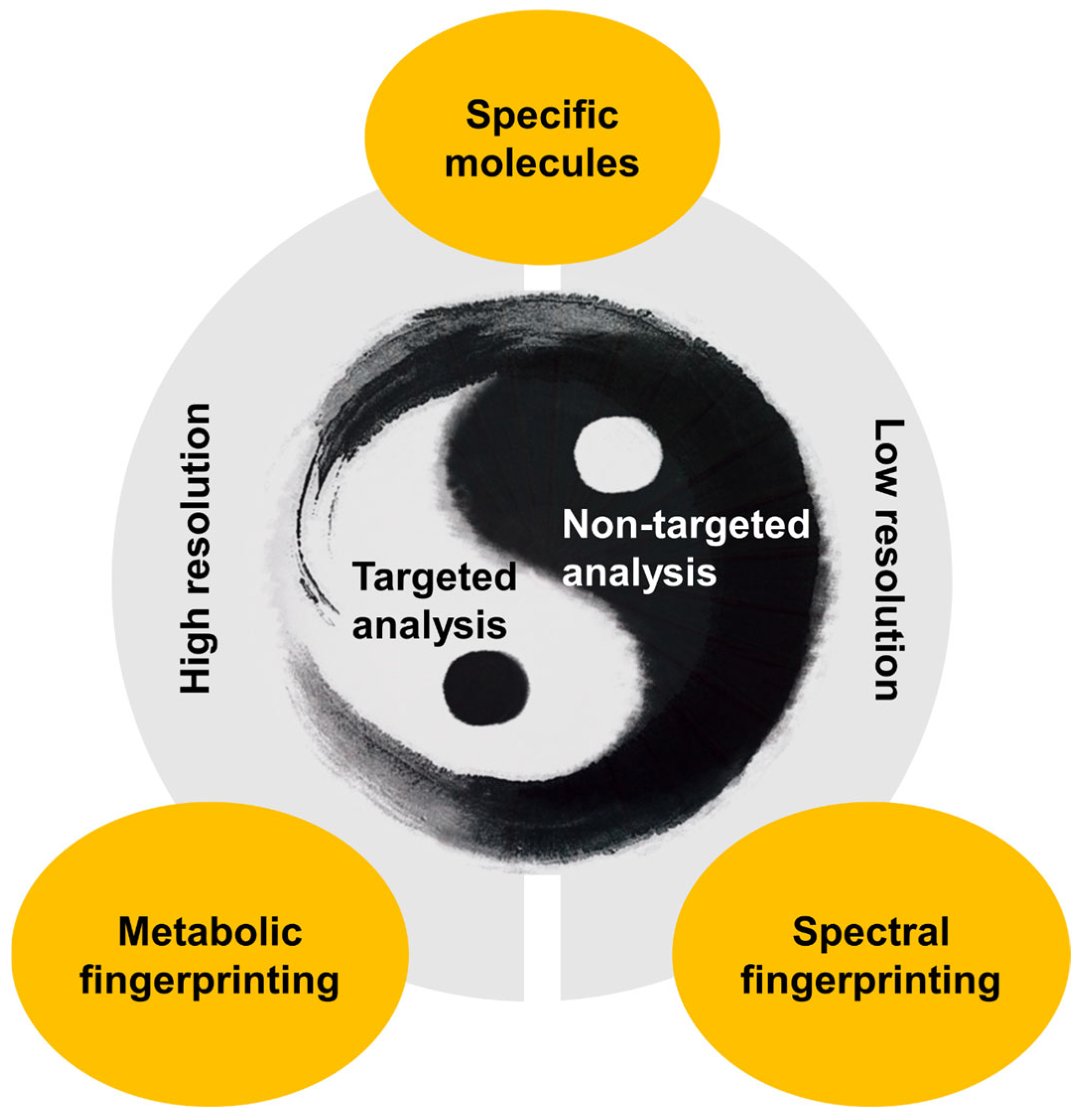
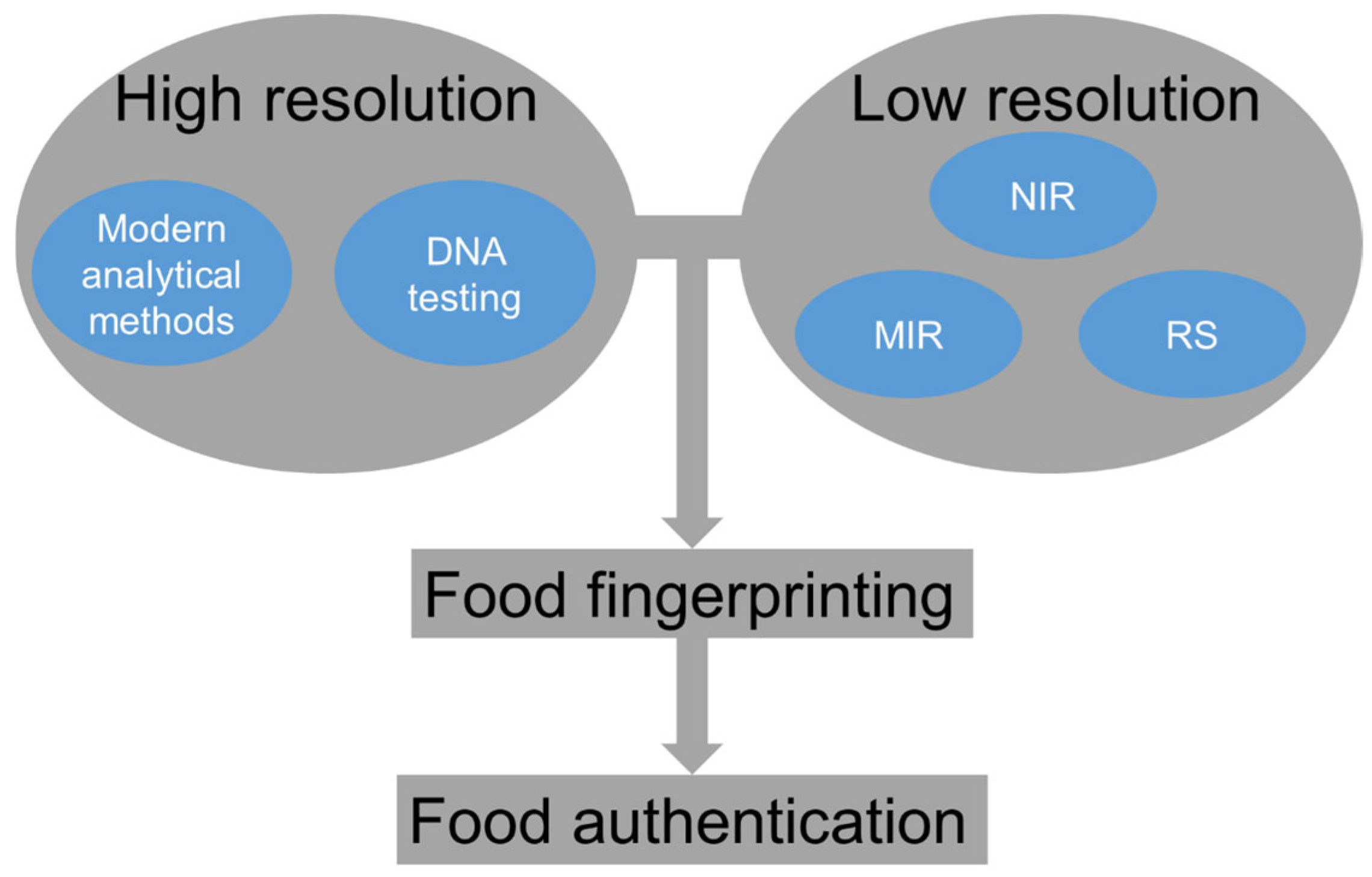
2. Food Fingerprinting and Vibrational Spectroscopy Techniques

Analytical methods that generate extensive, nonselective data for food characterization or authentication are commonly referred to as “fingerprinting” techniques; this broad category includes a variety of analytical approaches [8,9,24]. Terms such as “metabolite profiling”, which emphasizes specific metabolites or molecular components; “metabolomic profiling”; and “infrared fingerprinting” have been introduced by various researchers to describe related concepts. Fundamentally, a food fingerprint refers to a distinctive set of molecular indicators ranging from specific substances to broader chemical traits that collectively characterize the unique physicochemical and functional profile of a given food item. This framework offers a robust approach to identifying, categorizing, and tracing food ingredients and products with precision. Ultimately, effectively tackling the complexities of food fingerprinting and enhancing authentication methodologies rely heavily on the ability to detect and characterize either individual molecular indicators or comprehensive marker profiles within the food matrix.
Vibrational spectroscopy encompasses several techniques, including IR spectroscopy in both the NIR and MIR regions, as well as Raman spectroscopy. A unifying characteristic of these methods is their capacity to probe molecular structures and detect specific molecular species by analyzing the vibrational states of individual molecules or molecular groups; they capture the vibrational states of individual molecules or groups of molecules [23,25,26,27,28,29]. Nonetheless, it is essential to recognize that these techniques may exhibit varying sensitivity to different vibrational modes and bond types within a molecule. Consequently, integrating multiple vibrational spectroscopy methods can provide complementary insights, offering a more comprehensive understanding of the composition and characteristics of food samples.
Recent developments in Fourier transform instruments have resulted in advanced devices capable of analyzing a wide range of molecules in different forms, including solutions, powders, and even complex matrices such as food. A significant breakthrough has been the integration of attenuated total reflectance (ATR) modules with Fourier infrared instruments, which has notably improved their analytical performance. The growing number of applications using ATR-MIR technology highlights its effectiveness for food analysis. ATR has proven to be a valuable alternative for food sample analysis using MIR spectroscopy, providing a more efficient method. In ATR spectroscopy, the incident light is introduced into a high-refractive-index crystal, where it reflects internally at the crystal–sample interface. A portion of the evanescent wave extends a short distance beyond the surface into the sample, and if the sample is properly in contact, it selectively absorbs energy at characteristic wavenumbers, resulting in specific energy attenuation corresponding to molecular vibrations [20]. The penetration depth is influenced by factors such as the wavenumber, the refractive index of the crystal, and the incident radiation angle. Key benefits of ATR include the need for little or no sample preparation and its versatility in analyzing various food forms, including liquids, gels, solids, and powders [30]. However, when analyzing powders and solid samples, it is critical to ensure proper contact between the sample and the ATR crystal surface to obtain accurate results.
NIR spectroscopy is one of the most widely used techniques in food analysis. It works by measuring the absorption of energy from chemical bonds found in functional groups like C–H, O–H, N–H, and C=O, which are directly related to the chemical makeup and structural properties of food products [29,31,32]. NIR spectroscopy is highly adaptable and can be used to analyze a variety of samples, including those with high moisture content, powders, whole fruits, grains, individual kernels, and different plant parts. This method is applicable at various points in the food supply chain, from the farm and harvesting stages to in-line processing, transportation, storage, and even retail environments such as supermarkets.
In addition to infrared spectroscopy, Raman spectroscopy has become a valuable tool in food analysis. This technique detects vibrations caused by changes in the electrical polarizability of bonds, with bonds such as C–C typically exhibiting stronger activity compared to less polarizable bonds like O–H. Raman spectroscopy is particularly sensitive to molecular functional groups exhibiting high polarizability, such as carbon–carbon double and triple bonds (C=C and C≡C) and carbon–nitrogen triple bonds (C≡N), distinguishing it from infrared spectroscopy, which more strongly absorbs highly polar bonds, like those found in hydroxyl (O–H) and carbonyl (C=O) groups [27,31]. The spectral features observed in Raman arise from vibrational transitions analogous to those in the infrared region; however, stretching vibrations tend to be more pronounced in Raman spectra. Fundamentally, Raman analysis relies on the inelastic scattering of photons as they interact with molecular vibrations. Despite its advantages, a notable limitation is the inherently weak Raman signal, which results in relatively low sensitivity when applied to food samples [33]. To overcome the inherent limitations of weak Raman signals, multiple advanced signal enhancement strategies have been developed. These include techniques such as stimulated Raman scattering (SRS), coherent anti-Stokes Raman scattering (CARS), resonance Raman spectroscopy (RRS), and surface-enhanced Raman spectroscopy (SERS), all of which aim to amplify signal intensity and improve detection sensitivity [34,35]. A notable benefit of Raman spectroscopy, particularly in the context of aqueous food systems, is its minimal sensitivity to water. Unlike infrared spectroscopy, where water exhibits strong absorption bands, Raman spectra are largely unaffected by water, thus allowing clearer detection of solute-specific molecular features. Nevertheless, interferences from fluorescence and other instrumentation-related issues remain major limitations of Raman spectroscopy. Recently, there has been a rise in portable instruments aimed at mitigating some of these challenges, positioning Raman spectroscopy as a complementary technique to both NIR and MIR spectroscopy.
While both NIR and MIR spectroscopy provide point-based spectral data, they lack spatial information during analysis. In response to this limitation, hyperspectral imaging has emerged as a technique that combines point-based spectral analysis with spatial data, allowing for the simultaneous collection of digital images alongside molecular information from a sample. Hyperspectral imaging integrates the strengths of spectral analysis with spatial resolution, making it an effective tool for evaluating food quality and safety [36,37,38,39]. The primary advantage of hyperspectral imaging is its ability to provide both spatial and spectral information from the sample. This technique utilizes various wavelengths within the electromagnetic spectrum, including the visible and NIR ranges, and may also incorporate Raman spectroscopy [40]. Hyperspectral imaging has emerged as a cutting-edge analytical approach with strong potential for real-time surveillance and control within food production environments. However, despite its technical merits, such as the ability to simultaneously capture spatial and spectral data, its broader implementation in industrial settings faces notable limitations. Chief among these are the substantial financial investments required for certain high-performance systems, the relatively small number of commercial manufacturers offering robust solutions, and the considerable computational resources and time demanded for image acquisition, data processing, and model development, particularly for tasks such as classification and predictive analysis [41,42]. However, advancements in electronic components, improvements in camera technology, and more efficient algorithms are expected to enhance the speed of image acquisition and processing, facilitating the implementation of hyperspectral imaging systems in food authenticity applications.
The terahertz (THz) frequency range occupies the space between the end of the IR spectrum and the start of the microwave region. Although the use of THz technology in food analysis is still in its infancy, it holds significant potential. One of the key benefits of THz spectroscopy is its ability to probe far-IR vibrational modes in food, particularly for measuring water content. The resonance of THz waves with hydrogen bond vibrations makes this technique highly sensitive to water, which is an advantage when assessing moisture levels in food [43]. However, this heightened sensitivity to water also presents a challenge, as it can complicate the detection of other compounds in food samples that have high moisture content.
Beyond traditional spectroscopic approaches and hyperspectral imaging, integrating vibrational spectroscopic techniques, such as near-infrared, mid-infrared, and Raman spectroscopy, with advanced optical microscopy tools, including confocal and conventional light microscopes, has proven highly effective for visualizing and characterizing a broad spectrum of food substrates, ranging from individual grains to complex cereal structures [32]. This integration enables the analysis of the chemical composition of food samples without requiring staining or complex sample preparation, making it a more efficient and non-invasive method.

3. Data Analysis, Experimental Design, and Method Validation

3.1. Chemometrics, Preprocessing, and Data Fusion

Effectively employing vibrational spectroscopy for food fingerprinting within the context of foodomics depends heavily on the incorporation of chemometric modeling. In authentication tasks, spectral data, which are typically in the form of wavelength-dependent signals, serve as input variables, while corresponding output targets are defined during model construction, whether for categorical classification or quantitative regression purposes. Once the model is trained, new input data can be processed to identify clusters, patterns, or concentrations. The model’s accuracy is heavily reliant on the quality of the input variables, including the wavelength range, the signal-to-noise ratio, and the way the samples are presented [44,45].
Two primary approaches are used in applying these techniques: targeted and untargeted analyses. Targeted methods focus on identifying specific properties or characteristics in food, often to detect known ingredients or contaminants. On the other hand, untargeted analysis involves interpreting signals from various instrumental techniques to gain a comprehensive understanding of the sample, without any predefined assumptions about its composition [46,47].
A wide range of algorithms is available to support various types of analysis, many of which have been extensively used and documented. These algorithms include principal component analysis (PCA), partial least squares (PLS) regression, support vector machines (for both classification and regression), artificial neural networks, K-nearest neighbors, and convolutional neural networks [48,49]. Among multivariate analysis methods, PCA serves as a fundamental tool for data exploration and dimensionality reduction. It allows researchers to visualize variance patterns and identify outliers in high-dimensional spectral data. PLS, on the other hand, is especially valuable for developing regression models that correlate spectral data with physicochemical properties. Variants such as PLS discriminant analysis (PLS-DA) extend these capabilities to classification tasks. The interpretability and predictive strength of these methods make them indispensable in chemometric workflows involving vibrational spectroscopy.
Recent developments in food fingerprinting have emphasized the potential of data fusion techniques, which have shown promising results in creating models that integrate data from multiple instruments or methods [50]. Although this approach appears relatively simple, it continues to pose significant challenges in data analysis within food chemometrics. Moreover, various methods and algorithms are available for analyzing data from IR sensors, and preprocessing steps, such as baseline correction and derivative calculations, are crucial for developing accurate calibration and classification models [51,52,53,54].

3.2. Experimental Design and Sampling Strategies

A carefully planned experimental design and a robust sampling protocol are essential before developing and implementing applications for ensuring food authenticity [55,56]. However, the importance of defining these strategies is often overlooked, which can lead to challenges during data collection. Such oversights may negatively impact the development and interpretation of calibration or modeling processes. To enhance the generalizability and practical relevance of vibrational spectroscopy applications, it is essential to incorporate a wide range of food matrices. This includes less processed products such as fresh fruits, vegetables, and grains that may present complex or variable spectral features. Moreover, food products influenced by seasonal or regional variation (e.g., herbs, honey, and wines) should be considered, as they reflect real-world diversity and present important challenges for developing robust authentication models.

3.3. Validation and Method Monitoring

The validation of a developed model, whether for calibration or classification, is a crucial step in the application of any vibrational spectroscopy technique. Many studies often rely solely on cross-validation to evaluate the model’s performance, both qualitatively and quantitatively. However, independent validation is highly recommended and necessary to ensure the model’s robustness. This can be performed by incorporating samples that were not part of the model’s development process, such as those from different geographical regions or harvests. After the model is both developed and validated, continuous monitoring of its accuracy and reliability is essential. This ongoing evaluation should be part of the model’s lifecycle, as variations in sample characteristics or processing conditions may significantly influence the predicted results [57]. Cross-validation is commonly employed to estimate the performance of calibration and classification models when external validation sets are not available. Among various approaches, k-fold cross-validation and leave-one-out cross-validation (LOOCV) are widely used. These methods reduce the risk of overfitting and offer robust error estimation, especially in studies with a limited sample size. Furthermore, independent validation using external datasets, such as samples from different regions, harvest periods, or food batches, provides an additional layer of reliability. Model robustness should also be periodically re-evaluated to account for potential shifts in food matrix characteristics over time.

4. Vibrational Spectroscopic Techniques for Non-Invasive Food Authentication

Different spectroscopic techniques operate within defined frequency ranges, which are determined by the specific processes being studied and the energy transitions associated with them. Methods like FTIR, MIR, Raman spectroscopy, and HSI have demonstrated their effectiveness as quick and reliable tools for verifying food authenticity and evaluating the quality of various agro-food products. These techniques offer several benefits, including their non-destructive nature and relatively low analysis costs. Additionally, they are versatile and suitable for both qualitative and quantitative assessments of food and agricultural products, providing an efficient alternative to traditional wet chemical methods, which tend to be labor-intensive and time-consuming [58,59]. To contextualize the advancements in vibrational spectroscopy for food authentication, we analyzed 248 peer-reviewed studies (2010–2024) from databases like Scopus and Web of Science. Table S1 illustrates the research distribution of studies by technique and food matrix.

4.1. NIR Spectroscopy for Non-Invasive Food Authentication

NIR spectroscopy has found widespread application in both compositional assessments and classification tasks within the food and agricultural sectors, largely owing to its capability to evaluate large sample volumes with minimal or no preparation requirements. Initially, it served as a supplementary tool alongside other optical instruments, but by the 1980s, dedicated NIR systems emerged for the chemical analysis of products. Advances in both hardware and software significantly expanded its capabilities. Early NIR spectrometers were dispersive, measuring wavelengths one at a time, which required repositioning the grating for each measurement. This method was slow, and the detector was often a major source of noise. To overcome these limitations, FTIR spectroscopy was introduced. FTIR systems use a prism or a moving grating to separate frequencies from the NIR source, allowing the detector to simultaneously capture energy passing through the sample at multiple frequencies. This approach speeds up the analysis and produces a unique signal that encodes all infrared frequencies. Depending on their optical architecture, near-infrared spectrometers are generally divided into three principal categories: (i) stepwise scanning systems that acquire spectral information sequentially via monochromators or optical filters; (ii) interferometric designs, such as Fourier transform-based configurations, which capture comprehensive spectral data simultaneously through interferogram processing; and (iii) array-based or multichannel systems, in which distinct detectors concurrently monitor absorption at different wavelengths [59,60].
Advancements in instrumentation have been accompanied by significant progress in analytical methods and software, particularly those incorporating mathematical and chemometric techniques, which have greatly improved data storage and interpretation efficiency. These developments have made NIR spectroscopy an increasingly reliable, efficient, and standardized method for quality control. As a result, NIR spectroscopy is now widely adopted across a variety of industries, including food and pharmaceuticals, as well as the petrochemical and chemical sectors.
In NIR spectroscopy, different spectral modes can be applied to assess both the external and internal characteristics of samples. These modes include reflectance (specular and diffuse), transmission, interactance, and transflectance, each suited for different types of samples. Reflectance or interactance is typically used for solid samples, while transmission is ideal for liquids, and transflectance is applied to thin or transparent materials. The choice of mode depends on the specific properties of the sample, which need to be understood in detail. In reflectance mode, the light that is reflected or scattered from the sample’s surface is measured. Specular reflection occurs on smooth surfaces, while rough surfaces result in diffuse reflection. Specular reflectance offers limited information about the sample’s composition, whereas diffuse reflectance provides more detailed insights into the chemical and physical properties of the sample. This latter mode has gained significant attention in the agro-food sector, especially for food analysis, due to its ability to offer valuable data. In recent years, diffuse reflectance NIR techniques have been widely used in food adulteration detection. Studies have successfully identified meat muscles [61], detected adulteration in beef hamburgers [62], and identified adulteration in crabmeat [63,64]. Furthermore, high classification accuracy has been achieved in distinguishing between different types of meat, and diffuse reflectance NIR (1100–2500 nm) achieved 95% accuracy in detecting horsemeat adulteration in beef [65]. This technique has also been effectively used to detect melamine adulteration in milk powder and soybean meal. PLS-DA models for milk powder adulteration with melamine showed RMSECV < 0.5%, [66,67], and it has also been used to identify protein adulteration in yogurt [68]. NIR reflectance spectra within the wavelength range of 1100 to 2498 nm provide a straightforward method for detecting 10% pulp wash in orange juice and sugar–acid mixtures, achieving an accuracy of 90% [69]. Additionally, visible and NIR reflectance spectroscopy has been utilized to detect the adulteration of strawberry and raspberry purees with apples [70].
The transmission mode of NIR spectroscopy is highly effective for analyzing both the external and internal properties of a sample, as the transmitted light provides information about its internal composition. This method is one of the simplest and most versatile sampling techniques, applicable to solid, liquid, and gaseous samples. Transmission mode has been employed to assess the authenticity and detect adulteration in a variety of substances, including oils [71] and juices [72]. Additionally, transmission mode has been utilized to differentiate white wine samples based on their varietal origin [73] and predict the fatty acid composition of beef and chicken [63,74].
The interactance mode of NIR spectroscopy offers a balanced approach between reflectance and transmission modes, making it particularly effective for obtaining internal information from samples, such as fruits and vegetables, where transmission measurements may be difficult [75]. In this mode, the light source and detector are placed close to each other, ensuring that specular reflection does not directly enter the detector. The light travels through the outer layer of the sample, where part of it is reflected from the surface, and the rest is either scattered by internal structures or captured by the detector. This setup results in a higher signal-to-noise ratio, improving measurement accuracy [76].
Transflectance mode in NIR spectroscopy integrates features of both transmission and reflectance, making it ideal for analyzing thin or transparent samples. Although it is not as commonly used as the other modes, transflectance proves effective for assessing liquid samples, particularly when paired with optical bundle probes. A variety of studies have explored the use of NIR transflectance spectroscopy for evaluating the authenticity and quality of honey [77,78,79]. Beyond honey, NIR spectroscopy has been applied to evaluate the quality and authenticity of various food products, including oils [80], cow milk [81], and meats [82]. Additionally, it has been utilized for the geographical classification of wines [83]. However, it must be pointed that water-rich matrices (e.g., fruits) required spectral preprocessing to mitigate scattering effects [45].

4.2. FTIR Spectroscopy for Non-Invasive Food Authentication

FTIR spectroscopy, like NIR spectroscopy, is highly versatile and can be applied to solids, liquids, and gases using various measurement techniques, including ATR, diffuse reflectance, high-throughput transmission (HTT), and transmission cell methods. Among the various FTIR sampling techniques, ATR is the predominant method employed in food authenticity and quality assessments, accounting for approximately 72% of reported applications. This technique relies on guiding infrared radiation through a high-refractive-index crystal, where the beam undergoes multiple internal reflections at the interface with the sample [84]. A key advantage of ATR-FTIR spectroscopy is its ability to perform both qualitative and quantitative analyses with minimal or no sample preparation. Additionally, several accessories, such as single-bounce ATR (SBATR), micro-ATR (mATR), and horizontal ATR (HATR), offer varying internal reflection geometries, making ATR effective for characterizing materials that absorb infrared light strongly. FTIR transmission spectroscopy, whether through HTT or transmission cell methods, also finds extensive use in the food sector. For instance, HTT is employed to identify fungi responsible for food spoilage [85] and to conduct metabolic fingerprinting of legume silage [86].
Comparative studies of FTIR sampling methods have demonstrated the advantages of ATR for analyzing microbial spoilage in milk [87] and screening beer quality using HATR, mATR, and HTT (Figure 3) [88]. These studies highlighted that ATR provides superior accuracy and reliability in assessing food quality and authenticity. This increased precision is likely due to the multi-bounce crystal in ATR, which allows the infrared light to reflect multiple times within the sample, enhancing absorbance. While HTT also yielded satisfactory results, it is particularly useful for rapidly analyzing a large number of samples. Transmission mode is preferred for liquids, as the infrared radiation passes through the entire sample, providing a higher signal-to-noise ratio. Moreover, the automation of the filling and cleaning process in transmission measurements streamlines sample preparation, improving efficiency compared to ATR.
FTIR spectroscopy, along with the aforementioned sampling techniques, has shown considerable promise in detecting honey authenticity and adulteration. It has been successfully used to identify adulteration in honey with various substances, such as corn syrup, beet sugar, cane sugar [89], and sugar solutions [90]. ATR-FTIR with PCA-LDA detected syrup adulterants at 0.5% w/w [89]. Additionally, numerous studies have utilized FTIR spectroscopy to determine the sugar content in fruit juices [91].
The application of FTIR spectroscopy for the authentication of red wines based on grape variety achieved an impressive 95% classification accuracy [92]. Moreover, Greek red wines were successfully distinguished by grape variety through the comparison of FTIR spectra from samples with existing reference spectra in a database, where HATR-FTIR achieved 95% varietal classification [93]. FTIR has also proven to be a simple and cost-effective method for quality control and authenticity verification in beer [81]. Other notable uses of FTIR in food quality assessments include detecting adulteration in chocolate products [94]; monitoring meat spoilage through total viable bacterial counts, sensory quality, and pH [95]; and identifying added sugar and citric acid in fruit jams [96]. Additionally, FTIR has been utilized as an alternative method for characterizing and identifying microorganisms when coupled with a light microscope [97]. This approach has facilitated the discrimination and classification of enterococci [98] and the identification of bacteria in complex matrices [97]. FTIR spectroscopy combined with chemometrics for the detection, quantification, and classification of bacteria, including foodborne pathogens, indicates its promise as a powerful tool in microbiological analysis. FTIR spectroscopy is a highly effective analytical tool with diverse applications in the agro-food sector, and its sampling techniques have continuously progressed over the years. Both NIR and FTIR spectroscopy are capable of achieving similar analytical objectives; however, each technique may offer specific advantages in certain contexts. When selecting a method for sample analysis, it is essential to weigh the strengths and weaknesses of both techniques, considering factors such as sensitivity, sample preparation requirements, and the nature of the samples being analyzed.

4.3. Raman Spectroscopy for Non-Invasive Food Authentication

Raman spectroscopy is a highly effective analytical tool for identifying sample matrices, especially for analyzing key compounds like lipids, proteins, and carbohydrates. It is particularly sensitive to trace components, such as microorganisms, that contribute to food spoilage and contamination [99]. Though both IR and Raman spectroscopy are forms of vibrational spectroscopy, they operate based on different molecular interaction mechanisms. While IR absorption spectroscopy measures the absorption of light, Raman spectroscopy relies on the exchange of photon energy between the light and the molecule. The Raman effect occurs when light interacts with the electron cloud of a molecule’s bonds, causing the molecule to undergo a transition to a virtual state. As the molecule returns to a lower vibrational energy state, Raman scattering is produced. If the molecule was already in a higher vibrational state, the scattering is referred to as anti-Stokes scattering. In spontaneous Raman spectroscopy, the majority of the incident photons, approximately 99.999%, undergo elastic Rayleigh scattering, which does not provide useful information for molecular analysis because it does not change the molecule’s quantum state. Rayleigh scattering is an elastic interaction where light interacts with matter but leaves the material unchanged. Only a very small fraction—around 0.001%—of the incident light produces inelastic Raman scattering, which is the useful signal for molecular characterization. However, since spontaneous Raman scattering is relatively weak, it can be difficult to isolate the Raman signal from the much stronger Rayleigh scattering. To overcome this challenge, various optical components, such as notch filters, laser stop apertures, and tunable filters, are used to suppress Rayleigh scattering and enhance the quality of the Raman spectra. Furthermore, different illumination and sampling techniques are employed to boost the Raman signal. Methods such as SRS, CARS, RRS, and SERS all provide advantages in terms of sensitivity and signal amplification (Table 1), further improving the capabilities of Raman spectroscopy for detailed analysis.
Recent advancements in instrumentation have positioned Raman spectroscopy as an increasingly valuable tool in the food industry. One of the notable techniques, CARS spectroscopy, enables label-free chemical imaging with high sensitivity and excellent spatial resolution. However, a limitation of CARS is the presence of a nonresonant electronic background, which can obscure the chemical signals of interest [100]. In contrast, SRS spectroscopy provides enhanced sensitivity and avoids the issue of nonresonant interference, making it particularly advantageous for precise molecular analysis [101]. Both SRS and CARS are commonly applied in the agro-food industry, particularly for the microscopic examination of various products, thereby allowing for the detailed analysis of food components at the molecular level [102]. RRS represents a distinct Raman technique in which the excitation light is carefully aligned with an electronic absorption band of the target molecule. This resonance condition enhances the vibrational signal by promoting electronic excitation, thereby intensifying both Stokes and anti-Stokes scattering features in the resulting spectrum [103]. However, RRS typically requires visible or ultraviolet excitation, which can lead to interference from fluorescence, complicating the analysis. Despite this challenge, RRS has been successfully used for the characterization of microorganisms in various food products, aiding in the detection and analysis of microbial contamination [104]. These advanced Raman techniques are helping to push the boundaries of food analysis, offering powerful, non-destructive tools for improving food quality control, authenticity testing, and contamination detection.
SERS is a technique that merges Raman spectroscopy with nanotechnology by utilizing metallic nanosubstrates to significantly enhance the sensitivity and capabilities of conventional Raman analysis. By employing materials such as gold or silver nanoparticles, SERS can amplify the Raman signal of a molecule adsorbed on the surface of these metals, leading to signal enhancements by factors of 5 to 6 orders of magnitude compared to the same molecule in its bulk form. For example, with gold nanosubstrates, the enhancement factor can exceed three orders of magnitude. This dramatic improvement in sensitivity makes SERS a powerful analytical tool for the characterization of a wide array of inorganic substances and biologically relevant compounds. As a result, SERS is becoming an increasingly popular technique for chemical analysis in various fields, including food science, where it is used for detecting contaminants and adulterants and analyzing food composition [105]. The ability to obtain high sensitivity with minimal sample preparation makes SERS particularly valuable for food quality control and authenticity testing.
Raman spectroscopy is a highly effective tool for analyzing muscle foods, offering detailed structural information about key components such as proteins, water, and lipids. This capability has made it a valuable technique in the food industry, especially for detecting meat authenticity and assessing quality. For instance, it has been successfully used to predict sensory attributes like texture, tenderness, and juiciness in beef samples [106,107,108]. Additionally, Raman spectroscopy has been applied to determine the fatty acid composition of unextracted adipose tissue in various meat types and to assess the fat content in fish muscle [109]. The technique also has applications in monitoring changes in muscle protein during storage, such as in detecting muscle fiber tissue [110]. Furthermore, Raman spectroscopy can be used to track protein structure in muscle foods, which is crucial for understanding the textural qualities of meat [111]. Raman spectroscopy is not just limited to evaluating chemical and physical properties; it also has the ability to detect microbial spoilage in meat [112], offering significant advantages for food safety. A notable application in food authenticity is its use in detecting beef adulteration with horsemeat.
There has been a steady growth in research output of technological adoption for NIR, FTIR, and Raman spectroscopy in food authentication from 2010 to 2024, with significant acceleration post-2018, particularly in portable and in-field devices. Table 2 summarizes the key advantages, limitations, and common applications of each technique.
Recent advances in wearable spectroscopic devices and AI have significantly expanded the scope of food authentication. Wearable or handheld sensors enable real-time, on-site analysis, providing rapid decision-making tools for field or retail applications. In parallel, AI-driven algorithms, particularly deep learning models such as convolutional neural networks (CNNs) and recurrent neural networks (RNNs), are increasingly being used to handle high-dimensional spectral data, automating feature extraction and improving classification accuracy. These technologies represent a promising frontier for non-invasive and intelligent food quality monitoring.

5. Limitation of Vibrational Spectroscopy for Non-Invasive Food Authentication

Despite the growing popularity and success of vibrational spectroscopy techniques, such as NIR, FTIR, and Raman spectroscopy, for food authentication [113,114], several significant limitations must be acknowledged to ensure the development of reliable and scalable analytical protocols.
First, spectral complexity and overlapping signals represent a persistent challenge. Many food matrices are chemically complex and heterogeneous, resulting in overlapping spectral bands that complicate data interpretation. This is particularly problematic when trying to identify low-concentration adulterants or contaminants within samples containing dominant matrix signals, such as fats or water. For example, water exhibits strong absorption in the IR region, which can mask the subtle spectral features of other components, especially in high-moisture products like fruits, dairy, or beverages.
Second, fluorescence interference in Raman spectroscopy is a well-known limitation, especially in colored or processed food samples. The presence of pigments or phenolic compounds can induce strong background fluorescence, which overwhelms the weak Raman signal and reduces detection sensitivity. Although strategies such as shifted excitation Raman difference spectroscopy, SERS, and the use of near-infrared lasers have been explored to mitigate this effect, fluorescence remains a critical obstacle in practical applications.
Third, instrumental and methodological limitations hinder widespread industrial deployment. For instance, while portable spectrometers have improved accessibility and field use, they often exhibit lower resolution, sensitivity, and reproducibility compared to benchtop systems. Calibration transfer between instruments or laboratories is non-trivial due to differences in optical configurations, detector types, and spectral response functions. These variations necessitate robust standardization protocols and cross-platform calibration models, which are currently underdeveloped.
Fourth, sample heterogeneity and surface variability pose challenges for reproducibility and model generalization. In solid or semi-solid samples, differences in particle size, texture, temperature, and surface morphology can introduce significant scattering effects and baseline shifts, affecting model accuracy. Although spectral preprocessing techniques such as multiplicative scatter correction, standard normal variate, and derivatives can mitigate some of these effects, they cannot completely eliminate variability caused by inconsistent sample presentation or environmental factors.
Fifth, the limited availability of reference databases and authentic standards restricts the development of robust classification and calibration models. Most models are built using narrowly defined sample sets that may not capture the natural variation present in real-world scenarios, including regional, seasonal, and process-related variability. Without comprehensive and curated databases of authenticated reference samples, models are prone to overfitting and may fail to generalize across diverse production conditions.
Sixth, challenges in chemometric modeling and data fusion continue to limit the practical implementation of vibrational spectroscopy. The complexity of high-dimensional spectral data requires expertise in multivariate statistics, machine learning, and model validation. However, many studies rely on relatively simple chemometric approaches and lack external validation using independent datasets. Moreover, data fusion—although promising in combining multiple sensors or modalities—is computationally intensive and demands the careful synchronization of acquisition protocols, preprocessing strategies, and model architectures.
Lastly, while AI and deep learning models (e.g., CNNs and RNNs) have shown promise in enhancing classification accuracy and reducing dependence on feature engineering, they require large, diverse, and well-annotated datasets to avoid overfitting. The black-box nature of many AI models also raises concerns about interpretability, reliability, and regulatory acceptance, particularly in high-stakes food safety and traceability applications.
In summary, while vibrational spectroscopy offers substantial advantages as a non-invasive, rapid, and cost-effective tool for food authentication [76,113,114,115], its practical deployment requires overcoming several technical, analytical, and operational limitations. Future research should prioritize developing standardized protocols for sample handling, spectral acquisition, and model validation; creating shared spectral databases of authenticated food products; integrating real-time quality assurance systems into food supply chains; enhancing instrument robustness and portability without sacrificing analytical performance; and advancing hybrid AI-chemometric frameworks to improve model interpretability and deployment readiness.

6. Conclusions

The scientific literature and practical applications of vibrational spectroscopy have consistently highlighted the significant role these techniques play in monitoring food authenticity. However, it is crucial to recognize the limitations of vibrational spectroscopy, along with a comprehensive understanding of the data analytics necessary for developing both quantitative and qualitative models to prevent misleading results. Continued advancements in instrumentation, such as enhancements in portability and robustness, alongside progress in mathematical modeling and chemometrics, are expected to improve the reliability of these methods. Additionally, proper training in the application and interpretation of these models will further contribute to standardizing techniques and systems across the food supply and value chains.

Supplementary Materials

The following supporting information can be downloaded at https://www.mdpi.com/article/10.3390/app15115948/s1, Table S1. Distribution of studies by technique and food matrix (2010–2024).

Author Contributions

W.H.: writing—original draft. Q.Z.: validation. All authors have read and agreed to the published version of the manuscript.

Funding

This research was funded by Collaborative Innovation Project of “Vice General Manager of Technology” of Liaocheng grant number 2024XT02.

Data Availability Statement

Data will be provided by W.H. upon request.

Conflicts of Interest

The authors declare no conflicts of interest.

References

  1. Echegaray, N.; Hassoun, A.; Jagtap, S.; Tetteh-Caesar, M.; Kumar, M.; Tomasevic, I.; Goksen, G.; Lorenzo, J.M. Meat 4.0: Principles and applications of industry 4.0 technologies in the meat industry. Appl. Sci. 2022, 12, 6986. [Google Scholar] [CrossRef]
  2. Hassoun, A.; Aït-Kaddour, A.; Abu-Mahfouz, A.M.; Rathod, N.B.; Bader, F.; Barba, F.J.; Biancolillo, A.; Cropotova, J.; Galanakis, C.M.; Jambrak, A.R.; et al. The fourth industrial revolution in the food industry-Part I: Industry 4.0 technologies. Crit. Rev. Food Sci. Nutr. 2023, 63, 6547–6563. [Google Scholar] [CrossRef]
  3. Chapman, J.; Power, A.; Netzel, M.E.; Sultanbawa, Y.; Smyth, H.E.; Truong, V.K.; Cozzolino, D. Challenges and opportunities of the fourth revolution: A brief insight into the future of food. Crit. Rev. Food Sci. Nutr. 2022, 62, 2845–2853. [Google Scholar] [CrossRef]
  4. Hitzmann, B.; Hauselmann, R.; Niemoeller, A.; Sangi, D.; Traenkle, J.; Glassey, J. Process analytical technologies in food industry-challenges and benefits: A status report and recommendations. Biotechnol. J. 2015, 10, 1095–1100. [Google Scholar] [CrossRef]
  5. Konur, S.; Lan, Y.; Thakker, D.; Morkyani, G.; Polovina, N.; Sharp, J. Towards design and implementation of Industry 4.0 for food manufacturing. Neural Comput. Appl. 2023, 35, 23753–23765. [Google Scholar] [CrossRef]
  6. Hassoun, A.; Siddiqui, S.A.; Smaoui, S.; Ucak, I.; Arshad, R.N.; Garcia-Oliveira, P.; Prieto, M.A.; Ait-Kaddour, A.; Perestrelo, R.; Câmara, J.S.; et al. Seafood processing, preservation, and analytical techniques in the age of Industry 4.0. Appl. Sci. 2022, 12, 1703. [Google Scholar] [CrossRef]
  7. Islam, S.; Cullen, J.M. Food traceability: A generic theoretical framework. Food Control 2021, 123, 107848. [Google Scholar] [CrossRef]
  8. Ellis, D.I.; Brewster, V.L.; Dunn, W.B.; Allwood, J.W.; Golovanov, A.P.; Goodacre, R. Fingerprinting food: Current technologies for the detection of food adulteration and contamination. Chem. Soc. Rev. 2012, 41, 5706–5727. [Google Scholar] [CrossRef]
  9. Kharbach, M.; Alaoui Mansouri, M.; Taabouz, M.; Yu, H. Current application of advancing spectroscopy techniques in food analysis: Data handling with chemometric approaches. Foods 2023, 12, 2753. [Google Scholar] [CrossRef]
  10. Huang, Y.-T.; Fan, L.-Y.; Liu, Y.-F.; Zheng, X.-H. High-performance MoO3 nanosheets gas sensors for triethylamine detection: A rapid approach for assessing fish freshness. Ceram. Int. 2025, 51, 9912–9922. [Google Scholar] [CrossRef]
  11. Alvarado, V.L.S.; Diaz, F.J.; Parra, L.; Lloret, J.; Aldana, C.; Saavedra, Y. Proposal for a gas sensor device to classify hydrobiological species and estimate non-refrigeration time. IEEE Sens. J. 2025, 25, 18015–18022. [Google Scholar] [CrossRef]
  12. Noto, G.; Coletta, L.; Vainieri, M. Measuring the performance of collaborative governance in food safety management: An Italian case study. Public Money Manag. 2022, 42, 627–636. [Google Scholar] [CrossRef]
  13. Shi, Y.; Yu, Y.; Zhang, J.; Yin, C.; Chen, Y.; Men, H. Origin traceability of agricultural products: A lightweight collaborative neural network for spectral information processing. Food Res. Int. 2025, 208, 116131. [Google Scholar] [CrossRef] [PubMed]
  14. Carcea, M.; Brereton, P.; Hsu, R.; Kelly, S.; Marmiroli, N.; Melini, F.; Soukoulis, C.; Ding, W.P. Food authenticity assessment: Ensuring compliance with food legislation and traceability requirements. Qual. Assur. Saf. Crops Foods 2009, 1, 93–100. [Google Scholar] [CrossRef]
  15. Fritsche, J. Recent developments and digital perspectives in food safety and authenticity. J. Agric. Food Chem. 2018, 66, 7562–7567. [Google Scholar] [CrossRef] [PubMed]
  16. Gallo, M.; Ferranti, P. The evolution of analytical chemistry methods in foodomics. J. Chromatogr. A 2016, 1428, 3–15. [Google Scholar] [CrossRef]
  17. Wolfender, J.L.; Marti, G.; Thomas, A.; Bertrand, S. Current approaches and challenges for the metabolite profiling of complex natural extracts. J. Chromatogr. A 2015, 1382, 136–164. [Google Scholar] [CrossRef]
  18. Cuadros-Rodríguez, L.; Ruiz-Samblás, C.; Valverde-Som, L.; Pérez-Castaño, E.; González-Casado, A. Chromatographic fingerprinting: An innovative approach for food ‘identitation’ and food authentication—A tutorial. Anal. Chim. Acta 2016, 909, 9–23. [Google Scholar] [CrossRef]
  19. Mialon, N.; Roig, B.; Capodanno, E.; Cadiere, A. Untargeted metabolomic approaches in food authenticity: A review that showcases biomarkers. Food Chem. 2023, 398, 133856. [Google Scholar] [CrossRef]
  20. Karoui, R. Chapter 2—Spectroscopic technique: Mid-infrared (MIR) and Fourier transform mid-infrared (FT-MIR) spectroscopies. In Modern Techniques for Food Authentication, 2nd ed.; Sun, D.-W., Ed.; Academic Press: Cambridge, MA, USA, 2018; pp. 23–50. [Google Scholar]
  21. Cortés, V.; Blasco, J.; Aleixos, N.; Cubero, S.; Talens, P. Monitoring strategies for quality control of agricultural products using visible and near-infrared spectroscopy: A review. Trends Food Sci. Technol. 2019, 85, 138–148. [Google Scholar] [CrossRef]
  22. Cozzolino, D. Foodomics and infrared spectroscopy: From compounds to functionality. Curr. Opin. Food Sci. 2015, 4, 39–43. [Google Scholar] [CrossRef]
  23. Arendse, E.; Nieuwoudt, H.; Magwaza, L.S.; Nturambirwe, J.F.I.; Fawole, O.A.; Opara, U.L. Recent advancements on vibrational spectroscopic techniques for the detection of authenticity and adulteration in horticultural products with a specific focus on oils, juices and powders. Food Bioprocess Technol. 2021, 14, 1–22. [Google Scholar] [CrossRef]
  24. Cozzolino, D. Low resolution food fingerprinting: Vibrational spectroscopic methods for nondestructive food authentication. Curr. Opin. Food Sci. 2024, 60, 101229. [Google Scholar] [CrossRef]
  25. Bec, K.B.; Huck, C.W. Breakthrough potential in near-infrared spectroscopy: Spectra simulation. A review of recent developments. Front. Chem. 2019, 7, 48. [Google Scholar] [CrossRef]
  26. Manley, M. Near-infrared spectroscopy and hyperspectral imaging: Non-destructive analysis of biological materials. Chem. Soc. Rev. 2014, 43, 8200–8214. [Google Scholar] [CrossRef]
  27. Thygesen, L.G.; Løkke, M.M.; Micklander, E.; Engelsen, S.B. Vibrational microspectroscopy of food. Raman vs. FT-IR. Trends Food Sci. Technol. 2003, 14, 50–57. [Google Scholar] [CrossRef]
  28. Su, W.H.; He, H.J.; Sun, D.W. Non-destructive and rapid evaluation of staple foods quality by using spectroscopic techniques: A review. Crit. Rev. Food Sci. Nutr. 2017, 57, 1039–1051. [Google Scholar] [CrossRef]
  29. Li, H.; Wang, J.; Kim, H.; Peng, X.; Yoon, J. Activatable near-infrared versatile fluorescent and chemiluminescent dyes based on the dicyanomethylene-4H-pyran scaffold: From design to imaging and theranostics. Angew. Chem. Int. Ed. 2024, 63, e202311764. [Google Scholar] [CrossRef]
  30. Wu, L.M.; Xu, D.G.; Wang, Y.Y.; Ge, M.L.; Li, H.B.; Wang, Z.L.; Yao, J.Q. Common path continuous terahertz reflection and attenuated total reflection imaging. Acta Phys. Sin. 2021, 70, 118701. [Google Scholar] [CrossRef]
  31. Arjunan, V.; Sakiladevi, S.; Marchewka, M.K.; Mohan, S. FTIR, FT-Raman, FT-NMR and quantum chemical investigations of 3-acetylcoumarin. Spectrochim. Acta Part A Mol. Biomol. Spectrosc. 2013, 109, 79–89. [Google Scholar] [CrossRef]
  32. Gullifa, G.; Barone, L.; Papa, E.; Giuffrida, A.; Materazzi, S.; Risoluti, R. Portable NIR spectroscopy: The route to green analytical chemistry. Front. Chem. 2023, 11, 1214825. [Google Scholar] [CrossRef] [PubMed]
  33. Pan, L.R.; Zhang, P.; Daengngam, C.; Peng, S.L.; Chongcheawchamnan, M. A review of artificial intelligence methods combined with Raman spectroscopy to identify the composition of substances. J. Raman Spectrosc. 2022, 53, 6–19. [Google Scholar] [CrossRef]
  34. Batista, A.M.; Foiani, L.; Champeau, M.; Martinho, H. Advances in tissues and cells characterization by Raman micro-spectroscopy, atomic force microscopy, and tip-enhanced Raman spectroscopy. J. Raman Spectrosc. 2022, 53, 1848–1860. [Google Scholar] [CrossRef]
  35. Liu, G.K.; Zheng, H.; Lu, J.L. Recent progress and perspective of trace antibiotics detection in aquatic environment by surface-enhanced Raman spectroscopy. Trends Environ. Anal. Chem. 2017, 16, 16–23. [Google Scholar] [CrossRef]
  36. Sendin, K.; Williams, P.J.; Manley, M. Near infrared hyperspectral imaging in quality and safety evaluation of cereals. Crit. Rev. Food Sci. Nutr. 2018, 58, 575–590. [Google Scholar] [CrossRef]
  37. Amodio, M.L.; Chaudhry, M.M.A.; Colelli, G. Spectral and hyperspectral technologies as an additional tool to increase information on quality and origin of horticultural crops. Agronomy 2020, 10, 7. [Google Scholar] [CrossRef]
  38. Ahmad, M.; Vitale, R.; Silva, C.S.; Ruckebusch, C.; Cocchi, M. A novel proposal to investigate the interplay between the spatial and spectral domains in near-infrared spectral imaging data by means of image decomposition, encoding and localization (IDEL). Anal. Chim. Acta 2022, 1191, 339285. [Google Scholar] [CrossRef] [PubMed]
  39. Ma, J.J.; Zheng, B.Y.; He, Y. Applications of a hyperspectral imaging system used to estimate wheat grain protein: A review. Front. Plant Sci. 2022, 13, 837200. [Google Scholar] [CrossRef]
  40. Wolf, S.; Popp, J.; Frosch, T. Multifocal hyperspectral Raman imaging setup for multi-well plates. Sens. Actuators B Chem. 2023, 375, 132949. [Google Scholar] [CrossRef]
  41. Wang, Z.X.; Xu, P.; Liu, B.H.; Cao, Y.K.; Liu, Z.; Liu, Z.J. Hyperspectral imaging for underwater object detection. Sens. Rev. 2021, 41, 176–191. [Google Scholar] [CrossRef]
  42. Bai, H.X.; Xue, Q.S.; Hao, X.J.; Li, H.; Huang, L.Y.; Li, C.; Yang, J.Y. Underwater hyperspectral imaging system with dual-scanning mode. Appl. Opt. 2022, 61, 4226–4237. [Google Scholar] [CrossRef] [PubMed]
  43. Han, C.; Qu, F.; Wang, X.; Zhai, X.; Li, J.; Yu, K.; Zhao, Y. Terahertz spectroscopy and imaging techniques for herbal medicinal plants detection: A comprehensive review. Crit. Rev. Anal. Chem. 2024, 54, 2485–2499. [Google Scholar] [CrossRef] [PubMed]
  44. Saeys, W.; Do Trong, N.N.; Van Beers, R.; Nicolai, B.M. Multivariate calibration of spectroscopic sensors for postharvest quality evaluation: A review. Postharvest Biol. Technol. 2019, 158, 110981. [Google Scholar] [CrossRef]
  45. Agelet, L.E.; Hurburgh, C.R. A Tutorial on Near Infrared Spectroscopy and Its Calibration. Crit. Rev. Anal. Chem. 2010, 40, 246–260. [Google Scholar] [CrossRef]
  46. McGrath, T.F.; Haughey, S.A.; Patterson, J.; Fauhl-Hassek, C.; Donarski, J.; Alewijn, M.; van Ruth, S.; Elliott, C.T. What are the scientific challenges in moving from targeted to non-targeted methods for food fraud testing and how can they be addressed?—Spectroscopy case study. Trends Food Sci. Technol. 2018, 76, 38–55. [Google Scholar] [CrossRef]
  47. Musio, B.; Ragone, R.; Todisco, S.; Rizzuti, A.; Iorio, E.; Chirico, M.; Pisanu, M.E.; Meloni, N.; Mastrorilli, P.; Gallo, V. Non-targeted nuclear magnetic resonance analysis for food authenticity: A comparative study on tomato samples. Molecules 2024, 29, 4441. [Google Scholar] [CrossRef]
  48. Szymanska, E.; Gerretzen, J.; Engel, J.; Geurts, B.; Blanchet, L.; Buydens, L.M.C. Chemometrics and qualitative analysis have a vibrant relationship. TrAC Trends Anal. Chem. 2015, 69, 34–51. [Google Scholar] [CrossRef]
  49. Skov, T.; Honoré, A.H.; Jensen, H.M.; Næs, T.; Engelsen, S.B. Chemometrics in foodomics: Handling data structures from multiple analytical platforms. TrAC Trends Anal. Chem. 2014, 60, 71–79. [Google Scholar] [CrossRef]
  50. Ferreira, M.M.; Marins-Goncalves, L.; De Souza, D. An integrative review of analytical techniques used in food authentication: A detailed description for milk and dairy products. Food Chem. 2024, 457, 140206. [Google Scholar] [CrossRef]
  51. Cozzolino, D.; Williams, P.J.; Hoffman, L.C. An overview of pre-processing methods available for hyperspectral imaging applications. Microchem. J. 2023, 193, 109129. [Google Scholar] [CrossRef]
  52. Oliveri, P.; Malegori, C.; Simonetti, R.; Casale, M. The impact of signal pre-processing on the final interpretation of analytical outcomes—A tutorial. Anal. Chim. Acta 2019, 1058, 9–17. [Google Scholar] [CrossRef] [PubMed]
  53. Dayananda, B.; Owen, S.; Kolobaric, A.; Chapman, J.; Cozzolino, D. Pre-processing applied to instrumental data in analytical chemistry: A brief review of the methods and examples. Crit. Rev. Anal. Chem. 2023, 54, 2745–2753. [Google Scholar] [CrossRef]
  54. Wang, L.; Meng, Q.X.; Ren, L.P.; Yang, J.S. Near infrared reflectance spectroscopy (NIRS) and its application in the determination for the quality of animal feed and products. Spectrosc. Spectr. Anal. 2010, 30, 1482–1487. [Google Scholar] [CrossRef]
  55. Cozzolino, D. The sample, the spectra and the maths-The critical pillars in the development of robust and sound applications of vibrational spectroscopy. Molecules 2020, 25, 3674. [Google Scholar] [CrossRef] [PubMed]
  56. Brereton, R.G.; Jansen, J.; Lopes, J.; Marini, F.; Pomerantsev, A.; Rodionova, O.; Roger, J.M.; Walczak, B.; Tauler, R. Chemometrics in analytical chemistry-part I: History, experimental design and data analysis tools. Anal. Bioanal. Chem. 2017, 409, 5891–5899. [Google Scholar] [CrossRef]
  57. Brereton, R.G.; Jansen, J.; Lopes, J.; Marini, F.; Pomerantsev, A.; Rodionova, O.; Roger, J.M.; Walczak, B.; Tauler, R. Chemometrics in analytical chemistrypart II: Modeling, validation, and applications. Anal. Bioanal. Chem. 2018, 410, 6691–6704. [Google Scholar] [CrossRef]
  58. Lohumi, S.; Mo, C.; Kang, J.-S.; Hong, S.-J.; Cho, B.-K. Nondestructive evaluation for the viability of watermelon (citrullus lanatus) seeds using Fourier transform near infrared spectroscopy. J. Biosyst. Eng. 2013, 38, 312–317. [Google Scholar] [CrossRef]
  59. Lohumi, S.; Lee, S.; Lee, H.; Cho, B.-K. A review of vibrational spectroscopic techniques for the detection of food authenticity and adulteration. Trends Food Sci. Technol. 2015, 46, 85–98. [Google Scholar] [CrossRef]
  60. Rodriguez-Saona, L.E.; Allendorf, M.E. Use of FTIR for rapid authentication and detection of adulteration of food. Annu. Rev. Food Sci. Technol. 2011, 2, 467–483. [Google Scholar] [CrossRef]
  61. Cozzolino, D.; Murray, I. Identification of animal meat muscles by visible and near infrared reflectance spectroscopy. LWT-Food Sci. Technol. 2004, 37, 447–452. [Google Scholar] [CrossRef]
  62. Ding, H.B.; Xu, R.J. Near-infrared spectroscopic technique for detection of beef hamburger adulteration. J. Agric. Food Chem. 2000, 48, 2193–2198. [Google Scholar] [CrossRef] [PubMed]
  63. Gayo, J.; Hale, S.A.; Blanchard, S.M. Quantitative analysis and detection of adulteration in crab meat using visible and near-infrared spectroscopy. J. Agric. Food Chem. 2006, 54, 1130–1136. [Google Scholar] [CrossRef]
  64. Gayo, J.; Hale, S.A. Detection and quantification of species authenticity and adulteration in crabmeat using visible and near-infrared spectroscopy. J. Agric. Food Chem. 2007, 55, 585–592. [Google Scholar] [CrossRef] [PubMed]
  65. Alander, J.T.; Bochko, V.A.; Martinkauppi, B.; Saranwong, S.; Mantere, T. A review of optical nondestructive visual and near-infrared methods for food quality and safety. Int. J. Spectrosc. 2013, 2013, 341402. [Google Scholar] [CrossRef]
  66. Haughey, S.A.; Graham, S.F.; Cancouët, E.; Elliott, C.T. The application of near-infrared reflectance spectroscopy (NIRS) to detect melamine adulteration of soya bean meal. Food Chem. 2013, 136, 1557–1561. [Google Scholar] [CrossRef]
  67. Lu, C.H.; Xiang, B.R.; Hao, G.; Xu, J.P.; Wang, Z.W.; Chen, C.Y. Rapid detection of melamine in milk powder by near infrared spectroscopy. J. Near Infrared Spectrosc. 2009, 17, 59–67. [Google Scholar] [CrossRef]
  68. Xu, L.; Yan, S.-M.; Cai, C.-B.; Wang, Z.-J.; Yu, X.-P. The feasibility of using near-infrared spectroscopy and chemometrics for untargeted detection of protein adulteration in yogurt: Removing unwanted variations in pure yogurt. J. Anal. Methods Chem. 2013, 2013, 201873. [Google Scholar] [CrossRef]
  69. Twomey, M.; Downey, G.; McNulty, P.B. The potential of NIR spectroscopy for the detection of the adulteration of orange juice. J. Sci. Food Agric. 1995, 67, 77–84. [Google Scholar] [CrossRef]
  70. Contal, L.; León, V.; Downey, G. Detection and quantification of apple adulteration in strawberry and raspberry purées using visible and near infrared spectroscopy. J. Near Infrared Spectrosc. 2002, 10, 289–299. [Google Scholar] [CrossRef]
  71. Sinelli, N.; Casale, M.; Di Egidio, V.; Oliveri, P.; Bassi, D.; Tura, D.; Casiraghi, E. Varietal discrimination of extra virgin olive oils by near and mid infrared spectroscopy. Food Res. Int. 2010, 43, 2126–2131. [Google Scholar] [CrossRef]
  72. Xie, L.-J.; Ye, X.-Q.; Liu, D.-H.; Ying, Y.-B. Application of principal component-radial basis function neural networks (PC-RBFNN) for the detection of water-adulterated bayberry juice by near-infrared spectroscopy. J. Zhejiang Univ. Sci. B 2008, 9, 982–989. [Google Scholar] [CrossRef]
  73. Hou, X.; Xue, Y.; Liu, C.; Li, Z.; Xu, Z. Dual NIR-channel fluorescent probe for detecting ONOO in vitro and vivo. Spectrochim. Acta Part A Mol. Biomol. Spectrosc. 2024, 320, 124665. [Google Scholar] [CrossRef] [PubMed]
  74. Veloso Tropia, N.; Reis Vilela, R.S.; de Sales Silva, F.A.; Andrade, D.R.; Costa, A.C.; Cidrini, F.A.A.; de Souza Pinheiro, J.; Pucetti, P.; Chizzotti, M.L.; de Campos Valadares Filho, S. Regression models from portable NIR spectra for predicting the carcass traits and meat quality of beef cattle. PLoS ONE 2024, 19, e0303946. [Google Scholar] [CrossRef]
  75. Wang, Y.; Xing, L.; He, H.-J.; Zhang, J.; Chew, K.W.; Ou, X. NIR sensors combined with chemometric algorithms in intelligent quality evaluation of sweetpotato roots from ‘Farm’ to ‘Table’: Progresses, challenges, trends, and prospects. Food Chem. X 2024, 22, 101449. [Google Scholar] [CrossRef]
  76. Cozzolino, D. The ability of near infrared (NIR) spectroscopy to predict functional properties in foods: Challenges and opportunities. Molecules 2021, 26, 6981. [Google Scholar] [CrossRef] [PubMed]
  77. Xia, Y.; Li, D.; Wang, Y.; Xi, Q.; Jiao, T.; Wei, J.; Chen, X.; Chen, Q.; Chen, Q. Rapid identification of cod authenticity based on hyperspectral imaging technology. Spectrochim. Acta Part A Mol. Biomol. Spectrosc. 2024, 326, 125258. [Google Scholar] [CrossRef] [PubMed]
  78. Fu, H.Y.; Shi, Q.; Wei, L.N.; Xu, L.; Guo, X.M.; Hu, O.; Lan, W.; Xie, S.P.; Yang, T.M. Rapid recognition of geoherbalism and authenticity of a Chinese herb by data fusion of near-infrared spectroscopy (NIR) and mid-infrared (MIR) spectroscopy combined with chemometrics. J. Spectrosc. 2019, 2019, 2467185. [Google Scholar] [CrossRef]
  79. Parastar, H.; van Kollenburg, G.; Weesepoel, Y.; van den Doel, A.; Buydens, L.; Jansen, J. Integration of handheld NIR and machine learning to “Measure & Monitor” chicken meat authenticity. Food Control 2020, 112, 107149. [Google Scholar]
  80. Zhao, L.L.; Zhang, M.; Wang, H.X.; Mujumdar, A.S. Monitoring of free fatty acid content in mixed frying oils by means of LF-NMR and NIR combined with BP-ANN. Food Control 2022, 133, 108599. [Google Scholar] [CrossRef]
  81. Peng, D.; Xu, R.; Zhou, Q.; Yue, J.; Su, M.; Zheng, S.; Li, J. Discrimination of milk freshness based on synchronous two-dimensional visible/near-infrared correlation spectroscopy coupled with chemometrics. Molecules 2023, 28, 5728. [Google Scholar] [CrossRef]
  82. Cozzolino, D.; Bures, D.; Hoffman, L.C. Evaluating the use of a similarity index (SI) combined with near infrared (NIR) spectroscopy as method in meat species authenticity. Foods 2023, 12, 182. [Google Scholar] [CrossRef] [PubMed]
  83. Martelo-Vidal, M.J.; Vázquez, M. Classification of red wines from controlled designation of origin by ultraviolet-visible and near-infrared spectral analysis. Cienc. Tec. Vitivinic. 2014, 29, 35–43. [Google Scholar] [CrossRef]
  84. Ma, J.; Qi, Y.; Lei, M.; Xuan, H.; Li, X.; Lu, W.; Guo, J.; Chen, H. Analysis and discrimination of adhesive species using ATR-FTIR combined with Raman, and HS-GC-IMS together with multivariate statistical analysis. J. Chromatogr. A 2024, 1736, 465402. [Google Scholar] [CrossRef]
  85. Nittari, G.; Roy, P.; Martinelli, I.; Bellitto, V.; Tomassoni, D.; Traini, E.; Tayebati, S.K.; Amenta, F. Rodent models of Huntington’s disease: An overview. Biomedicines 2023, 11, 3331. [Google Scholar] [CrossRef] [PubMed]
  86. Soureshjani, H.K.; Bahador, M.; Tadayon, M.R.; Dehkordi, A.G. Modeling seed germination of quinoa (Chenopodium quinoa Willd.) at different temperatures and water potentials. Acta Physiol. Plant. 2022, 44, 102. [Google Scholar] [CrossRef]
  87. Nicolaou, N.; Goodacre, R. Rapid and quantitative detection of the microbial spoilage in milk using Fourier transform infrared spectroscopy and chemometrics. Analyst 2008, 133, 1424–1431. [Google Scholar] [CrossRef] [PubMed]
  88. Polshin, E.; Aernouts, B.; Saeys, W.; Delvaux, F.; Delvaux, F.R.; Saison, D.; Hertog, M.; Nicolaï, B.M.; Lammertyn, J. Beer quality screening by FT-IR spectrometry: Impact of measurement strategies, data pre-processings and variable selection algorithms. J. Food Eng. 2011, 106, 188–198. [Google Scholar] [CrossRef]
  89. Sivakesava, S.; Irudayaraj, J.M.K. Detection of inverted beet sugar adulteration of honey by FTIR spectroscopy. J. Sci. Food Agric. 2001, 81, 683–690. [Google Scholar] [CrossRef]
  90. Kelly, J.D.; Downey, G. Detection of sugar adulterants in apple juice using fourier transform infrared spectroscopy and chemometrics. J. Agric. Food Chem. 2005, 53, 3281–3286. [Google Scholar] [CrossRef]
  91. Yajima, Y.; Wakabayashi, H.; Suehara, K.-I.; Kameoka, T.; Hashimoto, A. Simultaneous content determination of mono-, di-, and fructo-oligosaccharides in citrus fruit juices using an FTIR-PLS method based on selected absorption bands. Int. J. Food Sci. 2024, 2024, 9265590. [Google Scholar] [CrossRef]
  92. Garcia-Hernandez, C.; Salvo-Comino, C.; Martin-Pedrosa, F.; Garcia-Cabezon, C.; Rodriguez-Mendez, M.L. Analysis of red wines using an electronic tongue and infrared spectroscopy, correlations with phenolic content and color parameters. LWT 2020, 118, 108785. [Google Scholar] [CrossRef]
  93. Tarantilis, P.A.; Troianou, V.E.; Pappas, C.S.; Kotseridis, Y.S.; Polissiou, M.G. Differentiation of Greek red wines on the basis of grape variety using attenuated total reflectance Fourier transform infrared spectroscopy. Food Chem. 2008, 111, 192–196. [Google Scholar] [CrossRef]
  94. Alkurd, R.A.; Al-Akayleh, F.T.; Al-Remawi, M.M.; Hasan, M.R.; Maqousi, A.; Ramadan, K.; Arafat, T.A. Evaluation of a Jordanian commercial chocolate brand fortified with micronutrients of vitamins B12 and D3, iron and zinc. Food Sci. Technol. 2023, 43, e105122. [Google Scholar] [CrossRef]
  95. Dashti, A.; Weesepoel, Y.; Muller-Maatsch, J.; Parastar, H.; Kobarfard, F.; Daraei, B.; Yazdanpanah, H. Assessment of meat authenticity using portable Fourier transform infrared spectroscopy combined with multivariate classification techniques. Microchem. J. 2022, 181, 107735. [Google Scholar] [CrossRef]
  96. Kurz, C.; Leitenberger, M.; Carle, R.; Schieber, A. Evaluation of fruit authenticity and determination of the fruit content of fruit products using FT-NIR spectroscopy of cell wall components. Food Chem. 2010, 119, 806–812. [Google Scholar] [CrossRef]
  97. Wang, Y.D.; Li, X.L.; Liu, Z.X.; Zhang, X.X.; Hu, J.; Lü, J.H. Discrimination of foodborne pathogenic bacteria using synchrotron FTIR microspectroscopy. Nucl. Sci. Tech. 2017, 28, 49. [Google Scholar] [CrossRef]
  98. Teng, A.S.J.; Habermehl, P.E.; van Houdt, R.; de Jong, M.D.; van Mansfeld, R.; Matamoros, S.P.F.; Spijkerman, I.J.B.; van Meer, M.P.A.; Visser, C.E. Comparison of fast Fourier transform infrared spectroscopy biotyping with whole genome sequencing-based genotyping in common nosocomial pathogens. Anal. Bioanal. Chem. 2022, 414, 7179–7189. [Google Scholar] [CrossRef]
  99. Liu, Q.; Dong, P.; Fengou, L.-C.; Nychas, G.-J.; Fowler, S.M.; Mao, Y.; Luo, X.; Zhang, Y. Preliminary investigation into the prediction of indicators of beef spoilage using Raman and Fourier transform infrared spectroscopy. Meat Sci. 2023, 200, 109168. [Google Scholar] [CrossRef]
  100. Song, S.W.; Jang, J.I.; Park, C.R.; Kim, H.M. Optically pumped and matrix-assisted anti-Stokes Raman spectroscopy. J. Raman Spectrosc. 2022, 53, 924–933. [Google Scholar] [CrossRef]
  101. Xu, Z.C.; Oguchi, K.; Taguchi, Y.; Sano, Y.; Miyawaki, Y.; Cheon, D.; Katoh, K.; Ozeki, Y. Stimulated Raman scattering spectroscopy with quantum-enhanced balanced detection. Opt. Express 2022, 30, 18589–18598. [Google Scholar] [CrossRef]
  102. Calisan Kinter, R.; Ozbaran, B.; Inal Kaleli, I.; Kose, S.; Bildik, T.; Ghaziuddin, M. The sensory profiles, eating behaviors, and quality of life of children with autism spectrum disorder and avoidant/restrictive food intake disorder. Psychiatr. Q. 2024, 95, 85–106. [Google Scholar] [CrossRef] [PubMed]
  103. Mohammed, T.A.; Moores, R.R., Jr.; Hendricks-Munoz, K.D.; Romfh, P.; Rozycki, H.J. Resonance Raman spectroscopy tissue oxygenation measurements in neonates. Neonatology 2023, 120, 363–370. [Google Scholar] [CrossRef]
  104. Hwang, J.-E.; Park, J.-Y.; Jung, M.H.; Eom, K.; Moon, H.S.; Joung, H.; Kim, Y.J. Evaluation of a commercial device based on reflection spectroscopy as an alternative to resonance Raman spectroscopy in measuring skin carotenoid levels: Randomized controlled trial. Sensors 2023, 23, 7654. [Google Scholar] [CrossRef] [PubMed]
  105. Jiang, L.; Hassan, M.M.; Ali, S.; Li, H.H.; Sheng, R.; Chen, Q.S. Evolving trends in SERS-based techniques for food quality and safety: A review. Trends Food Sci. Technol. 2021, 112, 225–240. [Google Scholar] [CrossRef]
  106. Chuesiang, P.; Ryu, V.; Siripatrawan, U.; He, L.L.; McLandsborough, L. Aptamer-based surface enhanced Raman spectroscopy (SERS) for the rapid detection of Salmonella Enteritidis contaminated in ground beef. LWT 2021, 150, 111937. [Google Scholar] [CrossRef]
  107. Gukowsky, J.C.; He, L.L. Development of a portable SERS method for testing the antibiotic sensitivity of foodborne bacteria. J. Microbiol. Methods 2022, 198, 106496. [Google Scholar] [CrossRef] [PubMed]
  108. Lu, X.; Ma, Y.; Jiang, S.; Wang, Z.; Yu, Q.; Ji, C.; Guo, J.; Kong, X. Quantitative monitoring ofloxacin in beef by TLC-SERS combined with machine learning analysis. Spectrochim. Acta Part A Mol. Biomol. Spectrosc. 2024, 308, 123790. [Google Scholar] [CrossRef]
  109. Zhao, Y.; Song, Y.Y.; Zhang, Y.Y.; Fan, Y.X.; Lai, K.Q. Ultra sensitive detection of malachite green in fish muscle with gold nanoparticles and graphene oxide hybrid as a substrate for surface enhanced Raman scattering. J. Food Meas. Charact. 2020, 14, 658–667. [Google Scholar] [CrossRef]
  110. Pan, C.Y.; Zhang, S.Y.; Xiong, X.L.; Li, Z.; Ai, B.W.; Qian, W.P.; Dong, J. Dynamically monitoring pH in living organisms based on a SERS-active optical fiber. Adv. Mater. Interfaces 2022, 9, 2200328. [Google Scholar] [CrossRef]
  111. Purwidyantri, A.; Karina, M.; Hsu, C.H.; Srikandace, Y.; Prabowo, B.A.; Lai, C.S. Facile bacterial cellulose nanofibrillation for the development of a plasmonic paper sensor. ACS Biomater. Sci. Eng. 2020, 6, 3122–3131. [Google Scholar] [CrossRef]
  112. Guo, Q.; Peng, Y.; Qin, J.; Chao, K.; Zhao, X.; Yin, T. Advance in detection technique of lean meat powder residues in meat using SERS: A review. Molecules 2023, 28, 7504. [Google Scholar] [CrossRef] [PubMed]
  113. Chapman, J.; Gangadoo, S.; Truong, V.K.; Cozzolino, D. Spectroscopic approaches for rapid beer and wine analysis. Curr. Opin. Food Sci. 2019, 28, 67–73. [Google Scholar] [CrossRef]
  114. He, H.J.; Sun, D.W. Hyperspectral imaging technology for rapid detection of various microbial contaminants in agricultural and food products. Trends Food Sci. Technol. 2015, 46, 99–109. [Google Scholar] [CrossRef]
  115. Walsh, K.B.; McGlone, V.A.; Han, D.H. The uses of near infra-red spectroscopy in postharvest decision support: A review. Postharvest Biol. Technol. 2020, 163, 111139. [Google Scholar] [CrossRef]
Figure 1. An overview of the essential steps and key considerations in applying vibrational spectroscopy for food fingerprinting and authenticity assessments.
Applsci 15 05948 g001
Figure 2. Integration of high-resolution and low-resolution techniques with chemometrics for food fingerprinting analysis.
Applsci 15 05948 g002
Figure 3. Schematic of typical FTIR and Raman spectra from whole milk samples and molecular vibrational states along with a simplified Jablonski diagram [8].
Applsci 15 05948 g003
Table 1. Comparison of Raman techniques for food authentication.
TechniqueEnhancement FactorFood ApplicationKey Limitation
SERS105−106Pesticides in fruitsSubstrate reproducibility
CARS103−104Lipid oxidation in meatNonresonant background
Resonance Raman102−103Carotenoids in seafoodUV-induced fluorescence
Table 2. Comparative summary of NIR, FTIR, and Raman spectroscopy.
FeatureNIRFTIRRaman
Water InterferenceModerateHighLow
Sample PrepMinimalMinimal–ModerateMinimal
SensitivityMediumHighVery High (esp. SERS)
Instrument CostMediumMediumHigh
PortabilityHighMediumIncreasing
Major ApplicationsBulk analysis and adulterationMolecular fingerprinting and sugar detectionProtein/lipid analysis and contaminant detection
Disclaimer/Publisher’s Note: The statements, opinions and data contained in all publications are solely those of the individual author(s) and contributor(s) and not of MDPI and/or the editor(s). MDPI and/or the editor(s) disclaim responsibility for any injury to people or property resulting from any ideas, methods, instructions or products referred to in the content.

