Article

A Statistical Procedure for Exploring a Skeletal Age-Explicative Tool for Growing Patients

by
Michele Tepedino
1,
Rosa Esposito
1,2,*,
Maurizio Delvecchio
1,
Domenico Ciavarella
2,
Giuseppe Rofrano
3 and
Francesco Masedu
1
1
Department of Biotechnological and Applied Clinical Sciences, University of L’Aquila, 67100 L’Aquila, Italy
2
Department of Clinical and Experimental Medicine, School of Dentistry, University of Foggia, 71122 Foggia, Italy
3
Experimental Zooprophylactic Institute of the South, National Reference Center for the Analysis and Study of the Correlation Between Environment, Animals and Humans, 80055 Portici, Italy
*
Author to whom correspondence should be addressed.
Appl. Sci. 2025, 15(10), 5593; https://doi.org/10.3390/app15105593
Submission received: 22 April 2025 / Revised: 13 May 2025 / Accepted: 15 May 2025 / Published: 16 May 2025
(This article belongs to the Special Issue Orthodontics: Advanced Techniques, Methods and Materials)

Abstract

:
Background: Skeletal age estimation plays a fundamental role in orthopedic treatments. Since the most reliable methods are based on ionizing radiation, this study aimed to use machine learning techniques to explore a skeletal age assessment method not based on additional radiographies. Methods: Patients aged between 6 and 16 years old whose clinical records included orthopantomography, radiographs of the second phalanx of the third finger, and biometric data were enrolled for the study. The radiographs were analyzed to estimate the maturation degree of the left lower first premolars, the midpalatal suture, and the second phalanx of the third finger. Both an explicative data analysis and a multivariate analysis were performed. Results: The sample comprised 111 subjects. The multivariate analysis revealed an explanatory role for sex (p < 0.01) and chronological age (p < 0.01). The ordinal tool showed how the use of height (p = 0.02) and weight (p = 0.03) was explicative of skeletal age against a loss of statistical significance corresponding to the use of body mass index (p = 0.6). The median palatine suture (p = 0.01) was explicative. Conclusions: The combined evaluation of weight, height, sex, chronological age, and grade of maturation of the midpalate suture provides an explicative tool for assessing skeletal age without additional radiographic exams, besides a routine orthopantomography.

1. Introduction

The assessment of skeletal age is one of the most important determinants of treatment outcomes in growing patients whenever the targets are skeletal structures, like in orthopedics, orthodontics, and pediatrics [1]. In such disciplines, the problem of skeletal age estimation plays a fundamental role [2]. Although the literature provides contradictory evidence about the actual possibility of affecting growth’s driving factors, the debate is still open, and many clinicians prefer to start orthodontic treatment during an active growth phase to take advantage of it. A representative example is the intervention for skeletal class II malocclusions, whose treatment yields optimal results when carried out around the pubertal growth spurt, during which the peak of mandibular growth is also observed [3]. In many cases, the orthodontists must be able to determine whether the subject is in a slow- or fast-growing phase and for how long the growth can be expected to last in order to plan the intervention [4,5]. Therefore, many methods for skeletal age assessment have been developed and described.
The simplest indicator of skeletal maturity is chronological age. Unfortunately, the literature shows that this simple indicator is also the worst. Indeed, puberty starts at very different ages in different individuals, even for subjects of the same sex [6,7]; therefore, skeletal age does not always correspond to chronological age [8,9]. On the other hand, the method considered to be the gold-standard is the evaluation of the level of bone maturation of the phalanges and wrist bones, using technique described by different authors [6,10]. Because this requires an additional radiograph of the hand, which has no other clinical value for orthodontic diagnosis, authors have tried to identify less invasive methods. An example is the evaluation of the skeletal maturation stage of the second, third, and fourth cervical vertebrae on lateral cephalograms [11,12]. The so-called “cervical vertebral maturation method” showed an acceptable correlation with the hand–wrist maturation method [12]. Although useful in reducing the patient’s radiation exposure [13], some authors questioned its precision in assessing the skeletal maturation stage.
A simpler method for the assessment of bone maturation is based on radiographic analysis of the second phalanx of the middle finger [14,15,16,17,18] instead of the whole hand. Another technique for the estimation of skeletal age was proposed by Demjran et al. and is based on dental maturity (root and crown) assessed on orthopantomography (OPG) [19].
Saraç et al. proposed another method that was based on mandibular morphometric measurements, like condylar height and tangential ramus height (the height of the ramus measured a line passing from the top of the condyle and intersecting with a line tangential to the gonion of the mandible), which were reported to be correlated with chronological age [20]. Moreover, due to the correlation found between dental age and skeletal age, some authors suggested that dental age assessment could be a supplementary tool for estimating a patient’s pubertal spurt in Class II malocclusion cases, which is considered the best period for the treatment [21], providing significant improvement also airway dimensions as well [22].
Despite all these attempts, the greatest precision is still offered by the evaluation of the hand–wrist radiograph or the evaluation of the sole second phalanx of the middle finger, but at the cost of additional radiographs [23]. The scientific question—and the clinical purpose—behind the present study was to investigate if the combination of multiple non-invasive predictors of skeletal age could provide a better estimate, comparable to the gold standard, but at a lower biological cost.
Therefore, the primary objective of the present study was to create a categorical decision-making tool, relying on OPGs and biometric data, that allows for a probabilistic score to suggest the skeletal age in a clinically practical way, which is non-invasive, does not require exposure to additional radiation, and is easy to measure. A secondary objective of the study was to identify the statistical significance and the weight of measured covariates to be explicatively relevant to the skeletal age.

2. Materials and Methods

This observational and retrospective study was approved by the Ethical Committee of the University of L’Aquila (protocol 135261, ID 22/2023). The research was performed in accordance with the Declaration of Helsinki from 1975 and subsequent revisions, and written informed consent was obtained from every suitable subject before collecting data.
Participants were recruited among patients treated at the Orthodontic Clinic of the Department of Biotechnological and Applied Clinical Sciences, University of L’Aquila from January 2011 to December 2019. They were selected after screening, in chronological order, according to the following inclusion criteria:
  • Age between 6 and 16 years old;
  • OPG;
  • X-ray of the second phalanx of the third finger of the right hand;
  • Collection of biometric data such as height, weight, and wrist circumference;
  • All data must have been collected within a time window of a maximum of three months.
The exclusion criteria were:
  • Systemic pathologies affecting growth;
  • Bone pathologies and/or previous fractures of the upper limbs;
  • Multiple dental agenesia or oligodontia.
Maturation of the second phalanx of the third finger
One of the data collected was the grade of maturation of the second phalanx of the third finger, using a well-established method. The grade of maturation of the second phalanx of the third finger is a validated method used in dentistry for assessing skeletal age. It was introduced by Hagg and Taranger in 1980 [13], who defined five stages considering the changes in the shape of the epiphysis and metaphysis of the second phalanx of the middle finger. Lately, it was improved by Rajagopal and Kansal in 2002 [18,24] with the addition of another stage, and finally, the staging was more precisely described by Perinetti et al. [2], who defined six stages as described in Table 1 and represented in Figure 1.
Therefore, it is a categorical assessment method. Over the years, different authors have demonstrated the reliability of this method [15,16,17,18], and since the radiography of the middle phalanx of the third finger is practical and easy to record, many dental clinicians use it routinely to assess skeletal age. Indeed, this radiogram can be taken with the intraoral radiographic machine, which is present in any dental clinic. Therefore, with a single intraoral radiography of the third finger of the hand, the clinician can have information about the skeletal maturation of the patient in a very quick and practical way without requiring additional examination (for instance X-ray of left hand and wrist).
  • Data collection
The evaluation of radiographs was performed by three operators (MT, RE, MD). Two operators performed the categorization, while a third author was asked to arbitrate when the first two operators were not in agreement. This was performed for both the radiograph of the second phalanx of the third finger and the OPGs.
Firstly, the radiograph of the second phalanx of the third finger was examined to assign one out of the six categories described by the classification of Perinetti et al. [2].
To simplify the statistical analysis and reduce the number of covariates, the six stages of maturation of the second phalanx of the third finger were merged into the following four categories: MPS-1, MPS-2, MPS-3, MPS-4, as represented in Table 2. Because stages MPS1 and MPS2, as well as MPS5 and MPS6, represent very similar developmental stages from a clinical point of view [2]—i.e., an early skeletal maturation phase well apart from the pubertal growth spurt, and a later stage when residual growth is negligible to obtain significant clinical results—it was decided to merge those categories to reduce the total number of statistical variables and increase the power of the analysis (Table 2).
Similarly, OPGs were evaluated to determine the degree of tooth maturation according to the Demirjian method. The Demirjian method is used for defining the degree of dental development and involves the evaluation of the stage of formation for each of the permanent teeth examined separately. Each tooth is assigned a stage from A to H based on the appearance of the first centers of calcification to the closure of the tips of the roots, taking into account the maturation of the crown and root, when present (Figure 2) [19].
It is accepted that teeth are usually symmetrical in their development between the left and right sides of the jaw and between the maxillary and mandibular teeth. Therefore, a specific tooth on the left side of the mandible is usually at the same level of formation as its contralateral homologous, and, similarly, a maxillary tooth and its counterpart in the mandible are similar in formation stages. Evaluating one side of one jaw is therefore sufficient to measure maturity, and, as the mandibular teeth are more easily visualized on a radiograph, these teeth have been the most studied [25]. In this research article, to simplify the statistical analysis by reducing the number of studied variables, only the first premolars of the left lower jaw were analyzed, as done in previous studies [26].
Moreover, the degree of ossification of the midpalate suture was assessed on OPGs using the classification of the BOKA Grading System, where four categories are defined according to the radiographic appearance of the suture (Figure 3) [27].
During the clinical examination, the height, weight, and wrist circumference of each patient were collected. Weight was assessed using a professional mechanical balance (SECA 700, Hammer Steindamm 3-25, Hamburg, Germany); during the measurements, the patient wore only socks and underwear with a weighing scale previously adjusted to zero [28]. Height was assessed using a balance with an altimeter (SECA 700, Hammer Steindamm 3-25, Hamburg, Germany), with patients having taken off their shoes, and staying in position against a wall totally upright and immobile to allow the clinicians to precisely measure height. Then, the circumference of the right wrist was collected with the subject in an upright position, with the arm flexed and the palm facing forward. The meter was slid just below the styloid and radial processes of the ulna, located palpatorially [29]. Finally, body mass index (BMI) was calculated, using height, weight, and the following mathematical formula made by Quetelet A. in 1832. BMI = [weight (kg)/height (m)2] × 100 [30].
Therefore, during the collection of radiographic records, all the biometric data of the patient, such as height, weight, and wrist circumference, were collected too, and all the data were organized into a dataset and then analyzed.
  • Statistical analysis
The analysis was divided into two parts. Preliminarily, an explicative data analysis assessed the association between explicators, such as age and sex, and skeletal age. Sample double frequency distributions have been calculated, and the association of skeletal age with ordinal explicators has been assessed by performing the Cuzik rank test. Odds ratios have been estimated by carrying out univariate ordinal logistic regressions. The results provided suggestions for setting up the overall explication tool for skeletal age. The variance inflation factor (VIF) analysis has been paralleled by the estimations of variables, focusing on those variables that suggested possible multicollinearity. This feature has been of relevant interest for the variables BMI, weight, height, dental degree of development of teeth on the right and left mandibular first premolar, and midpalate suture degree of maturation. The information collected was summarized into an ordinal logistic tool, which provided a good fit when assessed using the likelihood ratio test. The McFadden R2 was 0.53. The McFadden R2, in the linear regression tool, synthesizes the proportion of variance in the dependent variable associated with the predictive and independent variables, with wider R2 values indicating that increased variation is explained by the tool, to a maximum of 1 [31]. The statistical analysis was performed using the statistical software STATA version 17 (STATA, College Station, TX, USA).

3. Results

One hundred eleven subjects were selected for the study (44.1% female; 55.9% male).
In particular, the distribution of gender within the different skeletal maturation categories was the following:
  • SM1: 27.3% female; 62.7% male;
  • SM2: 52.4% female; 47.6% male;
  • SM3: 66.7% female 33.3% male;
  • SM4: 64.0% female; 36.0% male.
Descriptive statistics are reported in Table 3.
The results of Spearman’s Rank Correlation according to the biometric data are reported in Table 4.
The multivariate analysis reported a statistically significant effect of sex (ß = −3.64, SE = 0.04, p < 0.001) and of chronological age (ß = 1.23, SE = 0.35; p < 0.001). Indeed, the variable sex showed a high explicative value, meaning that being female correlates with a higher score on the assessment of skeletal age, while being male correlates with a lower score.
In the explicative ordinal tool, both the explicators height (ß = 0.1; SE = 0.04; p = 0.02) and weight (ß = 0.12; SE = 0.06; p = 0.03) turned out to be statistically significant. On the other hand, when height and weight were substituted in the explicative ordinal tool by BMI, this index was not explicative of skeletal age (ß = 1.03; SE = 0.7; p = 0.6). The dental maturation of the lower left first premolar (ß = −2.39; SE = 0.88; p = 0.6) was not a significant explicator (Table 5).
The degree of skeletal maturation and ossification of the median palatine suture was also explicative: the tool reported a statically significant effect on skeletal age when the suture showed a maturation score of 3, which is indicative of a higher skeletal age (i.e., SM3 or SM4) (Table 5) while it is not significant for all the other evaluated variables.

4. Discussion

Orthodontists always face the demanding task of identifying and foreseeing the pubertal growth spurt or recognizing the final phases of skeletal growth and explaining the distance in time from those developmental periods [11,32]. The methods currently used for the estimation of skeletal age require exposing the patients to ionizing radiation. Since children are more sensitive to ionizing radiation due to their high radiosensitivity [33], having a method that allows reducing patients’ exposure to ionizing radiation, following the ALARA principle [34], is of great importance. This is in accordance with the implementation of Directive 2013/59/Euratom, which outlines basic safety standards for protection against the dangers arising from exposure to ionizing radiation and repealing directives. Even if the intraoral radiograph provides a mean effective dose of 1.32 (0.60–2.56) μSv [35], according to the ALARA principle, it would be better to avoid them. This goal should be achievable using the method described in the present research article, because clinicians could obtain the same diagnostic results derived from the radiograph of the second phalanx of the third finger, but using only biometric data and the information given by an OPG, avoiding further radiographic exams. As said in the introduction chapter, another method for assessing skeletal age is the evaluation of the cervical vertebrae maturation method on lateral cephalograms [3]. This method was discarded in the present study because some studies questioned its reliability [36], and because lateral cephalograms—unlike OPGs that are a routinary screening exam—need precise indications to justify their request: this would have been in contrast with the aim of the study of finding an alternative method not relying on additional radiographs. The assessment of skeletal age is of great importance to determine treatment timing in orthodontic clinical practice, even though it is a challenging task because growth spans over a long time and is characterized by several events with complex interactions [34,37,38]. In fact, the majority of orthodontic and orthopedic treatments are time dependent. The literature confirms that the determination of the optimal timing for early orthodontic treatment requires a comprehensive assessment of clinical manifestations, dental age, and skeletal age; therefore, chronological age by itself is not enough [39]. For example, early treatment of skeletal Class II requires identification of the pubertal growth spurt to take advantage of a faster growth; early treatment of maxillary transverse deficiency or skeletal Class III requires identification of early stages of skeletal maturation to take advantage of immature sutures; and adolescent treatment may also require the assessment of some residual growth, which may be expected [40]. It is known that the maximum acceleration of growth that is found in puberty leads to the maturation of primary and secondary sexual characteristics. There are huge differences in the pubertal spurts of girls and boys: they begin earlier in females than in males [1,41,42], and they are accompanied by the development of secondary sexual characteristics [43]. However, those features are not unequivocally related to skeletal age, thus influencing the assessment of skeletal age and consequently the definition of the appropriate treatment timing. For these reasons, finding a reliable variable for skeletal age assessment would be advisable and desirable.
In the present research article, different biometric and radiographic variables were analyzed to investigate a method for skeletal age assessment. The simplest variables studied were age and sex. It is known that chronological age is not a reliable explicator of skeletal age [9,16]. Although the multivariate tool (Table 3) showed a significant effect of chronological age, the univariate tool confirmed that the chronological age alone has inadequate explicative power for the estimation of the skeletal age.
When evaluating skeletal age, it is of utmost importance to consider the sex-related differences that are typical of these age groups and are related to sex hormones [40]. Indeed, sex showed a significant explicative value, as the odds ratio for females was indicative of a higher degree of skeletal maturation [44].
Other biometric data included in the present evaluation were height and weight. These parameters are useful when evaluating patients’ development and growth. It has been stated that early menarche in girls is constantly associated with higher adult BMI because circulating hormones, such as leptin and insulin, provide signals reflecting body fat stores [42,45]. In the current evaluation, variables such as height and weight were also represented with BMI. Indeed, the statistical analysis highlighted that height and weight were significant explicators of skeletal maturity; however, when those two variables were combined to calculate BMI, their explicative value decreased remarkably. It could be inferred that weight and height are less related to each other in this age group, thus explaining why BMI performs worse than weight and height taken individually. This is a remarkable finding, because BMI is a commonly used index in clinical practice and research to reduce the number of variables (because BMI is a synthesis of two variables). The present results suggest that this should be performed with caution, due to the possibility of type II errors.
Finally, among the biometric data considered, the wrist circumference was also statistically significant in the univariate analysis, but it loses its significance in the multivariate analyses. This finding suggests that this variable should not be considered explanatory for the estimation of skeletal age.
Apart from biometric data, radiographic images were evaluated in this study. It is known that radiographs are particularly useful for skeletal age assessment, too. Dental radiology is the most frequent diagnostic radiological investigation in the industrialized world, representing one-third of all radiological examinations in Europe [46,47]. Among these, the most frequent imaging techniques are the OPG and periapical radiographs [43]. Considering that OPG provides a good overview of the most important dental and skeletal structures of the maxillary complex with a low radiation dose, it is recognized as an adequate tool for oral health screening [48,49]. Indeed, it is always recommended to have an OPG of the patient for formulating the correct orthodontic diagnosis. Since the OPG is the most common dental radiograph, the rationale for the present study was to optimize the information that can be obtained from it. One of the variables considered for the estimation of skeletal age in this research article was the dental maturation stage. This method was precisely defined by Demjiran et al. [19] and used by different authors for assessing skeletal age. All the previous articles using this method considered only the right or left side of the jaws; in the current research study, to contain the number of variables to be inserted in the tool, a single tooth from the left side was considered. The lower left premolars are the teeth whose development encompasses the entire timeframe considered hereby and seemed suitable to be used for skeletal age assessment; moreover, the first premolars were preferred over the second premolars because the latter can be relatively frequently missing because of agenesia, thus impairing the generalizability of the current explicative tool. The dental maturation stage is a very easy and quick variable to evaluate, since only an OPG is required, which is virtually always available for every patient. However, the statistical analysis showed that the dental maturation stage was not a statistically significant explicator because the assessment of skeletal age had a very low explicative value. The reason for this result could be ascribed to the first premolars always showing a high degree of maturation in patients aged [13,14,15,16,17,18,19], like in the present sample. Therefore, the results of the present study are partially supported by recent research of Hedge et al., who assessed that dental age is a reliable marker for chronological age, and skeletal maturation aligns closely with chronological age. While BMI appears to be weakly correlated with skeletal and dental growth, further research is necessary to fully understand its impact [50]. Finally, another variable studied in the present research article was the staging of the skeletal maturation of the midpalate suture. Methods have been proposed using either 3D images [27] or occlusal radiographs [51]; for the present study, the “BOKA Grading System” was used, because it is a method based on OPG and thus does not require additional radiographs. The obtained results suggested that the midpalate suture’s maturation has a role in the definition of skeletal age when stage 3 of maturation is observed. In other words, this result should be interpreted as stage 3 midplatal suture maturation being an indicator of a higher stage of skeletal age, such as SM3 or SM4. This is a very important outcome since midpalate suture maturation has never been correlated to skeletal age assessment before, apart from a study by Hezenc et al. that demonstrated a correlation between the cervical vertebrae maturation and the fusion of the midpalate suture, which seems to occur after the pubertal growth spurt when the cervical vertebrae maturation is at stage four (the first maturation stage after the pubertal spurt) [52].
The limitations of the present study are represented by the retrospective sampling, even though care was taken to avoid selection biases by enrolling the patients in a rigid chronological order. Due to the cross-sectional design, it was not possible to verify the actual distance of the patient from the pubertal growth spurt, which is easily identifiable in females with menarche, but it is more difficult in males. Therefore, the evaluation of the second phalanx of the third finger was considered as a reference, but only a longitudinal study over a very long period would allow us to confirm and validate the skeletal age estimation from the proposed methods. Moreover, since the radiograph of the second phalanx of the third finger is useful in pubertal subjects, most of the subjects enrolled in the study were in SM2 or SM3, while there were fewer subjects in SM1 and SM4. Finally, there are other methods available in the pediatric field for the assessment of the skeletal age [53,54,55] which were not considered in this study, like the cervical maturation method on cephalograms. However, sometimes the estimate of skeletal maturation should be performed prior to taking a cephalogram, or to identify when to take a cephalogram to start planning a treatment. Therefore, it was preferred to evaluate a statistical tool that relied only on screening exams, like OPGs.

5. Conclusions

Through the evaluation of different chronological, biometrical, and radiographical data, the present research article assessed that:
  • Sex has a high explicative role for the assessment of skeletal age;
  • Height and weight are explicative of skeletal age, while their combination in BMI is not significant for skeletal maturation explication;
  • The degree of skeletal maturation and ossification of the median palatine suture is also indicative, suggesting a higher score for skeletal age.
The proposed ordinal tool, using age, sex, height, weight, wrist circumference, dental maturation stage of the lower left first premolar, and midpalate suture maturation, showed a very good fit and was explicative of skeletal age. Further long-term studies on larger samples would be advisable to validate this tool and build an explicative tool that will allow to estimate skeletal age using only an OPG and biometric data that could be not only theoretical but also clinically useful.

Author Contributions

Conceptualization, M.T., F.M. and R.E.; methodology, M.T., F.M. and R.E.; software, F.M. and M.T.; validation, M.T., F.M. and R.E.; formal analysis, F.M.; investigation, M.T. and R.E.; resources, M.T. and R.E.; data curation, M.T., F.M. and R.E.; writing—original draft preparation, M.T. and R.E.; writing—review and editing M.D., D.C. and G.R.; visualization M.D., D.C. and G.R.; supervision, F.M. and D.C.; project administration M.T. and F.M. All authors have read and agreed to the published version of the manuscript.

Funding

This study was funded by the intramural DISCAB GRANT 2023 code 07_DG_2023_21 awarded by the Department of Biotechnological and Applied Clinical Sciences, University of L’Aquila.

Institutional Review Board Statement

The study was conducted in accordance with the Declaration of Helsinki, and approved by the Institutional Review Board of the University of L’Aquila protocol 135261, ID 22/2023.

Informed Consent Statement

Informed consent was obtained from all subjects involved in the study.

Data Availability Statement

The datasets used and/or analyzed during the current study are available from the corresponding author on reasonable request.

Conflicts of Interest

The authors declare no conflict of interest.

References

  1. Perinetti, G.; Contardo, L. Reliability of Growth Indicators and Efficiency of Functional Treatment for Skeletal Class II Malocclusion: Current Evidence and Controversies. Biomed Res. Int. 2017, 2017, 1367691. [Google Scholar] [CrossRef] [PubMed] [PubMed Central]
  2. Perinetti, G.; Perillo, L.; Franchi, L.; Di Lenarda, R.; Contardo, L. Maturation of the middle phalanx of the third finger and cervical vertebrae: A comparative and diagnostic agreement study. Orthod. Craniofacial Res. 2014, 17, 270–279. [Google Scholar] [CrossRef] [PubMed]
  3. Baccetti, T.; Franchi, L.; McNamara, A.J. The cervical vertebral maturation (CVM) method for the assessment of optimal treatment timing in dentofacial orthopedics. Semin. Orthod. 2005, 11, 119–129. [Google Scholar] [CrossRef]
  4. Malmgren, O.; Ömblus, J.; Hägg, U.; Pancherz, H. Treatment with an appliance system in relation to treatment intensity and growth periods. Am. J. Orthod. Dentofac. Orthop. 1987, 91, 143–151. [Google Scholar] [CrossRef]
  5. El Refaei, A.K.; Fayed, M.M.; Heider, A.M.; Mostafa, Y.A. Treatment of a complex malocclusion in a growing skeletal Class II patient. J. Clin. Orthod. 2014, 48, 181–189. [Google Scholar] [PubMed]
  6. Tanner, J.M. Normal growth and techniques of growth assessment. Clin. Endocrinol. Metab. 1986, 15, 411–451. [Google Scholar] [CrossRef] [PubMed]
  7. Frysz, M.; Gregory, J.S.; Aspden, R.M.; Paternoster, L.; Tobias, J.H. The effect of pubertal timing, as reflected by height tempo, on proximal femur shape: Findings from a population-based study in adolescents. Bone 2020, 131, 115179. [Google Scholar] [CrossRef] [PubMed] [PubMed Central]
  8. Malina, R.M. Skeletal age and age verification in youth sport. Sports Med. 2011, 41, 925–947. [Google Scholar] [CrossRef] [PubMed]
  9. Alkhal, H.A.; Wong, R.W.; Rabie, A.B. Correlation between chronological age, cervical vertebral maturation and Fishman’s skeletal maturity indicators in southern Chinese. Angle Orthod. 2008, 78, 591–596. [Google Scholar] [CrossRef] [PubMed]
  10. Greulich, W.W.; Pyle, S.I. Radiographic Atlas of Skeletal Development of the Hand and Wrist; Stanford University Press: Redwood City, CA, USA, 1959. [Google Scholar]
  11. Franchi, L.; Baccetti, T.; McNamara, J.A., Jr. Mandibular growth as related to cervical vertebral maturation and body height. Am. J. Orthod. Dentofac. Orthop. 2000, 118, 335–340. [Google Scholar] [CrossRef] [PubMed]
  12. Baccetti, T.; Franchi, L.; McNamara, J.A., Jr. An improved version of the cervical vertebral maturation (CVM) method for the assessment of mandibular growth. Angle Orthod. 2002, 72, 316–323. [Google Scholar] [CrossRef] [PubMed]
  13. Bulut, M.; Hezenci, Y. Is hand-wrist radiography still necessary in orthodontic treatment planning? BMC Oral Health 2024, 24, 616. [Google Scholar] [CrossRef] [PubMed] [PubMed Central]
  14. Szeląg, E.; Paradowska-Stolarz, A.; Noga, L.; Pietruszka, K.; Szumko, M.; Ogiński, T. Does the Baccetti’s Method of Establishing of Skeletal Age Have Clinical importance? Dent. Med. Probl. 2013, 50, 449–453. [Google Scholar]
  15. Hashim, H.A.; Mansoor, H.; Mohamed, M.H.H. Assessment of Skeletal Age Using Hand-Wrist Radiographs following Bjork System. J. Int. Soc. Prev. Community Dent. 2018, 8, 482–487. [Google Scholar] [CrossRef] [PubMed] [PubMed Central]
  16. Hägg, U.; Taranger, J. Maturation indicators and the pubertal growth spurt. Am. J. Orthod. 1982, 82, 299–309. [Google Scholar] [CrossRef] [PubMed]
  17. Fishman, L.S. Chronological versus skeletal age, an evaluation of craniofacial growth. Angle Orthod. 1979, 49, 181–189. [Google Scholar] [CrossRef] [PubMed]
  18. Rajagopal, R.; Kansal, S. A comparison of modified MP3 stages and the cervical vertebrae as growth indicators. J. Clin. Orthod. 2002, 36, 398–406. [Google Scholar] [PubMed]
  19. Demirjian, A.; Goldstein, H.; Tanner, J.M. A new system of dental age assessment. Hum. Biol. 1973, 45, 211–227. [Google Scholar] [PubMed]
  20. Saraç, F.; Baydemir Kılınç, B.; Çelikel, P.; Büyüksefil, M.; Yazıcı, M.B.; Şimşek Derelioğlu, S. Correlations between Dental Age, Skeletal Age, and Mandibular Morphologic Index Changes in Turkish Children in Eastern Anatolia and Their Chronological Age during the Pubertal Growth Spurt Period: A Cross-Sectional Study. Diagnostics 2024, 14, 887. [Google Scholar] [CrossRef] [PubMed] [PubMed Central]
  21. Ghergie, M.; Ciobotaru, C.D.; Pop, R.; Colceriu-Șimon, I.; Bunta, O.; Pastrav, M.; Feștilă, D. Correlation Between Dental Age, Chronological Age, and Cervical Vertebral Maturation in Patients with Class II Malocclusion: A Retrospective Study in a Romanian Population Group. Children 2025, 12, 398. [Google Scholar] [CrossRef] [PubMed] [PubMed Central]
  22. Tahmasbi, S.; Seifi, M.; Soleymani, A.A.; Mohamadian, F.; Alam, M. Comparative study of changes in the airway dimensions following the treatment of Class II malocclusion patients with the twin-block and Seifi appliances. Dent. Med. Probl. 2023, 60, 247–254. [Google Scholar] [CrossRef] [PubMed]
  23. Bala, M.; Pathak, A.; Jain, R.L. Assessment of skeletal age using MP3 and hand-wrist radiographs and its correlation with dental and chronological ages in children. J. Indian Soc. Pedod. Prev. Dent. 2010, 28, 95–99. [Google Scholar] [CrossRef] [PubMed]
  24. Tiegs-Heiden, C.A.; Howe, B.M. Imaging of the Hand and Wrist. Clin. Sports Med. 2020, 39, 223–245. [Google Scholar] [CrossRef] [PubMed]
  25. Liversidge, H.M. The assessment and interpretation of Demirjian, Goldstein and Tanner’s dental maturity. Ann. Hum. Biol. 2012, 39, 412–431. [Google Scholar] [CrossRef] [PubMed]
  26. Jourieh, A.; Khan, H.; Mheissen, S.; Assali, M.; Alam, M.K. The dorrelation between dental stages and skeletal maturity stages. Biomed. Res. Int. 2021, 2021, 9986498. [Google Scholar] [CrossRef] [PubMed] [PubMed Central]
  27. Angelieri, F.; Cevidanes, L.H.; Franchi, L.; Gonçalves, J.R.; Benavides, E.; McNamara, J.A., Jr. Midpalatal suture maturation: Classification method for individual assessment before rapid maxillary expansion. Am. J. Orthod. Dentofac. Orthop. 2013, 144, 759–769. [Google Scholar] [CrossRef] [PubMed] [PubMed Central]
  28. Tanner, J.M.; Whitehouse, R.H.; Takaishi, M. Standards from birth to maturity for height, weight, height velocity, and weight velocity: British children, 1965. I. Arch. Dis. Child. 1966, 41, 454–471. [Google Scholar] [CrossRef]
  29. Bedogni, G.; Borghi, A.; Battistini, N.C. Manuale di valutazione antropometrica dello stato nutrizionale. Edra 2001, 41, 454–471. [Google Scholar] [CrossRef] [PubMed] [PubMed Central]
  30. Eknoyan, G. Adolphe Quetelet (1796–1874)--the average man and indices of obesity. Nephrol. Dial. Transplant. 2008, 23, 47–51. [Google Scholar] [CrossRef] [PubMed]
  31. McFadden, D. Conditional logit analysis of qualitative choice behavior. Front. Econom. 1974, 1, 105–142. [Google Scholar]
  32. Franchi, L.; Pavoni, C.; Faltin, K., Jr.; McNamara, J.A., Jr.; Cozza, P. Long-term skeletal and dental effects and treatment timing for functional appliances in Class II malocclusion. Angle Orthod. 2013, 83, 334–340. [Google Scholar] [CrossRef] [PubMed] [PubMed Central]
  33. Hedesiu, M.; Marcu, M.; Salmon, B.; Pauwels, R.; Oenning, A.C.; Almasan, O.; Roman, R.; Baciut, M.; Jacobs, R.; DIMITRA Research Group. Irradiation provided by dental radiological procedures in a pediatric population. Eur. J. Radiol. 2018, 103, 112–117. [Google Scholar] [CrossRef] [PubMed]
  34. Winkler, N.T. ALARA concept-now a requirement. Radiol. Technol. 1980, 51, 525. [Google Scholar] [PubMed]
  35. Lee, H.; Badal, A. A Review of Doses for Dental Imaging in 2010–2020 and Development of a Web Dose Calculator. Radiol. Res. Pract. 2021, 2021, 6924314. [Google Scholar] [CrossRef] [PubMed] [PubMed Central]
  36. Zhao, X.G.; Lin, J.; Jiang, J.H.; Wang, Q.; Ng, S.H. Validity and reliability of a method for assessment of cervical vertebral maturation. Angle Orthod. 2012, 82, 229–234. [Google Scholar] [CrossRef] [PubMed] [PubMed Central]
  37. Grippaudo, M.M.; Quinzi, V.; Manai, A.; Paolantonio, E.G.; Valente, F.; La Torre, G.; Marzo, G. Orthodontic treatment need and timing: Assessment of evolutive malocclusion conditions and associated risk factors. Eur. J. Paediatr. Dent. 2020, 21, 203–208. [Google Scholar] [CrossRef] [PubMed]
  38. Fleming, P.S.; Scott, P.; DiBiase, A.T. Compliance: Getting the most from your orthodontic patients. Dent. Update 2007, 34, 565–566, 569–570, 572. [Google Scholar] [CrossRef] [PubMed]
  39. Zhou, X.; Chen, S.; Zhou, C.; Jin, Z.; He, H.; Bai, Y.; Li, W.; Wang, J.; Hu, M.; Cao, Y.; et al. Expert consensus on early orthodontic treatment of class III malocclusion. Int. J. Oral Sci. 2025, 17, 20. [Google Scholar] [CrossRef] [PubMed] [PubMed Central]
  40. Proffit, W.R.; Fields, H.W. Modern Orthodontics, 2nd ed.; Masson: Milano, Italy, 2001. [Google Scholar]
  41. Sizonenko, P.C. Physiology of puberty. J. Endocrinol. Investig. 1989, 12 (Suppl. S3), 59–63. [Google Scholar] [PubMed]
  42. Satoh, M.; Hasegawa, Y. Factors affecting prepubertal and pubertal bone age progression. Front. Endocrinol. 2022, 13, 967711. [Google Scholar] [CrossRef] [PubMed] [PubMed Central]
  43. Karlberg, J.; Kwan, C.W.; Gelander, L.; Albertsson-Wikland, K. Pubertal growth assessment. Horm. Res. 2003, 60 (Suppl. S1), 27–35. [Google Scholar] [CrossRef] [PubMed]
  44. Lewis, A.B.; Roche, A.F.; Wagner, B. Pubertal spurts in cranial base and mandible. Comparisons within individuals. Angle Orthod. 1985, 55, 17–30. [Google Scholar] [CrossRef] [PubMed]
  45. Bauman, D. Impact of obesity on female puberty and pubertal disorders. Best Pract. Res. Clin. Obstet. Gynaecol. 2023, 91, 102400. [Google Scholar] [CrossRef] [PubMed]
  46. European Commission. Radiation Protection 180, Medical Radiation Exposure for the European Population; Office for Official Publications of the European Communities: Luxembourg, 2014. Available online: https://ec.europa.eu/energy/en/radiation-protection-publications (accessed on 1 December 2014).
  47. Horner, K. Review article: Radiation protection in dental radiology. Br. J. Radiol. 1994, 67, 1041–1049. [Google Scholar] [CrossRef] [PubMed]
  48. Masthoff, M.; Gerwing, M.; Masthoff, M.; Timme, M.; Kleinheinz, J.; Berninger, M.; Heindel, W.; Wildgruber, M.; Schülke, C. Dental imaging—A basic guide for the radiologist. Rofo 2019, 191, 192–198. [Google Scholar] [CrossRef] [PubMed]
  49. Posadzy, M.; Desimpel, J.; Vanhoenacker, F. Cone beam CT of the musculoskeletal system: Clinical applications. Insights Imaging 2018, 9, 35–45. [Google Scholar] [CrossRef] [PubMed] [PubMed Central]
  50. Hegde, R.J.; Shigli, A.L.; Gawali, P.N.; Mune, B.B. Assessment of the relationship between body mass index, dental age, chronological age, and skeletal maturity among 6-12-year children in Pune, India. J. Indian Soc. Pedod. Prev. Dent. 2025, 43, 57–63. [Google Scholar] [CrossRef] [PubMed]
  51. Ballanti, F.; Lione, R.; Fiaschetti, V.; Fanucci, E.; Cozza, P. Low-dose CT protocol for orthodontic diagnosis. Eur. J. Paediatr. Dent. 2008, 9, 65–70. [Google Scholar] [PubMed]
  52. Hezenci, Y.; Bulut, M. Correlation of skeletal development and midpalatal suture maturation. Eur. J. Med. Res. 2024, 29, 461. [Google Scholar] [CrossRef] [PubMed] [PubMed Central]
  53. David, S.M.N.; Castilho, J.C.M.; Ortolani, C.L.F.; David, A.F.; Manhães Junior, L.R.C.; Matsui, R.H. Evaluation and measurement of midpalatal suture through the digitalized occlusal radiography in patients submitted to rapid maxillary expansion. Rev. Dent. Press Ortod. Ortopedia Facial 2009, 14, 62–68. [Google Scholar] [CrossRef]
  54. Gilli, G. The assessment of skeletal maturation. Horm Res. 1996, 45 (Suppl. S2), 49–52. [Google Scholar] [CrossRef] [PubMed]
  55. Figueiro, G.; Irurita Olivares, J.; Alemán Aguilera, I. Age estimation in infant skeletal remains by measurements of the pars lateralis. Int. J. Leg. Med. 2022, 136, 1675–1684. [Google Scholar] [CrossRef] [PubMed]
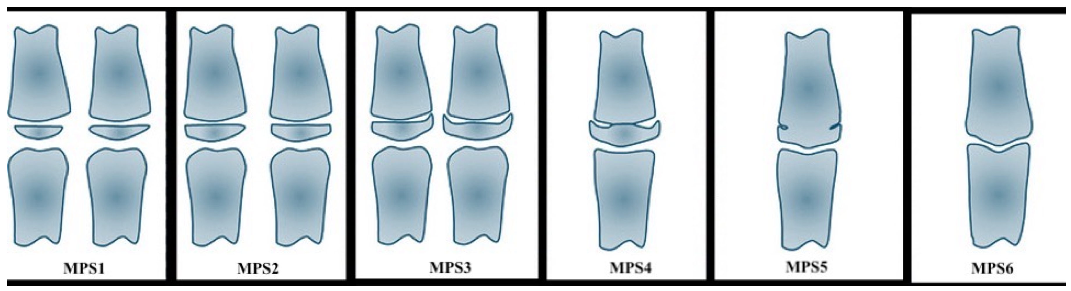
Figure 1. Representation of the six stages of skeletal maturation of the middle phalanx of the third finger according to Perinetti et al. [1].
Applsci 15 05593 g001
Figure 2. Representation and classification of the height stages (A–H) defined by Demirjian and used for the evaluation of the OPGs.
Applsci 15 05593 g002
Figure 3. Representation and classification of four categories of ossification of the midpalate suture, defined according to the radiographic appearance of the suture on OPGs.
Applsci 15 05593 g003
Table 1. Stadiation of the middle phalanx of the third finger according to Perinetti et al. [1].
StageBones’ MaturationPhase of the Growth
MPS1the epiphysis is narrower than the metaphysisthe patient is more than one year before the pubertal growth spurt
MPS2the epiphysis is at least as wide as the metaphysisthe patient is one year before the onset of the pubertal growth spurt
MPS3the epiphysis is either as wide as or wider than the metaphysisthe patient is at coincidence of the pubertal growth spurt
MPS4the epiphysis begins to fuse with the metaphysisthe patient is during the deceleration of the curve of growth
MPS5the epiphysis is mostly, but not completely fused with the metaphysisthe patient toward the end of the pubertal growth spurt
MPS6the epiphysis totally fused with the metaphysisthe patient is at the end of the pubertal growth spurt
Table 2. Scheme of the correspondence between the six stages of maturation of the second phalanx of the third finger, and the four categories that were used for the statistical analysis.
MPS Method CategoriesCategories Used for the Present Analysis
MPS-1SM1
MPS-2
MPS-3SM2
MPS-4SM3
MPS-5SM4
MPS-6
Table 3. Descriptive statistics of the analyzed variables.
VariablesMeanSD
Age137.425.6
Height (cm)149.113.5
Weight (kg)45.913.7
BMI20.34.2
Wrist circumference1.861.113
Table 4. Biometrical data analyzed with Spearman’s Rank Correlation test.
Skeletal Age
Weightρ: 0.5903 **
p: <0.001
Heightρ: 0.6188 **
p: <0.001
BMIρ: 0.3165 **
p: <0.001
Wrist circumferenceρ: 0.3881 **
p: <0.001
** Statistically significant for p < 0.01.
Table 5. Correlation of skeletal age with the variables analyzed, considering the weight and height separately.
Skeletal AgeOdds RatioSDzp > |z|Confidence Interval 95%
Lower BoundUpper Bound
Sex0.030.02−4.57<0.001−5.2−2.08
Age3.421.213.470.0010.531.93
Height (cm)1.10.0472.330.020.020.18
Weight (kg)1.130.062.110.0350.010.23
Wrist circumference0.790.33−0.570.569−1.060.58
Midpalate suture
23.522.561.730.084−0.172.68
315.2515.582.670.0080.724.73
40.810.86−0.20.845−2.291.87
Lower first premolar −343.232.681.410.16−0.462.80
Skeletal age < cut 120.696.62--7.7133.68
cut1 < Skeletal age < cut223.646.89--10.1337.16
Cut2 < Skeletal age < cut324.196.94--10.59 37.78
Disclaimer/Publisher’s Note: The statements, opinions and data contained in all publications are solely those of the individual author(s) and contributor(s) and not of MDPI and/or the editor(s). MDPI and/or the editor(s) disclaim responsibility for any injury to people or property resulting from any ideas, methods, instructions or products referred to in the content.

