Article

Color Variation in 3D-Printed Orthodontic Aligners as a Compliance Indicator: A Prospective Pilot Study

Postgraduate School of Orthodontics, University of Ferrara, Via Luigi Borsari 46, 44121 Ferrara, Italy
*
Author to whom correspondence should be addressed.
Appl. Sci. 2025, 15(12), 6409; https://doi.org/10.3390/app15126409
Submission received: 25 April 2025 / Revised: 3 June 2025 / Accepted: 4 June 2025 / Published: 6 June 2025
(This article belongs to the Special Issue Orthodontics: Advanced Techniques, Methods and Materials)

Abstract

Patient compliance remains a significant challenge in orthodontic treatment with clear aligners, as adherence to prescribed wear time is often suboptimal. This study investigated the potential of colorimetric analysis as a method to assess compliance with NOXI 3D-printed night-time aligners. Specifically, it evaluated color variations in polyamide aligners due to thermo-oxidation, using the RGB (Red, Green, Blue) color model as a non-invasive indicator. In total, 10 patients participated in this prospective study, wearing aligners for either 7 or 12 h daily over a 14-day period. Colorimetric measurements were collected via a smartphone-based application, and statistical analyses examined correlations between wear duration and color changes. The results revealed a significant association between a longer wear time and increased discoloration (p < 0.001), supporting the feasibility of RGB-based monitoring as a reliable compliance tool. However, individual variability in saliva composition, diet, and oral hygiene may have influenced the results, highlighting the need for further research into potential confounding variables. These findings underscore the promise of integrating digital monitoring technologies to improve adherence tracking and patient management in orthodontics. Future studies should refine the methodology and validate its efficacy in larger, more diverse populations.

1. Introduction

Clear aligner therapy has undergone substantial advancements in recent years, offering advantages such as reduced treatment durations and fewer emergency appointments compared to fixed appliances, particularly for mild to moderate malocclusions [1]. Nevertheless, patient compliance remains the primary limitation of clear aligner treatment, as optimal outcomes rely on wearing these aligners for approximately 22 h per day [2]. This requirement is dictated by the mechanical behavior of the materials used; insufficient wear time results in suboptimal force application, thereby compromising the efficiency of tooth movement [3].
Several studies have shown that adherence to the prescribed wear time for removable orthodontic devices is significantly lower than the recommended level [4]. A systematic review by Al-Moghrabi et al. on compliance with removable orthodontic appliances revealed that patient adherence is generally suboptimal, as they wear their appliances for significantly less time than prescribed and often overreport their actual wear duration [5,6]. Specifically, Timm et al. found that among aligner users, only 36% exhibited full compliance, while 38.3% showed moderate compliance and 25.7% demonstrated poor compliance [7].
In response, recent research has increasingly focused on the development of innovative 3D-printed materials for aligners, aiming to reduce the required daily wear time by exploiting the distinct mechanical properties of these materials [8,9]. A reduced wear time may also provide aesthetic benefits [10,11]. Although clear aligners are inherently transparent, Försch et al. demonstrated that the presence of aligners still draws significant visual attention to the mouth, as evidenced by eye-tracking studies comparing gaze patterns across individuals with and without orthodontic appliances [12].
One promising innovation is the development of night-time aligners, designed to be worn for only 10–12 h per day. NOXI (Sweden & Martina, Due Carrare, Padova, Italy) night-time aligners, developed at the University of Ferrara, are 3D printed using selective laser sintering from a polyamide material. This polymer has been demonstrated to deliver higher and more sustained orthodontic forces due to its reduced stress relaxation properties [13]. Another key feature lies in the ability to create differential thicknesses across various regions of the arch, as well as between the cervical and incisal sections of the aligner. This design flexibility enhances the fit and allows for more effective force delivery, particularly by increasing the force applied to selected teeth where needed [14,15,16]. Such mechanical advantages justify the clinical rationale behind the limited daily use of this aligner, offering a more discreet and patient-friendly alternative to full-time aligner wear.
Despite these material innovations, patient compliance remains a persistent challenge for all removable orthodontic systems [4,5,17,18]. A systematic review published in 2024 sought to further investigate the complex issue of patient compliance in orthodontics. After screening and analyzing over 3000 articles, the authors concluded that there is currently no clear evidence regarding which factors effectively influence patient compliance, nor how such compliance can be reliably improved [19]. Addressing this gap, the present study proposes a novel, objective method to determine whether a patient has adhered to the prescribed wear protocol. By providing a quantifiable and non-invasive marker of cooperation, this approach also removes the ambiguity that may otherwise exist in the patient–clinician relationship—namely, the clinician’s inability to verify the accuracy of self-reported compliance.
Colorimetry has long been employed in dentistry to assess the aesthetic and functional stability of dental materials. It provides quantitative data that eliminate subjectivity in visual shade selection and is commonly used in restorative dentistry, prosthodontics, and, increasingly, in orthodontics [20]. Colorimetric analysis has been applied to evaluate the staining susceptibility of clear aligners and thermoformed retainers after exposure to various staining agents. For example, studies have employed colorimetric and spectrophotometric measurements to assess the discoloration levels in aligner materials subjected to substances like coffee, red wine, and tea, demonstrating their utility in understanding both material behavior and external influences over time [21]. These applications highlight the relevance of colorimetry as a diagnostic tool not only for aesthetic evaluation, but also for monitoring the aging and functional performance of orthodontic appliances in the oral environment [22].
In this context, the aim of this study was to evaluate patient compliance with NOXI 3D-printed night-time aligners (Sweden&Martina, Due Carrare, Padova, Italy) through an innovative method based on material discoloration. A well-documented effect of thermo-oxidative degradation in aliphatic polyamides (PAs) is progressive yellowing, attributed to the formation of chromophoric structures—specifically, α-ketoamide groups—resulting from the oxidation of methylene units adjacent to the amide carbonyl group [23,24,25]. This phenomenon was exploited in the present study as an indirect yet objective indicator of intraoral wear time.
Accordingly, this study was designed to test the null hypothesis that different durations of intraoral wear (7 h vs. 12 h per day) would not result in a statistically significant difference in the degree of color change in polyamide aligners, as measured by RGB analysis. This hypothesis was tested over a standardized 14-day period using quantitative colorimetric measurements. Rejection of the null hypothesis would support the potential of RGB-based measurements as a reliable, non-invasive indicator for monitoring patient compliance.

2. Materials and Methods

In this prospective study conducted at the Postgraduate School of Orthodontics at the University of Ferrara, a total of 10 patients (3 males and 7 females; mean age: 28 years) were selected based on defined inclusion and exclusion criteria (Table 1). The study protocol was approved by the University of Ferrara Postgraduate School Ethics Committee (registration number 6/2024), and the research was conducted in conformity with the Declaration of Helsinki.
Following the collection and review of each participant’s diagnostic records, a customized digital setup was developed for each case by a certified orthodontic specialist. This process included 3D intraoral scanning, individualized treatment planning, and aligner design using dedicated orthodontic CAD software (Rhinoceros 3D, Version 6.0, Robert McNeel & Associates, Seattle, WA, USA). The final models were used to fabricate the aligners via selective laser sintering (SLS) 3D printing, using a medical-grade polyamide material, following the proprietary production protocol established by the manufacturer (NOXI, Sweden & Martina, Due Carrare, Padova, Italy).
Each patient was instructed to wear two pairs of NOXI aligners (Sweden & Martina, Due Carrare, Padova, Italy)—one for the upper and one for the lower arch—over a 14-day period (T0–T14), with assignment to one of the following two distinct wear-time protocols:
  • First pair: worn for 7 h per day (7 H protocol)
  • Second pair: worn for 12 h per day (12 H protocol)
For the purpose of this study, we considered compliant wear as 12 h per day, in line with the clinical protocol of NOXI aligners, while 7 h per day was considered a non-compliant scenario.
All patients initially followed the 7 h (7 H) protocol for 14 days. After a two-week transition period, they began the 12 h (12 H) protocol. The 7 H protocol was scheduled first because a daily wear time of 7 h falls below the 10–12 h required for NOXI aligners to exert a therapeutic effect. This preliminary phase allowed patients to adjust to the material, screening for any issues, and exclusion of those who were unsuitable for the study. After the transition period, patients began the actual treatment. The first set of aligners worn during this phase (also for 14 days) was collected and analyzed as part of the 12 H protocol.
Upon removal, the patients were instructed to rinse the aligners under cold running water and dry them with paper towels, ensuring that non-abrasive methods were employed. They were also advised to avoid any cleaning solutions containing abrasive microparticles or oxidizing agents, as these could potentially alter the material’s surface properties and compromise subsequent colorimetric analysis. After cleaning, the aligners were stored in a designated container provided to each patient and kept away from direct sunlight in a stable, room-temperature environment to prevent potential degradation of the polyamide.
Color variations were assessed using the Colorimeter app (Version 14; Copyright© Smyk Serhii), available for iPhone, iPad, Mac, and Apple Vision. The application uses the RGB color model to detect and quantify real-time color changes through the device’s built-in camera (Figure 1). The app provides precise color data across various formats, such as HEX codes (for web design), RGB, CMYK, and HSL (Hue, Saturation, Lightness) values.
The RGB model functions by combining three primary light colors—red, green, and blue—at intensities ranging from 0 to 255, producing up to 16.777.216 unique colors (2563).
The maximum intensity of all three colors results in white (255, 255, 255), while zero intensity results in black (0, 0, 0). As an additive light model, RGB quantifies brightness through the addition of light rather than its subtraction (as in print-based models). Its sensitivity to minor chromatic variations makes it particularly suitable for evaluating subtle discoloration in polymeric materials. To quantify color changes, the Euclidean distance between the RGB values at T0 and T14 was calculated using the following formula:
Δ ERGB = R 2 R 1 2 + G 2 G 1 2 + B 2 B 1 2
This metric captures the overall magnitude of color variation across all three channels, providing a robust and perceptually relevant indicator of discoloration.
All aligners were analyzed by the same operator using an iPhone 15 Pro (Apple Inc., Cupertino, CA, USA) under standardized environmental and external lighting conditions. To ensure consistency in data acquisition, all photographs were taken by the same operator, in the same room, at the same time of day, under identical lighting conditions, using the same device. A custom-designed True White Box (Sweden & Martina, Due Carrare, Padova, Italy) was used to maintain a fixed distance between the camera and the aligner, with the smartphone stably positioned on the upper edge of the box during each capture. The True White Box consisted of a container featuring a white base made from the same polyamide material as the aligners themselves. The color of the base was verified before each measurement to ensure consistency, as RGB values are sensitive to ambient lighting conditions. This control step was essential to standardize measurements and minimize environmental variability. To assess intra-operator repeatability, each color value was recorded twice by the same operator.
Color readings for each aligner were taken at the following three specific anatomical locations:
  • Upper arch (Figure 2):
    -
    Marginal ridge of tooth 14;
    -
    Marginal ridge of tooth 24;
    -
    Interproximal contact point between teeth 21 and 22.
  • Lower arch (Figure 3):
    -
    Marginal ridge of tooth 34;
    -
    Marginal ridge of tooth 44;
    -
    Interproximal contact point between teeth 41 and 42.

Statistical Analysis
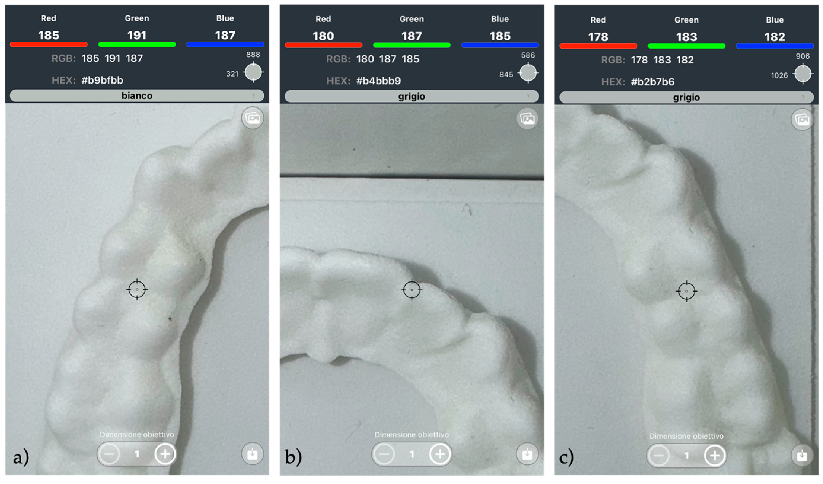
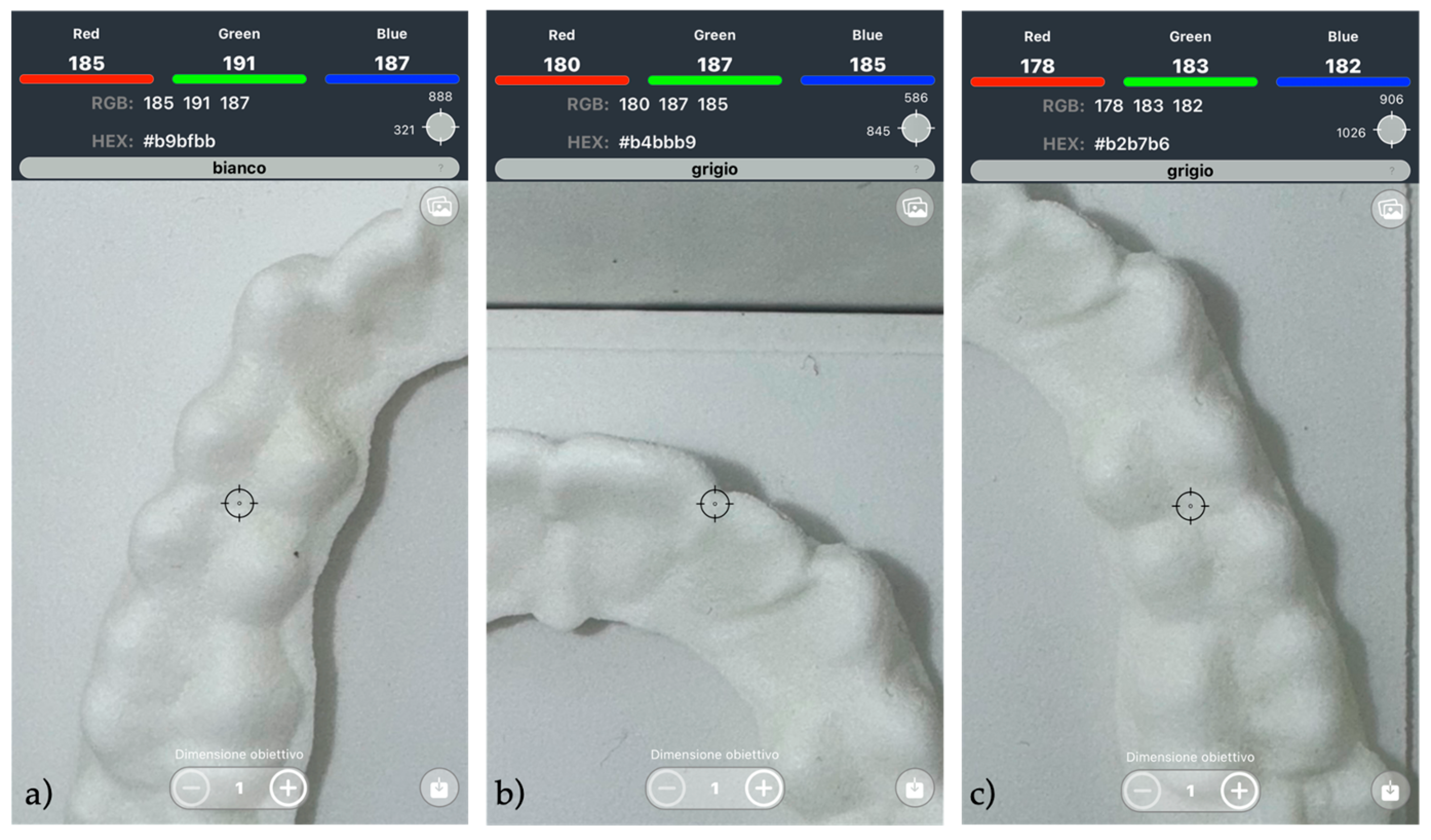
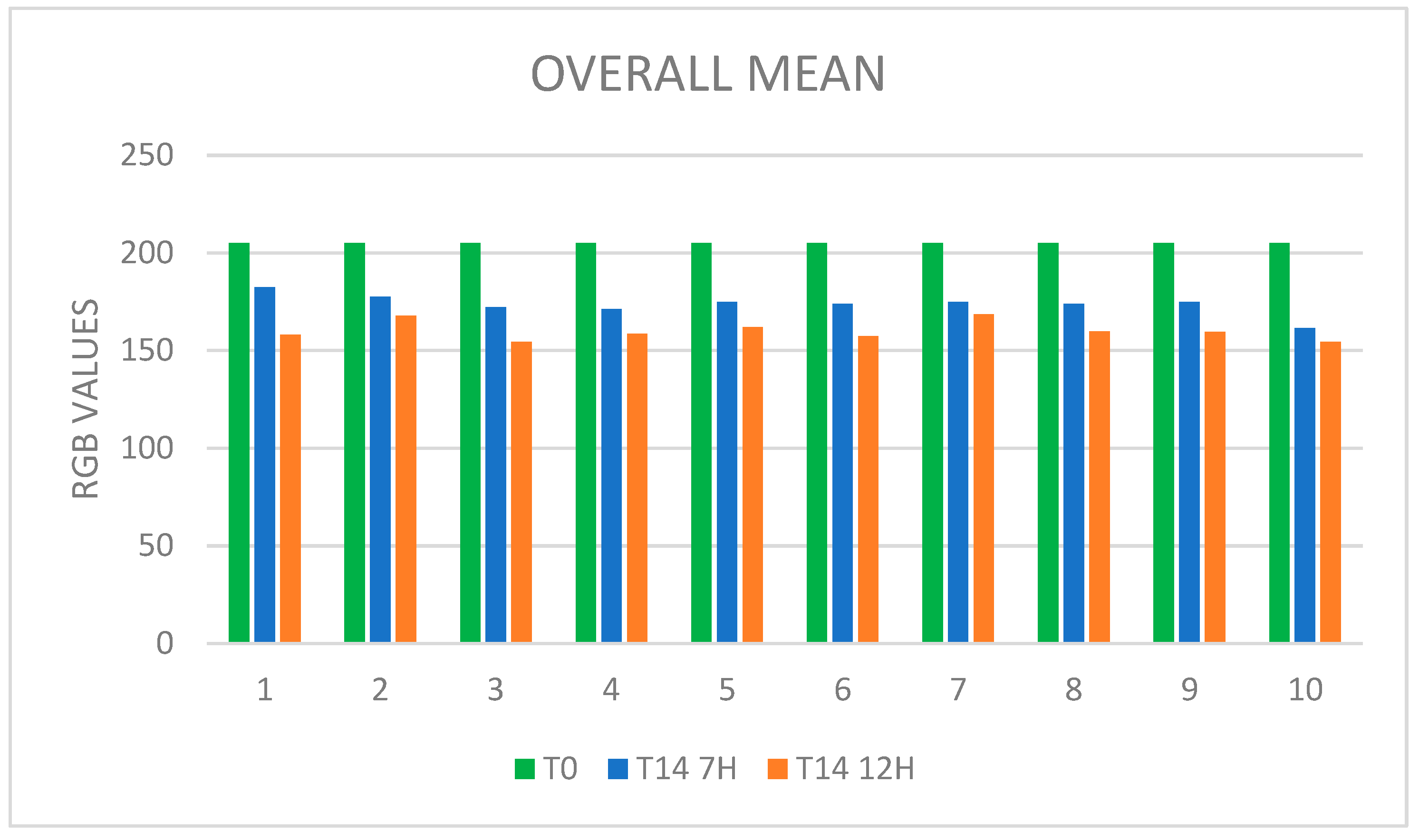
A total of 160 measurements were collected and recorded in an Excel spreadsheet (Microsoft Excel 2015; Microsoft Corp., Redmond, WA, USA). The control group consisted of a fixed reference value (205) with no variance and was, therefore, excluded from normality testing. The distribution of ΔRGB values in the experimental groups was assessed using the Shapiro–Wilk test. All experimental datasets showed a normal distribution (p > 0.05), allowing for the use of parametric statistical tests for further analysis. Paired t-tests were used to compare the color changes between T0 and T14 for both the 7 H and the 12 H wear protocols. Subsequently, paired t-tests were also conducted to directly compare the color change values between the two wear-time protocols. Additionally, a post hoc power analysis was conducted based on the mean difference and standard deviation of the paired ΔRGB values observed between the 7 H and 12 H protocols. The effect size (Cohen’s d) was calculated and used to estimate statistical power using a paired t-test model with α = 0.05. To assess measurement repeatability, Pearson’s correlation coefficient was calculated between the first and second measurement sets for each aligner, in order to demonstrate the consistency of the results (p < 0.001).

3. Results

Of the 10 participants, no patients were classified as dropouts, and all completed the 8-week study. Statistically significant differences (p < 0.001) were observed in the total, upper, and lower arch measurements. These results support the statistical rejection of the null hypothesis, indicating a clear association between an increased wear time and greater discoloration of the aligners.

3.1. Descriptive Statistics

Table 2, Table 3, Table 4, Table 5, Table 6 and Table 7 present the descriptive statistics of the color variations observed in the aligners subjected to both the 7 H and 12 H daily usage protocols. Mean values and standard deviations are reported to illustrate the distribution and variability of the data within each experimental group. The data show a progressive decline in RGB values with an increased wear time.
As illustrated in Figure 4, the RGB values show a progressive decline correlated with the number of hours of daily use. Specifically, the 7 H protocol resulted in a mean reduction of 16.61% in RGB values, whereas the 12 H protocol showed a greater average decrease of approximately 22.05%.

3.2. Paired t-Test

First, a paired t-test was performed to assess whether there were statistically significant changes in aligner color after 14 days of use under the two protocols. As shown in Table 8 and Table 9, the paired t-tests revealed that, for both protocols, there were statistically significant differences in the recorded RGB values following 14 days of wear.
Subsequently, we conducted a comparison between the 7 H protocol and the 12 H protocol to determine whether the differences observed were associated with the number of hours of wear. As shown in Table 10 and Figure 4, the paired t-test revealed statistically significant differences between the two protocols, highlighting a correlation between the number of hours of wear and the color change of the aligners.
Finally, we investigated potential color differences between upper and lower aligners within the same protocol. As shown in Table 11, no statistically significant differences were observed between the chromatic variations of upper and lower aligners under the same wear regimen (neither at 7 h nor at 12 h). In conclusion, the findings support the statistical rejection of the null hypothesis.
A post hoc power analysis was performed to assess the strength of the statistical findings. The analysis was based on the observed mean difference in ΔRGB values between the 7 H and 12 H protocols (mean difference = 13.63; standard deviation = 5.36; n = 10). The calculated effect size (Cohen’s d) was 2.55, corresponding to a statistical power of 1.0 (100%) at a significance level of α = 0.05. These results confirm that the study had sufficient power to detect the observed difference, despite the small sample size.

4. Discussion

To the best of our knowledge, no prior studies have directly investigated the use of a colorimetric system to monitor aligner discoloration as an objective indicator of patient compliance in orthodontic treatment. This study represents the first attempt to integrate such a technique into orthodontics, offering an innovative and non-invasive approach to assess patient compliance. Specifically, the application of the RGB color model to evaluate discoloration in night-time aligners stands out as a novel contribution. The RGB system—widely employed in digital displays such as monitors, televisions, and smartphones—provides a high sensitivity and precision in detecting chromatic changes, thereby enabling the development of personalized diagnostic tools.
Although this study marks the first application of the RGB system in this specific context, the existing literature includes related studies employing different methodologies to explore similar themes. For instance, two different studies by Schott et al. and Tuncay et al. examined the use of compliance indicators with Invisalign Teen aligners, incorporating a food dye (erioglaucine disodium salt) into the aligner polymer matrix. This dye underwent degradation when exposed to oral fluids, resulting in a color change intended to reflect wear duration. However, due to significant variability in color fading, it remains unclear whether the observed variations were the result of differences in wear time—resulting from poor patient compliance—or whether unknown factors influenced the fading of the dye [26,27]. In a different line of inquiry, Marks et al. investigated whether remote monitoring using the Dental Monitoring system could improve patient adherence, compared to conventional in-office follow-up. Their findings revealed no statistically significant differences in compliance between the two groups [28]. In contrast, a 2022 study by Timm et al. demonstrated that incorporating electronic reminders into remote monitoring protocols significantly enhanced patient compliance. These reminders served as behavioral prompts to wear the aligners regularly, reinforcing adherence [29]. However, this approach is not applicable to the NOXI Aligner system, which is specifically designed for night-time use only. Given its limited daily wear time, integrating electronic reminders is not a practical or effective strategy for this specific type of aligner.
In the present study, the data demonstrated a consistent and statistically significant correlation between aligner wear duration and RGB value reduction, reflecting a progressive loss of whiteness. This decline was directly proportional to the level of discoloration: as RGB values decreased, the material assumed a more yellowish hue. Specifically, aligners worn for 12 h per day exhibited significantly greater reductions in RGB values compared to those worn for 7 h per day (p < 0.001). These findings allow us to reject the null hypothesis, which posited no difference in discoloration between the two protocols.
However, variables such as differences in salivary flow or composition, pH levels, and dietary habits may alter the chemical reactions involved in the thermos-oxidative degradation of polyamide, potentially affecting the outcomes of colorimetric assessments. [30,31].
Parallel findings from other materials research support our observations: Wieckiewicz et al. investigated the color stability of polyamide-12, a material used in dental prosthetics after immersion in staining agents such as coffee and red wine [32]. The authors employed the CIE Lab∗ chromaticity system and their findings confirmed that exposure to pigmented substances significantly affected the chromatic stability of polyamide. Although not directly focused on patient compliance, their study highlights the relevance of color changes as an indicator of material aging and external exposure. Importantly, their results also suggest that factors such as oral hygiene and patient behavior may play substantial roles in the extent of discoloration.
The results of the present study underscore the potential of colorimetric evaluations as a diagnostic tool for patient compliance monitoring. Future directions should include the refinement and standardization of chromatic indicators as adjunctive tools in aligner therapy. With continued development, this approach may enable more precise, real-time, and non-invasive monitoring. Furthermore, the use of mobile applications instead of more advanced equipment, such as spectrophotometers or colorimeters, would make this tool more accessible and user-friendly for clinicians. Indeed, integrating RGB-based data collection with remote health platforms or mobile applications could allow clinicians to track aligner use remotely, reducing the need for frequent in-office visits and enabling timely intervention when non-compliance is detected.
Another promising avenue for future research could be the investigation of changes in material porosity and surface roughness following intraoral use. Surface characteristics such as increased roughness may potentially influence the extent of discoloration by facilitating the retention of staining agents or altering light reflection. These changes could serve as confounding variables in the interpretation of colorimetric data. Nonetheless, in the case of polyamide aligners, the discoloration observed in this study appears to be primarily driven by thermo-oxidative degradation, as evidenced by the consistent and homogeneous patterns of color change observed under controlled cleaning protocols [23,25]. Additionally, it is important to note that the NOXI Aligner is designed for 10–12 h of wear per day, primarily during the night. This limited wear time results in significantly less exposure to staining agents, cigarette smoke, and contact with oral fluids, reducing this exposure by almost 50% compared to aligners worn for 22 h a day. Further studies employing advanced characterization techniques such as scanning electron microscopy (SEM) or surface profilometry could elucidate the relationship between surface morphology and chromatic stability, thereby providing a more comprehensive understanding of material aging mechanisms in the oral environment.
A primary limitation of this study may be its relatively small sample size (n = 10), which limits the generalization of the findings. Future studies should involve larger and more heterogeneous populations and should consider additional behavioral and environmental variables that may affect material discoloration. Such studies are essential for enhancing the reliability of this methodology and for supporting clinicians in optimizing patient management protocols.

5. Conclusions

The main findings can be summarized as follows:
  • A strong and statistically significant correlation was observed between aligner wear duration and polyamide discoloration, as indicated by decreasing RGB values.
  • The progressive reduction in RGB values reflected increased yellowing of the aligner, supporting the potential use of this method as an indirect indicator of wear time.
  • This method is readily implementable through smartphone-based applications, enabling the accessible and real-time monitoring of patient compliance without the need for invasive procedures.
From a clinical perspective, this method may enhance orthodontic case management by allowing practitioners to objectively verify compliance and promptly address non-adherence, especially in cases where treatment effectiveness is not progressing as expected.

Author Contributions

Conceptualization, F.C. and L.L.; methodology, F.C.; software, F.C.; validation, L.L. and F.C.; formal analysis, F.C.; investigation, G.C.; resources, L.L.; data curation, F.P.; writing—original draft preparation, G.C.; writing—review and editing, F.C.; visualization, L.L.; supervision, F.C.; project administration, L.L.; funding acquisition, L.L. All authors have read and agreed to the published version of the manuscript.

Funding

This research received no external funding.

Institutional Review Board Statement

The study was conducted in accordance with the Declaration of Helsinki and approved by the Ethics Committee of the Ferrara University Postgraduate School of Orthodontics (Via Luigi Borsari 46, Ferrara, Italy; approval number 6/2024; date of approval 18 June 2024).

Informed Consent Statement

Informed consent was obtained from all subjects involved in the study.

Data Availability Statement

The datasets used and analyzed during the current study are available from the corresponding author on reasonable request.

Conflicts of Interest

The authors declare no conflicts of interest.

Abbreviations

The following abbreviations are used in this manuscript:
PAAliphatic polyamide
RGBRed, green, blue
CMYKCyan, Magenta, Yellow, Key
HEXHexadecimal
HSLHue, Saturation, Lightness

References

  1. Weir, T. Clear Aligners in Orthodontic Treatment. Aust. Dent. J. 2017, 62, 58–62. [Google Scholar] [CrossRef]
  2. Robertson, L.; Kaur, H.; Fagundes, N.C.F.; Romanyk, D.; Major, P.; Flores Mir, C. Effectiveness of Clear Aligner Therapy for Orthodontic Treatment: A Systematic Review. Orthod. Craniofacial Res. 2020, 23, 133–142. [Google Scholar] [CrossRef] [PubMed]
  3. Kohda, N.; Iijima, M.; Muguruma, T.; Brantley, W.A.; Ahluwalia, K.S.; Mizoguchi, I. Effects of Mechanical Properties of Thermoplastic Materials on the Initial Force of Thermoplastic Appliances. Angle Orthod. 2013, 83, 476–483. [Google Scholar] [CrossRef]
  4. Cremonini, F.; Karami Shabankare, A.; Guiducci, D.; Lombardo, L. Compliance with Headgear Evaluated by Force- and Temperature-Sensitive Monitoring Device: A Case-Control Study. Bioengineering 2024, 11, 789. [Google Scholar] [CrossRef] [PubMed]
  5. Al-Moghrabi, D.; Salazar, F.C.; Pandis, N.; Fleming, P.S. Compliance with Removable Orthodontic Appliances and Adjuncts: A Systematic Review and Meta-Analysis. Am. J. Orthod. Dentofac. Orthop. 2017, 152, 17–32. [Google Scholar] [CrossRef] [PubMed]
  6. Schott, T.C.; Meyer-Gutknecht, H.; Mayer, N.; Weber, J.; Weimer, K. A Comparison Between Indirect and Objective Wear-Time Assessment of Removable Orthodontic Appliances. Eur. J. Orthod. 2017, 39, 170–175. [Google Scholar] [CrossRef]
  7. Timm, L.H.; Farrag, G.; Baxmann, M.; Schwendicke, F. Factors Influencing Patient Compliance During Clear Aligner Therapy: A Retrospective Cohort Study. J. Clin. Med. 2021, 10, 3103. [Google Scholar] [CrossRef]
  8. Jindal, P.; Juneja, M.; Siena, F.L.; Bajaj, D.; Breedon, P. Mechanical and Geometric Properties of Thermoformed and 3D Printed Clear Dental Aligners. Am. J. Orthod. Dentofac. Orthop. 2019, 156, 694–701. [Google Scholar] [CrossRef]
  9. Tartaglia, G.M.; Mapelli, A.; Maspero, C.; Santaniello, T.; Serafin, M.; Farronato, M.; Caprioglio, A. Direct 3D Printing of Clear Orthodontic Aligners: Current State and Future Possibilities. Materials 2021, 14, 1799. [Google Scholar] [CrossRef]
  10. Skidmore, K.J.; Brook, K.J.; Thomson, W.M.; Harding, W.J. Factors Influencing Treatment Time in Orthodontic Patients. Am. J. Orthod. Dentofac. Orthop. 2006, 129, 230–238. [Google Scholar] [CrossRef]
  11. Alansari, R.A. Youth Perception of Different Orthodontic Appliances. Patient Prefer. Adherence 2020, 14, 1011–1019. [Google Scholar] [CrossRef] [PubMed]
  12. Försch, M.; Krull, L.; Hechtner, M.; Rahimi, R.; Wriedt, S.; Wehrbein, H.; Jacobs, C.; Jacobs, C. Perception of Esthetic Orthodontic Appliances: An Eye Tracking and Cross-Sectional Study. Angle Orthod. 2020, 90, 109–117. [Google Scholar] [CrossRef]
  13. Cremonini, F.; Brucculeri, L.; Pepe, F.; Palone, M.; Lombardo, L. Comparison of Stress Relaxation Properties between 3-Dimensional Printed and Thermoformed Orthodontic Aligners: A Pilot Study of In Vitro Simulation of Two Consecutive 8-Hours Force Application. APOS Trends Orthod. 2024, 14, 225–234. [Google Scholar] [CrossRef]
  14. Palone, M.; Longo, M.; Arveda, N.; Nacucchi, M.; De Pascalis, F.; Spedicato, G.A.; Siciliani, G.; Lombardo, L. Micro-Computed Tomography Evaluation of General Trends in Aligner Thickness and Gap Width After Thermoforming Procedures Involving Six Commercial Clear Aligners: An In Vitro Study. Korean J. Orthod. 2021, 51, 135–141. [Google Scholar] [CrossRef] [PubMed]
  15. Proffit, W.R.; Henry, W.F.; David, M.S.; James, L.A. Contemporary Orthodontics, 5th ed.; Elsevier: Amsterdam, The Netherlands, 2013. [Google Scholar]
  16. Hahn, W.; Dathe, H.; Fialka-Fricke, J.; Fricke-Zech, S.; Zapf, A.; Kubein-Meesenburg, D.; Sadat-Khonsari, R. Influence of Thermoplastic Appliance Thickness on the Magnitude of Force Delivered to a Maxillary Central Incisor During Tipping. Am. J. Orthod. Dentofac. Orthop. 2009, 136, 12.e1–12.e7. [Google Scholar] [CrossRef] [PubMed]
  17. Tsomos, G.; Ludwig, B.; Grossen, J.; Pazera, P.; Gkantidis, N. Objective Assessment of Patient Compliance with Removable Orthodontic Appliances: A Cross-Sectional Cohort Study. Angle Orthod. 2014, 84, 56–61. [Google Scholar] [CrossRef]
  18. Arreghini, A.; Trigila, S.; Lombardo, L.; Siciliani, G. Objective Assessment of Compliance with Intra- and Extraoral Removable Appliances. Angle Orthod. 2017, 87, 88–95. [Google Scholar] [CrossRef]
  19. van der Bie, M.R.; Bos, A.; Joseph Mathieu Bruers, J.; Edwin Gaston Jonkman, R. ARTICLE OPEN Patient Adherence in Orthodontics: A Scoping Review. BDJ Open 2024, 10, 58. [Google Scholar] [CrossRef]
  20. Ragain, J.C. A Review of Color Science in Dentistry: Colorimetry and Color Space. J. Dent. Oral. Disord. Ther. 2016, 4, 1–5. Available online: www.symbiosisonlinepublishing.com (accessed on 12 September 2024). [CrossRef]
  21. Olteanu, N.D.; Taraboanta, I.; Panaite, T.; Balcos, C.; Rosu, S.N.; Vieriu, R.M.; Dinu, S.; Zetu, I.N. Color Stability of Various Orthodontic Clear. Aligner Systems After Submersion in Different Staining Beverages. Materials 2024, 17, 4009. [Google Scholar] [CrossRef]
  22. Ajwa, N.; Radhi, F.; Aloraini, R.; AlSaydalani, G. Comparative Color Stability Assessment of Orthodontic Clear Aligners: An In Vitro Study. Sci. Rep. 2025, 15, 2041. [Google Scholar] [CrossRef] [PubMed]
  23. Filippone, G.; Carroccio, S.C.; Mendichi, R.; Gioiella, L.; Dintcheva, N.T.; Gambarotti, C. Time-Resolved Rheology as a Tool to Monitor the Progress of Polymer Degradation in the Melt State—Part I: Thermal and Thermo-Oxidative Degradation of Polyamide 11. Polymer 2015, 72, 134–141. [Google Scholar] [CrossRef]
  24. Powell, A.W.; Stavrinadis, A.; Christodoulou, S.; Quidant, R.; Konstantatos, G. On-Demand Activation of Photochromic Nanoheaters for High Color Purity 3D Printing. Nano Lett. 2020, 20, 3485–3491. [Google Scholar] [CrossRef] [PubMed]
  25. Meissner, H.; Vacquier, M.; Kresse-Walczak, K.; Boening, K. Mechanical, Optical and Surface Properties of 3D-Printed and Conventionally Processed Polyamide 12. Dent. Med. Probl. 2024, 61, 729–738. [Google Scholar] [CrossRef]
  26. Schott, T.C.; Göz, G. Color Fading of the Blue Compliance Indicator Encapsulated in Removable Clear Invisalign Teen® Aligners. Angle Orthod. 2011, 81, 185–191. [Google Scholar] [CrossRef]
  27. Tuncay, O.C.; Jay Bowman, S.; Nicozisis, J.L.; Amy, B.D. Effectiveness of a Compliance Indicator for Clear Aligners. J. Clin. Orthod. 2009, 43, 263–268. Available online: www.jco-online.com (accessed on 19 December 2024).
  28. Marks, J.; Freer, E.; Ong, D.; Lam, J.; Miles, P. Evaluating Dental Monitoring Effectiveness Compared with Conventional Monitoring of Clear Aligner Therapy Using the Peer Assessment Rating Index. Am. J. Orthod. Dentofac. Orthop. 2024, 166, 350–355. [Google Scholar] [CrossRef]
  29. Timm, L.H.; Farrag, G.; Wolf, D.; Baxmann, M.; Schwendicke, F. Effect of Electronic Reminders on Patients’ Compliance During Clear Aligner Treatment: An Interrupted Time Series Study. Sci. Rep. 2022, 12, 16652. [Google Scholar] [CrossRef]
  30. Eliades, T.; Bourauel, C. Intraoral Aging of Orthodontic Materials: The Picture We Miss and Its Clinical Relevance. Am. J. Orthod. Dentofac. Orthop. 2005, 127, 403–412. [Google Scholar] [CrossRef]
  31. Nicita, F.; D’Amico, C.; Filardi, V.; Spadaro, D.; Aquilio, E.; Mancini, M.; Fiorillo, L. Chemical-Physical Characterization of PET-G-Based Material for Orthodontic Use: Preliminary Evaluation of Micro-Raman Analysis. Eur. J. Dent. 2023, 18, 228–235. [Google Scholar] [CrossRef]
  32. Wieckiewicz, M.; Opitz, V.; Richter, G.; Boening, K.W. Physical Properties of Polyamide-12 Versus PMMA Denture Base Material. Biomed Res. Int. 2014, 2014, 150298. [Google Scholar] [CrossRef] [PubMed]
Figure 1. ‘Colorimeter app’ (Copyright© Smyk Serhii).
Applsci 15 06409 g001
Figure 2. Measurement points for the upper arch; (a) marginal ridge of tooth 14; (b) interproximal contact point between teeth 21 and 22; and (c) marginal ridge of tooth 24.
Applsci 15 06409 g002
Figure 3. Measurement points for the lower arch; (a) marginal ridge of tooth 44; (b) interproximal contact point between teeth 41 and 42; and (c) marginal ridge of tooth 34.
Applsci 15 06409 g003
Figure 4. Average RGB values for the 10 patients at T0 and T14 for both protocols, showing a decreasing trend associated with material yellowing over time.
Applsci 15 06409 g004
Table 1. Inclusion and exclusion criteria.
Inclusion CriteriaExclusion Criteria
Adult patients (aged from 25 to 31 years)Patients with systemic diseases
Good systemic healthHeavy smokers (>7 cigarettes/day)
Patients not undergoing orthodontic treatmentPatients undergoing treatment with antihistamines or anticholinergic drugs
Good oral hygienePatients with habitual consumption of pigmented beverage
Patients unable to perform home oral hygiene procedures
Patients with hyposalivation
Table 2. Measurements related to the upper arch for the 7 H protocol.
PatientControlTooth 24Tooth 14Tooth 21MeanSDMedian
12051791851821823182
220516518517517510175
3205185167160170.6712.9167
4205183172163172.6710.02172
5205168183176175.677.51176
6205165163180169.339.29165
7205167176186176.339.5176
8205163188167172.6713.43167
92051681801741746174
10205148178153159.6716.07153
Table 3. Measurements related to the lower arch for the 7 H protocol.
PatientControlTooth 34Tooth 41Tooth 44MeanSDMedian
1205177187184182.675.13184
22051851751801805180
3205170170181173.676.35170
4205171166172169.673.21171
52051801731691745.57173
6205179173184178.675.51179
7205174163183173.3310.02174
82051801721731754.36173
92051741701841767.21174
10205161157172163.337.68161
Table 4. Measurements related to the upper arch for the 12 H protocol.
PatientControlTooth 24Tooth 14Tooth 21MeanSDMedian
120516516414515811.27164
22051591621651623162
3205150149152150.331.53150
42051671531661627.81166
52051571651641624.36164
6205164151140151.6712.01151
7205165180170171.677.64170
82051591621531584.58159
9205168159155160.676.66159
1020517614414815617.44148
Table 5. Measurements related to the lower arch for the 12 H protocol.
PatientControlTooth 34Tooth 41Tooth 44MeanSDMedian
12051551531661587155
2205178163180173.679.29178
3205163148165158.679.29163
4205158161147155.337.37158
52051601631631621.73163
62051541731621639.53162
7205161166170165.674.51166
8205173150162161.6711.5162
9205158163155158.674.04158
10205144156158152.677.57156
Table 6. Measurements related to the overall mean for the 7 H protocol.
PatientControlMeanSDMedian
1205182.333.77183
2205177.57.58177.5
3205172.179.24170
4205171.176.85171.5
5205174.835.98174.5
62051748.53176
7205174.838.88175
8205173.839.02172.5
92051756.03174
10205161.511.47159
Table 7. Measurements related to the overall mean for the 12 H protocol.
PatientControlMeanSDMedian
12051588.39159.5
2205167.838.89164
3205154.57.5151
4205158.677.7159.5
52051622.97163
6205157.3311.52158
7205168.676.5168
8205159.838.08160.5
9205159.675.05158.5
10205154.3312.16152
Table 8. Comparison between T0 and T1 for the 7 H protocol for each of the three conditions: overall mean, upper arch, and lower arch.
Control7 H
Average
Average of
Differences
SD of
Differences
Test Statisticp Value
Overall Mean205173.72−31.285.28−18.73<0.001
Upper Arch205172.8−32.25.78−17.62<0.001
Lower Arch205174.64−30.375.45−17.63<0.001
Table 9. Comparison between T0 and T1 for the 12 H protocol for each of the three conditions: overall mean, upper arch, and lower arch.
Control12 H
Average
Average of
Differences
SD of
Differences
Test Statisticp Value
Overall Mean205160.08−44.924.9−29.01<0.001
Upper Arch205159.23−45.776.05−23.92<0.001
Lower Arch205160.93−44.075.86−23.78<0.001
Table 10. Paired t-test for each of the three conditions: overall mean, upper arch, and lower arch.
7 H
Average
12 H
Average
Average of
Differences
SD of
Differences
Test
Statistic
p Value
Overall Mean173.72160.09−13.63335.3563−8.0489<0.001
Upper Arch172.8159.23−13.56676.3129−6.7958<0.001
Lower Arch174.64160.93−13.75.1889−8.3492<0.001
Table 11. Paired t-test between upper and lower arch within the same protocol.
Upper ArchLower ArchAverage of
Differences
SD of
Differences
Test Statisticp Value
7 H172.8174.631.83333.82411.52>0.1
12 H159.23160.931.76.78310.7925>0.1
Disclaimer/Publisher’s Note: The statements, opinions and data contained in all publications are solely those of the individual author(s) and contributor(s) and not of MDPI and/or the editor(s). MDPI and/or the editor(s) disclaim responsibility for any injury to people or property resulting from any ideas, methods, instructions or products referred to in the content.

