Review

Biomechanical Impact of Vertebral Augmentation Techniques: Clinical and Radiological Results in the Literature

by
Eliodoro Faiella
1,2,
Federica Vaccarino
1,2,*,
Domiziana Santucci
1,2,
Elva Vergantino
1,2,
Bruno Beomonte Zobel
1,2 and
Rosario Francesco Grasso
1,2
1
Operative Research Unit of Radiology and Interventional Radiology, Fondazione Policlinico Universitario Campus Bio-Medico di Roma, Via Alvaro del Portillo 200, 00128 Rome, Italy
2
Research Unit of Radiology and Interventional Radiology, Department of Medicine and Surgery, Università Campus Bio-Medico di Roma, Via Alvaro del Portillo 21, 00128 Rome, Italy
*
Author to whom correspondence should be addressed.
Appl. Sci. 2025, 15(1), 426; https://doi.org/10.3390/app15010426
Submission received: 5 November 2024 / Revised: 12 December 2024 / Accepted: 2 January 2025 / Published: 5 January 2025
(This article belongs to the Special Issue Biomechanics and Biofluidodynamics in Biomedical Engineering)

Abstract

:
Vertebral augmentation techniques have advanced significantly, offering improved outcomes in the treatment of vertebral compression fractures. This review explores third-generation devices such as the SpineJack®, Vertebral Body Stenting System® (VBS), and OsseoFix®, which offer enhanced vertebral height restoration, stability, and reduced complications. These devices have been shown to outperform traditional methods like vertebroplasty and balloon kyphoplasty, particularly in reducing the risk of cement leakage and ensuring long-term vertebral stability. Biomechanical studies confirm the efficacy of these systems in promoting spinal recovery. Many of these studies have utilized indirect parameters, such as the Beck Index and kyphotic angles (α and γ) measured by the Cobb method, to evaluate improvements in vertebral deformity. Furthermore, preclinical studies indicate that third-generation devices like SpineJack® enhance vertebral height restoration and stability, with performance influenced by bone quality and implant positioning, and have demonstrated superior initial and sustained height maintenance compared to kyphoplasty. While the higher costs of third-generation systems could be justified by reduced revision rates and better patient outcomes, further research is needed to optimize patient selection and assess long-term benefits. Overall, these devices could represent a significant advancement in vertebral fracture treatment, improving clinical outcomes and biomechanical stability.

1. Introduction

Vertebral augmentation procedures are efficient, offering the benefit of being minimally invasive and performed under local anesthesia. Introduced by Galibert et al. in 1987, vertebroplasty (VP) is the first percutaneous vertebral augmentation system (PVAS) [1]. It involves injecting low viscosity polymethyl methacrylate (PMMA) bone cement at high pressure into the collapsed vertebra, which has been shown to restore vertebral strength and help prevent further kyphotic deformities. Although VP effectively relieves pain, it does not restore vertebral height and carries a risk of cement leakage into surrounding tissues, including intervertebral disks, and, in rare cases, the spinal canal [2].
The challenges of first-generation PVASs led to the development of balloon kyphoplasty [3]. In this second-generation technique, a balloon is inflated inside the collapsed vertebra to regain its height, followed by the stabilization of the fracture using bone cement, which can be injected a at lower pressure, reducing the risk of leakage [3,4]. However, vertebral height recovery may be temporary, as partial or complete collapse can occur after balloon deflation [5].
The challenges with height restoration led to the development of third-generation PVAS devices, which use mechanical kyphoplasty without traditional balloons to provide long-term vertebral body height restoration [6]. These spinal implants consist of expandable titanium mesh cages, designed for the reduction and stabilization of vertebral fractures. The cage’s expansion mechanism allows for compression of the trabecular bone, restoring vertebral height and correcting kyphosis. The internal cavity of the cage is filled with PMMA bone cement, which promotes interaction with the host bone, contributing to the healing process and structural integrity of the implant [7].
The most commonly used devices include the Vertebral Body Stenting System®, SpineJack®, and OsseoFix Spinal Fracture Reduction System® [8]. While these systems serve similar purposes, they differ in their technical specifications. The aim of this review is to evaluate and compare the efficacy, safety, and technical advancements of third-generation PVASs, with a particular focus on their ability to restore vertebral height, minimize cement leakage, and provide long-term stabilization in patients with vertebral compression fractures. Additionally, the rationale of this review is to provide a critical synthesis of the most recent developments in the context of the existing literature, analyzing the effectiveness and biomechanical impact of third-generation devices, and any potential combined treatments, and how these may influence future clinical practice.

2. Methodology and Literature Search Strategy

This review was conducted through a comprehensive literature search utilizing established academic databases, including PubMed, Scopus, and Web of Science, to identify relevant studies. The search employed combinations of keywords such as “vertebral augmentation devices”, “vertebroplasty”, “kyphoplasty”, “vertebral compression fracture”, “cement leakage”, and “biomechanical stability”. The timeframe included all articles published in English up to November 2024. The search strategy was specifically designed to capture both preclinical and clinical studies, ensuring a thorough evaluation of the biomechanical impact and clinical outcomes associated with third-generation vertebral augmentation systems such as the SpineJack®, Vertebral Body Stenting System® (VBS), and OsseoFix®. Relevant data on clinical and radiological outcomes were qualitatively synthesized to provide a comprehensive analysis of the advancements and limitations of these technologies. Studies were excluded if they were not published in English or lacked sufficient methodological detail.

3. Specific Devices

3.1. SpineJack®

The SpineJack® is a mechanical kyphoplasty device made of titanium, designed for restoring vertebral body height in osteoporotic vertebral fractures, bone tumors (primary or secondary), and some traumatic fractures [9]. It is inserted through a bilateral transpedicular approach from T10 to L5 and gradually expanded, reducing the fracture by distracting the vertebra, particularly affecting the anterior longitudinal ligament. Once the vertebra is restored and stabilized in 3D, PMMA is injected to secure the reduction and support axial compression [10]. The use of two devices symmetrically positioned within the vertebral body ensures even distribution of PMMA, reducing the risk of cement leakage [9,10,11]. The device’s expansion directs the flow of PMMA in a controlled manner, and the interlocking of the cement creates a broad contact area that provides stability to the vertebral body. This system also lowers the risk of leakage into the intervertebral disk space, decreasing the chances of fractures in adjacent vertebrae. Additionally, the device is equipped with a self-locking mechanism that halts further expansion under excessive load forces, significantly reducing the risk of vertebral endplate fractures [10,11]. By allowing for controlled reduction of the superior endplate, the system enhances the functional recovery of the injured disk. Studies have shown that the SpineJack® is a safe and effective method for treating vertebral fractures.
The SpineJack® is not only effective for treating osteoporotic vertebral fractures, but also for addressing traumatic fractures and primary or secondary bone tumors. The system generates substantial elevation force, making it suitable for reducing older vertebral fractures, known as inveterate collapses, where bone marrow edema is still visible on MRI scans. The SpineJack® stands out as the only third-generation PVAS device capable of treating such fractures. In conclusion, this device provides a safer and more efficient restoration of vertebral body height compared to conventional balloon kyphoplasty [8,10].
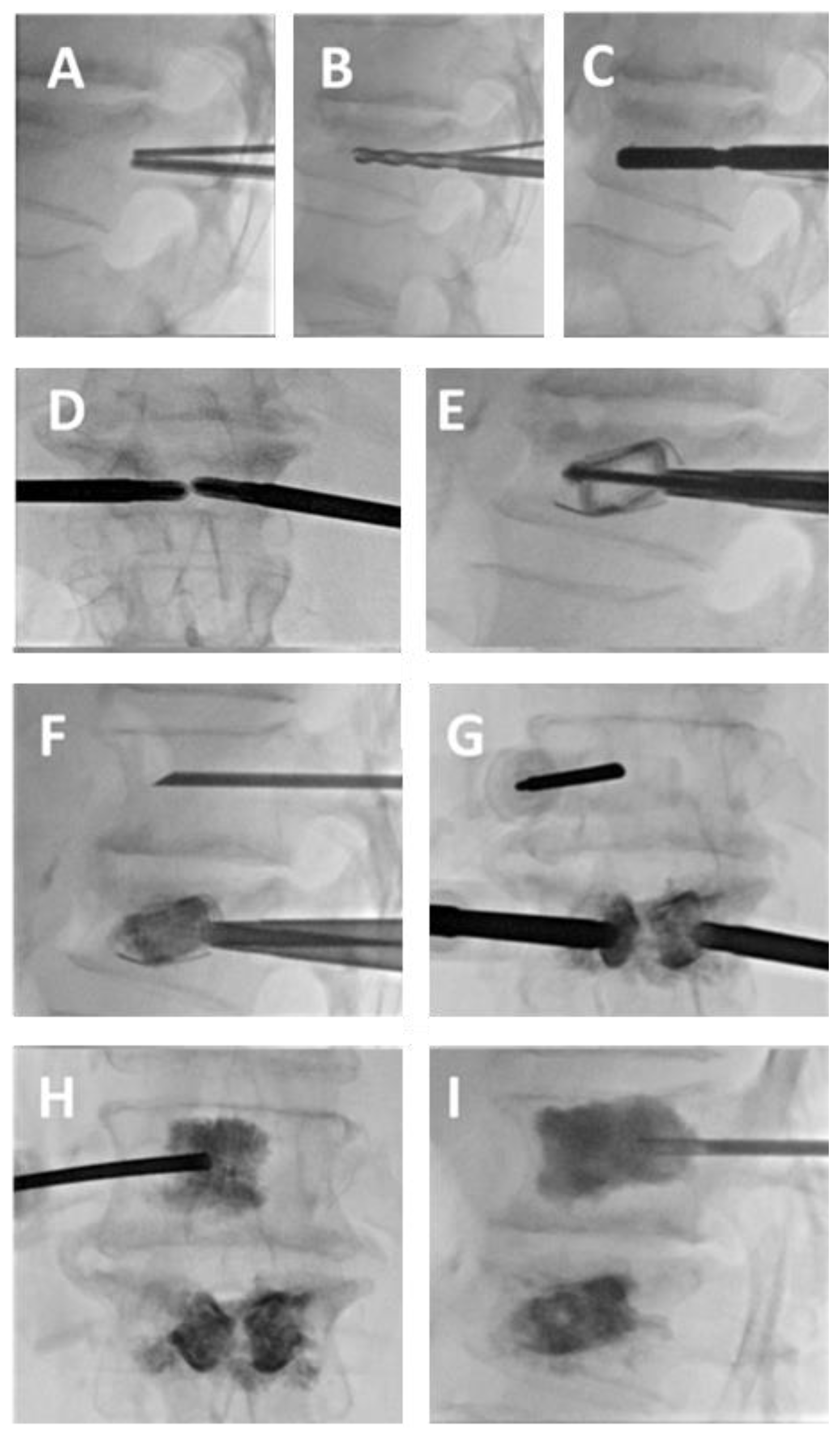
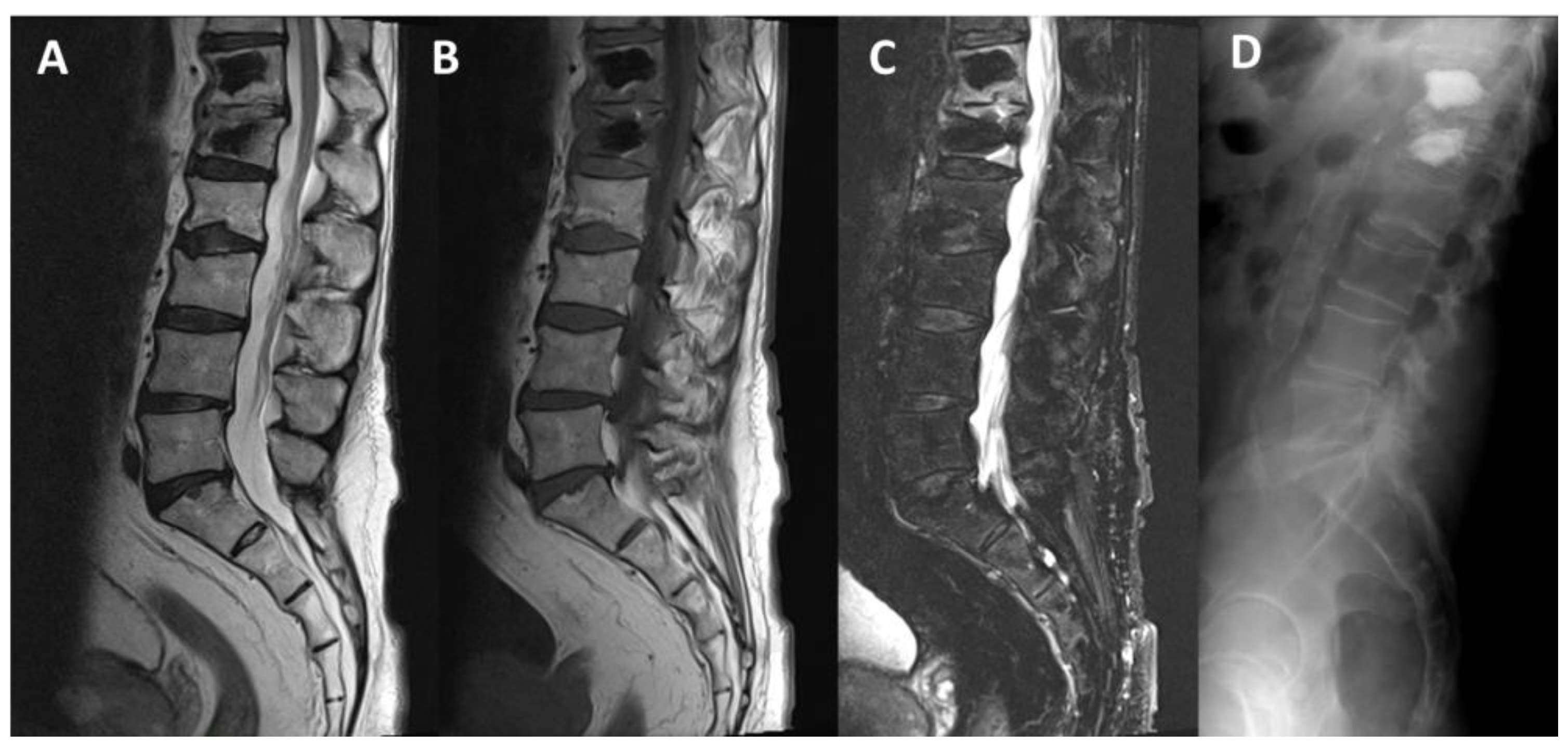
In the case shown in Figure 1, Figure 2 and Figure 3, a patient with collapse of the L1 vertebral body due to suspected metastasis in a case of non-Hodgkin lymphoma, with no evidence of peripheral neurological deficits, is presented (Figure 1). The patient was subsequently subjected to a vertebral biopsy, which did not show any disease localization. The patient then underwent a vertebral reconstruction procedure of the L1 vertebral body with the placement of a SpineJack, followed by vertebroplasty of the D12 vertebral body. Following a bi-pedicular approach to L1, and after a preliminary needle biopsy, the procedure began with the preparation of bone tunnels for pre-implantation, followed by the implantation of 5 mm SpineJack devices. This allowed for partial recovery of the height of the collapsed superior endplate and realignment of the posterior wall displacement. The procedure was completed with vertebroplasty of the D12 vertebral body via a left-sided monopedicular approach (Figure 2). At follow-up imaging approximately two weeks after the procedure, MRI and X-ray confirmed the successful outcome of the recent treatment, showing stability of the treated vertebral segments, partial recovery of vertebral height, and realignment of the posterior wall at L1. No cement leakage was detected on the final CBCT scan. The postoperative course was smooth, with the patient reporting a good recovery of functionality (Figure 3).

3.2. Vertebral Body Stenting System®

The Vertebral Body Stenting System® (VBS) is a titanium device designed for percutaneous vertebral augmentation. It aims to relieve pain and restore the height of fractured vertebrae, helping to correct spinal curvature [12]. The system uses a balloon, similar to standard kyphoplasty, which can expand up to 400% within the vertebral body. By employing ligamentotaxis, the fracture is reduced, restoring vertebral height and creating space for the injection of highly viscous PMMA. After expansion, the balloon is deflated and removed, while the stent remains in place to prevent height loss [13].
VBS is indicated for treating osteoporotic vertebral compression fractures from T10 to L5, particularly in cases without posterior vertebral edge involvement. It is suitable for Genant grade 2 and 3 fractures with kyphotic angulation greater than 15°, as well as fractures classified as A1.1, A1.2, A1.3, and A3.1 under the AO classification [11,14]. Contraindications include fracture types A2, A3.3, B1.1, B2.1, B3, and C.
Extensive mechanical testing has demonstrated VBS’s ability to minimize height loss after balloon deflation, performing better than balloon kyphoplasty [15]. VBS maintains pre-fracture vertebral height and shows improved clinical outcomes, with significant reductions in pain (VAS down by 6.4 points) and disability (ODI decreased by 41.7%) at 12 months. Additionally, vertebral body height improved by 15.3%, and kyphotic correction reached 4.5°. The risk of adjacent vertebral fractures was around 9%, similar to or slightly lower than balloon kyphoplasty rates. Overall, VBS is a safe and effective technique for treating vertebral fractures, with a low incidence of adverse events [12,15].

3.3. OsseoFix®

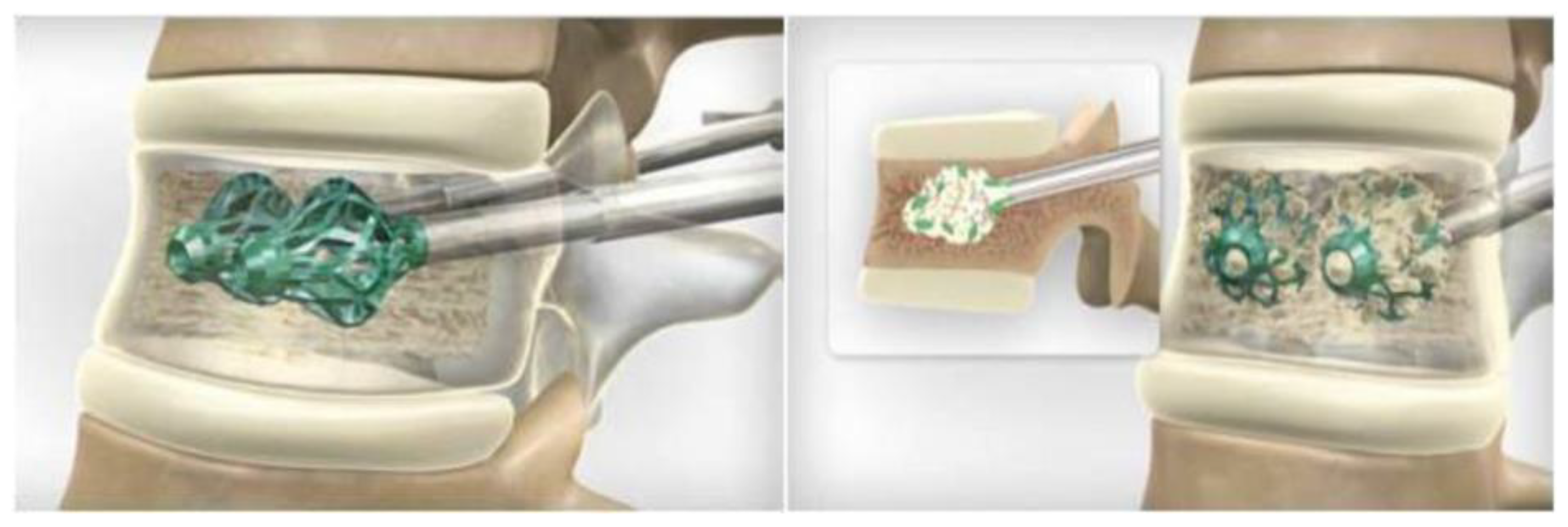
The OsseoFix® Spine Fracture Reduction System (AlphaTec Spine Inc., Carlsbad, CA, USA) is an expandable titanium device predominantly used in the treatment of symptomatic osteoporotic vertebral stable compression fractures. It consists of a titanium mesh that expands within the vertebral body, helping to reduce fractures and correct kyphotic deformities by compacting the surrounding trabecular bone, as seen in Figure 4 [16,17]. OsseoFix® is suitable for treating vertebral compression fractures from T6 to L5, particularly in stable fractures classified as A1.1 to A1.3 or A3.1 according to the AO classification. It is also effective in managing acute, stable traumatic vertebral fractures of the same type in younger patients. Contraindications include vertebral fractures with retropulsed fragments, dural sac or spinal cord compression, previous treatment at the same level, systemic or local infections, anaphylactic reactions to iodine-based compounds, cancer, irreversible coagulopathies, pre-existing calcium disorders, renal failure, or psychiatric disorders [17,18].
Its technical success, particularly with regard to the extremely low rate of complications and cement leakage, has been documented in several studies, including one by Gandham et al., who evaluated the treatment of multi-level vertebral compression fractures caused by multiple myeloma and found no clinically significant cement leakages among the 152 implants used, resulting in a 0% leakage rate [19]. In fact, OsseoFix® and similar expandable titanium implants require significantly less cement for vertebral reinforcement. Despite using less cement, they offer comparable biomechanical strength to kyphoplasty, reducing the risk of cement leakage into surrounding tissues in vertebrae with compromised posterior walls, as highlighted also by Upasani et al. [20]. Thus, the subsequently inserted PMMA interdigitates with the titanium mesh and surrounding bone, enhancing system stability [21].
The main characteristics of third-generation vertebral augmentation devices are summarized in Table 1.

4. Discussion

The biomechanical impact of vertebral augmentation techniques continues to evolve with the introduction of newer devices and methods. Our review highlights the significant advantages and limitations for vertebral compression fractures of various third-generation PVAS devices, like the SpineJack®, Vertebral Body Stenting System® (VBS), and OsseoFix®. These devices, compared to traditional balloon kyphoplasty and vertebroplasty, offer improved vertebral height restoration, stability, and reduced rates of complications, such as cement leakage, which is a common concern in the use of earlier techniques.

4.1. Biomechanical Impact

In the literature, nearly all studies available demonstrate an improvement in spinal stability following the implantation of third-generation PVAS devices. For the assessment of biomechanical functional recovery of the spine, many studies have relied on indirect parameters to evaluate vertebral deformity improvement, particularly focusing on the “Beck Index” (the ratio of anterior to posterior vertebral height) [22], and the vertebral kyphotic angle and the regional kyphotic angle (α-angle and γ-angle, respectively), as determined by the Cobb method [23]. For example, in a study conducted by Ender et al. which examined the use of titanium mesh cages (OsseoFix®) for unstable osteoporotic thoracolumbar burst fractures in 15 patients, the average kyphotic angle, measured using the Cobb method, significantly decreased from 9 degrees preoperatively to 8 degrees after 12 months (p < 0.05) [21]. Furthermore, Upasani et al. conducted a biomechanical comparison between OsseoFix® and the kyphoplasty balloon, finding that the titanium mesh implant provided superior height restoration while requiring a smaller volume of injected cement [20]. On the other hand, Ghofrani et al. conducted an in vitro biomechanical study using human cadaveric vertebrae to compare kyphoplasty with third-generation PVAS implants, both with and without cement. Their findings revealed that the titanium mesh, regardless of cement usage, exhibited comparable biomechanical properties to balloon kyphoplasty [24]. Additionally, in a prospective study involving 32 patients with symptomatic osteoporotic or tumorous fractures, OsseoFix® demonstrated significant improvements in kyphotic angle and was effective in controlling pain and reducing the Oswestry Disability Index score [19]. For Eschler et al., in a study that addressed unstable thoracolumbar burst fractures in elderly patients using percutaneously applied titanium mesh cages and a transpedicular fixation system with expandable screws, the postoperative kyphotic angle and Cobb angle showed significant improvements, with the kyphotic angle decreasing from 13.7° to 7.4° (p < 0.001) and the Cobb angle from 9.6° to 6.0° (p < 0.002) [25].
Regarding osteoporotic vertebral compression fractures, a preclinical study investigated the relationship between applied forces and the kinematics of a deployable implant for percutaneous vertebral augmentation. Using ex vivo and cadaveric models equipped with strain gauges and sensors for precise measurements, the researchers found a proportional relationship between the actuator force and the implant deployment. The study also highlighted how bone condition (normal vs. osteoporotic) and implant positioning significantly affect the force required [26]. Furthermore, according to Krüger et al., in an analysis of 36 human cadaveric vertebrae, the SpineJack®, demonstrated superior performance compared to kyphoplasty in restoring and maintaining vertebral height. It achieved higher anterior and central height restoration (up to 96.20% and 101.13%, respectively) and maintained better results after cyclic loading, surpassing kyphoplasty in both initial and long-term outcomes [27].
One of the potential advantages of the SpineJack device is its ability to direct the force required for fracture reduction along the craniocaudal axis. In contrast, during balloon kyphoplasty, the direction of the force is determined by the patient’s specific anatomy and the balloon itself. In this regard, a pre-clinical study by Krüger et al. investigated the biomechanical performance and height restoration achieved with the SpineJack device in comparison to balloon kyphoplasty using cadaveric models. The study found that the SpineJack device achieved greater restoration of anterior, central, and posterior height compared to balloon kyphoplasty (p < 0.05) [11]. In another study conducted by Noriega et al., comparing balloon kyphoplasty with the SpineJack, the latter demonstrated a shorter procedure time, similar improvements in pain relief and function, and a substantial increase in vertebral height and vertebral body angle [28]. So far, biomechanical and clinical comparative studies versus percutaneous kyphoplasty or vertebroplasty have reported non-inferiority of spine implants with a reduced volume of injected cement [29].
Instead, concerning preclinical studies on the biomechanical impact of these devices in traumatic vertebral compression fractures, a study conducted on 28 cadaveric spine segments with induced burst fractures demonstrated that the combination of an intravertebral reduction device, with or without cement augmentation, was more effective in restoring central endplates and reducing spinal canal narrowing compared to dorsal instrumentation alone [30]. Additionally, Rotter et al. demonstrated that in a study involving 36 fresh cadaveric vertebrae, the SpineJack system was more effective than balloon kyphoplasty in maintaining vertebral height after reducing traumatic wedge fractures. The study showed that with the SpineJack, it is possible to reduce the cement volume to 10% of the vertebral body volume without compromising height restoration, unlike kyphoplasty, which requires a 30% cement volume to achieve similar results. Moreover, the SpineJack better preserved intraoperative height gain, with an average loss of 1% compared to 16% with kyphoplasty [31].

4.2. PVAS Devices in Tumoral Vertebral Compression Fractures

In recent years, some studies have evaluated the outcomes of using PVAS devices in vertebral compression fractures caused by malignant pathologies, such as multiple myeloma (MM). MM is a clonal disorder of B-cells characterized by the proliferation and accumulation of B-lymphocytes and plasma cells within bone marrow. The spine is particularly affected, because vertebrae, with their high hematopoietic marrow content, are a preferred site for plasma cell neoplasms to infiltrate and grow [32]. Osteoclast activation disrupts bone remodeling, leading to localized osteoporosis and increasing the risk of vertebral compression fractures which are further exacerbated by high-dose steroids [33,34]. For MM vertebral fractures, pharmacological options consist of bisphosphonates, denosumab, teriparatide, and estrogen therapy. Non-invasive approaches to vertebral fractures involve the use of bracing, physical therapy, and pain management strategies. Interventional radiology is crucial in managing this condition, particularly for alleviating pain caused by secondary vertebral fractures. The primary techniques used are percutaneous kyphoplasty and VP, as recommended by the 2017 CIRSE guidelines [35,36]. Vertebral deformity and adjacent level fractures are significant long-term complications associated with vertebroplasty and kyphoplasty in osteoporotic patients. While VP effectively reduces pain in the short and long term, it often fails to fully restore vertebral height or correct the kyphotic angle, leading to deformities and an increased risk of adjacent fractures. To address these issues, various percutaneous implant techniques were developed. These techniques aim to minimize the secondary loss of vertebral height after balloon deflation during kyphoplasty and to maintain vertebral height and kyphotic angle restoration. SpineJack® use in patients with multiple myeloma was reported by Pusceddu et al., who retrospectively evaluated its feasibility and effectiveness for treating and stabilizing painful vertebral compression fractures. In their study, 39 patients with 49 fractures underwent vertebroplasty with SpineJack® implants. Results showed a 100% technical success rate, with no major complications. Pain significantly decreased by 96.3% (VAS score), and functional mobility improved by 47.8% over a 6-month follow-up. The procedure proved safe and effective, offering long-term pain relief and vertebral height restoration without major complications or new fractures [37]. They also noted that the combined CT–Fluoroscopy technique is especially beneficial for patients with significant pre-existing spinal deformities. This approach, in fact, allowed for more accurate implant positioning and provided easier access to the vertebra through CT guidance.

4.3. Pain Relief and Complication Rate

Nearly all studies on third-generation PVAS devices report significant reduction in pain scores and disability, predominantly measured through the Visual Analog Scale (VAS) and the Oswestry Disability Index (ODI), as well as improvements in quality of life and lower postoperative complication rates. Ender et al., for example, demonstrated a significant reduction in pain intensity (VAS), dropping from a preoperative score of 8.0 to 1.6 after, along with significant improvement in activity level (ODI) from a preoperative 79.0 to 30.5% after 12 months [21]. Eschler et al. treated unstable thoracolumbar burst fractures in elderly patients using titanium mesh cages combined with a transpedicular fixation system with expandable screws, resulting in significant pain reduction, maintained fracture alignment, and a low complication rate [25]. In most of the studies reviewed, follow-up imaging confirmed partial recovery of the vertebral height and stability of the treated segments, reinforcing the notion that third-generation devices provide lasting biomechanical support.
Although the current literature indicates that third-generation VPAS are associated with a lower overall complication rate, certain issues remain. Cement leakage, while less common than in earlier techniques, can still occur, particularly in cases involving compromised vertebral endplates or suboptimal implant positioning. Adjacent vertebral fractures, likely due to altered biomechanical forces post-procedure, have also been observed, albeit at low rates. Furthermore, the success of these devices heavily depends on accurate positioning. Improper placement can result in uneven height restoration, inadequate fracture reduction, or suboptimal biomechanical outcomes.

4.4. Combined Treatments

In another study conducted by Pusceddu et al., a novel technique for treating pathological vertebral compression fractures was assessed, combining microwave ablation (MWA), bilateral expandable titanium SpineJack® implants, and vertebroplasty. The retrospective study included 28 patients with vertebral metastases, all of whom had a history of primary tumors. The procedure achieved a 100% technical success rate with no major complications. Vertebral height was restored in 58% of the treated vertebrae, and pain levels showed a significant reduction, with the VAS score dropping by 93.65% at the 6-month follow-up. Imaging studies (CT and MRI) conducted post-procedure confirmed no implant displacement, local recurrence, or new fractures. This innovative combination of MWA, SpineJack® implants, and vertebroplasty proved to be a safe and effective method for treating pathological vertebral fractures, offering long-term pain relief, enhanced mobility, and effective tumor control [38].

4.5. Cost and Justification, Limitations and Future Directions

The costs of third-generation PVAS devices are significantly higher compared to traditional methods such as vertebroplasty and balloon kyphoplasty. This increased cost is primarily attributed to the advanced materials (e.g., titanium components) and the sophisticated engineering of these devices. These factors can be a barrier to widespread adoption, especially in resource-limited settings. Furthermore, their deployment often requires advanced technical expertise, and operator inexperience may increase the risk of complications or diminish the effectiveness of the intervention.
However, the higher costs of these systems can be justified by their long-term benefits. Third-generation devices have been shown to reduce revision rates, lower the risk of re-fractures and adjacent vertebral fractures, and improve patient outcomes overall. Additionally, their use in complex cases, such as those involving multiple myeloma or older fractures with marrow edema, expands the range of treatable conditions, further supporting their adoption in clinical practice.
Despite the clear advantages, there remain areas for improvement. One key issue is patient selection, as not all fractures may benefit equally from the same device. Future studies should focus on refining criteria for selecting the most appropriate device based on fracture type, patient age, and comorbidities. Furthermore, long-term studies comparing the efficacy of these devices in preventing adjacent fractures and maintaining height restoration are needed to fully assess their impact on patient outcomes.

5. Conclusions

Third-generation PVAS devices, such as the SpineJack®, Vertebral Body Stenting System®, and OsseoFix®, have demonstrated significant advancements in the treatment of vertebral compression fractures. These devices offer improved restoration of vertebral height and correction of kyphotic angles, thereby enhancing biomechanical stability, while also reducing the risk of complications such as cement leakage. Each device presents unique advantages depending on the type of fracture and patient characteristics. However, the studies currently available in the literature involve relatively small patient cohorts, highlighting the need for larger, more comprehensive trials to further refine patient selection criteria and optimize long-term outcomes.

Author Contributions

Conceptualization, E.F. and F.V.; methodology, E.F. and F.V.; validation, D.S. and R.F.G.; formal analysis, E.F.; investigation, F.V.; resources, E.V.; data curation, E.V.; writing—original draft preparation, E.F. and F.V.; writing—review and editing, E.F. and D.S.; visualization, B.B.Z. and R.F.G.; supervision, E.F. and R.F.G.; project administration, E.F. All authors have read and agreed to the published version of the manuscript.

Funding

This research received no external funding.

Conflicts of Interest

The authors declare no conflicts of interest.

Abbreviations

VPVertebroplasty
PVASPercutaneous vertebral augmentation system
MMMultiple myeloma
RTRadiotherapy
PMMApolymethyl methacrylate
VASVisual Analog Scale
ODIOswestry Disability Index

References

  1. Galibert, P.; Deramond, H.; Rosat, P.; Le Gars, D. Preliminary note on the treatment of vertebral angioma by percutaneous acrylic vertebroplasty. Neurochirurgie 1987, 33, 166–168. [Google Scholar] [PubMed]
  2. Saracen, A.; Kotwica, Z. Complications of percutaneous vertebroplasty: An analysis of 1100 procedures performed in 616 patients. Medicine 2016, 95, e3850. [Google Scholar] [CrossRef] [PubMed]
  3. McGirt, M.J.; Parker, S.L.; Wolinsky, J.-P.; Witham, T.F.; Bydon, A.; Gokaslan, Z.L. Vertebroplasty and kyphoplasty for the treatment of vertebral compression fractures: An evidenced-based review of the literature. Spine J. 2009, 9, 501–508. [Google Scholar] [CrossRef]
  4. Luo, J.; Adams, M.A.; Dolan, P. Vertebroplasty and Kyphoplasty Can Restore Normal Spine Mechanics Following Osteoporotic Vertebral Fracture. J. Osteoporos. 2010, 2010, 729257. [Google Scholar] [CrossRef]
  5. Feltes, C.; Fountas, K.N.; Machinis, T.; Nikolakakos, L.G.; Dimopoulos, V.; Davydov, R.; Kassam, M.; Johnston, K.W.; Robinson, J.S. Immediate and early postoperative pain relief after kyphoplasty without significant restoration of vertebral body height in acute osteoporotic vertebral fractures. Neurosurg. Focus 2005, 18, e5. [Google Scholar] [CrossRef]
  6. Fields, A.J.; Lee, G.L.; Keaveny, T.M. Mechanisms of initial endplate failure in the human vertebral body. J. Biomech. 2010, 43, 3126–3131. [Google Scholar] [CrossRef]
  7. Schützenberger, S.; Schwarz, S.M.; Greiner, L.; Holub, O.; Grabner, S.; Huf, W.; Sailler, A.; Fialka, C. Is vertebral body stenting in combination with CaP cement superior to kyphoplasty? Eur. Spine J. 2018, 27, 2602–2608. [Google Scholar] [CrossRef]
  8. Vanni, D.; Galzio, R.; Kazakova, A.; Pantalone, A.; Grillea, G.; Bartolo, M.; Salini, V.; Magliani, V. Third-generation percutaneous vertebral augmentation systems. J. Spine Surg. 2016, 2, 13–20. [Google Scholar] [CrossRef]
  9. Noriega, D.; Krüger, A.; Ardura, F.; Hansen-Algenstaedt, N.; Hassel, F.; Barreau, X.; Beyerlein, J. Clinical Outcome After the Use of a New Craniocaudal Expandable Implant for Vertebral Compression Fracture Treatment: One Year Results from a Prospective Multicentric Study. BioMed Res. Int. 2015, 2015, 927813. [Google Scholar] [CrossRef]
  10. Vanni, D.; Pantalone, A.; Bigossi, F.; Pineto, F.; Lucantoni, D.; Salini, V. New perspective for third generation percutaneous vertebral augmentation procedures: Preliminary results at 12 months. J. Craniovertebral Junction Spine 2012, 3, 47–51. [Google Scholar] [CrossRef]
  11. Krüger, A.; Oberkircher, L.; Figiel, J.; Floßdorf, F.; Bolzinger, F.; Noriega, D.C.; Ruchholtz, S. Height restoration of osteoporotic vertebral compression fractures using different intravertebral reduction devices: A cadaveric study. Spine J. 2015, 15, 1092–1098. [Google Scholar] [CrossRef] [PubMed]
  12. Disch, A.C.; Schmoelz, W. Cement augmentation in a thoracolumbar fracture model: Reduction and stability after balloon kyphoplasty versus vertebral body stenting. Spine 2014, 39, E1147–E1153. [Google Scholar] [CrossRef] [PubMed]
  13. Fürderer, S.; Anders, M.; Schwindling, B.; Salick, M.; Düber, C.; Wenda, K.; Urban, R.; Glück, M.; Eysel, P. Vertebral body stenting. A method for repositioning and augmenting vertebral compression fractures. Orthopade 2002, 31, 356–361. [Google Scholar] [CrossRef]
  14. Joseph, R.N.; Swift, A.J.; Maliakal, P.J. Single centre prospective study of the efficacy of percutaneous cement augmentation in the treatment of vertebral compression fractures. Br. J. Neurosurg. 2013, 27, 459–464. [Google Scholar] [CrossRef]
  15. Werner, C.M.L.; Osterhoff, G.; Schlickeiser, J.; Jenni, R.; Wanner, G.A.; Ossendorf, C.; Simmen, H.-P. Vertebral body stenting versus kyphoplasty for the treatment of osteoporotic vertebral compression fractures: A randomized trial. J. Bone Jt. Surg. Am. 2013, 95, 577–584. [Google Scholar] [CrossRef]
  16. Ender, S.A.; Wetterau, E.; Ender, M.; Kühn, J.-P.; Merk, H.R.; Kayser, R. Percutaneous Stabilization System Osseofix® for Treatment of Osteoporotic Vertebral Compression Fractures—Clinical and Radiological Results after 12 Months. PLoS ONE 2013, 8, e65119. [Google Scholar] [CrossRef]
  17. Eschler, A.; Ender, S.A.; Ulmar, B.; Herlyn, P.; Mittlmeier, T.; Gradl, G. Cementless fixation of osteoporotic VCFs using titanium mesh implants (OsseoFix): Preliminary results. BioMed Res. Int. 2014, 2014, 853897. [Google Scholar] [CrossRef]
  18. Ender, S.A.; Gradl, G.; Ender, M.; Langner, S.; Merk, H.R.; Kayser, R. Osseofix® system for percutaneous stabilization of osteoporotic and tumorous vertebral compression fractures—Clinical and radiological results after 12 months. Rofo 2014, 186, 380–387. [Google Scholar] [CrossRef]
  19. Gandham, S.; Islim, A.; Alhamad, S.; Thambiraj, S. The outcome of expandable titanium mesh implants for the treatment of multi-level vertebral compression fractures caused by multiple myeloma. SICOT J. 2021, 7, 28. [Google Scholar] [CrossRef]
  20. Upasani, V.V.; Robertson, C.; Lee, D.; Tomlinson, T.; Mahar, A.T. Biomechanical comparison of kyphoplasty versus a titanium mesh implant with cement for stabilization of vertebral compression fractures. Spine 2010, 35, 1783–1788. [Google Scholar] [CrossRef]
  21. Ender, S.A.; Eschler, A.; Ender, M.; Merk, H.R.; Kayser, R. Fracture care using percutaneously applied titanium mesh cages (OsseoFix®) for unstable osteoporotic thoracolumbar burst fractures is able to reduce cement-associated complications—Results after 12 months. J. Orthop. Surg. Res. 2015, 10, 175. [Google Scholar] [CrossRef] [PubMed]
  22. Beck, E. Radiographic measuring methods in vertebral fractures. Hefte Unfallheilkunde 1971, 108, 36–37. [Google Scholar]
  23. Outline for the Study of Scoliosis—ScienceOpen. Available online: https://www.scienceopen.com/book?vid=76a12f1e-c7ef-4cc2-8aec-0904b520cd98 (accessed on 30 September 2024).
  24. Ghofrani, H.; Nunn, T.; Robertson, C.; Mahar, A.; Lee, Y.; Garfin, S. An evaluation of fracture stabilization comparing kyphoplasty and titanium mesh repair techniques for vertebral compression fractures: Is bone cement necessary? Spine 2010, 35, E768–E773. [Google Scholar] [CrossRef] [PubMed]
  25. Eschler, A.; Ender, S.A.; Schiml, K.; Mittlmeier, T.; Gradl, G. Bony healing of unstable thoracolumbar burst fractures in the elderly using percutaneously applied titanium mesh cages and a transpedicular fixation system with expandable screws. PLoS ONE 2015, 10, e0117122. [Google Scholar] [CrossRef]
  26. Bolzinger, F.; Oglaza, J.F.; Krüger, A.; Swider, P. Measurement of implant deployment and related forces in kyphoplasty by percutaneous approach. Clin. Biomech. 2014, 29, 463–467. [Google Scholar] [CrossRef]
  27. Krüger, A.; Baroud, G.; Noriega, D.; Figiel, J.; Dorschel, C.; Ruchholtz, S.; Oberkircher, L. Height restoration and maintenance after treating unstable osteoporotic vertebral compression fractures by cement augmentation is dependent on the cement volume used. Clin. Biomech. 2013, 28, 725–730. [Google Scholar] [CrossRef]
  28. Noriega, D.C.; Ramajo, R.H.; Lite, I.S.; Toribio, B.; Corredera, R.; Ardura, F.; Krüger, A. Safety and clinical performance of kyphoplasty and SpineJack® procedures in the treatment of osteoporotic vertebral compression fractures: A pilot, monocentric, investigator-initiated study. Osteoporos. Int. 2016, 27, 2047–2055. [Google Scholar] [CrossRef]
  29. Filippiadis, D.K.; Marcia, S.; Ryan, A.; Beall, D.P.; Masala, S.; Deschamps, F.; Kelekis, A. New Implant-Based Technologies in the Spine. CardioVascular Interv. Radiol. 2018, 41, 1463–1473. [Google Scholar] [CrossRef]
  30. Krüger, A.; Schmuck, M.; Noriega, D.C.; Ruchholtz, S.; Baroud, G.; Oberkircher, L. Percutaneous Dorsal Instrumentation of Vertebral Burst Fractures: Value of Additional Percutaneous Intravertebral Reposition-Cadaver Study. BioMed Res. Int. 2015, 2015, 434873. [Google Scholar] [CrossRef]
  31. Rotter, R.; Schmitt, L.; Gierer, P.; Schmitz, K.-P.; Noriega, D.; Mittlmeier, T.; Meeder, P.-J.; Martin, H. Minimum cement volume required in vertebral body augmentation—A biomechanical study comparing the permanent SpineJack device and balloon kyphoplasty in traumatic fracture. Clin. Biomech. 2015, 30, 720–725. [Google Scholar] [CrossRef]
  32. Tosi, P. Diagnosis and treatment of bone disease in multiple myeloma: Spotlight on spinal involvement. Scientifica 2013, 2013, 104546. [Google Scholar] [CrossRef] [PubMed]
  33. Hoes, J.N.; Jacobs, J.W.G.; Hulsmans, H.M.J.; De Nijs, R.N.J.; Lems, W.F.; Bruyn, G.A.W.; Geusens, P.P.M.M.; Bijlsma, J.W.J. High incidence rate of vertebral fractures during chronic prednisone treatment, in spite of bisphosphonate or alfacalcidol use. Extension of the alendronate or alfacalcidol in glucocorticoid-induced osteoporosis-trial. Clin. Exp. Rheumatol. 2010, 28, 354–359. [Google Scholar] [PubMed]
  34. Kulkarni, A.G.; Patel, A. Denosumab: A potential new treatment option for recurrent Aneurysmal Bone Cyst of the spine. SICOT J. 2019, 5, 10. [Google Scholar] [CrossRef] [PubMed]
  35. Tsoumakidou, G.; Too, C.W.; Koch, G.; Caudrelier, J.; Cazzato, R.L.; Garnon, J.; Gangi, A. CIRSE Guidelines on Percutaneous Vertebral Augmentation. CardioVascular Interv. Radiol. 2017, 40, 331–342. [Google Scholar] [CrossRef]
  36. Martikos, K.; Greggi, T.; Faldini, C.; Vommaro, F.; Scarale, A. Osteoporotic thoracolumbar compression fractures: Long-term retrospective comparison between vertebroplasty and conservative treatment. Eur. Spine J. 2018, 27, 244–247. [Google Scholar] [CrossRef]
  37. Pusceddu, C.; Faiella, E.; Derudas, D.; Ballicu, N.; Melis, L.; Zedda, S.; Marsico, S. Re-expansion of vertebral compression fractures in patients with multiple myeloma with percutaneous vertebroplasty using spinejack implants: A preliminary and retrospective study. Front. Surg. 2023, 10, 1121981. [Google Scholar] [CrossRef]
  38. Pusceddu, C.; Marsico, S.; Derudas, D.; Ballicu, N.; Melis, L.; Zedda, S.; de Felice, C.; Calabrese, A.; De Francesco, D.; Venturini, M.; et al. Percutaneous Vertebral Reconstruction (PVR) Technique of Pathological Compression Fractures: An Innovative Combined Treatment of Microwave Ablation, Bilateral Expandable Titanium SpineJack Implants Followed by Vertebroplasty. J. Clin. Med. 2023, 12, 4178. [Google Scholar] [CrossRef]
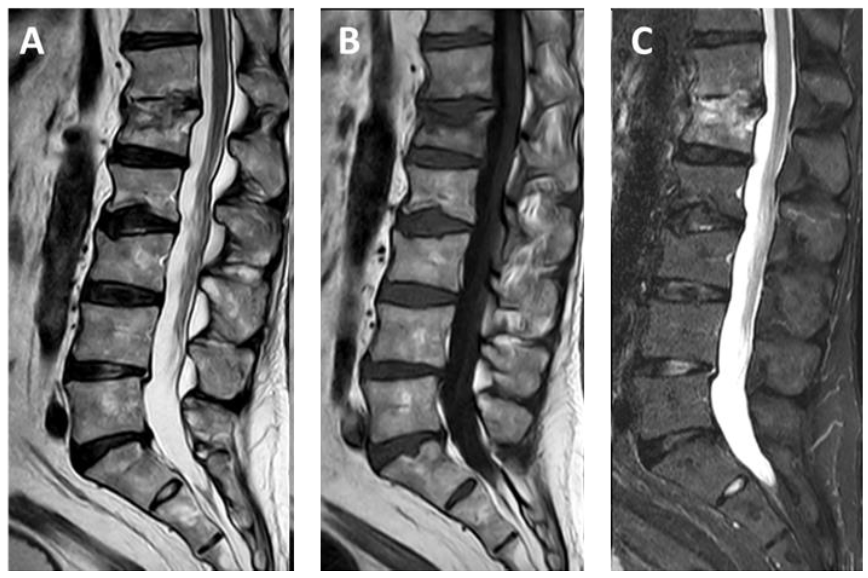
Figure 1. Patient with non-metastatic collapse of the L1 vertebral body in a case of non-Hodgkin lymphoma, with no evidence of peripheral neurological deficits. Image showing TSE sequences obtained with a 1.5 Tesla magnet in the sagittal plane of the lumbosacral spine of the patient, before treatment: (A) T2-weighted sequence, (B) T1-weighted sequence, and (C) Short Tau Inversion Recovery sequence.
Applsci 15 00426 g001
Figure 2. Images depicting the stages of a vertebral reconstruction procedure in which the patient underwent vertebral body reconstruction of L1 with the placement of a SpineJack and vertebroplasty of the D12 vertebral body. (A) Transpedicular approach to L1. (B) Drilling of bone tunnels for pre-implantation. (C) Insertion of the SpineJack device into the vertebral body. (D) Positioning and adjustment of the devices for optimal height recovery and realignment. (E) Partial recovery of the height of the collapsed superior endplate and realignment of the posterior wall displacement. Completion of the procedure with vertebroplasty of the D12 vertebral body via a left-sided monopedicular approach. (F) Left-sided approach to the D12 vertebral body. (G) Needle insertion into the D12 body for vertebroplasty. (H) Distribution of cement material within the vertebral body of D12. (I) Final image showing the successful filling of the D12 vertebral body with cement, securing structural stability.
Applsci 15 00426 g002
Figure 3. Follow-up imaging approximately two weeks after the procedure, obtained with a 1.5 Tesla magnet and TSE sequences: (A) T2-weighted sagittal MRI, (B) T1-weighted sagittal MRI, and (C) T2-weighted fat-saturated (fat sat) sagittal MRI. (D) Laterolateral projection X-ray of the lumbar spine.
Applsci 15 00426 g003
Figure 4. Osseofix™ implant is inserted, compacting the trabecular bone and improving cement interdigitation for enhanced stability. Reprinted from Gandham et al. [19].
Applsci 15 00426 g004
Table 1. Characteristics of Third-Generation Vertebral Augmentation Devices.
Main UseMaterialOperating Principle
KyphonTreatment of recent fractures, osteoporosis, and metastasisElastomerBalloon-based
SpineJack®Manages both recent and older fractures, osteoporosis, metastasisTitaniumDeformable metallic structure
Vertebral Body Stenting® (VBS)Recent fractures, osteoporosis, metastasisTitaniumBalloon combined with a deformable metallic component
OsseoFix®Osteoporotic vertebral collapseTitaniumDeformable metallic structure
Disclaimer/Publisher’s Note: The statements, opinions and data contained in all publications are solely those of the individual author(s) and contributor(s) and not of MDPI and/or the editor(s). MDPI and/or the editor(s) disclaim responsibility for any injury to people or property resulting from any ideas, methods, instructions or products referred to in the content.