Review

A Review of the Impacts of Implant Stiffness on Fracture Healing

1
Department of Orthopaedic Surgery, Graduate School of Medicine, Tohoku University, Sendai 980-8574, Japan
2
Department of Radiology, Graduate School of Medicine, Akita University, Akita 010-8543, Japan
3
Institute for Material Research, Tohoku University, Sendai 980-8577, Japan
*
Author to whom correspondence should be addressed.
Appl. Sci. 2024, 14(6), 2259; https://doi.org/10.3390/app14062259
Submission received: 13 February 2024 / Revised: 5 March 2024 / Accepted: 6 March 2024 / Published: 7 March 2024
(This article belongs to the Special Issue Advances in Mechanics of Biomaterials)

Abstract

:

Featured Application

Interfragmentary movement is crucial in promoting callus formation during secondary fracture healing. Fixation stiffness is a critical factor, with excessive flexibility and rigidity hindering optimal healing. The concepts of flexible fixation, techniques for regulating implant stiffness, and material selection, favoring a lower Young’s modulus like titanium alloys, are emphasized. A novel TiNbSn alloy with a low Young’s modulus has been introduced as a potential solution that has shown efficacy in promoting osteosynthesis and superior mechanical properties.

Abstract

The bone healing process is influenced by various physiological factors. Fracture fixation traditionally relied on rigid metallic implants. However, excessively rigid constructs can lead to complications, necessitating revision surgery. This review focuses on approaches to improve bone healing by introducing adequate interfragmentary movement (IFM) at the fracture site. IFM promotes secondary fracture healing and callus formation. Studies suggest that rigid fixation may impair fracture healing by inhibiting callus formation and causing stress shielding. Titanium alloy locking plates have been shown to be biomechanically superior to stainless steel. Flexible fixation and techniques to regulate implant stiffness are crucial for managing fractures with bridge plating. Materials with a lower Young’s modulus balance biomechanical properties. A novel TiNbSn alloy with a low Young’s modulus has been developed to address stress shielding issues. It is effective in promoting osteosynthesis, bone healing, and superior mechanical properties compared with materials with higher Young’s moduli. The enhanced formation of bone and callus associated with TiNbSn alloy suggests its promise for use in fracture treatment plates. Understanding the biomechanics of fracture healing, optimizing fixation stiffness, and exploring innovative materials like TiNbSn alloys, are crucial for advancing approaches to accelerate and enhance bone healing.

1. Introduction

Bone healing is intricate and involves various physiological factors. Physical stimulation techniques, including mechanical stimulation methods, play a crucial role in the fracture healing process [1,2,3], alongside other significant factors such as blood flow, the supply of mesenchymal stem cells [4], and physiological inflammation at the fracture site [5]. Many studies have documented the impact of mechanical stimulation on fracture healing in both experimental animals and humans. Previous studies have demonstrated that modulating the mechanical forces at the fracture site can significantly influence the healing rate and dictate whether healing proceeds via the intramembranous or endochondral ossification pathway [6,7,8,9]. Collectively, these elements contribute to the complex biological and mechanical environment necessary for effective bone regeneration and repair. The external mechanical conditions closely regulate the healing response and the formation of calluses that can affect the transmission of loads to the callus tissue, encompassing the fracture geometry, gap size, fixation type, fixation stability, and the magnitude and direction of interfragmentary movement (IFM). Furthermore, these mechanical forces directly impact the localized stress and strain at the fracture site, consequently affecting a complex and sequential series of biological pathways involved in the healing process.
Optimizing the speed and strength of fracture healing is challenging. However, adjusting the mechanical parameters of the implants can enhance bone healing and alleviate stress shielding concerns. This has spurred the development of implants with reduced stiffness, dynamic features, and functionally graded characteristics [10,11,12].
Internal fracture fixation has long depended on the use of metallic implants, whose mechanical properties stay fairly unchanged over their duration of use. Throughout much of the 20th century, fracture reconstruction commonly utilized plates equipped with non-locking screws. These devices necessitate a substantial degree of bone integrity, relying on the friction generated between the plate, screws, and bone to promote primary bone healing. However, the introduction of locking plate technology in the 1990s marked a significant shift in the approach to fracture management. This innovation provided stability that does not depend on the condition of the bone, making it especially useful for patients with osteoporosis or for treating highly fragmented fractures [3,10]. The substantial rigidity associated with locking plate reconstruction has given rise to distinct failure mechanisms, notably screw cutout, which frequently necessitates revision surgery [13].
Figure 1 shows a case of fusion failure caused by excessively rigid fixation using locking plates. Cutoff occurrences are typically observed in older patients, indicating that excessively rigid constructs may pose challenges in the context of compromised bone quality. Adequate autologous bone grafting is often necessary for the treatment of comminuted fractures, but the invasiveness of autologous bone grafting may be problematic in fractures in elderly patients. Insufficient autologous bone grafting and gaps between bone fragments limit bone healing and, further, excessively rigid implants lead to inadequate IFM, ultimately causing stress shielding and unfavorable remodeling [14]. Effective secondary fracture healing requires controlled IFM at the fracture site. Independent studies have shown that rigid locking plates do not create an environment conducive to this process [15,16]. The inherent axial flexibility characteristic of long and narrow plates permits bending; yet, this predominantly initiates IFM at the cortex farthest from the fracture gap, limiting the movement directly underneath the plate [15]. Undesirable heterogeneous healing at the fracture site is evident through the emergence of asymmetric and insufficient callus formation, fixation failure, and delayed non-union [17,18].
The aim of this review is to compile and analyze the current understanding and research findings on the biomechanical principles and contemporary clinical practices that enhance bone healing through the introduction of suitable IFM at the site of a fracture. Additionally, it explores the clinical utility of innovative low Young’s modulus titanium alloys in the development of fracture treatment plates, which are instrumental in optimizing IFM to facilitate better healing outcomes. This review also addresses the challenges that must be overcome to fully realize the potential of these advanced materials in improving fracture management.

2. Importance of Interfragmentary Movement in Fracture Healing

The stiffness of the fracture fixation and weight bearing dictates the mechanical environment. The healing process may be compromised if fixation is excessively flexible or overly rigid. Specifically, when high-strain forces are applied at a fracture site (indicating inadequate stability), fibrous tissue persists, and the formation of a stabilizing bony callus is impeded.
Persisting under such conditions for a prolonged period may result in the formation of fibrous non-unions [19,20]. Earlier studies suggested that rigid fixation yields the most favorable clinical outcomes, subsequently contributing to the widespread adoption of nailing and plating to achieve stable internal fixation [8]. Nonetheless, several studies have indicated that overly rigid fixation can paradoxically hinder the process of fracture healing [21]. The rigidity required for fixation is also affected by the size of the gap between bone fragments. A study using live sheep determined that for larger fracture gaps (2 mm or 6 mm), a more rigid fixation method was preferred, while for a smaller gap (1 mm), better healing occurred under higher interfragmentary strain [20]. In contrast, a more recent computer simulation suggested that higher interfragmentary strain could enhance callus formation, which is crucial for indirect fracture healing in larger gaps (3 mm), but the same level of strain in smaller gaps (1 mm) might result in excessive fibrous tissue formation, possibly leading to non-union [22]. The primary concerns raised revolve around potential hindrances to external callus formation, the exacerbation of maintaining a fracture gap due to bone end resorption, and overprotection of the healing bone from regular stress, which can lead to adverse remodeling (stress shielding). Consequently, these studies have identified numerous mechanical factors that influence fracture healing, such as the magnitude and direction of the IFM, type of fracture, and fracture geometry [23,24].
IFM denotes the displacement occurring between fracture fragments as a consequence of both physiological and external loads applied to the fractured limb. Upon loading a fractured bone, the fracture fragments undergo displacement relative to one another, producing a multiaxial strain that exhibits spatial variation across the fracture gap [3]. IFM plays a pivotal role in the process of fracture healing, being significantly influenced by the stability of the fixation and the load applied by the musculoskeletal system. Moderate levels of IFM facilitate successful bone healing, marked by the formation of a fracture callus [25]. In contrast, excessive IFM can hinder the healing process or may even lead to the development of non-unions [26,27]. Various combinations of axial forces, bending, and torsional moments can occur, depending on the load sharing and muscular strain. The two primary factors influencing IFM are the rigidity of the fracture stabilization implant and the superficial area on the fracture fragments. These factors determine tissue strain and cellular responses within the fracture site. The magnitude and direction of the IFM within the fracture site are contingent on the load due to weight bearing, muscle forces, and stiffness characteristics of the selected device [9,28,29]. The strain induced by IFM is dispersed across the fracture surface and varies depending on the fracture geometry. Comminuted fractures, for example, can accommodate relatively higher motion levels since the strain is distributed over a larger surface area of the fractured fragments. The extent of callus formation during the healing process is contingent upon the magnitude of the IFM [1,20]. When the IFM surpasses a critical level, the blood vessels formed at the fracture site face repeated disruption, impeding their establishment and preventing the development of stable tissues [20]. For instance, a specific degree of mechanical instability results in increased IFM accompanied by cartilage formation, leading to endochondral healing, which is the predominant mechanism for healing most fractures [9]. These findings suggest that appropriate IFM promotes fracture healing. However, optimization of IFM is difficult to implement in clinical practice, and actual studies have been limited to preclinical trials [30]. There are anticipations for the development of techniques that enable surgeons to directly assess IFM and interfragmentary movement strain across fracture fragments in real-time during surgical procedures.

3. Clinical Problems in Bridge Plating for Fracture Treatment

The locking plate system was developed to enhance stability and minimize compression between the bone and the plate [16]. Furthermore, biological bridge plating aids in preserving the blood supply, facilitating a functional reduction in complex fractures [31]. Some types of locking plates offer a comparatively stable fixation for fractures while allowing adjustments to reduce the stiffness [32,33]. Fracture treatment devices characterized by excessive stiffness have been reported to result in suboptimal outcomes, including delayed bone healing and failure of the healing process [17,34].
Despite the recent focus on biologically favorable, indirect reduction techniques and the use of submuscular implants, clinical series published over the past decade have reported fracture non-union rates in the distal femur of up to 20% of patients [17,18,35]. The recent identification of several risk factors has been associated with an increased risk of non-union. A previous study examined 283 distal femoral fractures treated with locked plating [36]. Obesity, open fracture, infection occurrence, and the use of stainless steel plates were identified as prognostic risk factors for non-union. Another study reported that 64 of 335 patients with distal femoral fractures required reoperation to promote union [35].
Several studies comparing the effects of stainless steel versus titanium plates were identified. Two articles by Rodriguez et al., which analyzed data from the same group of patients, found a significant link between the use of stainless steel plates and the occurrence of non-union, with a highly significant p-value [36,37]. Conversely, three other studies found no significant difference in outcomes between the two types of metal [38,39,40]. Specifically, Henderson et al. observed an increase in callus formation in patients who received titanium plates as opposed to steel plates at 6 and 12 weeks after surgery, with a statistically significant difference. However, this difference in callus formation was not considered statistically significant at the 24-week mark [39]. Although the results of these findings may vary when compared to plates made of metallic materials with greatly different Young’s moduli, there are currently no clinically usable low Young’s modulus fracture treatment plates. The most successful healing rates for unstable distal femoral fractures have been achieved through the application of titanium locking plates. While the theoretical biomechanical benefits of CP-Ti and its alloys appear to contribute to clinical enhancements, no high-quality study has conclusively validated this assertion. In a previous study involving 21 patients, mostly comprising young individuals with 22 high-energy distal femur fractures, treatment with titanium locking plates was utilized. Furthermore, the study noted that all 22 fractures healed promptly, obviating the necessity for secondary surgeries [41]. It was suggested that pure titanium and titanium alloys, which have a lower Young’s modulus than high Young’s modulus materials, such as stainless steel, may be useful for fracture treatment plates.
Effectively managing fractures that are amenable to bridge plating involves understanding the concept of flexible fixation and techniques for regulating implant stiffness. The existing clinical and biomechanical data provide a foundation for the selection of suitable fixation methods. Additionally, when selecting an implant, it is important to consider titanium or titanium alloys because these metals have a lower Young’s modulus than that of stainless steel. Nevertheless, it is essential to balance the biomechanical advantages with the limitations of titanium, such as difficulties in contouring and the risk of cold welding. Multiple studies have not found a statistically significant correlation between the length of the plate and the incidence of non-union [36,37,39]. Specifically, within a population of low-energy fractures treated using the minimally invasive plate osteosynthesis (MIPO) technique, a trend was observed where longer plates were associated with a lower rate of non-union, although this trend did not achieve statistical significance. Factors such as the total number of plate holes, average bridging length, ratio of bridging length to plate length, and fixation crossing the fracture line were also not significantly associated with non-union. Comparative studies between MIPO and traditional locking compression plates were conducted in previous studies [38,42]. Hoffman et al. found a significant decrease in the rate of non-union for the MIPO group in comparison to the group treated with open reduction [38]. Meanwhile, the other study reported no significant difference in postoperative complications between the two methods [42]. MIPO has gained popularity as a technique for managing distal femoral fractures. Theoretically, MIPO allows for better preservation of the blood supply, maintenance of the fracture hematoma, and reduces soft tissue damage. It has been reported in a mouse fracture model that the starting point for fracture repair is mesenchymal cell proliferation from the periosteal area near the fracture site and differentiation into osteoblasts [4], and there is a consensus that maintenance of blood flow and protection of the periosteum is important. However, the effectiveness of MIPO for the treatment of other long bone fractures remains uncertain, with studies presenting mixed results. This indicates that additional research is needed to fully understand the potential advantages of MIPO. The preferred choice is the longest anatomically feasible plate, as these plates can be applied [14,35]. This approach preserves the biology, ensures a more uniform stress distribution, and diminishes implant fatigue. Using a greater number of screw fixings than necessary is not required. Moreover, it is crucial to maintain low screw-fill levels and minimize stress concentrations [14,43].
Rodriguez et al. found a significant link between higher rigidity scores and an increased likelihood of non-union, where rigidity score encompassed factors such as proximal screw density, plate material, and the number of screws traversing the fracture site [37]. Moreover, constructs utilizing solely locking screws were correlated with a greater incidence of non-union compared to hybrid constructs that combine locking and non-locking screws [40]. Notably, a higher density of proximal screws and overall screw density was significantly related to elevated non-union rates, specifically within the group of patients with low-energy fractures [44]. This particular relationship was not observed in other studies examining similar variables [36,38].
The plate construct’s effective length is defined as the distance between the initial screws located on either side of the fracture. It has been proposed that a longer effective length may lead to a reduction in non-union rates. A previous study found strong evidence supporting a significant correlation between a greater effective length and lower non-union rates [39]. This correlation was established by measuring the average number of unfilled holes next to the fracture. However, this relationship was not observed when measured in millimeters [39]. In contrast, other studies did not discover a significant connection between the effective length and the occurrence of non-union [38,40].

4. Clinical Applications of Low Young’s Modulus Metallic Materials to Accelerate Fracture Healing

Titanium alloy fracture treatment plates have been reported to have a higher fracture healing rate than stainless steel fracture treatments [39]. We will focus on the elastic modulus of Ti6Al4V alloy, a typical titanium alloy clinically applied in orthopedics owing to its excellent biocompatibility and corrosion resistance. Young’s moduli of the Ti6Al4V alloy and commercially pure titanium (CP-Ti) are 117 and 107 GPa, respectively, which are lower than those of stainless steel (205 GPa) [45,46]. Indeed, Young’s moduli of Ti6Al4V alloy and CP-Ti, although lower than that of stainless steel, are still considerably higher than that of the human cortical bone, which typically ranges from 11 GPa to 20 GPa [47]. The discrepancy in Young’s moduli between the prosthesis and cortical bone results in stress shielding, which impedes the efficient transfer of load from the implant material to the bone, thereby affecting the load-sharing dynamics [48]. Fracture treatment devices that exhibit excessive stiffness have been reported to lead to suboptimal outcomes, including delays and failures in the process of bone healing [17,34].
In contrast, previous studies have demonstrated that when evaluating fracture healing through imaging, intramedullary nails composed of titanium alloy with a low Young’s modulus facilitate the process of fracture healing and the restoration of bone strength [49,50]. A novel TiNbSn alloy characterized by Young’s modulus as less than 50 GPa was developed to address the issue of stress shielding [51]. The tensile strength of the TiNbSn alloy is equivalent to that of the Ti6Al4V alloy. Moreover, upon implantation into the rabbit femoral bone marrow cavity, the TiNbSn alloy exhibited biocompatibility that was comparable to that of the Ti6Al4V alloy [52]. Cell culture tests showed reduced cytotoxicity [52]. Previous studies have documented the excellent corrosion resistance of TiNbSn alloys [53,54].
Results from studies on rabbit and murine tibia fractures treated with intramedullary nails indicate that nails fabricated from the TiNbSn alloy are more efficacious in facilitating osteosynthesis compared to materials possessing higher Young’s moduli [55,56,57]. The effect of the TiNbSn alloy compared with CP-Ti in promoting osteosynthesis in a rabbit tibial osteotomy model is presented in Figure 2 [58].
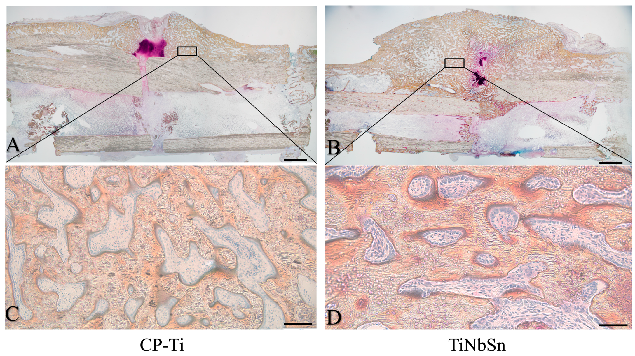
The results demonstrated that in the TiNbSn alloy group, the bone healing process was linked to heightened callus formation, improved structure of newly formed bone, greater bone strength in the early phase, and enhanced bone strength and stiffness during the late phase of bone healing. Adequate IFM and controlled axial motion are provided by elastic intramedullary nails composed of a TiNbSn alloy with a low Young’s modulus, and these motions may contribute to better bone healing and superior mechanical properties. Additionally, the impact of the fracture plate’s low Young’s modulus on osteosynthesis was examined. Radiological, histological, and mechanical evaluations have suggested the outstanding effectiveness of the TiNbSn alloy on bone repair compared to both CP-Ti and Ti6Al4V alloy [11,58]. The TiNbSn alloy is a titanium alloy with a low Young’s modulus and could be equipped with a locking mechanism [58]. Utilizing an implant fabricated from a material with a lower Young’s modulus, which facilitates more effective load sharing with the bone compared to an implant made of Ti6Al4V, could confer advantages for bone healing and potentially diminish the risk of stress shielding. Specifically, a TiNbSn alloy plate, characterized by its low Young’s modulus, demonstrated superior new bone formation and produced a stiffer callus at the osteotomy site, particularly during the initial phases of the healing process, in comparison to plates made from Ti6Al4V alloy and CP-Ti [11,58]. The TiNbSn alloy group exhibited a more robust formation of intramedullary callus with a well-developed bone structure than the Ti6Al4V alloy and CP-Ti groups. At the osteotomy site, the TiNbSn alloy group demonstrated a more advanced formation of mature bone, accompanied by improved orientation of the bone structure (Figure 3).
These improvements may promote bone repair. TiNbSn alloy has emerged as a promising biomaterial for fracture treatment plates. Excessive plate stiffness can potentially hinder callus formation and impede the process of bone healing [59]. The accelerated callus formation observed in the bone defect at the site of plate installation in the group receiving TiNbSn alloy plates is attributed to the effective load-sharing capabilities afforded by the low Young’s modulus of the TiNbSn alloy. Similarly, fracture treatment plates made from Ti29Nb13Ta4.6Zr alloy, which also possesses a low Young’s modulus, demonstrated a propensity to enhance osteosynthesis, even though a quantitative assessment was not conducted [49,60]. Studies on fracture treatment devices using low Young’s modulus titanium alloys are shown in Table 1.
Clinical applications of TiNbSn alloys with low Young’s moduli have been explored. A TiNbSn alloy stem for a hip prosthesis was developed, and the findings from the clinical trial indicated that TiNbSn alloy stems contributed to a reduction in stress shielding, which included improvements in thigh pain and a decrease in hip prosthesis loosening [61,62]. In addition to other TiNbSn alloys, TiNbZrTaSi alloys, TiNbZrSn alloys, TiMoZrSnNb alloys, and TiNbTaO alloys have been reported for their low Young’s moduli, biocompatibility, and osteoinductive effects [63,64,65,66,67]. However, there are few preclinical studies reporting their bone healing-promoting effects as fracture treatment plates or intramedullary nails. Although the usefulness of low Young’s TiCr alloys has been reported for implant rods for spinal fusion, their clinical application has not been realized [68,69,70]. Furthermore, there is no low Young’s modulus titanium alloy other than our TiNbSn alloy that has achieved clinical application in joint prostheses [61,62].
Considering the established safety, durability, and material availability of the TiNbSn alloy, future research should concentrate on the clinical utilization of this alloy in fracture treatment devices with the objective of reducing the duration of the fracture treatment. The efficacy of fracture fixation is determined not only by Young’s modulus of the plate material but also by its length and thickness. While the beneficial effects of the TiNbSn alloy on bone healing were observed in rabbit tibia studies [11,58], the optimal dimensions for plates—specifically their length and thickness—will vary for different long bones. It is also critical to carefully consider these dimensions when applying TiNbSn alloy plates to human patients in clinical settings. Despite this, the successful use of hip prostheses made from TiNbSn alloy suggests that the principles of design similar to Ti6Al4V prosthesis could be advantageous for developing fracture treatment plates. The authors consider that the stiffness of implants plays a critical role in fracture healing by optimizing IFM and load transmission to the bone. The TiNbSn alloy plate emerges as a promising alternative to the Ti6Al4V alloy plate, particularly in scenarios where a load greater than that which the Ti6Al4V alloy plate can accommodate is required, allowing the healed bone to bear a larger share of the load. However, the transition of TiNbSn alloy plates from experimental to clinical use faces several challenges, including cost considerations, indicating that multiple factors must be addressed before these materials can be widely adopted in clinical practice.

5. Issues in the Clinical Setting

In actual clinical practice, IFM cannot be verified in each individual case, so the method that is assumed to optimize IFM is selected. Surgical factors linked to increased fusion failure rates align with the growing body of literature indicating that excessively rigid implants can impede fracture fusion in distal femur fractures [37]. Various factors could relate to non-union, such as different metal materials, plate length, and other variables affecting bone fusion post-locking plate fixation. The prevalence of non-union associated with this procedure may vary due to factors like metal material variations, plate length, and other influencing factors. While the optimal surgical setup remains unclear, controlling factors like metal material and plate thickness to prevent overly rigid fixation and ensure optimal stability could help reduce non-union rates in managing distal femoral fractures.
Surgeons must navigate the delicate balance between preventing excessively rigid fixation constructs and allowing callus formation without compromising fixation stability. The TiNbSn alloy is a high-performance metal known for its exceptional combination of high strength and low Young’s modulus, qualities that have led to its clinical use in femoral stems [61,62]. It is anticipated that by manipulating variables other than Young’s modulus, such as the thickness and length of plates, it will be possible to engineer materials for fracture treatment that possess the ideal stiffness. This approach suggests a tailored strategy for material development, aiming to optimize the biomechanical environment for fracture healing. Further research is necessary to determine the ideal balance of intraoperative variables. Currently, only animal studies are approved to address these concerns, and the clinical application of novel materials is anticipated. Future studies should ideally employ randomized controlled trials to minimize confounding factors. If not possible, it is crucial to analyze homogeneous data adequately powered for analysis purposes to address surgical cohort analysis effectively.

6. Conclusions

This review discusses the biomechanical principles and clinical strategies for improving bone healing and emphasizes the importance of IFM. IFM is crucial in promoting callus formation during secondary fracture healing. Fixation stiffness is a critical factor, with excessive flexibility and rigidity hindering optimal healing. Titanium alloy locking plates have been highlighted for their biomechanical superiority over stainless steel. The concepts of flexible fixation, techniques for regulating implant stiffness, and material selection, favoring a lower Young’s modulus like titanium alloys, are emphasized. A novel TiNbSn alloy with a low Young’s modulus has been introduced as a potential solution that showed efficacy in promoting osteosynthesis and superior mechanical properties. Future research should aim to corroborate and augment these findings by exploring other animal models, assessing different plate geometries, and evaluating fatigue life and plate strength. Validation of the bone healing efficacy of TiNbSn alloy under varied experimental conditions, across different models, and with assorted TiNbSn alloy plate designs, as well as verification of the durability and strength of TiNbSn alloy plates, are essential steps towards their potential clinical application. The clinical applications of hip prostheses have indicated reduced stress shielding and improved outcomes, contributing to advancements in fracture treatments.

Author Contributions

Conceptualization, Y.M.; writing—original draft preparation, Y.M.; writing—review and editing, Y.M., M.K. (Masayuki Kamimura), K.I., M.K. (Masashi Koguchi), H.T., H.K., T.K., N.M. (Naoko Mori), N.M. (Naoya Masahashi) and T.A.; funding acquisition, Y.M. and N.M. (Naoya Masahashi). All authors have read and agreed to the published version of the manuscript.

Funding

This study was supported by the Japan Society for the Promotion of Science (No. 23K08604 to Yu Mori; No. 20H02458 to Naoya Masahashi).

Institutional Review Board Statement

Not applicable.

Informed Consent Statement

Not applicable.

Data Availability Statement

Not applicable.

Conflicts of Interest

The authors declare no conflicts of interest.

References

  1. Claes, L.E.; Heigele, C.A.; Neidlinger-Wilke, C.; Kaspar, D.; Seidl, W.; Margevicius, K.J.; Augat, P. Effects of mechanical factors on the fracture healing process. Clin. Orthop. Relat. Res. 1998, 355, S132–S147. [Google Scholar] [CrossRef]
  2. Foster, A.L.; Moriarty, T.F.; Zalavras, C.; Morgenstern, M.; Jaiprakash, A.; Crawford, R.; Burch, M.A.; Boot, W.; Tetsworth, K.; Miclau, T.; et al. The influence of biomechanical stability on bone healing and fracture-related infection: The legacy of Stephan Perren. Injury 2021, 52, 43–52. [Google Scholar] [CrossRef]
  3. Glatt, V.; Evans, C.H.; Tetsworth, K. A Concert between Biology and Biomechanics: The Influence of the Mechanical Environment on Bone Healing. Front. Physiol. 2016, 7, 678. [Google Scholar] [CrossRef]
  4. Mori, Y.; Adams, D.; Hagiwara, Y.; Yoshida, R.; Kamimura, M.; Itoi, E.; Rowe, D.W. Identification of a progenitor cell population destined to form fracture fibrocartilage callus in Dickkopf-related protein 3-green fluorescent protein reporter mice. J. Bone Miner. Metab. 2016, 34, 606–614. [Google Scholar] [CrossRef] [PubMed]
  5. Kamimura, M.; Mori, Y.; Sugahara-Tobinai, A.; Takai, T.; Itoi, E. Impaired Fracture Healing Caused by Deficiency of the Immunoreceptor Adaptor Protein DAP12. PLoS ONE 2015, 10, e0128210. [Google Scholar] [CrossRef] [PubMed]
  6. Carter, D.R.; Wong, M. The role of mechanical loading histories in the development of diarthrodial joints. J. Orthop. Res. 1988, 6, 804–816. [Google Scholar] [CrossRef] [PubMed]
  7. Carter, D.R.; Wong, M. Mechanical stresses and endochondral ossification in the chondroepiphysis. J. Orthop. Res. 1988, 6, 148–154. [Google Scholar] [CrossRef]
  8. Grundnes, O.; Reikeras, O. Effects of instability on bone healing. Femoral osteotomies studied in rats. Acta Orthop. Scand. 1993, 64, 55–58. [Google Scholar] [CrossRef] [PubMed]
  9. Augat, P.; Simon, U.; Liedert, A.; Claes, L. Mechanics and mechano-biology of fracture healing in normal and osteoporotic bone. Osteoporos. Int. 2005, 16, S36–S43. [Google Scholar] [CrossRef] [PubMed]
  10. Ganse, B.; Orth, M.; Roland, M.; Diebels, S.; Motzki, P.; Seelecke, S.; Kirsch, S.M.; Welsch, F.; Andres, A.; Wickert, K.; et al. Concepts and clinical aspects of active implants for the treatment of bone fractures. Acta Biomater. 2022, 146, 1–9. [Google Scholar] [CrossRef] [PubMed]
  11. Ito, K.; Mori, Y.; Kamimura, M.; Koguchi, M.; Kurishima, H.; Koyama, T.; Mori, N.; Masahashi, N.; Hanada, S.; Itoi, E.; et al. beta-type TiNbSn Alloy Plates With Low Young Modulus Accelerates Osteosynthesis in Rabbit Tibiae. Clin. Orthop. Relat. Res. 2022, 480, 1817–1832. [Google Scholar] [CrossRef] [PubMed]
  12. Schmidt, E.C.; Judkins, L.M.; Manogharan, G.; Mehta, S.; Hast, M.W. Current concepts in fracture healing: Temporal dynamization and applications for additive manufacturing. OTA Int. 2022, 5, e164. [Google Scholar] [CrossRef]
  13. Sudkamp, N.; Bayer, J.; Hepp, P.; Voigt, C.; Oestern, H.; Kaab, M.; Luo, C.; Plecko, M.; Wendt, K.; Kostler, W.; et al. Open reduction and internal fixation of proximal humeral fractures with use of the locking proximal humerus plate. Results of a prospective, multicenter, observational study. J. Bone Jt. Surg. Am. 2009, 91, 1320–1328. [Google Scholar] [CrossRef]
  14. Beltran, M.J.; Collinge, C.A.; Gardner, M.J. Stress Modulation of Fracture Fixation Implants. J. Am. Acad. Orthop. Surg. 2016, 24, 711–719. [Google Scholar] [CrossRef]
  15. Bottlang, M.; Lesser, M.; Koerber, J.; Doornink, J.; von Rechenberg, B.; Augat, P.; Fitzpatrick, D.C.; Madey, S.M.; Marsh, J.L. Far cortical locking can improve healing of fractures stabilized with locking plates. J. Bone Jt. Surg. Am. 2010, 92, 1652–1660. [Google Scholar] [CrossRef] [PubMed]
  16. Bottlang, M.; Tsai, S.; Bliven, E.K.; von Rechenberg, B.; Klein, K.; Augat, P.; Henschel, J.; Fitzpatrick, D.C.; Madey, S.M. Dynamic Stabilization with Active Locking Plates Delivers Faster, Stronger, and More Symmetric Fracture-Healing. J. Bone Jt. Surg. Am. 2016, 98, 466–474. [Google Scholar] [CrossRef]
  17. Henderson, C.E.; Kuhl, L.L.; Fitzpatrick, D.C.; Marsh, J.L. Locking plates for distal femur fractures: Is there a problem with fracture healing? J. Orthop. Trauma 2011, 25 (Suppl. S1), S8–S14. [Google Scholar] [CrossRef]
  18. Lujan, T.J.; Henderson, C.E.; Madey, S.M.; Fitzpatrick, D.C.; Marsh, J.L.; Bottlang, M. Locked plating of distal femur fractures leads to inconsistent and asymmetric callus formation. J. Orthop. Trauma 2010, 24, 156–162. [Google Scholar] [CrossRef]
  19. Carter, D.R.; Blenman, P.R.; Beaupre, G.S. Correlations between mechanical stress history and tissue differentiation in initial fracture healing. J. Orthop. Res. 1988, 6, 736–748. [Google Scholar] [CrossRef]
  20. Claes, L.; Augat, P.; Suger, G.; Wilke, H.J. Influence of size and stability of the osteotomy gap on the success of fracture healing. J. Orthop. Res. 1997, 15, 577–584. [Google Scholar] [CrossRef]
  21. Chao, E.Y.; Aro, H.T.; Lewallen, D.G.; Kelly, P.J. The effect of rigidity on fracture healing in external fixation. Clin. Orthop. Relat. Res. 1989, 241, 24–35. [Google Scholar] [CrossRef]
  22. Miramini, S.; Zhang, L.; Richardson, M.; Mendis, P.; Oloyede, A.; Ebeling, P. The relationship between interfragmentary movement and cell differentiation in early fracture healing under locking plate fixation. Australas. Phys. Eng. Sci. Med. 2016, 39, 123–133. [Google Scholar] [CrossRef]
  23. Augat, P.; Margevicius, K.; Simon, J.; Wolf, S.; Suger, G.; Claes, L. Local tissue properties in bone healing: Influence of size and stability of the osteotomy gap. J. Orthop. Res. 1998, 16, 475–481. [Google Scholar] [CrossRef]
  24. Augat, P.; Burger, J.; Schorlemmer, S.; Henke, T.; Peraus, M.; Claes, L. Shear movement at the fracture site delays healing in a diaphyseal fracture model. J. Orthop. Res. 2003, 21, 1011–1017. [Google Scholar] [CrossRef]
  25. Einhorn, T.A. The science of fracture healing. J. Orthop. Trauma 2005, 19, S4–S6. [Google Scholar] [CrossRef]
  26. Augat, P.; Merk, J.; Ignatius, A.; Margevicius, K.; Bauer, G.; Rosenbaum, D.; Claes, L. Early, full weightbearing with flexible fixation delays fracture healing. Clin. Orthop. Relat. Res. 1996, 328, 194–202. [Google Scholar] [CrossRef]
  27. Schell, H.; Thompson, M.S.; Bail, H.J.; Hoffmann, J.E.; Schill, A.; Duda, G.N.; Lienau, J. Mechanical induction of critically delayed bone healing in sheep: Radiological and biomechanical results. J. Biomech. 2008, 41, 3066–3072. [Google Scholar] [CrossRef]
  28. Klein, P.; Schell, H.; Streitparth, F.; Heller, M.; Kassi, J.P.; Kandziora, F.; Bragulla, H.; Haas, N.P.; Duda, G.N. The initial phase of fracture healing is specifically sensitive to mechanical conditions. J. Orthop. Res. 2003, 21, 662–669. [Google Scholar] [CrossRef]
  29. Epari, D.R.; Schell, H.; Bail, H.J.; Duda, G.N. Instability prolongs the chondral phase during bone healing in sheep. Bone 2006, 38, 864–870. [Google Scholar] [CrossRef]
  30. Rechenmacher, A.J.; Helmkamp, J.; Brown, M.; Paul, A.V.; Campbell, S.T.; Pean, C.A.; DeBaun, M.R. Interfragmentary strain measurement post-fixation to guide intraoperative decision making: A narrative review. Eur. J. Orthop. Surg. Traumatol. 2023, 33, 3299–3305. [Google Scholar] [CrossRef]
  31. Tsai, S.; Fitzpatrick, D.C.; Madey, S.M.; Bottlang, M. Dynamic locking plates provide symmetric axial dynamization to stimulate fracture healing. J. Orthop. Res. 2015, 33, 1218–1225. [Google Scholar] [CrossRef] [PubMed]
  32. Han, Z.; Wu, J.; Deng, G.; Bi, C.; Wang, J.; Wang, Q. Axial Micromotion Locking Plate Construct Can Promote Faster and Stronger Bone Healing in an Ovine Osteotomy Model. Front. Bioeng. Biotechnol. 2020, 8, 593448. [Google Scholar] [CrossRef]
  33. Madey, S.M.; Tsai, S.; Fitzpatrick, D.C.; Earley, K.; Lutsch, M.; Bottlang, M. Dynamic Fixation of Humeral Shaft Fractures Using Active Locking Plates: A Prospective Observational Study. Iowa Orthop. J. 2017, 37, 1–10. [Google Scholar]
  34. Ebraheim, N.A.; Martin, A.; Sochacki, K.R.; Liu, J. Nonunion of distal femoral fractures: A systematic review. Orthop. Surg. 2013, 5, 46–50. [Google Scholar] [CrossRef]
  35. Ricci, W.M.; Streubel, P.N.; Morshed, S.; Collinge, C.A.; Nork, S.E.; Gardner, M.J. Risk Factors for Failure of Locked Plate Fixation of Distal Femur Fractures: An Analysis of 335 Cases. J. Orthop. Trauma 2014, 28, 83–89. [Google Scholar] [CrossRef]
  36. Rodriguez, E.K.; Boulton, C.; Weaver, M.J.; Herder, L.M.; Morgan, J.H.; Chacko, A.T.; Appleton, P.T.; Zurakowski, D.; Vrahas, M.S. Predictive factors of distal femoral fracture nonunion after lateral locked plating: A retrospective multicenter case-control study of 283 fractures. Injury 2014, 45, 554–559. [Google Scholar] [CrossRef]
  37. Rodriguez, E.K.; Zurakowski, D.; Herder, L.; Hall, A.; Walley, K.C.; Weaver, M.J.; Appleton, P.T.; Vrahas, M. Mechanical Construct Characteristics Predisposing to Non-union After Locked Lateral Plating of Distal Femur Fractures. J. Orthop. Trauma 2016, 30, 403–408. [Google Scholar] [CrossRef]
  38. Hoffmann, M.F.; Jones, C.B.; Sietsema, D.L.; Tornetta, P., 3rd; Koenig, S.J. Clinical outcomes of locked plating of distal femoral fractures in a retrospective cohort. J. Orthop. Surg. Res. 2013, 8, 43. [Google Scholar] [CrossRef]
  39. Henderson, C.E.; Lujan, T.J.; Kuhl, L.L.; Bottlang, M.; Fitzpatrick, D.C.; Marsh, J.L. 2010 mid-America Orthopaedic Association Physician in Training Award: Healing complications are common after locked plating for distal femur fractures. Clin. Orthop. Relat. Res. 2011, 469, 1757–1765. [Google Scholar] [CrossRef]
  40. Harvin, W.H.; Oladeji, L.O.; Della Rocca, G.J.; Murtha, Y.M.; Volgas, D.A.; Stannard, J.P.; Crist, B.D. Working length and proximal screw constructs in plate osteosynthesis of distal femur fractures. Injury 2017, 48, 2597–2601. [Google Scholar] [CrossRef] [PubMed]
  41. Weight, M.; Collinge, C. Early results of the less invasive stabilization system for mechanically unstable fractures of the distal femur (AO/OTA types A2, A3, C2, and C3). J. Orthop. Trauma 2004, 18, 503–508. [Google Scholar] [CrossRef]
  42. Southeast Fracture, C. LCP Versus LISS in the Treatment of Open and Closed Distal Femur Fractures: Does it Make a Difference? J. Orthop. Trauma 2016, 30, e212–e216. [Google Scholar] [CrossRef]
  43. Cao, Y.; Zhang, Y.; Huang, L.; Huang, X. The impact of plate length, fibula integrity and plate placement on tibial shaft fixation stability: A finite element study. J. Orthop. Surg. Res. 2019, 14, 52. [Google Scholar] [CrossRef]
  44. Moloney, G.B.; Pan, T.; Van Eck, C.F.; Patel, D.; Tarkin, I. Geriatric distal femur fracture: Are we underestimating the rate of local and systemic complications? Injury 2016, 47, 1732–1736. [Google Scholar] [CrossRef] [PubMed]
  45. Long, M.; Rack, H.J. Titanium alloys in total joint replacement—A materials science perspective. Biomaterials 1998, 19, 1621–1639. [Google Scholar] [CrossRef] [PubMed]
  46. Zhan, X.; Li, S.; Cui, Y.; Tao, A.; Wang, C.; Li, H.; Zhang, L.; Yu, H.; Jiang, J.; Li, C. Comparison of the osteoblastic activity of low elastic modulus Ti-24Nb-4Zr-8Sn alloy and pure titanium modified by physical and chemical methods. Mater. Sci. Eng. C Mater. Biol. Appl. 2020, 113, 111018. [Google Scholar] [CrossRef] [PubMed]
  47. Bayraktar, H.H.; Morgan, E.F.; Niebur, G.L.; Morris, G.E.; Wong, E.K.; Keaveny, T.M. Comparison of the elastic and yield properties of human femoral trabecular and cortical bone tissue. J. Biomech. 2004, 37, 27–35. [Google Scholar] [CrossRef] [PubMed]
  48. Glassman, A.H.; Bobyn, J.D.; Tanzer, M. New femoral designs: Do they influence stress shielding? Clin. Orthop. Relat. Res. 2006, 453, 64–74. [Google Scholar] [CrossRef] [PubMed]
  49. Niinomi, M.; Nakai, M. Titanium-Based Biomaterials for Preventing Stress Shielding between Implant Devices and Bone. Int. J. Biomater. 2011, 2011, 836587. [Google Scholar] [CrossRef] [PubMed]
  50. Sha, M.; Guo, Z.; Fu, J.; Li, J.; Yuan, C.F.; Shi, L.; Li, S.J. The effects of nail rigidity on fracture healing in rats with osteoporosis. Acta Orthop. 2009, 80, 135–138. [Google Scholar] [CrossRef] [PubMed]
  51. Matsumoto, H.; Watanabe, S.; Hanada, S. Beta TiNbSn alloys with low young’s modulus and high strength. Mater. Trans. 2005, 46, 1070–1078. [Google Scholar] [CrossRef]
  52. Masahashi, N.; Mori, Y.; Tanaka, H.; Kogure, A.; Inoue, H.; Ohmura, K.; Kodama, Y.; Nishijima, M.; Itoi, E.; Hanada, S. Bioactive TiNbSn alloy prepared by anodization in sulfuric acid electrolytes. Mater. Sci. Eng. C Mater. Biol. Appl. 2019, 98, 753–763. [Google Scholar] [CrossRef]
  53. Zheng, Y.F.; Wang, B.L.; Wang, J.G.; Li, C.; Zhao, L.C. Corrosion behaviour of Ti-Nb-Sn shape memory alloys in different simulated body solutions. Mat. Sci. Eng. A Struct. 2006, 438, 891–895. [Google Scholar] [CrossRef]
  54. Rosalbino, F.; Maccio, D.; Scavino, G.; Saccone, A. In vitro corrosion behaviour of Ti-Nb-Sn shape memory alloys in Ringer’s physiological solution. J. Mater. Sci. Mater. Med. 2012, 23, 865–871. [Google Scholar] [CrossRef]
  55. Fujisawa, H.; Mori, Y.; Kogure, A.; Tanaka, H.; Kamimura, M.; Masahashi, N.; Hanada, S.; Itoi, E. Effects of intramedullary nails composed of a new beta-type Ti-Nb-Sn alloy with low Young’s modulus on fracture healing in mouse tibiae. J. Biomed. Mater. Res. B 2018, 106, 2841–2848. [Google Scholar] [CrossRef] [PubMed]
  56. Kogure, A.; Mori, Y.; Tanaka, H.; Kamimura, M.; Masahashi, N.; Hanada, S.; Itoi, E. Effects of elastic intramedullary nails composed of low Young’s modulus Ti-Nb-Sn alloy on healing of tibial osteotomies in rabbits. J. Biomed. Mater. Res. B Appl. Biomater. 2019, 107, 700–707. [Google Scholar] [CrossRef] [PubMed]
  57. Mori, Y.; Fujisawa, H.; Kamimura, M.; Kogure, A.; Tanaka, H.; Mori, N.; Masahashi, N.; Aizawa, T. Acceleration of Fracture Healing in Mouse Tibiae Using Intramedullary Nails Composed of β-Type TiNbSn Alloy with Low Young’s Modulus. Tohoku J. Exp. Med. 2021, 255, 135–142. [Google Scholar] [CrossRef] [PubMed]
  58. Koguchi, M.; Mori, Y.; Kamimura, M.; Ito, K.; Tanaka, H.; Kurishima, H.; Koyama, T.; Mori, N.; Masahashi, N.; Aizawa, T. Low Young’s Modulus TiNbSn Alloy Locking Plates Accelerate Osteosynthesis in Rabbit Tibiae. Tohoku J. Exp. Med. 2023, 261, 199–209. [Google Scholar] [CrossRef] [PubMed]
  59. Henschel, J.; Tsai, S.; Fitzpatrick, D.C.; Marsh, J.L.; Madey, S.M.; Bottlang, M. Comparison of 4 Methods for Dynamization of Locking Plates: Differences in the Amount and Type of Fracture Motion. J. Orthop. Trauma 2017, 31, 531–537. [Google Scholar] [CrossRef]
  60. Sumitomo, N.; Noritake, K.; Hattori, T.; Morikawa, K.; Niwa, S.; Sato, K.; Niinomi, M. Experiment study on fracture fixation with low rigidity titanium alloy: Plate fixation of tibia fracture model in rabbit. J. Mater. Sci. Mater. Med. 2008, 19, 1581–1586. [Google Scholar] [CrossRef] [PubMed]
  61. Chiba, D.; Yamada, N.; Mori, Y.; Oyama, M.; Ohtsu, S.; Kuwahara, Y.; Baba, K.; Tanaka, H.; Aizawa, T.; Hanada, S.; et al. Mid-term results of a new femoral prosthesis using Ti-Nb-Sn alloy with low Young’s modulus. BMC Musculoskelet. Disord. 2021, 22, 987. [Google Scholar] [CrossRef]
  62. Baba, K.; Mori, Y.; Chiba, D.; Kuwahara, Y.; Kurishima, H.; Tanaka, H.; Kogure, A.; Kamimura, M.; Yamada, N.; Ohtsu, S.; et al. TiNbSn stems with gradient changes of Young’s modulus and stiffness reduce stress shielding compared to the standard fit-and-fill stems. Eur. J. Med. Res. 2023, 28, 214. [Google Scholar] [CrossRef]
  63. Sun, Y.; Liu, Q.; Yu, Z.; Ren, L.; Zhao, X.; Wang, J. Study on Osseointegration Capability of beta-Type Ti-Nb-Zr-Ta-Si Alloy for Orthopedic Implants. Materials 2024, 17, 472. [Google Scholar] [CrossRef] [PubMed]
  64. Bahl, S.; Das, S.; Suwas, S.; Chatterjee, K. Engineering the next-generation tin containing beta titanium alloys with high strength and low modulus for orthopedic applications. J. Mech. Behav. Biomed. Mater. 2018, 78, 124–133. [Google Scholar] [CrossRef] [PubMed]
  65. Yang, X.; Wu, L.; Li, C.; Li, S.; Hou, W.; Hao, Y.; Lu, Y.; Li, L. Synergistic Amelioration of Osseointegration and Osteoimmunomodulation with a Microarc Oxidation-Treated Three-Dimensionally Printed Ti-24Nb-4Zr-8Sn Scaffold via Surface Activity and Low Elastic Modulus. ACS Appl. Mater. Interfaces 2024, 16, 3171–3186. [Google Scholar] [CrossRef] [PubMed]
  66. Liu, J.; Wang, K.; Li, X.; Zhang, X.; Gong, X.; Zhu, Y.; Ren, Z.; Zhang, B.; Cheng, J. Biocompatibility and osseointegration properties of a novel high strength and low modulus beta- Ti10Mo6Zr4Sn3Nb alloy. Front. Bioeng. Biotechnol. 2023, 11, 1127929. [Google Scholar] [CrossRef]
  67. Acharya, S.; Bahl, S.; Dabas, S.S.; Hassan, S.; Gopal, V.; Panicker, A.G.; Manivasagam, G.; Suwas, S.; Chatterjee, K. Role of aging induced alpha precipitation on the mechanical and tribocorrosive performance of a beta Ti-Nb-Ta-O orthopedic alloy. Mater. Sci. Eng. C Mater. Biol. Appl. 2019, 103, 109755. [Google Scholar] [CrossRef]
  68. Liu, H.; Niinomi, M.; Nakai, M.; Cho, K. beta-Type titanium alloys for spinal fixation surgery with high Young’s modulus variability and good mechanical properties. Acta Biomater. 2015, 24, 361–369. [Google Scholar] [CrossRef] [PubMed]
  69. Zhao, X.; Niinomi, M.; Nakai, M.; Hieda, J.; Ishimoto, T.; Nakano, T. Optimization of Cr content of metastable beta-type Ti-Cr alloys with changeable Young’s modulus for spinal fixation applications. Acta Biomater. 2012, 8, 2392–2400. [Google Scholar] [CrossRef]
  70. Warburton, A.; Girdler, S.J.; Mikhail, C.M.; Ahn, A.; Cho, S.K. Biomaterials in Spinal Implants: A Review. Neurospine 2020, 17, 101–110. [Google Scholar] [CrossRef]
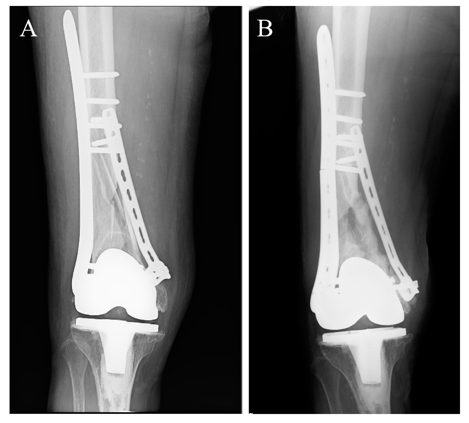
Figure 1. Radiographs of a distal femur fracture indicating non-union at 6 months postoperatively. The patient was a 79-year-old female who underwent a total knee replacement and had a distal femur fracture (A). A noticeable deficiency in callus formation is observed, which may be attributed in part to the rigidity of the bridging implant and the absence of motion. Observations included apparent fracture dislocation, bone atrophy, and, notably, plate failure (B). The corresponding author obtained written informed consent from the patient.
Applsci 14 02259 g001
Figure 2. Radiographs after osteotomy in rabbit tibia. (A) Commercially pure titanium (CP-Ti), 4 weeks after osteotomy; (B) TiNbSn alloy, 4 weeks after osteotomy; (C) CP-Ti, 6 weeks after osteotomy; (D) TiNbSn alloy, 6 weeks after osteotomy. Arrowheads indicate fracture callus. The scale bar indicates 10 mm. Reproduced from ref. [58] with permission [58].
Applsci 14 02259 g002
Figure 3. These images are from a non-demineralized specimen stained with Villanueva bone stain. (A) CP-Ti, (B) TiNbSn alloy at 4× magnification (scale bar indicates 2 mm), (C) CP-Ti, (D) TiNbSn alloy at 20× magnification (scale bar indicates 100 mm). The TiNbSn alloy group exhibited a more robust formation of intramedullary callus with a definite bone structure compared with that of the CP-Ti group. Beneath the plate at the osteotomy site, the TiNbSn alloy group demonstrated a more advanced formation of mature bone, accompanied by an improved orientation of the bone structure. Reproduced from ref. [58] with permission [58].
Applsci 14 02259 g003
Table 1. Experimental models for the development of fracture treatment devices using low Young’s modulus titanium alloys.
MaterialsImplant TypesReferences
1TiNbSn alloyIntramedullary nail[55,56,57]
Plate[11]
Locking plate[58]
2TiNbTaZr alloyIntramedullary nail[49]
Plate[60]
Disclaimer/Publisher’s Note: The statements, opinions and data contained in all publications are solely those of the individual author(s) and contributor(s) and not of MDPI and/or the editor(s). MDPI and/or the editor(s) disclaim responsibility for any injury to people or property resulting from any ideas, methods, instructions or products referred to in the content.