Article

Moderate-Intensity Swimming Alleviates Oxidative Injury in Ischemic Heart

Department of Physiology, Anatomy and Neuroscience, Faculty of Science and Informatics, University of Szeged, 6726 Szeged, Hungary
*
Author to whom correspondence should be addressed.
Appl. Sci. 2024, 14(5), 2073; https://doi.org/10.3390/app14052073
Submission received: 22 January 2024 / Revised: 26 February 2024 / Accepted: 28 February 2024 / Published: 1 March 2024

Abstract

:
The global burden of cardiovascular diseases is indisputable, as it claims nearly 18 million lives a year. In this current study, we aimed to prove that exercise, a cornerstone in cardiovascular disease management, emerges as a powerful tool in the pathology of myocardial ischemia. Male rats were divided into three groups: pre-swimming training + isoproterenol (ISO) treated, isoproterenol-treated, and control-sedentary. Myocardial infarction was induced by the subcutaneous injection of 1.0 mg/kg ISO. After the subsequent rest period, the animals swam for 3 weeks, every day for 25 min. At the end of the experiment, the serum levels of atrial natriuretic peptide (ANP) and B-type natriuretic peptide (BNP), as well as the cardiac concentrations of reactive oxygen species (ROS), catalase (CAT), glutathione peroxidase (GPx), and superoxide dismutase (SOD) were determined. Our results indicate that both cardiac injury biomarkers (ANP, BNP) and ROS levels were significantly lower in swimming rats compared to the sedentary animals. Moreover, the level of enzymatic components of the intracellular antioxidant system, CAT, SOD, and GPx were increased in swimming animals after ISO-induced myocardial infarction. Our findings support the fact that moderate-intensity swimming training can be efficiently used to prevent myocardial infarction-induced ischemic injury, by inhibiting ROS production and strengthening intracellular antioxidant defense.

1. Introduction

Cardiovascular diseases (CVDs) are the most challenging health problems that science is facing in this century. It is pivotal to understand the molecular and cellular nature of CVDs as treatment and prevention are based on this knowledge. A non-exhaustive list of CVDs includes cerebrovascular disease and coronary heart disease, which encompasses myocardial ischemia (MI) [1,2]. Myocardial ischemia, in particular, is associated with, among other things, extensive oxidative damage to the myocardium. Oxidative damage often develops during the so-called reperfusion injury (RI) after MI, which further aggravates the outcome of ischemia [3]. Oxidative stress is one of the major pathological mechanisms in reperfusion injury, causing myocyte death, inflammation, and endothelial dysfunction. It is characterized by the increased levels of reactive oxygen species (ROS) that can ultimately lead to the deterioration of lipids, DNA, and proteins and eventually irreversible cardiomyocyte damage [4]. To eliminate oxidative damage, the evolutionary antioxidant defense system ensures the integrity of the body through enzymatic and non-enzymatic reactions. Catalase (CAT), glutathione peroxidase (GPx), superoxide dismutase (SOD), and other enzymes that scavenge ROS and prevent their buildup have developed into a complex enzymatic antioxidant system in cells [5]. In addition to determining the degree of harm, oxidative stress markers as well as antioxidant enzymes can also be valuable as particular biomarkers in diagnostic and prognostic procedures. Likewise, cardiac biomarkers such as B-type natriuretic peptide (BNP) and atrial natriuretic peptide (ANP) serve as reliable indicators for cardiac insult [6,7]. To tackle cardiovascular-related challenges, we need to be aware of the fact that lifestyle habits are the pillars of cardiovascular health, hence, longevity. As sedentary lifestyle has been identified as a major risk factor for cardiovascular disease, it goes without saying that regular physical activity has long been shown to be an effective way to reduce cardiovascular mortality and morbidity. Exercise is known to improve blood flow to the heart and lower blood pressure and has also been shown to have antioxidant and cholesterol-lowering effects [8]. Exercise, by enhancing the function of antioxidant enzyme systems, can contribute to the improvement in adverse parameters following infarction [9,10]. Several types of exercise, e.g., treadmill training or voluntary wheel running, have been shown to have beneficial effects on the heart after MI, due to the elevation in antioxidant enzymes [11,12,13]. The wide-ranging benefits of physical activity have been known for a long time, but it is important to emphasize that besides being an exceptional treatment option, it has an outstanding potential in the prevention of life-threatening health conditions as well.
This article underscores the pivotal role of ongoing cardiovascular research in advancing our knowledge of myocardial ischemia and emphasizes the significance of exercise as a potent preventive strategy. We hypothesized that exercise protocol before infarction may be effective in alleviating the pathological processes that result from myocardial injury. Consequently, our research aimed to apply swimming as a non-pharmacological prevention method in non-invasively induced MI. The objective of the present study was to clarify how our exercise protocol affects cardiac biomarkers, antioxidants, and oxidative markers in the heart and systemically after MI/RI.

2. Materials and Methods

2.1. Animal Model

Nine-month-old male Harlan–Wistar rats were accommodated in a constant-temperature (25 °C) animal room, at the Institute of Biology, University of Szeged. Husbandry conditions such as regular light/dark cycle, ad libitum water, and standard chow were provided based on international standards (Directive 2010/63/EU).
During the initial phase of the study, the male rats (n = 24) were separated into three different groups: (1) pre-swimming training + ISO treated (PRE + ISO), (2) isoproterenol-treated (ISO), and (3) control-sedentary (CTRL). Myocardial infarction was induced by the subcutaneous injection of 1.0 mg/kg ISO (Sigma Chemicals Co., St. Louis, MO, USA) diluted in 1 mL physiological saline. Following the onset of the infarction, the animals had a three-week-long resting period (Figure 1). The 1 mg/kg dose of ISO was calculated and selected based on one of our previous studies, with the specific aim to cause myocardial injury while minimizing mortality.

2.2. Exercise Protocol

For the implementation of the swimming exercise, a specialized swimming apparatus, designed for the purpose of the experiment, was utilized. The glass apparatus was specifically tailored for swimming rats. It was filled with tap water and maintained at a 30–32 °C temperature and was monitored with a thermometer. In addition, besides the appropriate temperature, the purity of the water was also constantly surveyed. The 70 cm height of the apparatus ensured that the rats’ tails could not reach the bottom of the pool. The animals swam solitarily in separate chambers every morning for 25 min. Once the training was completed, the animals were carefully removed from the apparatus, towel-dried, and returned to their cages. Pre-swimming training meant that the animals swam for 3 weeks before ISO administration. CTRL and ISO animals did not perform the exercise. The swimming training was preceded by one week of adaptation to the water, to minimize the stress response. On the first day, the animals spent 5 min in the water, then with each passing day, the exposure time was increased by 5 min. The rats that showed no interest in swimming were subsequently removed from the experiment. Given that there was no extra weight attached to the animals’ tails, the intensity of the exercise can be defined as moderate. At the end of the experiment, rats were euthanized, and their blood specimens were collected with their excised hearts. Serum and cardiac tissues were used for several biochemical measurements. Every aspect of the experimental protocol was thoroughly reviewed and approved by the Welfare Committee of University of Szeged (XX./1405/2021).

2.3. ELISA Measurements

At the end of the experimental period, the concentration of several biomarkers from both heart tissue and serum was measured using an enzyme-linked immune sorbent assay (ELISA kit, GenAsia Biotech Co., Ltd., Shanghai, China). The molecules of interest included atrial natriuretic peptide, B-type natriuretic peptide, reactive oxygen species, catalase, glutathione peroxidase, and superoxide dismutase.
Tissue samples were homogenized (Ultra-Turrax T8, IKA Werke GmbH & Co., Staufen im Breisgau, Germany) in cold phosphate buffer (pH 7.4) for 2 × 30 s (with a 5 s cooling break in between), centrifuged at 2500 rpm for 20 min, and finally the supernatant was extracted. For the ELISA measurements supernatants (in the case of CAT, SOD, GPx, and ROS) and serum samples (in the case of ANP and BNP) were used. The first step of the ELISA protocol was to create a standard dilution series containing 5 different concentrations. Following this, 100 µL of standard diluent was used as a blank. The standards were added to the wells in pairs, with each well containing 50 µL of solution. The rest of the wells were each filled with 40 µL of sample and 10 µL of antibody labeled with biotin (the standards already contained the antibodies), and 50 µL streptavidin–HRP was added to the standards, as well as the samples, to achieve a final volume of 100 µL/well. After incubation (1 h, 37 °C) and a five-step washing process, 50-50 µL of both chromogen A and B solutions were added to the wells, followed by another incubation period (10 min, 37 °C). Lastly, 50 µL of stop solution was added to achieve color development. Absorbance (OD) was measured at 450 nm within 10 min (Benchmark Microplate reader, Bio-Rad, Hercules, CA, USA). The OD values were used to calculate the concentration of the corresponding samples, which were normalized to the protein content.

2.4. BCA Protein Measurements

To determine the total protein content of samples, bicinchoninic acid (BCA) protein assay kit (ThermoFisher Scientific Inc., Waltham, MA, USA) was used. A series of diluted Bovine Serum Albumin (BSA) standards were used to plot a standard curve. Samples and working reagents were pipetted into each well and then incubated for 20 min at 37 °C. Finally, absorbance was measured at 562 nm with a spectrophotometer, and protein concentrations were expressed as µg/mL.

2.5. Statistical Analysis

Statistical analysis was performed with SigmaPlot 12.0 (Systat Software Inc., San Jose, CA, USA). The normality of data and homogeneity of variances were checked with the Shapiro–Wilk test. In order to analyze the differences between groups, one-way ANOVA with Tukey post-testing was applied, and the Kruskal–Wallis test followed by Dunn’s test was chosen in the case of nonparametric data. Differences were considered significant when the p values were <0.05.

3. Results

3.1. Serum ANP and BNP Concentrations

To evaluate the severity of cardiac damage, serum ANP and BNP levels were determined. ANP and BNP concentrations were found to be lower in CTRL animals in comparison with the ISO group, due to myocardial ischemia. Regarding BNP, this change was found to be significant. In addition, we found a significant decrease in both ANP and BNP levels as a result of preventive swimming training compared to ISO groups. Data are presented in Figure 2a,b.

3.2. Cardiac CAT and SOD Concentrations

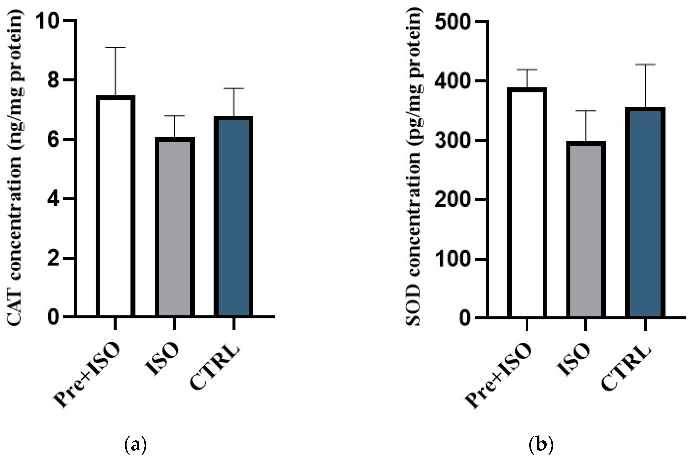
As shown in Figure 3a,b, antioxidant parameters such as CAT and SOD increased in the Pre + ISO group compared to the ISO group; however, these changes could not be considered significant. Three-week-long swimming training had a visible impact on antioxidant enzymes detected in the heart tissue. While the values of the ISO group were diminished as a result of myocardial damage; preventive exercise protocol before infarction was able to mitigate these adverse changes.

3.3. Cardiac GPx Concentrations

To complement the antioxidant parameters, cardiac GPx concentration was determined. ISO-resulted cardiac injury caused a significant decrease in GPx compared to CTRL animals; however, as an effect of moderate swimming training, GPx values were elevated despite ISO treatment. Thus, reduced antioxidant values precipitated by myocardial ischemia were compensated by 3 weeks of preventive exercise. Data are presented in Figure 4.

3.4. Cardiac ROS Concentrations

Assuming the cardiovascular protective effects of exercise, total ROS concentration was measured in heart tissues as seen in Figure 5. ISO-treated rats possessed significantly higher ROS levels in comparison with the CTRL group, whereas 3 weeks of the swimming protocol prior to infarction resulted in a significant mitigation of the elevated ROS concentrations compared to the sedentary ISO group.

4. Discussion

Our experimental study was aimed at examining the impact of pre-emptive moderate physical exercise, in the form of swimming, on cardiac parameters after myocardial infarction from the perspective of changes in antioxidant capacity and oxidative stress. The positive effects of physical exercise as a preconditioning method in myocardial ischemia and reperfusion injury models are well documented. Running in particular had been shown to improve post-ischemic cardiac output [13], and in one of our previous studies, we found that it also helps in reducing oxidative stress, through increased cardiac heme oxygenase and GSH activity as well as reduced myeloperoxidase activity [12]. Moreover, according to Glisic et al. [14], swimming before the onset of ischemia helps regulate heart rate and contractility after reperfusion. In addition, short-duration swimming as a part of a post-infarction treatment reduces mortality, improves left ventricular ejection fraction and fractional shortening, reduces interstitial fibrosis and myocardial apoptosis, improves mitochondrial size homogeneity, reduces ROS content, and inhibits SOD acetylation, thus increasing its enzymatic activity [15]. In our current study, we managed to provide further evidence that preconditioning via moderate-intensity swimming is an efficient method to reduce the severity of cardiac injury caused by myocardial ischemia, through the stimulation of the antioxidant system and the reduction in overall ROS production.
For the assessment of cardiac damage after ISO treatment, we decided to use serum atrial and brain natriuretic peptide concentrations as indicators for ischemic injury. Elevated BNP and N-terminal prohormone of BNP (NT-proBNP) levels in particular have long been considered, alongside cardiac troponin, to be one of the most reliable biomarkers in the clinical diagnostic process for heart failure associated with systolic and diastolic dysfunction, left ventricular hypertrophy, valvular heart disease, and ischemia [7]. Furthermore, animal studies focused on post-myocardial infarction also frequently use elevated serum BNP concentration as evidence for the presence of cardiac damage [16]. Similarly, ANP, which is synthesized, stored, and secreted by atrial cardiac cells, is also considered an important factor in establishing the severity of cardiac dysfunctions and heart injuries. Myocardial wall stretching in the atrium, a common pathological feature of acute and chronic congestive heart failure, induces the secretion of pro-ANP, which is converted into biologically active α-ANP by the serine protease corin, thus rapidly increasing the serum ANP concentration [17]. In animal studies conducted on rats with myocardial infarction-induced heart failure, both ANP and BNP expressions were repeatedly shown to be significantly increased [18,19]. Our results regarding the connection between ANP and BNP levels and myocardial infarction are in line with the consensus found in the associated literature. We found that both ANP and BNP serum levels were significantly increased after ISO treatment, suggesting post-ischemic cardiac damage; however, in rats preconditioned with moderate-intensity swimming training, these increases in the concentrations of cardiac injury biomarkers are not present.
Regarding the oxidative stress theory, the idea that oxidative damage and the alteration in the intracellular antioxidant system have a significant role in the development of pathologic changes followed by myocardial infarction and subsequent ischemia is a well-accepted one. Oxidative stress is most often defined as an imbalance between intracellular ROS production and the capacity to neutralize ROS, carried out by a system that includes, amongst others, CAT, SOD, GSH, and GPx. During an ischemic period, the efficiency of this system is drastically decreased, leading to cell death and cardiac dysfunction, and following reperfusion, the suddenly increased oxygen supply leads to the high levels of oxygen free radicals, causing lipid peroxidation, protein degradation, and DNA damage, further injuring the cardiac tissue [20]. In a study conducted on albino rats, Rao and Viswanath observed that ischemia–reperfusion-induced myocardial infarction is associated with significantly increased concentrations of cardiac injury and oxidative stress biomarkers (lactate dehydrogenase, creatine kinase, creatine kinase isoenzyme, lipid peroxidase) and reduced antioxidant capacity due to the reduced levels of CAT, SOD, and GSH [21]. The findings of Rostamzadeh et al. [22] present evidence that ISO-induced myocardial injury causes the elevated levels of malonaldehyde (MDA) and the decreased concentrations of SOD and GPx. The data regarding the effects of physical exercise and swimming in particular on maintaining cardiac antioxidant capacity after a myocardial infarction-induced ischemic injury are still fairly inconclusive. From one point of view, several studies focused on treadmill exercise found that this type of physical activity has a positive impact on maintaining the efficiency of the enzymatic antioxidant system through the improved levels of CAT, SOD, and GPx. On the other hand, some of these findings offer a positive but not significant correlation between exercise and antioxidant enzyme levels, and the significantly increased protein levels are usually restricted to mitochondria and cannot be found in the cytosol [23]. Endurance and resistance training have been shown to significantly increase GPx content and reduce myeloperoxidase and MDA activity, while having no significant impact on CAT concentration [24]. According to Venditti and Meo [25], there is a significant increase in GPx activity in rats who went through a swimming training protocol, compared to sedentary animals; however, the study did not focus on cardiac injury, only on adaptive changes between trained and untrained animals. In contrast, Ascensão et al. [26] studied the effects of preconditioning through swimming in mice with doxorubicin-induced cardiac oxidative damage but found no significant increase in CAT, SOD, or GPx levels that could be associated with the training.
In this regard, to the best of our knowledge, our present study is the first one to demonstrate that moderate-intensity swimming training can be efficiently used to prevent myocardial infarction-induced ischemic injury, by inhibiting ROS production and strengthening intracellular antioxidant defense. Our results indicate that both cardiac injury biomarkers (ANP and BNP) and ROS levels were significantly lower in rats who partook in swimming training, compared to the sedentary animals. Moreover, the enzymatic components of the intracellular antioxidant system also showed increased resilience in trained animals after ISO-induced myocardial infarction. GPx, in particular, presented significantly increased concentrations in the swimming animals. Although SOD and CAT levels in this group were also slightly elevated, these changes were not considered statistically significant. Exercise triggers a complex adaptive response in the body, enhancing endogenous antioxidant defenses and improving the overall redox balance. This adaptive response involves the upregulation of enzymatic antioxidants such as SOD, CAT, and GPx, contributing to a more robust defense against oxidative stress [27]. It is no coincidence that exercise proved to be more efficient against oxidative stress in comparison to antioxidants obtained from dietary sources or supplements for the simple reason that exercise may interact synergistically with endogenous antioxidant systems, creating a more effective defense against oxidative damage. This synergy is thought to be superior to the isolated use of exogenous antioxidants, as the body’s adaptive responses are engaged in a comprehensive and coordinated manner during physical activity [28].
Taking everything into consideration, we conclude that physical exercise in the form of moderate-intensity swimming has the potential to be considered as a part of cardiac conditioning training, with the specific aim of preventing myocardial infarction-induced heart injury, mediated by oxidative stress and dysfunctional antioxidant defense. While excessive ROS can lead to oxidative stress and damage cellular structures, balanced ROS levels play important roles in various physiological processes. Regular, moderate-intensity exercise seems to induce adaptive responses that exploit the benefits of ROS (e.g., triggering adaptive responses that enhance mitochondrial function and improve the efficiency of energy production), while minimizing the risk of oxidative damage. That being said, our findings represent only a small fraction of the currently available and often contradictory data associated with this topic. We suggest that further research should be conducted to elucidate the biochemical basis of the link between ischemia and oxidative cardiac damage, thus providing an opportunity to develop effective preventive and treatment methods.

5. Limitations

While the current study provides valuable insights, the variability in individual responses and the potential confounding factors necessitate further investigation to validate the broader applicability of this approach. In summary, while it is unquestionable that the potential benefits of swimming exercises in cardiac conditioning are promising, it is imperative to acknowledge the need for further research and highlight the individual variations among patients. A cautious and personalized approach, considering the limitations and potential differences in patient responses, will contribute to the development of more effective and tailored cardiac rehabilitation strategies.

Author Contributions

Conceptualization, D.B. and R.S.; methodology, R.S. and A.H.; investigation, M.V. and V.K.; writing—original draft preparation, D.B.; writing—review and editing, S.T. and N.A.; visualization, A.N.; supervision, R.S.; project administration, C.V. All authors have read and agreed to the published version of the manuscript.

Funding

This research received no external funding.

Institutional Review Board Statement

The animal study protocol was approved by the Animal Welfare Committee of University of Szeged (XX./1405/2021).

Informed Consent Statement

Not applicable.

Data Availability Statement

All data used to support the findings of this study are included within the article.

Conflicts of Interest

The authors declare no conflicts of interest.

References

  1. Edgardo Olvera Lopez, B.D.B.; Jan, A. Cardiovascular Disease; StatPearls Publishing: Treasure Island, FL, USA, 2022. [Google Scholar]
  2. Flora, G.D.; Nayak, M.K. A Brief Review of Cardiovascular Diseases, Associated Risk Factors and Current Treatment Regimes. Curr. Pharm. Des. 2019, 25, 4063–4084. [Google Scholar] [CrossRef] [PubMed]
  3. Kibel, A.; Lukinac, A.M.; Dambic, V.; Juric, I.; Selthofer-Relatic, K. Oxidative Stress in Ischemic Heart Disease. Oxid. Med. Cell. Longev. 2020, 2020, 6627144. [Google Scholar] [CrossRef] [PubMed]
  4. Kurian, G.A.; Rajagopal, R.; Vedantham, S.; Rajesh, M. The Role of Oxidative Stress in Myocardial Ischemia and Reperfusion Injury and Remodeling: Revisited. Oxid. Med. Cell Longev. 2016, 2016, 1656450. [Google Scholar] [CrossRef] [PubMed]
  5. Santos, C.X.; Raza, S.; Shah, A.M. Redox signaling in the cardiomyocyte: From physiology to failure. Int. J. Biochem. Cell Biol. 2016, 74, 145–151. [Google Scholar] [CrossRef] [PubMed]
  6. Suzuki, S.; Yoshimura, M.; Nakayama, M.; Mizuno, Y.; Harada, E.; Ito, T.; Nakamura, S.; Abe, K.; Yamamuro, M.; Sakamoto, T.; et al. Plasma level of B-type natriuretic peptide as a prognostic marker after acute myocardial infarction: A long-term follow-up analysis. Circulation 2004, 110, 1387–1391. [Google Scholar] [CrossRef] [PubMed]
  7. Wang, X.Y.; Zhang, F.; Zhang, C.; Zheng, L.R.; Yang, J. The Biomarkers for Acute Myocardial Infarction and Heart Failure. BioMed Res. Int. 2020, 2020, 2018035. [Google Scholar] [CrossRef] [PubMed]
  8. Borzsei, D.; Szabo, R.; Hoffmann, A.; Harmath, A.; Sebestyen, J.; Osman, J.; Juhasz, B.; Priksz, D.; Varga, C.; Posa, A. Multiple Applications of Different Exercise Modalities with Rodents. Oxid. Med. Cell. Longev. 2021, 2021, 3898710. [Google Scholar] [CrossRef]
  9. Elokda, A.S.; Nielsen, D.H. Effects of exercise training on the glutathione antioxidant system. Eur. J. Cardiovasc. Prev. Rehabil. 2007, 14, 630–637. [Google Scholar] [CrossRef]
  10. Yan, Z.; Spaulding, H.R. Extracellular superoxide dismutase, a molecular transducer of health benefits of exercise. Redox Biol. 2020, 32, 101508. [Google Scholar] [CrossRef] [PubMed]
  11. Gul, M.; Demircan, B.; Taysi, S.; Oztasan, N.; Gumustekin, K.; Siktar, E.; Polat, M.F.; Akar, S.; Akcay, F.; Dane, S. Effects of endurance training and acute exhaustive exercise on antioxidant defense mechanisms in rat heart. Comp. Biochem. Physiol. A Mol. Integr. Physiol. 2006, 143, 239–245. [Google Scholar] [CrossRef] [PubMed]
  12. Szabo, R.; Borzsei, D.; Karacsonyi, Z.; Gesztelyi, R.; Nemes, K.; Berko, A.M.; Veszelka, M.; Torok, S.; Kupai, K.; Varga, C.; et al. Postconditioning-like effect of exercis: New paradigm in experimental menopause. Am. J. Physiol. Heart Circ. Physiol. 2019, 316, H400–H407. [Google Scholar] [CrossRef] [PubMed]
  13. Bowles, D.K.; Farrar, R.P.; Starnes, J.W. Exercise training improves cardiac function after ischemia in the isolated, working rat heart. Am. J. Physiol. 1992, 263, H804–H809. [Google Scholar] [CrossRef] [PubMed]
  14. Glisic, M.; Nikolic Turnic, T.; Zivkovic, V.; Pindovic, B.; Chichkova, N.V.; Fisenko, V.P.; Nikolic, M.; Stijak, L.; Yurievna, L.E.; Veselinovic, M.; et al. The Enhanced Effects of Swimming and Running Preconditioning in an Experimental Model of Myocardial Ischemia/Reperfusion Injury. Medicina 2023, 59, 1995. [Google Scholar] [CrossRef] [PubMed]
  15. Zhao, D.; Sun, Y.; Tan, Y.; Zhang, Z.; Hou, Z.; Gao, C.; Feng, P.; Zhang, X.; Yi, W.; Gao, F. Short-Duration Swimming Exercise after Myocardial Infarction Attenuates Cardiac Dysfunction and Regulates Mitochondrial Quality Control in Aged Mice. Oxid. Med. Cell. Longev. 2018, 2018, 4079041. [Google Scholar] [CrossRef]
  16. Song, X.J.; Yang, C.Y.; Liu, B.; Wei, Q.; Korkor, M.T.; Liu, J.Y.; Yang, P. Atorvastatin inhibits myocardial cell apoptosis in a rat model with post-myocardial infarction heart failure by downregulating ER stress response. Int. J. Med. Sci. 2011, 8, 564–572. [Google Scholar] [CrossRef] [PubMed]
  17. Lyngbakken, M.N.; Myhre, P.L.; Rosjo, H.; Omland, T. Novel biomarkers of cardiovascular disease: Applications in clinical practice. Crit. Rev. Clin. Lab. Sci. 2019, 56, 33–60. [Google Scholar] [CrossRef]
  18. He, J.; Lu, Y.; Song, X.; Gong, X.; Li, Y. Inhibition of microRNA-146a attenuated heart failure in myocardial infarction rats. Biosci. Rep. 2019, 39, BSR20191732. [Google Scholar] [CrossRef]
  19. Horakova, D.; Azeem, K.; Benesova, R.; Pastucha, D.; Horak, V.; Dumbrovska, L.; Martinek, A.; Novotny, D.; Svagera, Z.; Hobzova, M.; et al. Total and High Molecular Weight Adiponectin Levels and Prediction of Cardiovascular Risk in Diabetic Patients. Int. J. Endocrinol. 2015, 2015, 545068. [Google Scholar] [CrossRef]
  20. Xiang, M.; Lu, Y.; Xin, L.; Gao, J.; Shang, C.; Jiang, Z.; Lin, H.; Fang, X.; Qu, Y.; Wang, Y.; et al. Role of Oxidative Stress in Reperfusion following Myocardial Ischemia and Its Treatments. Oxid. Med. Cell. Longev. 2021, 2021, 6614009. [Google Scholar] [CrossRef]
  21. Rao, P.R.; Viswanath, R.K. Cardioprotective activity of silymarin in ischemia-reperfusion-induced myocardial infarction in albino rats. Exp. Clin. Cardiol. 2007, 12, 179–187. [Google Scholar] [PubMed]
  22. Rostamzadeh, F.; Najafipour, H.; Aminizadeh, S.; Jafari, E. Therapeutic effects of the combination of moderate-intensity endurance training and MitoQ supplementation in rats with isoproterenol-induced myocardial injury: The role of mitochondrial fusion, fission, and mitophagy. Biomed. Pharmacother. 2024, 170, 116020. [Google Scholar] [CrossRef] [PubMed]
  23. Powers, S.K.; Sollanek, K.J.; Wiggs, M.P.; Demirel, H.A.; Smuder, A.J. Exercise-induced improvements in myocardial antioxidant capacity: The antioxidant players and cardioprotection. Free Radic. Res. 2014, 48, 43–51. [Google Scholar] [CrossRef] [PubMed]
  24. Mohammadkhani, R.; Ranjbar, K.; Salehi, I.; Komaki, A.; Zarrinkalam, E.; Amiri, P. Comparison of the preconditioning effect of different exercise training modalities on myocardial ischemia-reperfusion injury. PLoS ONE 2023, 18, e0295169. [Google Scholar] [CrossRef] [PubMed]
  25. Venditti, P.; Di Meo, S. Antioxidants, tissue damage, and endurance in trained and untrained young male rats. Arch. Biochem. Biophys. 1996, 331, 63–68. [Google Scholar] [CrossRef] [PubMed]
  26. Ascensao, A.; Magalhaes, J.; Soares, J.; Ferreira, R.; Neuparth, M.; Marques, F.; Oliveira, J.; Duarte, J. Endurance training attenuates doxorubicin-induced cardiac oxidative damage in mice. Int. J. Cardiol. 2005, 100, 451–460. [Google Scholar] [CrossRef] [PubMed]
  27. Radak, Z.; Taylor, A.W.; Ohno, H.; Goto, S. Adaptation to exercise-induced oxidative stress: From muscle to brain. Exerc. Immunol. Rev. 2001, 7, 90–107. [Google Scholar]
  28. Ristow, M.; Zarse, K.; Oberbach, A.; Kloting, N.; Birringer, M.; Kiehntopf, M.; Stumvoll, M.; Kahn, C.R.; Bluher, M. Antioxidants prevent health-promoting effects of physical exercise in humans. Proc. Natl. Acad. Sci. USA 2009, 106, 8665–8670. [Google Scholar] [CrossRef]
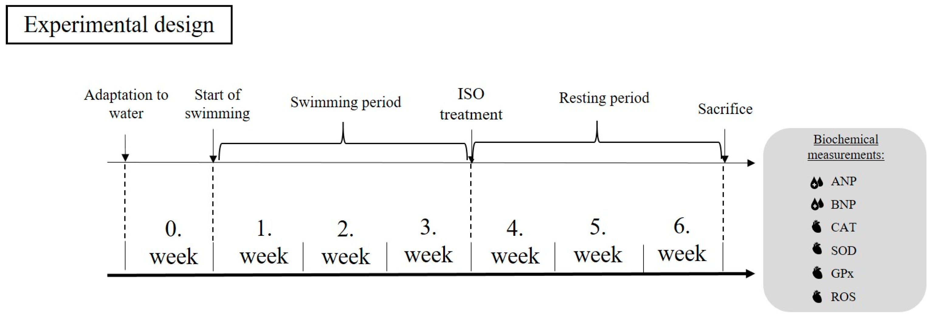
Figure 1. The experimental protocol of the study. ANP = atrial natriuretic peptide; BNP = brain natriuretic peptide; CAT = catalase; GPx = glutathione peroxidase; ISO = isoproterenol; ROS = reactive oxygen species; and SOD = superoxide dismutase.
Applsci 14 02073 g001
Figure 2. (a) The effects of swimming training on cardiac ANP concentration (ANP; expressed as ng/L). Results shown as means ± S.E.M. n = 5–7. *: p < 0.05 statistical comparison between ISO and Pre + ISO groups. (b) The effects of swimming training on cardiac BNP concentration (BNP; expressed as ng/L). Result shown as means ± S.E.M. n = 4–5. * p < 0.05 statistical comparison between ISO and Pre + ISO groups. #: p < 0.05 statistical comparison between ISO and CTRL groups. Pre + ISO = pre-swimming training + ISO treated; ISO = isoproterenol-treated; CTRL = control; ANP = atrial natriuretic peptide; and BNP = B-type natriuretic peptide.
Applsci 14 02073 g002
Figure 3. (a) The effects of swimming training on cardiac CAT concentration (CAT; expressed as ng/mg protein). Results are shown as means ± S.E.M. n = 6–7. (b) The effects of swimming training on cardiac SOD concentration (SOD; expressed as pg/mg protein). Results are shown as means ± S.E.M. n = 5. Pre + ISO = pre-swimming training + ISO treated; ISO = isoproterenol-treated; CTRL = control; CAT = catalase; and SOD = superoxide dismutase.
Applsci 14 02073 g003
Figure 4. The effects of swimming training on cardiac GPx concentration (GPx; expressed as U/mg protein). Results shown as means ± S.E.M. n = 4–6. *: p < 0.05 statistical comparison between ISO and Pre + ISO groups. #: p < 0.05 statistical comparison between ISO and CTRL groups. Pre + ISO = pre-swimming training + ISO treated; ISO = isoproterenol-treated; CTRL = control; and GPx = glutathione peroxidase.
Applsci 14 02073 g004
Figure 5. The effects of swimming training on cardiac ROS concentration (ROS; expressed as U/mg protein). Results shown as means ± S.E.M. n = 5–6. *: p < 0.05 statistical comparison between ISO and Pre + ISO groups. #: p < 0.05 statistical comparison between ISO and CTRL groups. Pre + ISO = pre-swimming training + ISO treated; ISO = isoproterenol-treated; CTRL = control; and ROS = reactive oxygen species.
Applsci 14 02073 g005
Disclaimer/Publisher’s Note: The statements, opinions and data contained in all publications are solely those of the individual author(s) and contributor(s) and not of MDPI and/or the editor(s). MDPI and/or the editor(s) disclaim responsibility for any injury to people or property resulting from any ideas, methods, instructions or products referred to in the content.