Article

Exploring the Interplay of Uric Acid and Advanced Oxidation Protein Products Following Myocardial Infarction

by
Joanna Sikora
1,*,
Tomasz Wybranowski
2,
Aleksandra Karczmarska-Wódzka
1,
Przemysław Sobczak
3,
Stefan Kruszewski
2 and
Zbigniew Włodarczyk
4
1
Research and Education Unit for Experimental Biotechnology, Department of Transplantology and General Surgery, Faculty of Medicine, Collegium Medicum in Bydgoszcz, Nicolaus Copernicus University in Toruń, 85-094 Bydgoszcz, Poland
2
Department of Biophysics, Faculty of Pharmacy, Collegium Medicum in Bydgoszcz, Nicolaus Copernicus University, 85-094 Bydgoszcz, Poland
3
Dr Jan Biziel’s University Hospital, Ujejskiego Street 75, 85-168 Bydgoszcz, Poland
4
Department of Transplantology and General Surgery, Faculty of Medicine, Collegium Medicum, Nicolaus Copernicus University, 85-094 Bydgoszcz, Poland
*
Author to whom correspondence should be addressed.
Appl. Sci. 2024, 14(5), 1983; https://doi.org/10.3390/app14051983
Submission received: 21 January 2024 / Revised: 23 February 2024 / Accepted: 26 February 2024 / Published: 28 February 2024

Abstract

:
Recent studies have underscored the potential elevation of Advanced Oxidation Protein Products (AOPP) and uric acid following myocardial infarction, suggesting their involvement in the development and progression of coronary artery disease and potentially influencing patient outcomes. This study focuses explicitly on examining uric acid and AOPP in the same patients to address the research gap in these biomarkers’ interplay. Recognizing the dual character of uric acid as both an antioxidant and a pro-oxidant, this study delves into its complex biological implications. An analysis was conducted on 40 patients who had experienced myocardial infarction. AOPP levels were quantified using absorbance at 340 nm. Results demonstrated significantly increased AOPP levels in myocardial infarction patients compared to healthy controls, especially in those with high serum uric acid. The serum uric acid and AOPP relationship exhibits a J-shaped curve, indicating a complex, multifactorial interaction. These findings offer new insights into the intricate relationship between serum uric acid and AOPP in myocardial infarction patients, underscoring the significance of these biomarkers in enhancing our understanding of clinical outcomes and informing targeted management strategies for coronary artery disease.

1. Introduction

Oxidative stress (OS) is defined as a disruption of the equilibrium between the generation of free radicals and the activity of antioxidant systems [1,2]. In a pathological state, many activities of the organism are disturbed, which influences the level of antioxidants, making it difficult to detect the causes of OS. A lot of research indicates elevated free radical production in many diseases [3,4,5]. Advanced oxidation protein products (AOPP) are a marker of oxidative stress first detected in the plasma of chronic uremic patients [3]. AOPP originates from reactions between free radicals, chlorinated oxidants, and plasma proteins [6], mainly albumin [7]. They can either escalate the OS or reactive oxygen species (ROS) generation [7,8,9]. AOPP is also associated with coronary artery disease (CAD) [10], atherosclerosis [11], and comorbidities, such as diabetes mellitus (DM), hypertension, metabolic syndrome [12], and lipid disorders (hypercholesterolemia and hypertriglyceridemia) [13]. The relationship between the level of AOPP and monocyte activation markers suggests that the oxidized proteins can contribute to the inflammatory process. An increased inflammatory response also causes an increased production of free radicals, which enhance OS and stimulate the production of AOPP. However, AOPP may be a valuable tool for measuring protein oxidative damage and a general level of OS.
Uric acid is a commonly known antioxidant detectable in human serum plasma [14,15]; however, some conditions can provoke and escalate OS [16]. This organic compound is the end product of purine metabolism [17], mainly regulated by xanthine oxidoreductase, converting hypoxanthine to xanthine and xanthine to uric acid. Many factors could increase serum uric acid levels: diet (alcohol consumption, meat, high fructose intake) [18], uncommon genetic disorders connected with enzymatic degradation pathways [16,18], metabolic syndrome, DM, hypertension, women after menopause, race, renal diseases, inflammatory process, diseases with high cell turnover, some drugs [19], obesity, and ischemia [16,17,18]. Evidence shows that the overproduction of uric acid by xanthine oxidase produces excessive free radicals, such as ROS [20,21]. The main ROS generators are NADPH oxidase, xanthine oxidase, mitochondrial enzymes, myeloperoxidase (MPO), lipoxygenase, and uncoupled nitric oxide (NO) synthase [22]. Myocardial infarction (MI), as well as post-infarction, is one of the most urgent situations in the health system, and needs follow-up due to the possibility of developing heart failure (HF) [23]. In this context, seeking therapeutic markers and links that can prevent or help in the control and management of the patient is of great importance. The pathophysiological interaction between acute coronary syndromes (ACS), including MI, and the appearance of inflammatory cells that produce oxygen radicals, proteases, and proinflammatory cytokines leading to endothelial damage and plaque rupture was proved [24,25]. The increase in OS likely reflects the effect of the accumulation of inflammatory cells in unstable atherosclerotic plaques. Moreover, free radicals are generated due to acidosis, decreased blood flow to heart muscle, and sodium and calcium pump dysfunction. Thus, the status of antioxidant defense systems in patients after MI could provide potentially relevant prognostic information [26]. This, however, needs to be verified. Still, there are no tests to measure OS in individual patients, nor are there effective methods to counteract these processes. The correct interpretation of markers of OS may be the basis for assessing prognosis and making appropriate treatment decisions.
This study aimed to evaluate the interplay of uric acid and AOPP in patients after MI, and verify the correlation between uric acid and AOPP as the markers of OS in patients with MI.

2. Materials and Methods

2.1. Study Design and Population
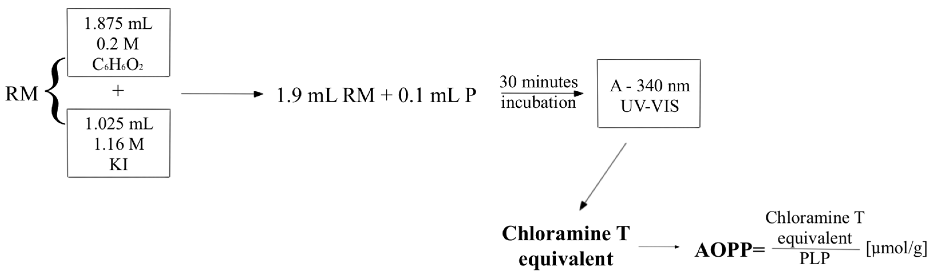
The study group consisted of 40 patients diagnosed with myocardial infarction hospitalized at the Department of Cardiology and Internal Medicine at University Hospital No. 1 in Bydgoszcz. Upon admission, all patients underwent a comprehensive series of basic laboratory tests to assess the progression of inflammation, cardiac function, lipid profiles, and protein levels, following the hospital’s established standard procedures. To ensure the accuracy and relevance of our findings, blood samples were collected explicitly within the first 24 h post-admission, a critical period for assessing the acute phase of MI and its impact on biochemical markers. This timing was meticulously chosen to capture the immediate physiological responses and biochemical changes triggered by the event. During this pivotal phase, a portion of the blood samples were designated for the analysis of AOPP and uric acid, allowing for a comprehensive evaluation of oxidative stress and its correlation with the pathophysiological processes of MI. Patients with any stage of chronic kidney disease (CKD) were excluded from the study to mitigate potential confounding effects on serum uric acid and AOPP levels, as CKD significantly influences the body’s ability to process and eliminate these compounds. Similarly, individuals presenting with abnormal creatinine levels were also excluded, given that creatinine is a crucial marker of renal function, and its abnormal levels could indicate underlying renal impairment not previously classified as CKD. This exclusion criterion was essential for ensuring the homogeneity of the study population and the validity of the association between uric acid, AOPP levels, and myocardial infarction outcomes. The complete list of inclusion/exclusion criteria is presented in Table 1.
The control group consisted of 30 healthy volunteers who routinely donated blood at the local blood donation center (Bydgoszcz, Poland). A licensed physician rigorously assessed the determination of a participant’s health status. Based on these comprehensive health evaluations, the decision to forward a patient’s blood sample for inclusion in our study was made solely by the assessing physician. We established a control group without coexisting diseases to examine the relationship between uric acid and AOPP. This decision aimed to minimize confounding variables, such as age-related changes and unrelated health conditions that could influence uric acid and AOPP levels, potentially masking the specific interaction we sought to examine. The protocol of the study was approved by the Ethics Committee of the Nicolaus Copernicus University in Toruń, Collegium Medicum in Bydgoszcz (approval number KB 406/2010). Each patient provided written informed consent to participate in the study before recruitment.

2.2. Methods

Blood samples were added to standard sterile polystyrene tubes containing ethylenediaminetetraacetic acid (EDTA) and centrifuged at 4000 rpm for 5 min. Plasma was stored at a temperature of −20 °C until AOPP was measured. All samples were assayed on the same day. A spectrophotometer UV-Vis JASCO V-550 was used to measure absorbance. The concentration of AOPP was determined by measuring absorbance at 340 nm according to the modified method described for the first time by Witko-Sarsat [3]. This method has been described previously [27]. Figure 1 shows the method of determining the AOPP concentration. Briefly, the reactant mixture for the AOPP assay contains 1.875 mL of 0.2 M citric acid and 0.025 mL of 1.16 M potassium iodide. Then, 1.9 mL of this mixture was added to 100 µL of the test sample, and after 30 min, the absorbance was recorded. The results were expressed as chloramine T equivalents and divided by the level of proteins. Compared to the original method, citric acid was used instead of acetic acid. This modified method is characterized by excellent stability over time.
The Benecheck PLUS Multi Monitoring Device measured serum uric acid (SUA) for both the healthy group and post-myocardial infarction patients. Our study aimed to investigate the potential relationship between uric acid and AOPP among patients experiencing MI. Grounded in the hypothesis that varying serum uric acid levels could differentially correlate with oxidative stress, we strategically categorized MI patients based on their serum uric acid concentrations. To conduct a nuanced analysis, patients were divided into two groups: those with serum uric acid levels greater than 8 mg/dL and those with levels less than 8 mg/dL. This categorization criterion was determined by reviewing the existing literature and our preliminary observations, which suggested a significant shift in the relationship between uric acid levels and AOPP beyond the 8 mg/dL threshold. The threshold of 8 mg/dL was specifically chosen as it is generally recognized as the upper limit of normal, with values exceeding this point considered indicative of hyperuricemia, a condition associated with increased risk for various pathological states, including cardiovascular diseases.

2.3. Statistical Analysis

In the study, the normality of data was initially assessed using the Shapiro–Wilk test. For variables that demonstrated a normal distribution (indicated by Shapiro–Wilk p > 0.05), students’ t-tests were applied to compare means. Conversely, the Wilcoxon rank sum test was utilized for median comparisons for variables not adhering to a normal distribution (Shapiro–Wilk p ≤ 0.05). Additionally, regression analysis was conducted, employing either the Pearson correlation coefficient or Spearman’s rank correlation, depending on the normality of the data.

3. Results

3.1. Population Characteristics
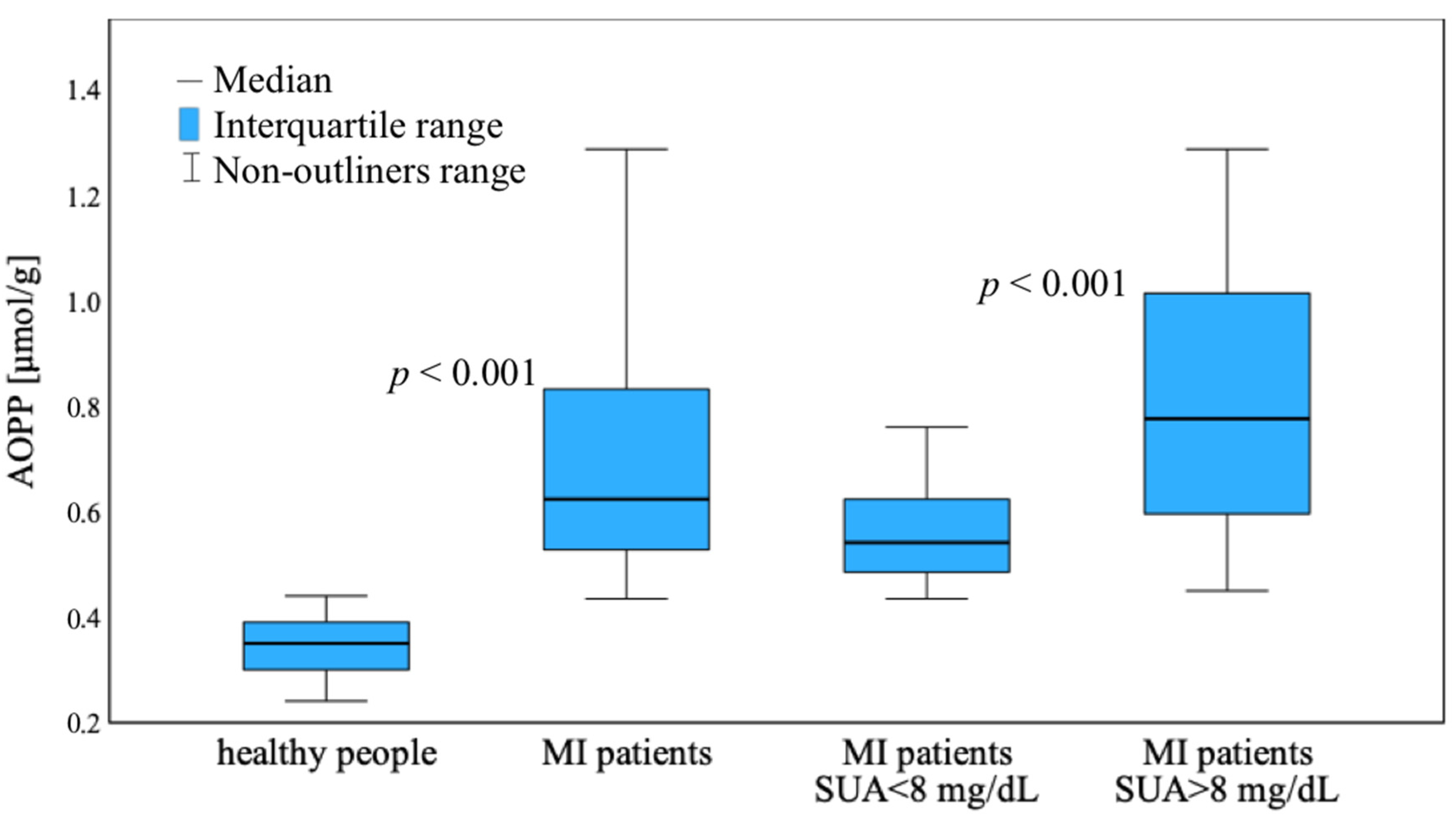
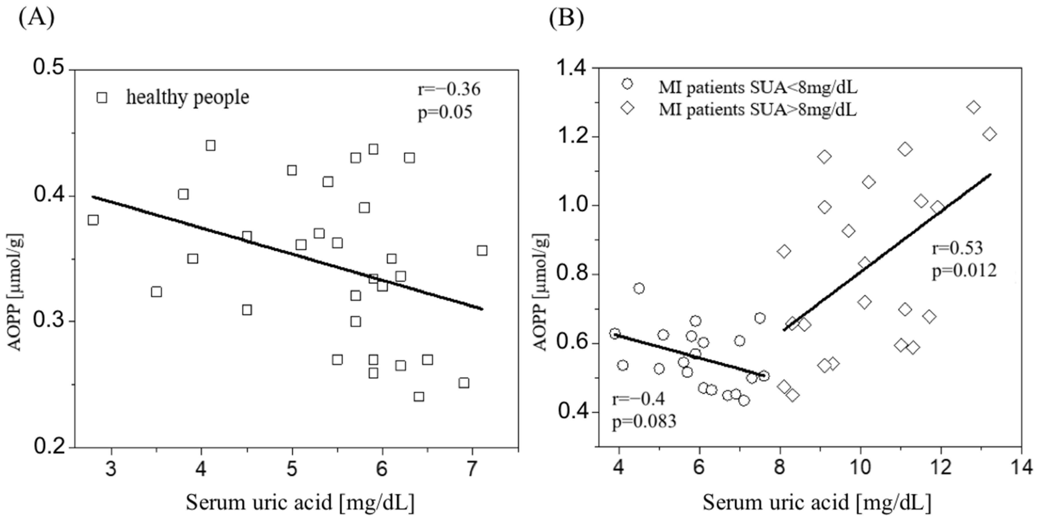
The place of recruitment of patients was the Department of Cardiology and Internal Medicine, Antoni Jurasz University Hospital No. 1 in Bydgoszcz, Poland. Baseline characteristics are presented in the table below (Table 2). There were no significant differences in blood test results between MI patients with elevated and normal uric acid levels. It can be seen that parameters related to myocardial damage, such as troponin I (5.04 ± 7.505) and CK-MB (28.23 ± 20.917), are elevated in these patients. No correlation was observed between uric acid levels, AOPP and CRP, or troponins and CK-MB in patients with MI. Still, patients in the acute phase of infarction were not recruited.
The control group comprised 30 donors from Bydgoszcz Regional Blood Donation and Blood Treatment Centre. The baseline characteristics of the control group are as follows: age 45.33 ± 6.348, with 63% being male and 37% being female.

3.2. AOPP Results

Figure 2 shows the values of AOPP for healthy people and patients with MI. Patients with MI were divided into groups because of the concentration of uric acid. Blood samples from people after MI have higher levels of AOPP in comparison to healthy people (0.696 ± 0.187 vs. 0.342 ± 0.048 µmol/g, p < 0.001), especially for samples with high concentrations of serum uric acid (0.822 ± 0.223 µmol/g).
The values of AOPP in dependence on serum uric acid for healthy subjects and patients with MI are shown in Figure 3A,B, respectively. Considering the normal distribution of the data, Pearson’s correlation was employed for regression analysis to investigate these relationships further. A weak negative correlation between AOPP and uric acid was found in healthy people (r = −0.36, p = 0.05) and patients with MI with uric acid levels less than 8 mg/dL (r = −0.40, p = 0.083). For patients with MI with higher values of serum uric acid (>8 mg/dL), a positive correlation was observed (r = 0.53, p = 0.012).
Figure 3 also indicates that the relationship between AOPP and uric acid shows a “J-shaped curve” in all uric acid ranges instead of a weak negative correlation for smaller uric acid values and a strong positive correlation for higher values. The values of AOPP in patients after MI were significantly higher than in healthy subjects and were associated with the concentration of uric acid. A J-curve showing the relationship between uric acid and AOPP obtained for people after an MI was observed. This observation may have important clinical implications and will allow us to determine the valid value of uric acid.

4. Discussion

Our study elucidates the connection between uric acid levels and AOPP in patients with MI, revealing significant insights into the oxidative stress mechanisms underlying cardiovascular diseases. Notably, we observed that AOPP levels are elevated in MI patients compared to healthy subjects, with a pronounced increase in individuals presenting high concentrations of serum uric acid. However, the crux of our investigation extends beyond comparing AOPP levels between healthy individuals and MI patients; it delves into the intricate dynamics of the relationship between uric acid and AOPP. To precisely examine the relationship between uric acid and AOPP, we established a control group of younger individuals without coexisting diseases. This strategic decision aimed to minimize confounding variables, such as age-related changes and unrelated health conditions that could influence uric acid and AOPP levels, potentially masking the specific interaction we sought to examine. Studying the relationship between uric acid and AOPP in healthy individuals is essential, as it could provide valuable data that will aid in interpreting results post-MI. This methodological choice was informed by the need to establish a clear baseline for the SUA-AOPP interaction in a relatively homogeneous group.
While it is well acknowledged that a subset of post-MI patients presents elevated uric acid levels and AOPP due to the influence of coexisting diseases and other factors, these increments are generally more pronounced as a direct result of the MI itself [28,29,30]. Nonetheless, we recognize the need for further investigation to substantiate this presumption and differentiate the specific impacts of MI on uric acid and AOPP levels from those attributed to coexisting conditions. To ensure the observed changes in uric acid were primarily associated with MI rather than other health conditions or medication use, we diligently strived to exclude patients with elevated uric acid due to pre-existing diseases or drug intake. However, it is essential to acknowledge that despite our rigorous approach, the possibility remains that some illnesses influencing uric acid levels may have evaded detection. Moreover, the challenge of identifying uric acid as an independent risk factor for MI is compounded by the variety of confounding factors, such as hypertension, dyslipidemia, smoking, and alcohol consumption, which can all independently affect the risk and outcomes of heart attacks [31,32]. This acknowledgment underscores the complexity of disentangling the multifaceted influences on uric acid levels in the context of our study.
The relationship between uric acid levels and AOPP after MI appears to assume a J-curve form (Figure 3B), which can be explained by the dual role of uric acid as both an antioxidant and a pro-oxidant, depending on its concentration [33]. At the left side of the J-curve, where uric acid levels are low, we often observe high AOPP levels. This reflects an insufficient antioxidant defense due to the lower availability of uric acid. At physiological levels, uric acid is a scavenger of free radicals, protecting tissues from oxidative damage. Thus, its lower levels might lead to an inadequate response to the heightened oxidative stress that typically follows MI, resulting in increased oxidative protein damage indicated by elevated AOPP levels. Moving towards the bottom of the J-curve, where uric acid levels are moderate, there tends to be a correlation with lower or middle AOPP levels. This part of the curve suggests a balanced state where the antioxidant properties of uric acid are optimally utilized. In this state, uric acid effectively neutralizes free radicals, reducing OS and lowering AOPP levels. This balance might indicate better cellular functioning and an optimal response to the post-MI oxidative challenge. However, as we ascend the right side of the J-curve, characterized by high uric acid levels, we again observe an increase in AOPP levels. In this situation, the excessive concentration of uric acid may shift its role from being an antioxidant to a pro-oxidant. High uric acid levels can contribute to the production of ROS, increasing protein damage, as reflected by higher AOPP levels [33,34]. Elevated uric acid can induce inflammation and endothelial dysfunction, further contributing to the OS and the subsequent increase in AOPP levels [35,36]. In summary, the J-curve relationship between uric acid and AOPP post-MI reflects the complex balance between uric acid’s protective and harmful effects in oxidative stress. This interplay is crucial in understanding the pathophysiology of myocardial infarction and highlights the importance of maintaining an optimal level of uric acid for the best post-MI outcomes.
Elevated range of AOPP may potentially identify a group of people at high risk of death from cardiovascular causes [11]. However, recently, the relationship between the increase of uric acid by diuretics and the risk of ischemic heart disease was demonstrated [37]. Inflammation and OS are established risk factors for the formation of atherosclerosis. Some studies show that AOPP can accelerate the progression of atherosclerosis through OS and inflammation [38] by inhibiting high-density lipoprotein (HDL) receptor scavenger receptor class B type I-mediated HDL cholesterol reverse transport, leading to metabolic disturbances [39]. Some studies have suggested that AOPP can be involved in cardiovascular diseases (CVD) [40,41]. AOPP are capable of triggering oxidative responses in neutrophils, amplifying the generation of ROS, and facilitating the secretion of cytokines, which can contribute to the acceleration of damage to endothelial cells [42,43]. It has also been observed that AOPP can harm cardiomyocytes by increasing the expression of TRAF3 Interacting Protein 2 (TRAF3IP2), which plays a crucial role in activating signaling pathways associated with the apoptosis of heart cells [44].
According to our results in healthy people and patients after MI (serum uric acid < 8 mg/dL), uric acid is an antioxidant, which is confirmed by low AOPP levels. Ames et al. [45] proposed the antioxidant properties of uric acid; they demonstrated in their in vitro experiments that uric acid is a potent scavenger of peroxyl radicals, hydroxyl radicals, and singlet oxygen. Increasing the synthesis of serum uric acid is also associated with forming oxygen free radicals, which damage the heart’s structure. The consequence of growing the free radicals is an increase in damaged proteins. The rise in AOPP and uric acid in studied MI patients may have clinical significance because the decrease in OS or the serum uric acid level could probably improve the state of patients after MI. Uric acid contributes to vascular damage through a series of complex interactions. Key among these is its role in promoting oxidative stress and inflammation within the vascular system. This process involves the generation of ROS, which in turn triggers inflammation and proliferation in both endothelial and vascular smooth muscle cells [46]. Previous studies reported that exposure to serum uric acid in cells can lead to generation of ROS [47,48]. Uric acid can also impact vascular endothelial function significantly, largely through the downregulation of nitric oxide (NO) production [49]. This reduction in NO, a critical molecule for maintaining vascular health, can contribute to thrombosis [50].
A study designed by Omidvar et al. [51] shows that the role of uric acid depends on its concentration by assessing the relation between serum uric acid level and in-hospital and short-term mortality rates. Patients were divided into four groups according to gender and serum uric acid level. The mortality rate of male patients with serum uric acid concentrations of more than 7 mg/dL was 3.76 times higher than those with serum uric acid concentrations below 7 mg/dL during the 30 days after admission. Results of this study indicate that serum uric acid levels might have a prognostic role in in-hospital and short-term mortality. In another study, Ndrepepa et al. [52] included 5124 patients with ACS who underwent PCI (1629 with acute STEMI, 1332 with NSTEMI, and 2163 with unstable angina). All patients were divided into quartiles according to serum uric acid concentration. The primary endpoint was 1 year of mortality. Table 3 presents data on this study.
This study showed that elevated serum uric acid levels are an independent predictor of 1 year of mortality across the whole spectrum of patients with ACS. Ioachimescu et al. [53] obtained similar results from their study, which created a database cohort study with high-risk CVD patients (n = 3098). All patients were divided into quartiles according to serum uric acid concentration (Table 3). In this study, the mortality rates were also the highest in quartile 4. Moreover, they estimated that for every 1 mg/dL elevation in the serum uric acid level, the risk of death from all causes increased by 39%. Recently, there were established cut-off values for the optimal prediction of fatal MI and all-cause mortality of 5.7 mg/dL and 4.7 mg/dL, respectively. As previously mentioned, a J-curve showing the relationship between uric acid and AOPP was observed in individuals after an MI. This observation may have significant clinical implications, enabling the determination of a desirable value for uric acid in this context.
However, it is also necessary to consider other explanations. This part of the curve may be associated with an effect of the increase in uric acid and AOPP, directly resulting from severe MI. The intricate mechanisms involved in MI are not limited to direct cardiac tissue damage, but also encompass a series of biochemical and cellular responses. MI, commonly known as a heart attack, is characterized by the death of cardiac muscle cells, primarily due to the occlusion of coronary arteries [54]. This blockage initiates a cascade of events at the cellular level, central to the release of intracellular components from necrotic cardiac cells [55]. A key component among these is extracellular mitochondrial DNA (mtDNA), which is released significantly during cell death [56,57,58,59]. Due to its structural similarity to bacterial DNA, mtDNA is adeptly recognized by the immune system as a damage-associated molecular pattern (DAMP) [56,60]. This recognition triggers immune responses designed to mitigate the injury, a vital process in the body’s attempt to cope with the damage. Neutrophils play a noteworthy role in the immune response as early responders to damaged cardiac tissue in MI. Upon encountering extracellular DNA, including mtDNA, neutrophils undergo NETosis, releasing neutrophil extracellular traps (NETs) [61,62]. However, this also leads to the release of multiple cellular components, including DNA and several enzymes. In this context, myeloperoxidase (MPO) is a heme-containing enzyme abundantly found in the azurophilic granules of neutrophils [63]. MPO is crucial for the oxidative burst, generating ROS [64,65]. During MI, excessive MPO release can exacerbate oxidative stress within the cardiac tissue, leading to further cellular damage and initiating inflammatory responses. This oxidative stress is intimately linked to forming AOPP, primarily through the action of hypochlorous acid (HOCl) produced by MPO [66,67]. Our in vitro studies using human serum albumin (HSA) demonstrated that chloramine T, a source of active chlorine substituting unstable HOCl, induces the formation of AOPP-HSA in a dose-dependent exponential manner [68]. It is important to note that the breakdown of extracellular DNA, particularly mitochondrial DNA (mtDNA) released during necrotic and apoptotic cell processes and DNA liberated during the formation of NETs, can contribute to an increase in serum uric acid levels [69]. While this correlation suggests an essential intersection between cellular death processes and metabolic alterations after cardiac injury, the exact mechanisms by which mtDNA from necrotic cells influences uric acid levels are not fully understood. Continued research in this domain may reveal crucial insights into the metabolic and cellular responses activated during myocardial infarction and other forms of cardiac stress. One study has posited that uric acid may promote myocardial infarction injury by activating specific inflammatory pathways [70]. Hence, the reactions leading to AOPP formation and increased serum uric acid might establish a feedback loop, intensifying cardiac damage and complicating the inflammatory response. In summary, the relationship between extracellular DNA, serum uric acid, AOPP formation, and oxidative stress appears to form a complex network of responses following cardiac injury. This interconnectedness highlights the complexity of the immune response to cardiac tissue damage and its implications for inflammation and oxidative stress in the context of MI, as suggested by this hypothesis.
The increase in AOPP in this clinical situation may also be related to the mechanism of cardiomyocyte ischemia. During myocardial ischemia, oxygen supply to the heart muscle is significantly reduced or completely cut off due to blocked or narrowed coronary arteries [71]. Without sufficient oxygen, cardiac myocytes switch from aerobic metabolism, which utilizes oxygen, to anaerobic glycolysis, a less efficient form of energy production. Hypoxia-inducible factors (HIFs) are crucial in cellular responses to low oxygen levels during myocardial ischemia and reperfusion injury where HIF-1 alfa, a specific isoform of HIF, exerts a notable influence on neutrophil function, significantly impacting inflammation and tissue damage [72,73]. HIF-1 alfa’s role in modulating neutrophil function is noteworthy because it can potentially prolong the lifespan of neutrophils by affecting apoptosis (programmed cell death) and cell survival pathways [74]. This can lead to an increased number of neutrophils in the affected tissue. Consequently, the elevated presence of neutrophils, which are closely associated with inflammation and immune responses, may contribute to the rise of AOPP. Some studies suggest that the accumulation of AOPP may accelerate the progression of chronic kidney disease [75,76]. It has been shown that an increase in plasma AOPP levels in the remnant kidney model leads to kidney damage. This is manifested by increased tubular fibrosis and glomerulosclerosis, worsening proteinuria, and overall deterioration of renal function [75]. Furthermore, one study suggests that AOPP may disrupt the process of autophagy in renal tubular epithelial cells (RTECs) by activating the PI3K/AKT/mTOR signaling pathway. This inhibition of autophagy plays a significant role in AOPP-induced RTEC injury, potentially contributing to the deterioration of kidney function and the development of kidney diseases [76]. Consequently, uric acid levels may also rise due to impaired renal function, as the kidneys cannot excrete uric acid effectively.
Regrettably, our study did not reveal a significant correlation between uric acid or AOPP and CRP or other markers of cardiac injury, namely C troponins and CK-MB, in patients who have experienced an MI. This observation might stem from the distinct kinetics of release into the bloodstream post-infarction exhibited by troponins and CK-MB, which are established biomarkers for cardiac injury [77]. Their release patterns and the temporal dynamics of their elevation may differ significantly from those of oxidative stress markers, such as uric acid and AOPP. It is important to note that troponins and CK-MB are primarily utilized to confirm the occurrence of myocardial infarction [78,79].

5. Conclusions

Our study elucidates a nuanced J-curve relationship between serum uric acid levels and AOPP in the aftermath of myocardial infarction, underscoring the intricate interplay between oxidative stress and cardiovascular pathology. The dual role of uric acid, as both a protective antioxidant at physiological levels and a harmful pro-oxidant when elevated, highlights the complexity of its impact on post-MI outcomes. The findings suggest that both extremely low and high serum uric acid levels are associated with increased oxidative protein damage, as reflected by AOPP levels, pointing towards the need for a balanced uric acid level to mitigate post-MI oxidative stress. Future studies with larger cohorts and more refined methodologies are essential to validate these findings, explore the mechanistic pathways in greater depth, and assess the clinical applicability of modulating uric acid levels in myocardial infarction management.

Author Contributions

Methodology, J.S. and T.W.; Formal analysis, T.W.; Resources, A.K.-W. and P.S.; Data curation, T.W. and A.K.-W.; Writing—original draft, J.S., T.W. and P.S.; Writing—review & editing, A.K.-W., S.K. and Z.W.; Supervision, J.S., S.K. and Z.W. All authors have read and agreed to the published version of the manuscript.

Funding

This research received no external funding.

Institutional Review Board Statement

The study protocol was approved by the Ethics Committee of the Nicolaus Copernicus University in Toruń, Collegium Medicum in Bydgoszcz (approval number KB 406/2010; approval date 16 June 2012). The study was conducted according to the Declaration of Helsinki.

Informed Consent Statement

Any study-related procedures were undertaken only after obtaining informed consent from each participant to participate in the trial.

Data Availability Statement

The raw data supporting the conclusions of this article will be made available by the authors on request.

Conflicts of Interest

The authors declare no conflict of interest.

Abbreviations

Aabsorbance
ACSacute coronary syndrome
AOPPadvanced oxidation protein products
BMIBody mass index
CADcoronary artery disease
CHDcoronary heart disease
CK-MBcreatine phosphokinase-MB
CRPC-reactive protein
CVDcardiovascular diseases
DAMPsdamage-associated molecular patterns
DMdiabetes mellitus
DNAdeoxyribonucleic acid
EDTAethylenediaminetetraacetic acid
HDLhigh-density lipoprotein
HFheart failure
HIFhypoxia-inducible factors
HIF-1 alfahypoxia-inducible factors-1 alfa
HOClhypochlorous acid
LDLlow-density lipoprotein cholesterol
MIacute myocardial infarction
MPOmyeloperoxidase
mtDNAmitochondrial deoxyribonucleic acid
NETsneutrophil extracellular traps
NOnitric oxide
NSTEMINon-ST elevation MI
NYHANew York Heart Association Classification
OSoxidative stress
Pplasma
PCIpercutaneous coronary intervention;
PPCplasma protein concentration
PLPprotein level in plasma
RMreactant mixture
ROSreactive oxygen species
SDstandard deviation
STEMIST-segment elevation MI
SUAserum uric acid
UAuric acid

References

  1. Pizzino, G.; Irrera, N.; Cucinotta, M.; Pallio, G.; Mannino, F.; Arcoraci, V.; Squadrito, F.; Altavilla, D.; Bitto, A. Oxidative Stress: Harms and Benefits for Human Health. Oxid. Med. Cell Longev. 2017, 2017, 8416763. [Google Scholar] [CrossRef]
  2. Turell, L.; Carballal, S.; Botti, H.; Radi, R.; Alvarez, B. Oxidation of the Albumin Thiol to Sulfenic Acid and Its Implications in the Intravascular Compartment. Braz. J. Med. Biol. Res. 2009, 42, 305–311. [Google Scholar] [CrossRef]
  3. Witko-Sarsat, V.; Friedlander, M.; Capeillère-Blandin, C.; Nguyen-Khoa, T.; Nguyen, A.T.; Zingraff, J.; Jungers, P.; Descamps-Latscha, B. Advanced Oxidation Protein Products as a Novel Marker of Oxidative Stress in Uremia. Kidney Int. 1996, 49, 1304–1313. [Google Scholar] [CrossRef]
  4. Partyka, Ł.; Hartwich, J.; Drożdż, W.; Gruca, A.; Jopek, R.; Karcz, D.; Dembińska-Kieć, A. Changes in Oxidative Stress Parameters in Patients with Peripheral Vascular Disease in Response to Conservative and Surgical Treatment. Acta Angiol. 2001, 7, 29–41. [Google Scholar]
  5. Khansari, N.; Shakiba, Y.; Mahmoudi, M. Chronic Inflammation and Oxidative Stress as a Major Cause of Age-Related Diseases and Cancer. Recent. Pat. Inflamm. Allergy Drug Discov. 2009, 3, 73–80. [Google Scholar] [CrossRef]
  6. Gryszczyńska, B.; Formanowicz, D.; Budzyń, M.; Wanic-Kossowska, M.; Pawliczak, E.; Formanowicz, P.; Majewski, W.; Strzyzewski, K.W.; Kasprzak, M.P.; Iskra, M. Advanced Oxidation Protein Products and Carbonylated Proteins as Biomarkers of Oxidative Stress in Selected Atherosclerosis-Mediated Diseases. Biomed. Res. Int. 2017, 2017, 4975264. [Google Scholar] [CrossRef] [PubMed]
  7. Azouaou Toualbi, L.; Mounir, A.; Wafa, B.; Medina, A.; Abderrezak, K.; Chahine, T.; Henni, C.; Abdelghani, B.; Atmane, S. Implications of Advanced Oxidation Protein Products and Vitamin E in Atherosclerosis Progression. Arch. Med. Sci. Atheroscler. Dis. 2021, 6, 135–144. [Google Scholar] [CrossRef]
  8. Shang, X.L.; Fan, F.H.; Zhi, J.G.; Nagai, R.; Wei, R.Z.; Zhi, Q.L.; Zhan, M.Z.; Zhou, M.; Xie, D.; Guo, B.W.; et al. Advanced Oxidation Protein Products Accelerate Atherosclerosis Through Promoting Oxidative Stress and Inflammation. Arterioscler. Thromb. Vasc. Biol. 2006, 26, 1156–1162. [Google Scholar] [CrossRef]
  9. Servettaz, A.; Guilpain, P.; Goulvestre, C.; Chéreau, C.; Hercend, C.; Nicco, C.; Guillevin, L.; Weill, B.; Mouthon, L.; Batteux, F. Radical Oxygen Species Production Induced by Advanced Oxidation Protein Products Predicts Clinical Evolution and Response to Treatment in Systemic Sclerosis. Ann. Rheum. Dis. 2007, 66, 1202–1209. [Google Scholar] [CrossRef]
  10. Kaneda, H.; Taguchi, J.; Ogasawara, K.; Aizawa, T.; Ohno, M. Increased Level of Advanced Oxidation Protein Products in Patients with Coronary Artery Disease. Atherosclerosis 2002, 162, 221–225. [Google Scholar] [CrossRef]
  11. Descamps-Latscha, B.; Witko-Sarsat, V.; Nguyen-Khoa, T.; Nguyen, A.T.; Gausson, V.; Mothu, N.; London, G.M.; Jungers, P. Advanced Oxidation Protein Products as Risk Factors for Atherosclerotic Cardiovascular Events in Nondiabetic Predialysis Patients. Am. J. Kidney Dis. 2005, 45, 39–47. [Google Scholar] [CrossRef]
  12. Żurawska-Płaksej, E.; Grzebyk, E.; Marciniak, D.; Szymańska-Chabowska, A.; Piwowar, A. Oxidatively Modified Forms of Albumin in Patients with Risk Factors of Metabolic Syndrome. J. Endocrinol. Invest. 2014, 37, 819–827. [Google Scholar] [CrossRef]
  13. Klafke, J.Z.; Porto, F.G.; Batista, R.; Bochi, G.V.; Moresco, R.N.; da Luz, P.L.; Viecili, P.R.N. Association between Hypertriglyceridemia and Protein Oxidation and Proinflammatory Markers in Normocholesterolemic and Hypercholesterolemic Individuals. Clin. Chim. Acta 2015, 448, 50–57. [Google Scholar] [CrossRef]
  14. Holme, I.; Aastveit, A.H.; Hammar, N.; Jungner, I.; Walldius, G. Uric Acid and Risk of Myocardial Infarction, Stroke and Congestive Heart Failure in 417,734 Men and Women in the Apolipoprotein MOrtality RISk Study (AMORIS). J. Intern. Med. 2009, 266, 558–570. [Google Scholar] [CrossRef]
  15. Chen, J.H.; Chuang, S.Y.; Chen, H.J.; Wen-Ting, Y.E.H.; Wen-Harn, P.A.N. Serum Uric Acid Level as an Independent Risk Factor for All-Cause, Cardiovascular, and Ischemic Stroke Mortality: A Chinese Cohort Study. Arthritis Rheum. 2009, 61, 225–232. [Google Scholar] [CrossRef]
  16. Trkulja, V.; Car, S. On-Admission Serum Uric Acid Predicts Outcomes after Acute Myocardial Infarction: Systematic Review and Meta-Analysis of Prognostic Studies. Croat. Med. J. 2012, 53, 162–172. [Google Scholar] [CrossRef]
  17. Gagliardi, A.C.M.; Miname, M.H.; Santos, R.D. Uric Acid: A Marker of Increased Cardiovascular Risk. Atherosclerosis 2009, 202, 11–17. [Google Scholar] [CrossRef] [PubMed]
  18. Strazzullo, P.; Puig, J.G. Uric Acid and Oxidative Stress: Relative Impact on Cardiovascular Risk? Nutr. Metab. Cardiovasc. Dis. 2007, 17, 409–414. [Google Scholar] [CrossRef] [PubMed]
  19. Reyes, A.J. Cardiovascular Drugs and Serum Uric Acid. Cardiovasc. Drugs Ther. 2003, 17, 397–414. [Google Scholar] [CrossRef]
  20. Lippi, G.; Montagnana, M.; Franchini, M.; Favaloro, E.J.; Targher, G. The Paradoxical Relationship between Serum Uric Acid and Cardiovascular Disease. Clin. Chim. Acta 2008, 392, 1–7. [Google Scholar] [CrossRef]
  21. Sakai, H.; Tsutamoto, T.; Tsutsui, T.; Tanaka, T.; Ishikawa, C.; Horie, M. Serum Level of Uric Acid, Partly Secreted from the Failing Heart, Is a Prognostic Marker in Patients with Congestive Heart Failure. Circ. J. 2006, 70, 1006–1011. [Google Scholar] [CrossRef]
  22. Di Meo, S.; Reed, T.T.; Venditti, P.; Victor, V.M. Role of ROS and RNS Sources in Physiological and Pathological Conditions. Oxid. Med. Cell Longev. 2016, 2016, 1245049. [Google Scholar] [CrossRef]
  23. Jenča, D.; Melenovský, V.; Stehlik, J.; Staněk, V.; Kettner, J.; Kautzner, J.; Adámková, V.; Wohlfahrt, P. Heart Failure after Myocardial Infarction: Incidence and Predictors. ESC Heart Fail. 2021, 8, 222. [Google Scholar] [CrossRef]
  24. Wang, H.; Liu, Z.; Shao, J.; Lin, L.; Jiang, M.; Wang, L.; Lu, X.; Zhang, H.; Chen, Y.; Zhang, R. Immune and Inflammation in Acute Coronary Syndrome: Molecular Mechanisms and Therapeutic Implications. J. Immunol. Res. 2020, 2020, 4904217. [Google Scholar] [CrossRef]
  25. Zhang, Q.; Wang, L.; Wang, S.; Cheng, H.; Xu, L.; Pei, G.; Wang, Y.; Fu, C.; Jiang, Y.; He, C.; et al. Signaling Pathways and Targeted Therapy for Myocardial Infarction. Signal Transduct. Target. Ther. 2022, 7, 78. [Google Scholar] [CrossRef]
  26. Tomandlova, M.; Parenica, J.; Lokaj, P.; Ondrus, T.; Kala, P.; Miklikova, M.; Helanova, K.; Helan, M.; Malaska, J.; Benesova, K.; et al. Prognostic Value of Oxidative Stress in Patients with Acute Myocardial Infarction Complicated by Cardiogenic Shock: A Prospective Cohort Study. Free Radic. Biol. Med. 2021, 174, 66–72. [Google Scholar] [CrossRef]
  27. Hanasand, M.; Omdal, R.; Norheim, K.B.; Gøransson, L.G.; Brede, C.; Jonsson, G. Improved Detection of Advanced Oxidation Protein Products in Plasma. Clin. Chim. Acta 2012, 413, 901–906. [Google Scholar] [CrossRef]
  28. Cvetkovic, T.; Saric, S.; Stefanovic, N.; Stojiljkovic, V.; Djordjevic, B.; Stojanovic, D.; Cvetkovic, M.; Deljanin Ilic, M. Plasma Advanced Oxidation Products as an Additional Tool in Assessment of Post-Infarction Heart Failure. J. Int. Med. Res. 2022, 50, 030006052211397. [Google Scholar] [CrossRef]
  29. Skvarilová, M.; Bulava, A.; Stejskal, D.; Adamovská, S.; Bartek, J. Increased Level of Advanced Oxidation Products (AOPP) as a Marker of Oxidative Stress in Patients with Acute Coronary Syndrome. Biomed. Pap. Med. Fac. Univ. Palacky Olomouc Czech Repub. 2005, 149, 83–87. [Google Scholar] [CrossRef]
  30. Nadkar, M.Y.; Jain, V.I. Serum Uric Acid in Acute Myocardial Infarction. J. Assoc. Physicians India 2008, 56, 759–762. [Google Scholar]
  31. Gordon, T.; Kannel, W.B. Drinking and Its Relation to Smoking, BP, Blood Lipids, and Uric Acid. The Framingham Study. Arch. Intern. Med. 1983, 143, 1366–1374. [Google Scholar] [CrossRef] [PubMed]
  32. Conen, D.; Wietlisbach, V.; Bovet, P.; Shamlaye, C.; Riesen, W.; Paccaud, F.; Burnier, M. Prevalence of Hyperuricemia and Relation of Serum Uric Acid with Cardiovascular Risk Factors in a Developing Country. BMC Public Health 2004, 4, 9. [Google Scholar] [CrossRef] [PubMed]
  33. Sautin, Y.Y.; Johnson, R.J. Uric Acid: The Oxidant-Antioxidant Paradox. Nucleosides Nucleotides Nucleic Acids 2008, 27, 608. [Google Scholar] [CrossRef] [PubMed]
  34. Kurajoh, M.; Fukumoto, S.; Yoshida, S.; Akari, S.; Murase, T.; Nakamura, T.; Ishii, H.; Yoshida, H.; Nagata, Y.; Morioka, T.; et al. Uric Acid Shown to Contribute to Increased Oxidative Stress Level Independent of Xanthine Oxidoreductase Activity in MedCity21 Health Examination Registry. Sci. Rep. 2021, 11, 7378. [Google Scholar] [CrossRef] [PubMed]
  35. Ciarambino, T.; Crispino, P.; Giordano, M. Hyperuricemia and Endothelial Function: Is It a Simple Association or Do Gender Differences Play a Role in This Binomial? Biomedicines 2022, 10, 3067. [Google Scholar] [CrossRef] [PubMed]
  36. Cai, W.; Duan, X.M.; Liu, Y.; Yu, J.; Tang, Y.L.; Liu, Z.L.; Jiang, S.; Zhang, C.P.; Liu, J.Y.; Xu, J.X. Uric Acid Induces Endothelial Dysfunction by Activating the HMGB1/RAGE Signaling Pathway. Biomed. Res. Int. 2017, 2017, 391920. [Google Scholar] [CrossRef]
  37. Gherghina, M.E.; Peride, I.; Tiglis, M.; Neagu, T.P.; Niculae, A.; Checherita, I.A. Uric Acid and Oxidative Stress-Relationship with Cardiovascular, Metabolic, and Renal Impairment. Int. J. Mol. Sci. 2022, 23, 3188. [Google Scholar] [CrossRef]
  38. Marsche, G.; Frank, S.; Hrzenjak, A.; Holzer, M.; Dirnberger, S.; Wadsack, C.; Scharnagl, H.; Stojakovic, T.; Heinemann, A.; Oettl, K. Plasma-Advanced Oxidation Protein Products Are Potent High-Density Lipoprotein Receptor Antagonists in Vivo. Circ. Res. 2009, 104, 750–757. [Google Scholar] [CrossRef]
  39. Ou, H.; Huang, Z.; Mo, Z.; Xiao, J. The Characteristics and Roles of Advanced Oxidation Protein Products in Atherosclerosis. Cardiovasc. Toxicol. 2017, 17, 1–12. [Google Scholar] [CrossRef]
  40. Bagyura, Z.; Takács, A.; Kiss, L.; Dósa, E.; Vadas, R.; Nguyen, T.D.; Dinya, E.; Soós, P.; Szelid, Z.; Láng, O.; et al. Level of Advanced Oxidation Protein Products Is Associated with Subclinical Atherosclerosis. BMC Cardiovasc. Disord. 2022, 22, 5. [Google Scholar] [CrossRef]
  41. Villalpando Sánchez, D.C.; Alvarez Aguilar, C.; Gómez García, A. Advanced Oxidation Protein Products and Their Relationship with Cardiovascular Risk Factors in Young Apparently Healthy People. Clin. Investig. Arterioscler. 2017, 29, 209–215. [Google Scholar] [CrossRef] [PubMed]
  42. Witko-Sarsat, V.; Gausson, V.; Nguyen, A.T.; Touam, M.; Drüeke, T.; Santangelo, F.; Descamps-Latscha, B. AOPP-Induced Activation of Human Neutrophil and Monocyte Oxidative Metabolism: A Potential Target for N-Acetylcysteine Treatment in Dialysis Patients. Kidney Int. 2003, 64, 82–91. [Google Scholar] [CrossRef] [PubMed]
  43. Gorudko, I.V.; Grigorieva, D.V.; Shamova, E.V.; Kostevich, V.A.; Sokolov, A.V.; Mikhalchik, E.V.; Cherenkevich, S.N.; Arnhold, J.; Panasenko, O.M. Hypohalous Acid-Modified Human Serum Albumin Induces Neutrophil NADPH Oxidase Activation, Degranulation, and Shape Change. Free Radic. Biol. Med. 2014, 68, 326–334. [Google Scholar] [CrossRef] [PubMed]
  44. Yariswamy, M.; Yoshida, T.; Valente, A.J.; Kandikattu, H.K.; Sakamuri, S.S.V.P.; Siddesha, J.M.; Sukhanov, S.; Saifudeen, Z.; Ma, L.; Siebenlist, U.; et al. Cardiac-Restricted Overexpression of TRAF3 Interacting Protein 2 (TRAF3IP2) Results in Spontaneous Development of Myocardial Hypertrophy, Fibrosis, and Dysfunction. J. Biol. Chem. 2016, 291, 19425–19436. [Google Scholar] [CrossRef] [PubMed]
  45. Ames, B.N.; Cathcart, R.; Schwiers, E.; Hochstein, P. Uric Acid Provides an Antioxidant Defense in Humans against Oxidant- and Radical-Caused Aging and Cancer: A Hypothesis. Proc. Natl. Acad. Sci. USA 1981, 78, 6858–6862. [Google Scholar] [CrossRef] [PubMed]
  46. Corry, D.B.; Eslami, P.; Yamamoto, K.; Nyby, M.D.; Makino, H.; Tuck, M.L. Uric Acid Stimulates Vascular Smooth Muscle Cell Proliferation and Oxidative Stress via the Vascular Renin-Angiotensin System. J. Hypertens. 2008, 26, 269–275. [Google Scholar] [CrossRef] [PubMed]
  47. Shen, S.C.; He, F.; Cheng, C.; Xu, B.L.; Sheng, J.L. Uric Acid Aggravates Myocardial Ischemia–Reperfusion Injury via ROS/NLRP3 Pyroptosis Pathway. Biomed. Pharmacother. 2021, 133, 110990. [Google Scholar] [CrossRef]
  48. Lanaspa, M.A.; Sanchez-Lozada, L.G.; Choi, Y.J.; Cicerchi, C.; Kanbay, M.; Roncal-Jimenez, C.A.; Ishimoto, T.; Li, N.; Marek, G.; Duranay, M.; et al. Uric Acid Induces Hepatic Steatosis by Generation of Mitochondrial Oxidative Stress: Potential Role in Fructose-Dependent and -Independent Fatty Liver. J. Biol. Chem. 2012, 287, 40732–40744. [Google Scholar] [CrossRef]
  49. Papežíková, I.; Pekarová, M.; Kolářová, H.; Klinke, A.; Lau, D.; Baldus, S.; Lojek, A.; Kubala, L. Uric Acid Modulates Vascular Endothelial Function through the down Regulation of Nitric Oxide Production. Free Radic. Res. 2013, 47, 82–88. [Google Scholar] [CrossRef]
  50. Țăpoi, L.; Șalaru, D.L.; Sascău, R.; Stătescu, C. Uric Acid—An Emergent Risk Marker for Thrombosis? J. Clin. Med. 2021, 10, 2062. [Google Scholar] [CrossRef]
  51. Omidvar, B.; Ayatollahi, F.; Alasti, M. The Prognostic Role of Serum Uric Acid Level in Patients with Acute ST Elevation Myocardial Infarction. J. Saudi Heart Assoc. 2012, 24, 73–78. [Google Scholar] [CrossRef]
  52. Ndrepepa, G.; Braun, S.; Haase, H.U.; Schulz, S.; Ranftl, S.; Hadamitzky, M.; Mehilli, J.; Schömig, A.; Kastrati, A. Prognostic Value of Uric Acid in Patients with Acute Coronary Syndromes. Am. J. Cardiol. 2012, 109, 1260–1265. [Google Scholar] [CrossRef] [PubMed]
  53. Ioachimescu, A.G.; Brennan, D.M.; Hoar, B.M.; Hazen, S.L.; Hoogwerf, B.J. Serum Uric Acid Is an Independent Predictor of All-Cause Mortality in Patients at High Risk of Cardiovascular Disease: A Preventive Cardiology Information System (PreCIS) Database Cohort Study. Arthritis Rheum. 2008, 58, 623–630. [Google Scholar] [CrossRef] [PubMed]
  54. Saleh, M.; Ambrose, J.A. Understanding Myocardial Infarction. F1000Res 2018, 7, F1000 Faculty Rev-1378. [Google Scholar] [CrossRef] [PubMed]
  55. Frank, A.; Bonney, M.; Bonney, S.; Weitzel, L.; Koeppen, M.; Eckle, T. Myocardial Ischemia Reperfusion Injury—From Basic Science to Clinical Bedside. Semin. Cardiothorac. Vasc. Anesth. 2012, 16, 123–132. [Google Scholar] [CrossRef] [PubMed]
  56. Qin, C.; Gu, J.; Liu, R.; Xu, F.; Qian, H.; He, Q.; Meng, W. Release of Mitochondrial DNA Correlates with Peak Inflammatory Cytokines in Patients with Acute Myocardial Infarction. Anatol. J. Cardiol. 2017, 17, 224–228. [Google Scholar] [CrossRef] [PubMed]
  57. Krychtiuk, K.A.; Wurm, R.; Ruhittel, S.; Lenz, M.; Huber, K.; Wojta, J.; Heinz, G.; Hülsmann, M.; Speidl, W.S. Release of Mitochondrial DNA Is Associated with Mortality in Severe Acute Heart Failure. Eur. Heart J. Acute Cardiovasc. Care 2020, 9, 419–428. [Google Scholar] [CrossRef] [PubMed]
  58. Gaál Kovalčíková, A.; Janovičová, L.; Hodosy, J.; Bábíčková, J.; Vavrincová-Yaghi, D.; Vavrinec, P.; Boor, P.; Podracká, L.; Šebeková, K.; Celec, P.; et al. Extracellular DNA Concentrations in Various Aetiologies of Acute Kidney Injury. Sci. Rep. 2022, 12, 16812. [Google Scholar] [CrossRef] [PubMed]
  59. Barbalata, T.; Scarlatescu, A.I.; Sanda, G.M.; Toma, L.; Stancu, C.S.; Dorobantu, M.; Micheu, M.M.; Sima, A.V.; Niculescu, L.S. Mitochondrial DNA Together with MiR-142-3p in Plasma Can Predict Unfavorable Outcomes in Patients after Acute Myocardial Infarction. Int. J. Mol. Sci. 2022, 23, 9947. [Google Scholar] [CrossRef]
  60. Wang, L.; Zhang, Q.; Yuan, K.; Yuan, J. MtDNA in the Pathogenesis of Cardiovascular Diseases. Dis. Markers 2021, 2021, 7157109. [Google Scholar] [CrossRef]
  61. Korabecna, M.; Zinkova, A.; Brynychova, I.; Chylikova, B.; Prikryl, P.; Sedova, L.; Neuzil, P.; Seda, O. Cell-Free DNA in Plasma as an Essential Immune System Regulator. Sci. Rep. 2020, 10, 17478. [Google Scholar] [CrossRef] [PubMed]
  62. Singel, K.L.; Grzankowski, K.S.; Khan, A.N.M.N.H.; Grimm, M.J.; D’Auria, A.C.; Morrell, K.; Eng, K.H.; Hylander, B.; Mayor, P.C.; Emmons, T.R.; et al. Mitochondrial DNA in the Tumour Microenvironment Activates Neutrophils and Is Associated with Worse Outcomes in Patients with Advanced Epithelial Ovarian Cancer. Br. J. Cancer 2018, 120, 207–217. [Google Scholar] [CrossRef] [PubMed]
  63. Papayannopoulos, V. Neutrophil Extracellular Traps in Immunity and Disease. Nat. Rev. Immunol. 2017, 18, 134–147. [Google Scholar] [CrossRef] [PubMed]
  64. Arnhold, J. The Dual Role of Myeloperoxidase in Immune Response. Int. J. Mol. Sci. 2020, 21, 8057. [Google Scholar] [CrossRef] [PubMed]
  65. Rizo-Téllez, S.A.; Sekheri, M.; Filep, J.G. Myeloperoxidase: Regulation of Neutrophil Function and Target for Therapy. Antioxidants 2022, 11, 2302. [Google Scholar] [CrossRef] [PubMed]
  66. Descamps-Latscha, B.; Witko-Sarsat, V. Importance of Oxidatively Modified Proteins in Chronic Renal Failure. Kidney Int. Suppl. 2001, 78, S108–S113. [Google Scholar] [CrossRef] [PubMed]
  67. Capeillère-Blandin, C.; Gausson, V.; Descamps-Latscha, B.; Witko-Sarsat, V. Biochemical and Spectrophotometric Significance of Advanced Oxidized Protein Products. Biochim. Biophys. Acta Mol. Basis Dis. 2004, 1689, 91–102. [Google Scholar] [CrossRef]
  68. Wybranowski, T.; Napiórkowska, M.; Bosek, M.; Pyskir, J.; Ziomkowska, B.; Cyrankiewicz, M.; Pyskir, M.; Pilaczyńska-Cemel, M.; Rogańska, M.; Kruszewski, S.; et al. Study of Albumin Oxidation in COVID-19 Pneumonia Patients: Possible Mechanisms and Consequences. Int. J. Mol. Sci. 2022, 23, 10103. [Google Scholar] [CrossRef]
  69. Qi, Y.; Uchida, T.; Yamamoto, M.; Yamamoto, Y.; Kido, K.; Ito, H.; Ohno, N.; Asahara, M.; Yamada, Y.; Yamaguchi, O.; et al. Perioperative Elevation in Cell-Free DNA Levels in Patients Undergoing Cardiac Surgery: Possible Contribution of Neutrophil Extracellular Traps to Perioperative Renal Dysfunction. Anesthesiol. Res. Pract. 2016, 2016, 2794364. [Google Scholar] [CrossRef]
  70. Wu, H.; Dai, R.; Wang, M.; Chen, C. Uric Acid Promotes Myocardial Infarction Injury via Activating Pyrin Domain-Containing 3 Inflammasome and Reactive Oxygen Species/Transient Receptor Potential Melastatin 2/Ca2+pathway. BMC Cardiovasc. Disord. 2023, 23, 10. [Google Scholar] [CrossRef]
  71. Shohet, R.V.; Garcia, J.A. Keeping the Engine Primed: HIF Factors as Key Regulators of Cardiac Metabolism and Angiogenesis during Ischemia. J. Mol. Med. 2007, 85, 1309–1315. [Google Scholar] [CrossRef] [PubMed]
  72. Heck-Swain, K.L.; Koeppen, M. The Intriguing Role of Hypoxia-Inducible Factor in Myocardial Ischemia and Reperfusion: A Comprehensive Review. J. Cardiovasc. Dev. Dis. 2023, 10, 215. [Google Scholar] [CrossRef] [PubMed]
  73. Zheng, J.; Chen, P.; Zhong, J.; Cheng, Y.; Chen, H.; He, Y.; Chen, C. HIF-1α in Myocardial Ischemia-reperfusion Injury (Review). Mol. Med. Rep. 2021, 23, 352. [Google Scholar] [CrossRef] [PubMed]
  74. Walmsley, S.R.; Print, C.; Farahi, N.; Peyssonnaux, C.; Johnson, R.S.; Cramer, T.; Sobolewski, A.; Condliffe, A.M.; Cowburn, A.S.; Johnson, N.; et al. Hypoxia-Induced Neutrophil Survival Is Mediated by HIF-1α–Dependent NF-ΚB Activity. J. Exp. Med. 2005, 201, 105–115. [Google Scholar] [CrossRef] [PubMed]
  75. Hong, Y.L.; Fan, F.H.; Zhang, X.; Ping, Y.C.; Shang, X.L.; Jian, X.F.; Zhi, Q.L.; Yue, X.S.; Guo, B.W.; Zhan, M.Z.; et al. Advanced Oxidation Protein Products Accelerate Renal Fibrosis in a Remnant Kidney Model. J. Am. Soc. Nephrol. 2007, 18, 528–538. [Google Scholar] [CrossRef]
  76. Zhang, J.; Xiang, X.; Shu, S.; Zhang, C.; Liang, Y.; Jiang, T.; Zhang, W.; Guo, T.; Liang, X.; Tang, X. Advanced Oxidation Protein Products Inhibit the Autophagy of Renal Tubular Epithelial Cells. Exp. Ther. Med. 2018, 15, 3908–3916. [Google Scholar] [CrossRef]
  77. Wu, Y.W.; Ho, S.K.; Tseng, W.K.; Yeh, H.I.; Leu, H.B.; Yin, W.H.; Lin, T.H.; Chang, K.C.; Wang, J.H.; Wu, C.C.; et al. Potential Impacts of High-Sensitivity Creatine Kinase-MB on Long-Term Clinical Outcomes in Patients with Stable Coronary Heart Disease. Sci. Rep. 2020, 10, 5638–5639. [Google Scholar] [CrossRef]
  78. Babuin, L.; Jaffe, A.S. Troponin: The Biomarker of Choice for the Detection of Cardiac Injury. Can. Med. Assoc. J. 2005, 173, 1191–1202. [Google Scholar] [CrossRef]
  79. Aydin, S.; Ugur, K.; Aydin, S.; Sahin, İ.; Yardim, M. Biomarkers in Acute Myocardial Infarction: Current Perspectives. Vasc. Health Risk Manag. 2019, 15, 1–10. [Google Scholar] [CrossRef] [PubMed]
Figure 1. Method of determination of the advanced oxidation protein products concentration; A: Absorbance; RM: Reactant mixture; P: Plasma; PLP: protein level in plasma.
Applsci 14 01983 g001
Figure 2. Levels of advanced oxidation protein products for healthy people and patients after myocardial infarction; MI: myocardial infarction; AOPP: advanced oxidation protein products; SUA: serum uric acid.
Applsci 14 01983 g002
Figure 3. Advanced oxidative protein products in dependence on serum uric acid for healthy people (A) and myocardial infarction patients (B); MI: myocardial infarction; AOPP: advanced oxidation protein products; SUA: serum uric acid.
Applsci 14 01983 g003
Table 1. A list of inclusion and exclusion criteria for participation in the study.
Inclusion CriteriaExclusion Criteria
Men or womenDiabetes mellitus
Age 18–80 yearsObesity (BMI > 30)
Provision of informed consent before any study-specific proceduresPregnancy
Diagnosis of Myocardial InfarctionActive bleeding
History of moderate or severe hepatic impairment
History of major surgery or severe trauma (within three months)
Kidney disease requiring dialysis
Respiratory failure
History of severe chronic heart failure (NYHA class III or IV)
Taking drugs increasing uric acid production: diuretics, antitubercular drugs, immunosuppressant agents, testosterone, xylitol, nicotinic acid, and lactate infusion.
BMI: body mass index; NYHA: New York Heart Association Classification.
Table 2. The baseline characteristics of the study group included patients with post-myocardial infarction.
CharacteristicMean ± SDReference Values
Age69.36 ± 12.175not applicable
Gender (male)60%not applicable
Troponin I5.04 ± 7.505<0.04 ng/mL
CK-MB28.23 ± 20.917<25 U/L
Total protein6.49 ± 0.7026–8 g/dL
CRP30.85 ± 44.303<5.00 mg/L
Cholesterol204.09 ± 62.687<190 mg/dL
LDL cholesterol125.58 ± 48.677<115 mg/dL
HDL cholesterol45.12 ± 12.538>40 mg/dL
Triglycerides127.30 ± 108.171<150 mg/dL
Data are presented as mean ± standard deviation. SD: standard deviation; CK-MB: creatine phosphokinase-MB; CRP: C-reactive protein; LDL cholesterol: low-density lipoprotein; HDL cholesterol: high-density cholesterol.
Table 3. Summary of the studies: Ndrepepa et al. [52] and Ioachimescu et al. [53] (own preparation of statistical data).
VariableNdrepepa et al. [52]Ioachimescu et al. [53]
Q 1Q 2Q 3Q 4Q 1Q 2Q 3Q 4
Number of patients1271126113001292755802751790
UA level, [mg/dL]1.3–5.35.3–6.36.3–7.57.5–18.40.4–4.95.0–5.96.0–7.07.1–13.9
Primary endpoint, [1 year mortality]80 deaths77 deaths72 deaths221 deaths20 deaths28 deaths36 deaths71 deaths
Q: quartiles; UA: uric acid.
Disclaimer/Publisher’s Note: The statements, opinions and data contained in all publications are solely those of the individual author(s) and contributor(s) and not of MDPI and/or the editor(s). MDPI and/or the editor(s) disclaim responsibility for any injury to people or property resulting from any ideas, methods, instructions or products referred to in the content.

