Article

Seaweed Calliblepharis jubata and Fucus vesiculosus Pigments: Anti-Dermatophytic Activity

1
Marine Resources, Conservation and Technology, Marine Algae Lab, CFE—Centre for Functional Ecology, Science for People & Planet, Department of Life Sciences, University of Coimbra, Calçada Martim de Freitas, 3000-456 Coimbra, Portugal
2
CNC—Center for Neurosciences and Cell Biology, University of Coimbra, Rua Larga, 3004-504 Coimbra, Portugal
3
FMUC—Faculty of Medicine, University of Coimbra, Rua Larga, 3004-504 Coimbra, Portugal
*
Authors to whom correspondence should be addressed.
Appl. Sci. 2024, 14(4), 1456; https://doi.org/10.3390/app14041456
Submission received: 8 January 2024 / Revised: 30 January 2024 / Accepted: 5 February 2024 / Published: 10 February 2024

Abstract

:

Featured Application

This article reports the bioactivity activity of macroalgae pigments against fungal agents of human skin diseases. Since this topic has not been thoroughly explored, this article presents a compilation of techniques to obtain the highest pigment yield, optimizing the methodology for both the extraction and separation of pigments, together with the anti-dermatophytic activity.

Abstract

Seaweeds have been explored as a natural resource of compounds of interest due to their bioactivities. Although many studies report the interest and the application of seaweeds in various areas, from food or human health to the economy, these data mostly focus on raw extracts and not on specific compounds, such as seaweed pigments. Fungal infections of the skin, nails, and hair caused by dermatophytes are the most common fungal infections worldwide. These pathologies require long periods of topical and/or systemic treatment associated with adverse effects and increased antifungal resistance. So, this study had two objectives: the first was to isolate and characterize the pigments of the seaweeds Calliblepharis jubata and Fucus vesiculosus; the second was to assess their antifungal activity. The extraction of pigments was performed using a method of extraction by exhaustion, and the purification was achieved via column chromatography. Three techniques were used to characterize the pigments: thin-layer chromatography (TLC), UV–visible spectrophotometry, and Fourier-transform infrared spectroscopy (FTIR). The antifungal activity against the three most common dermatophytes, Trichophyton rubrum, Trichophyton mentagrophytes, and Microsporum canis, was evaluated using a microdilution methodology, following the EUCAST international standards. It was possible to observe that the extracts obtained from the seaweed C. jubata, corresponding to the purified pigment phycobiliprotein, and the crude extract (an enriched extract) showed antifungal activity against the three fungal agents of human skin infection.

1. Introduction

Seaweed extracts are a desirable substitute for traditional therapies due to several factors. One of these advantages is their degradability into less harmful substances for the environment [1]. Interest in seaweeds has been growing in the past few years due to their wide range of biological actions, including anticancer, anti-inflammatory, antioxidant, and anti-obesity [2,3,4].
Seaweeds can be divided into three phyla, and the principal characteristic responsible for that division is the different pigments that they possess (Table 1). Chlorophyll (Chl), carotenoids, and phycobiliproteins (PBPs) are the three main categories of pigments [5,6,7,8,9].
Calliblepharis jubata is a benthic seaweed that belongs to the phylum Rhodophyta, class Florideophyceae, genus Calliblepharis, and species Calliblepharis jubata [11]. C. jubata is a brownish-red seaweed with a thallus made up of holdfast with branches that grow into an upright frond and then spread out into a dichotomous or unevenly split blade. The frond’s shape might vary although it frequently has a cylindrical or hardly compressed stipe. It has blades that are 30 cm long, 6 mm wide, and have slender branching. The branches have a lengthy, tendril-like appearance. The blade surface and branch edges produce long branchlets or proliferations [11]. Around April, the female reproductive structures (cystocarps) of C. jubata, which have a diameter of 1–2 mm, start to emerge as noticeable globose formations on the branchlets. Tetrasporangia, which are known to occur in April, June, and July, are sexual reproductive structures. Although they occasionally appear sparsely on the blades, they are found on the chanceless. The tetrasporangia grow in the cortex with zonately organized tetraspores [11]. The studies on this seaweed have focused mostly on its polysaccharide composition. C. jubata has a hybrid iota or iota-kappa carrageenan with an extremely low content of kappa carrageenan [12].
F. vesiculosus belongs to the phylum Ochrophyta, class Phaeophyceae, genus Fucus, and species Fucus vesiculosus [11]. This seaweed is normally approximately 40 cm long (although fronds can grow longer) and has a color range of olive green to olive brown to reddish brown to almost black. It uses a tiny disc-shaped holdfast to attach to rocky superficies. Some studies have reported a few activities and application of this seaweed. F. vesiculosus can be used in agriculture as a fertilizer and in bioremediation as a biosorbent of heavy metals because of its high capacity to capture environmental pollution [12]. Despite having good quantities of proteins, minerals, and fatty acids, it is not advisable to ingest this seaweed due to its capacity to absorb heavy metals. In pharmacology, this seaweed has been reported to help with problems related to obesity and cellulite; it has anticancer potential and helps to reduce blood pressure pathologies [13,14].
Natural pigments exist in a wide range of hues and have been widely used throughout history in daily life. A few examples worth mentioning are food production, textile and paper industries, water science, and technology, as well as agricultural research and practice. Pigments exhibit advantageous biological activities such as antioxidants, antidiabetic, antiangiogenic, anti-inflammatory, and anticancer. These qualities make them suitable for use in these various industrial contexts [2,3,4]. As a result, they have a great chance of meeting current market needs, which increasingly focus on the health and biotechnology sectors in search of natural substances and products with established positive impacts on human health [15,16].
Seaweeds inhabit aqueous habitats with a rich diversity of microorganisms and consequently acquired some antibacterial qualities. They can be viewed as suitable alternatives to the resistance to traditional antimicrobials. In several publications, seaweeds have been suggested as potential sources of compounds with antimicrobial properties. However, knowledge about the antimicrobial activities of certain components of such pigments remains scarce, although seaweeds produce a wide range of pigments. Studies about these pigments have mostly focused on their antioxidant potential [6,17,18,19,20]. A few studies highlight the antibacterial properties of carotenoids, particularly the action of fucoxanthin against Gram-positive and Gram-negative bacteria [6,21,22,23]. It has also been shown that lutein has anti-viral properties against the hepatitis B virus (HBV) [24,25].
Antifungal resistance has been identified as a significant public health problem in the twenty-first century by the World Health Organization (WHO). This resistance mechanism may be due to clinical factors such as insufficient dosage, inaccurate diagnosis, inappropriate or irresponsible treatment, microbiological factors like genetic alterations, or both aspects [26].
Human cutaneous fungal infections due to dermatophytes, a group of keratinophilic fungi, are the most common fungal infections of the skin, nails, and hair. These fungi can be found in the environment and infect both humans and other animals [27]. The emerging resistance to conventional antifungals, the limited number of drugs available, and because these superficial mycoses require long periods of topical and systemic treatment, sometimes associated with adverse effects, contraindications, and interaction with other drugs, it is essential to screen for novel antifungal compounds. Natural compounds with antifungal activities have also received considerable attention during the past few decades, namely from terrestrial plants and seaweeds [27,28,29,30,31]. Due to the increasing number of fungal infections, the objective of the present work was to isolate and characterize C. jubata and F. vesiculosus pigments and extracts and to evaluate the pigments’ antifungal effect against fungi agents of skin infections.

2. Materials and Methods

2.1. Seaweed Harvesting Location

The seaweed specimens were harvested in the intertidal medio-littoral with a predominance of the rocky substrate with some intertidal pools, a little tilted and exposed to the waves. C. jubata was harvested in Buarcos Bay, located at 40°16′ N and 8°90′ W, Figueira da Foz, Portugal. F. vesiculosus was collected in Mondego Estuary, located at 40°08′ N, 8°50′ W. Both seaweeds were collected on the same day, 15 September 2021.

2.2. Biomass Treatment for the Pigment Extraction

After the harvesting, the samples were transported to the Marine Algae Laboratory hosted at Coimbra University to be cleaned. The biomass was washed with seawater collected on the site of harvesting to remove sands, stones, mollusks, and other specimens that may be mixed with the intended seaweed. In case the samples were not used immediately, they were put in plastic bags and stored in the freezer at −20 °C. For the biomass required to use immediately after being washed with seawater, this is rinsed with sterile distilled water to eliminate salts [32,33].
Once the salts of the biomass were eliminated, this was then dried. This step is important to remove all the water present in the samples. The seaweeds were then put in plastic trays and taken to the forced-air oven (Raypa DAF-135, R. Espinar S.L., Barcelona, Spain) at 40 °C for 48 h [34].

2.3. Preparation of the Sample for the Extraction of Pigments

The pigment extraction was adapted from various studies [35]. Several assays were performed to verify which method was more effective.
The first step in the extraction process is the grinding of the biomass. The method used was exhaustion, using two solvents. Samples were extracted with ethanol absolute (José Manuel Gomes dos Santos, Odivelas, Portugal) and pure acetone (Fisher Chemical, Merelbeke, Belgium). To obtain the extracts, 5 g of milled and dried seaweed was covered with 100 mL of acetone and ethanol, respectively. This step was repeated three times with 1 h between each extraction, and all the extractions were in constant agitation. After the three extractions, the extract was filtered with a Gouch funnel (porosity: G3) and then concentrated in a rotary vacuum evaporator (rotary evaporator: 2600000, Witeg, Wertheim, Germany) until approximately 20 mL [10,32,36].
For the extraction of phycobiliproteins, it was needed to prepare a solution of phosphate buffer of 0.1 M and pH 6.8. The extraction proceeded in the same way as the extraction of the other pigments; 5 g of seaweed was covered with 100 mL of phosphate-buffered solution. After 1 h, the sample was centrifuged for 20 min at 4000 rpm. The supernatant was collected, and the pellet was resuspended with another 100 mL of phosphate-buffered solution. This step was repeated three times [37,38].
All the extractions were performed at room temperature (25 °C) and then stored at 4 °C until further use.

2.4. Isolation and Characterization of Pigments

For the isolation, it was carried out using TLC and also, Column Chromatography. TLC and Ultraviolet and Visible (UV-Vis) spectrophotometry were used to characterize the purified pigment. The TLC method can be considered an isolation and characterization technique.

2.4.1. Chromatography Methods

For the separation and purification, a glass column of 50 mL was cleaned with acetone. After cleaning the column was added, to the bottom, a small piece of cotton and approximately 1 cm of sea sand. The stationary phase used was silica (silica gel, for chromatography, 0.060–0.200 mm, 60A, Acros Organics, Waltham, MA, USA); 30 g was used and mixed with 40 mL of n-hexane solvent. The silica does not dissolve in the n-hexane, so it needs to be well mixed and poured almost immediately into the column. The solvent was collected in the bottom and then poured again into the top of the column to guarantee that all the silica was removed from the sides of the column. After this, the column must be left undisturbed overnight for the proper binding and to prevent the existence of “holes” in the middle of the silica. Once the silica was set, 5 mL of the crude extract was carefully introduced at the top of the column, so the pigments adsorb to the silica. After being bound to the silica, the elution solvent n-hexane (Fisher chemical, Belgium)/acetone (7:3, v/v) was added. This eluent was poured frequently into the column for the separation of the pigments. As the extract runs along the column, it is possible to observe the separation of pigments due to the appearance of areas with different colors. After obtaining the fraction, they were stored in falcons at 4 °C until further use [36,39,40,41].

Thin-Layer Chromatography (TLC)

The affinity-based approach of TLC is used to separate the different fractions present in a sample. TLC is a flexible separation process that may be used to analyze both qualitative and quantitative samples [39,40,41]. In the development of this thesis, TLC was only used as a qualitative method. The TLC method was used for two reasons: (1) to evaluate if the fractions of pigments obtained from the column chromatography were, in fact, isolated and (2) to identify the retention factor (RF) the pigments present in which sample.
The TLC method was adapted from Cotas et al. [36] with some modifications. For this method, we used silica gel plates (TLC Silica gel 60 F254, Supelco, Gablingen, Germany).
After isolating the samples, the next step was to activate the plates of silica gel (60 F254, Supelco) in the incubator at 120 °C for 5 min. After this was applied, 30 μL of each fraction was obtained. After the application of all the samples, the plate was introduced in a chromatography chamber with petroleum ether (Chem-lab NV, Zedelgem, Belgium)/acetone (7:3, v/v). The TLC was removed from the chamber when the eluent reached 1 cm from the top. After the evaporation of the eluent at room temperature, the plate was observed in visible light. The pigments appear at different heights of the TLC due to their polarity and density. the retention factor (Rf) was calculated, which is defined as the ratio of the distance traveled by the spot of injection to the distance moved by the eluent [36,42].
R F = D i s t a n c e   m o v e d   b y   t h e   p i g m e n t D i s t a n c e   m o v e d   b y   s o l v e n t

2.4.2. Ultraviolet and Visible (UV-Vis) Spectrophotometry

Pigments only absorb particular wavelengths of visible light and reflect others. To be able to observe all the wavelengths and to have a clearer image, the absorption spectrum of the pigments was recorded from 350 to 800 nm [35,43]. The spectra were obtained from the spectrophotometer Spectra Plus (Molecular Devices, San Jose, CA, USA).
It was observed that the pigments are shown to be isolated in the TLC and the crude extracts of each seaweed. The pigments were observed in the respective solvents.

2.4.3. Fourier-Transform Infrared Spectroscopy (FTIR)

FTIR is a low-cost method for examining the chemical bond of dried extracts. This method may be used to examine polysaccharides, pigments, phenolic fractions, compound oxidation, and microplastics in seaweed. It is based on the vibration of chemical bonds. When compared to chromatography, it is less expensive and simpler to use. However, it is less useful for biochemical quantification and quality analysis [39].
For the FTIR-ATR, the extracts were lyophilized (Alpha 1–2 LD plus Christ) for 48 h. The FTIR-ATR spectra were recorded on Thermo Nicolet 380FTIR using a smart Orbit diamond ATR accessory. All spectra are the average of two independent measurements from 400 to 4000 cm−1 with 64 scans, each at a resolution of 2 cm−1.

2.5. Resuspension of the Pigment Fractions in Water

After the isolation and the selection of the pigments, they were resuspended in water to be used in the antifungal assays. This technique was based in the Cotas [35] and was realized in a rotary vacuum evaporator. Approximately 10 mL and 5 mL of distilled water were added to each faction of pigment. The water was added to the solvents for two reasons: (1) if the pigment was evaporated to exhaustion, the pigment would attached to the walls of the balloon, and only water would not be sufficient to resuspend them; (2) the point of ebullition of the water, n-hexane, and acetone are different (100, 69, and 56 °C, respectively) so, as the solvents evaporate, the extract of pigments began to dissolved in water since this is the last one to begin to evaporate.

2.6. Determination of the Concentration

For the determination of the pigment concentration and of the yield of the extraction process, 1 mL of pigment was weighed after drying at 50 °C for 4 days.

2.7. Sterility Assays

To guarantee that the pigment extracts were sterile, they were inoculated in Potato Dextrose Agar (PDA, Difco, Davenport, IA, USA) and Columbia agar plates and incubated for 7 days at 30 °C.

2.8. Antifungal Assays

2.8.1. Microorganism Culture and Growth Conditions

The fungal strains that were used in this study were T. rubrum IMF028, T. mentagrophytes IMF029, and M. canis IMF035, which belong to the Clinical Yeast Collection–University of Coimbra (CYC–UC) of the Institute of Microbiology of the Faculty of Medicine of the University of Coimbra. For fungal culture, T. mentagrophytes and T. rubrum were grown in PDA, and M. canis was grown in rice agar (20 g of ground rice and 20 g of agar for 1 L of medium) and incubated at 30 °C for 7 days.

2.8.2. Quantification of the Antifungal Activity

The assessment of the antifungal susceptibility was performed according to the EUCAST E.DEF 9.3.1 standards. The suspension of fungal spores was prepared from 7 day dermatophyte cultures as described before [44]. An amount of 100 μL of fungal spore suspension at a concentration of 2.5 × 10⁵ spores/mL in double-strength RPMI 2% glucose was added to 100 μL of the extract. Different concentrations of extract were tested in a 2-fold dilution. Itraconazole was used as a control. The multi-well plates inoculated with Trichophyton spp. were incubated at 35 °C, and the multi-well plates containing M. canis were incubated at 30 °C. The three plates were evaluated 4 days after the inoculation, and the Minimum Inhibitory Concentration (MIC) was determined as the lowest concentration at which an agent inhibits fungal growth. MIC50 and MIC100 stands for the minimal inhibitory concentration of extract that inhibited half and the total fungal growth, respectively. For this study, three different assays with the same pigments were conducted.

2.8.3. Minimum Fungicidal Concentration

The Minimum Fungicidal Concentration (MFC) is defined as the lowest concentration of an antifungal agent that induces 99–99.5% of fungi death [45]. To perform this assay, 30 μL of suspension of the well at the MIC, 2 × MIC, 4 × MIC, and growth control (extract-free medium) were inoculated and spread in solid medium (PDA for T. mentagrophytes and T. rubrum and rice medium for M. canis). The plates were left for 4 days, at 35 °C in the case of the T. mentagrophytes and T. rubrum and the M. canis at 30 °C. The MFC considered in this study was the lowest drug concentration for which no growth was observed.

3. Results

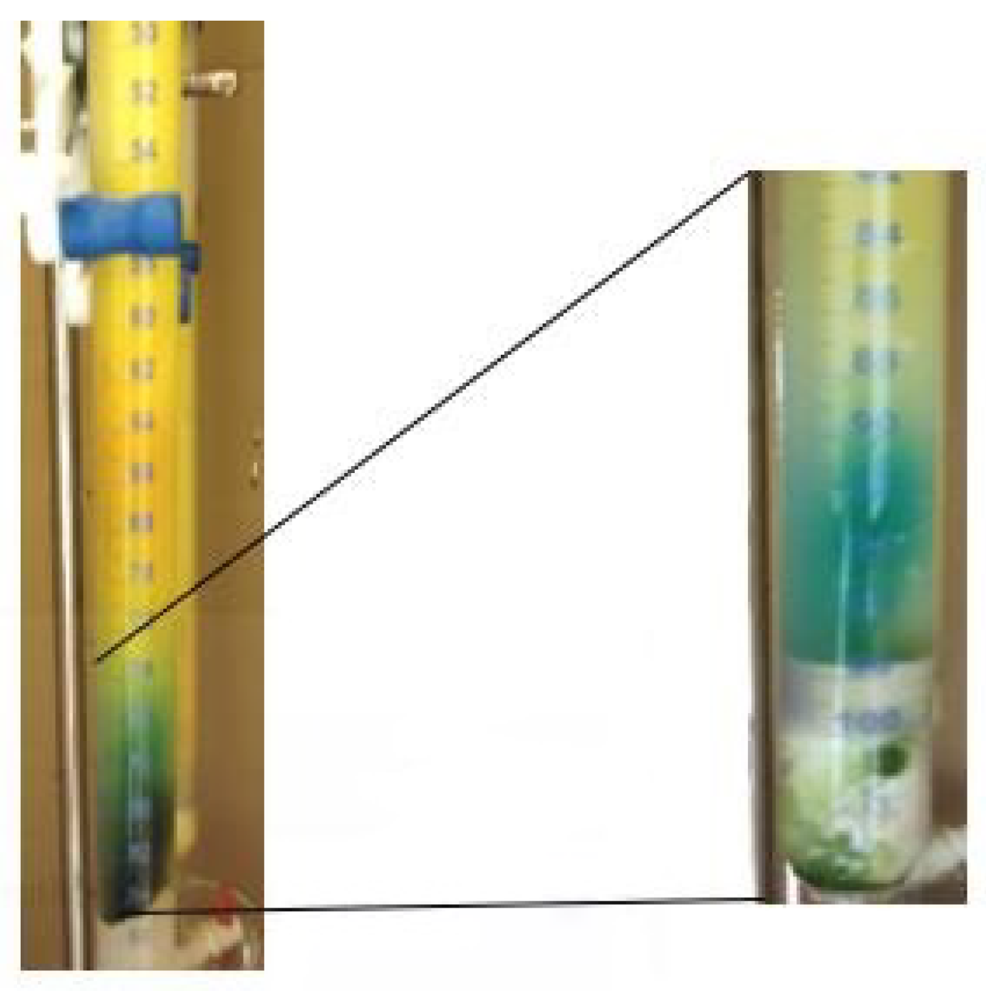
3.1. Pigment Purification

The purification of the pigments from crude extracts was achieved via column chromatography (CC). Throughout the experiments, several mobile phases were studied to observe which one was more efficient in the separation of the pigments. It tested four mobile phases: n-hexane/acetone (7:3, v/v), Petroleum ether/acetone (7:3, v/v), methanol/acetone (7:3), and n-hexane/methanol (7:3, v/v). Of all mobile phases studied, the only one that showed to be able to separate the pigments was n-hexane/acetone (7:3, v/v); with this, the pigment separation occurred more efficiently (Figure 1. In fact, it was possible to see the separation of pigments with different “colors”. This technique is based on the observation of the naked eye because it is possible to observe the different fragments via the column. In the selected fragment, it was possible to see the start and the end of the yellow, aqua-blue, and green pigments.

3.2. Pigment Characterization

3.2.1. Pigment Characterization by Thin Layer Chromatography (TLC)

This method was used to evaluate the yield of pigments present in each fraction collected in the column chromatography and to identify them based on the Retention factor (Rf) value assigned in the literature. This enables us to decide which pigments to use for further studies. The ones chosen to proceed in the subsequent phases of the work are presented in Table 2 and Table 3. The extracts chosen were the crude extracts and the ones that appeared to be isolated or with a maximum of two pigments. Table 2 and Table 3 also present the retention factors calculated for the pigment of each extract used.

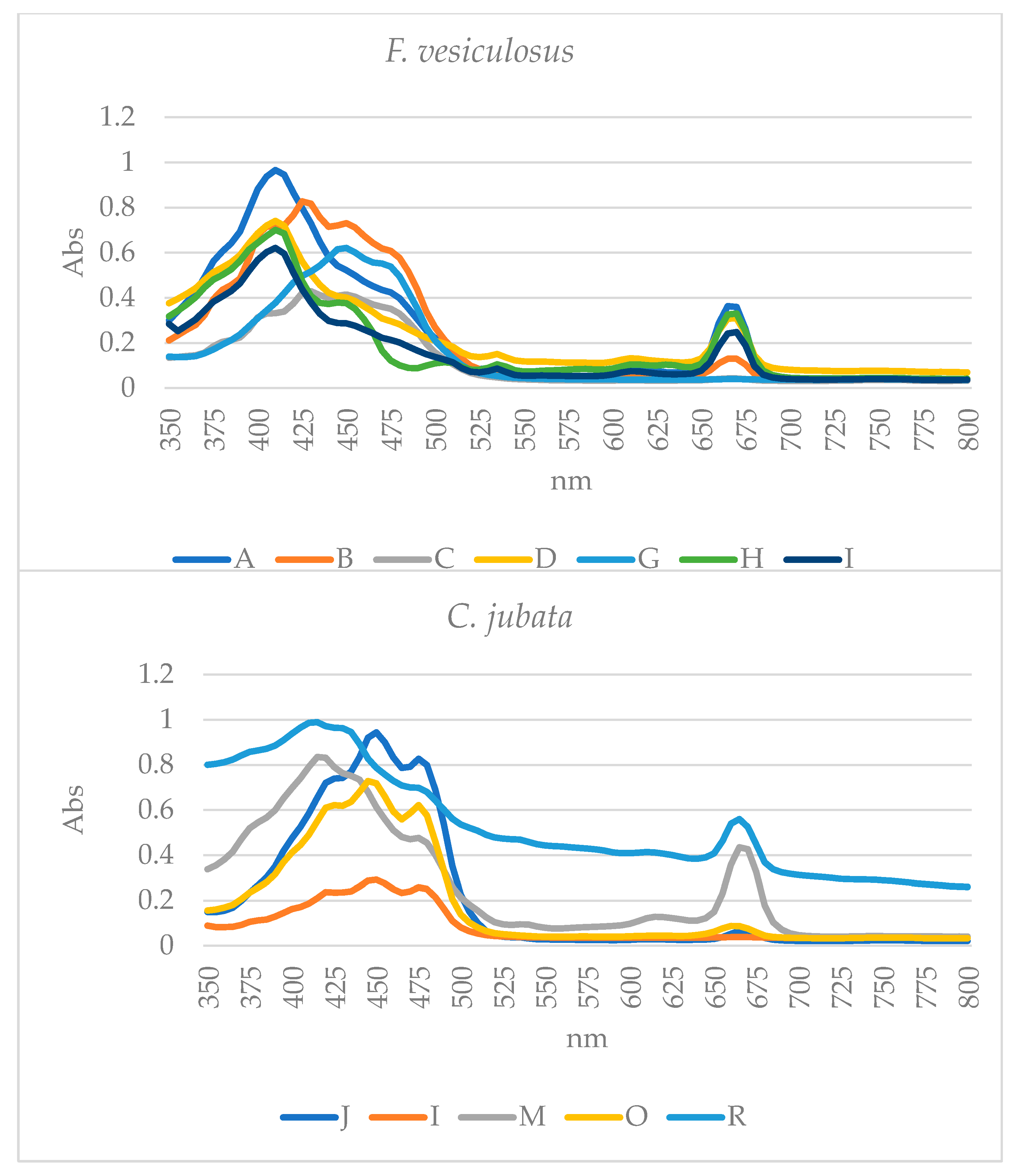
3.2.2. Ultraviolet and Visible (UV-Vis) Spectrophotometry

The UV-Vis spectrophotometry (Figure 2) was only performed for the pigments that appear to be isolated by TLC. The analysis of the spectra and the identifications of the pigments was based on the absorbance wavelength found in the literature and is presented in Table 4. Via analysis of the spectra, it was possible to understand that the Chl a was present in several fractions of the extracts. The extracts D and I were the crude extracts of F. vesiculosus, and based on the spectrophotometric data, these extracts only had the characteristic peaks of Chl a. In the case of C. jubata, the crude extracts M and R already showed several peaks, showing to be more enriched extracts by spectrophotometry.

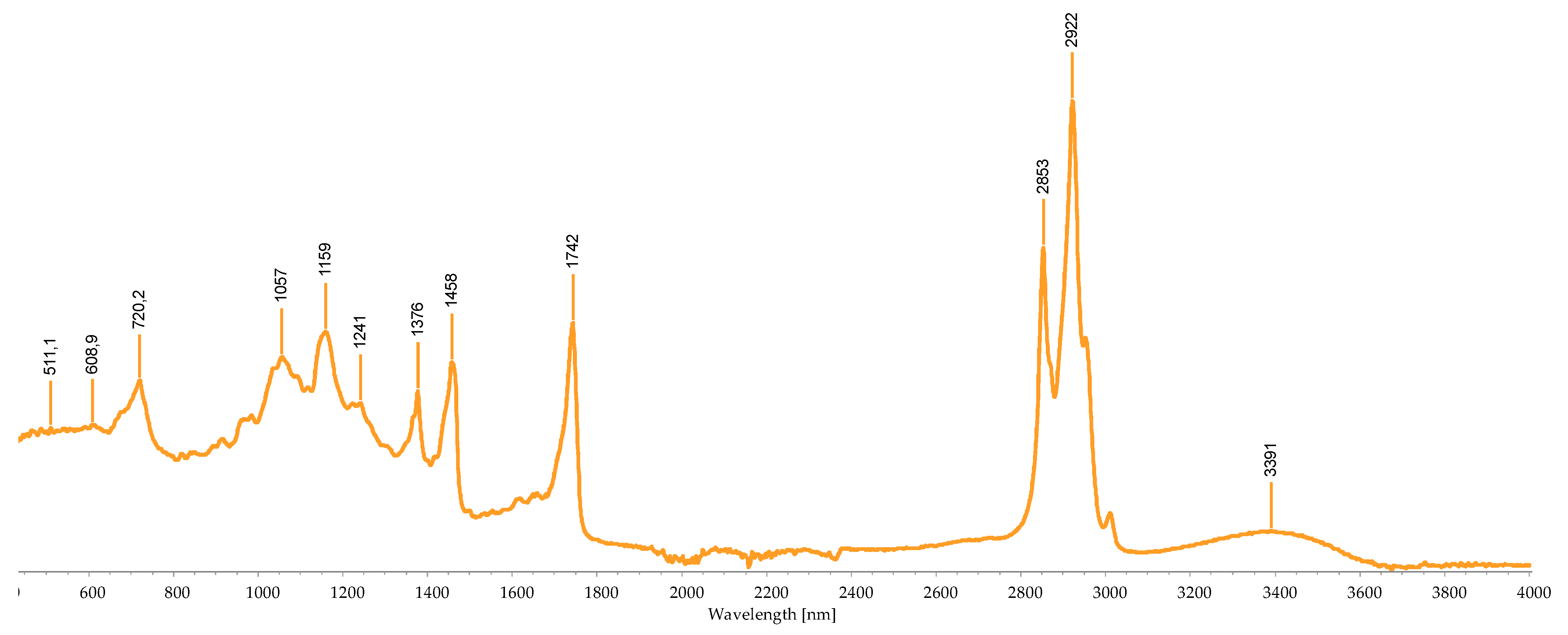
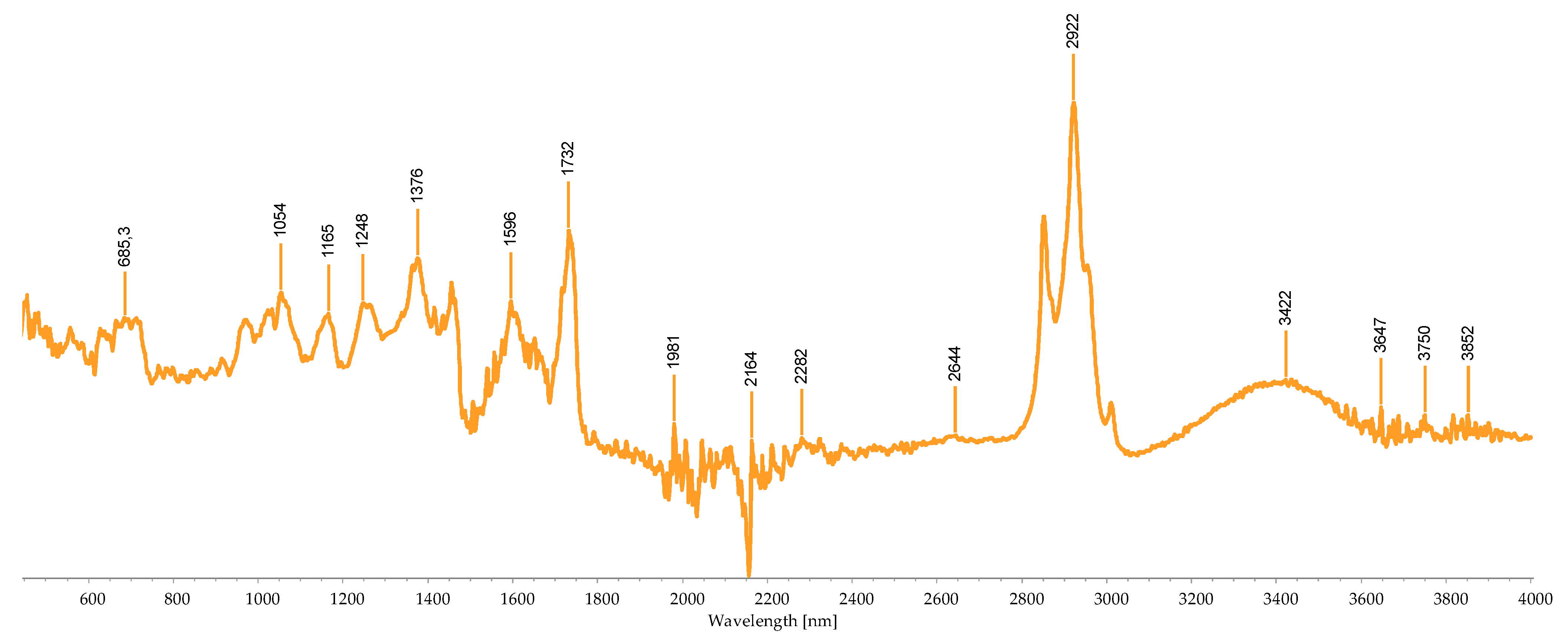
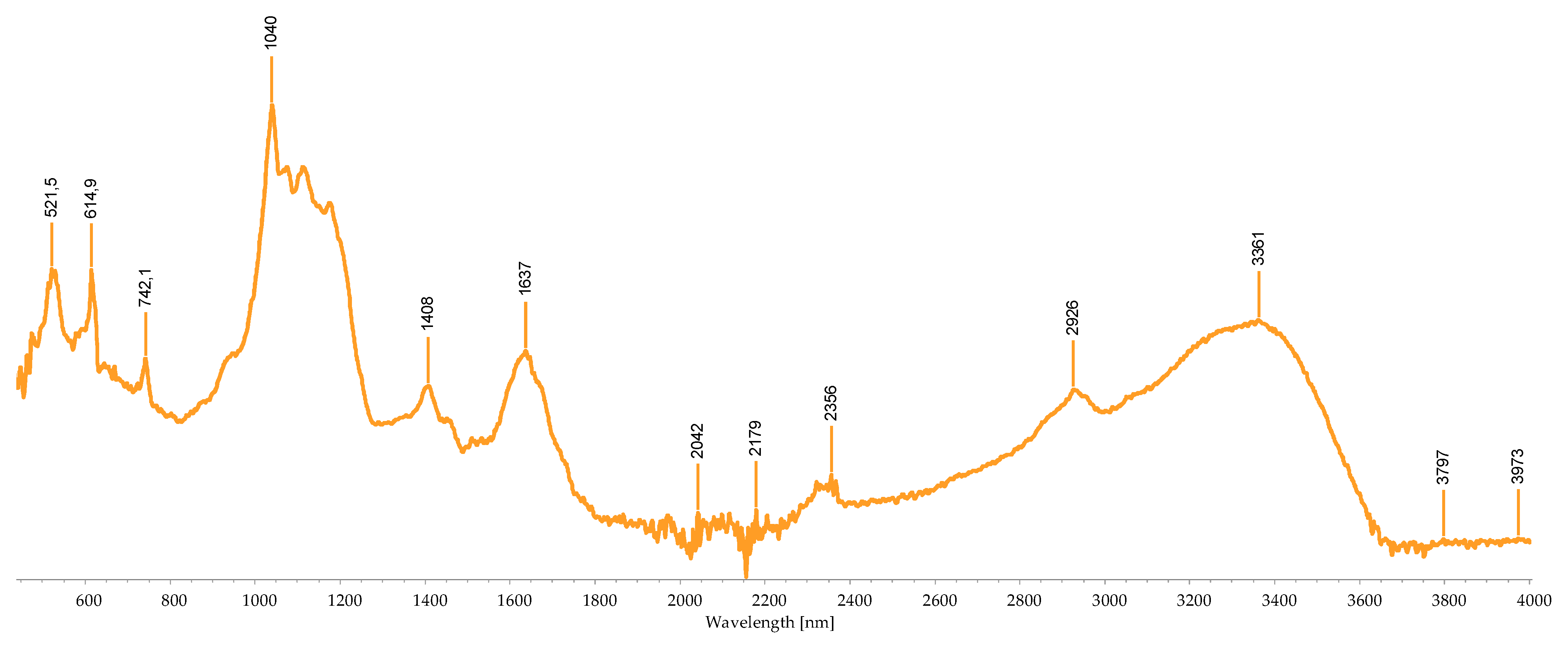
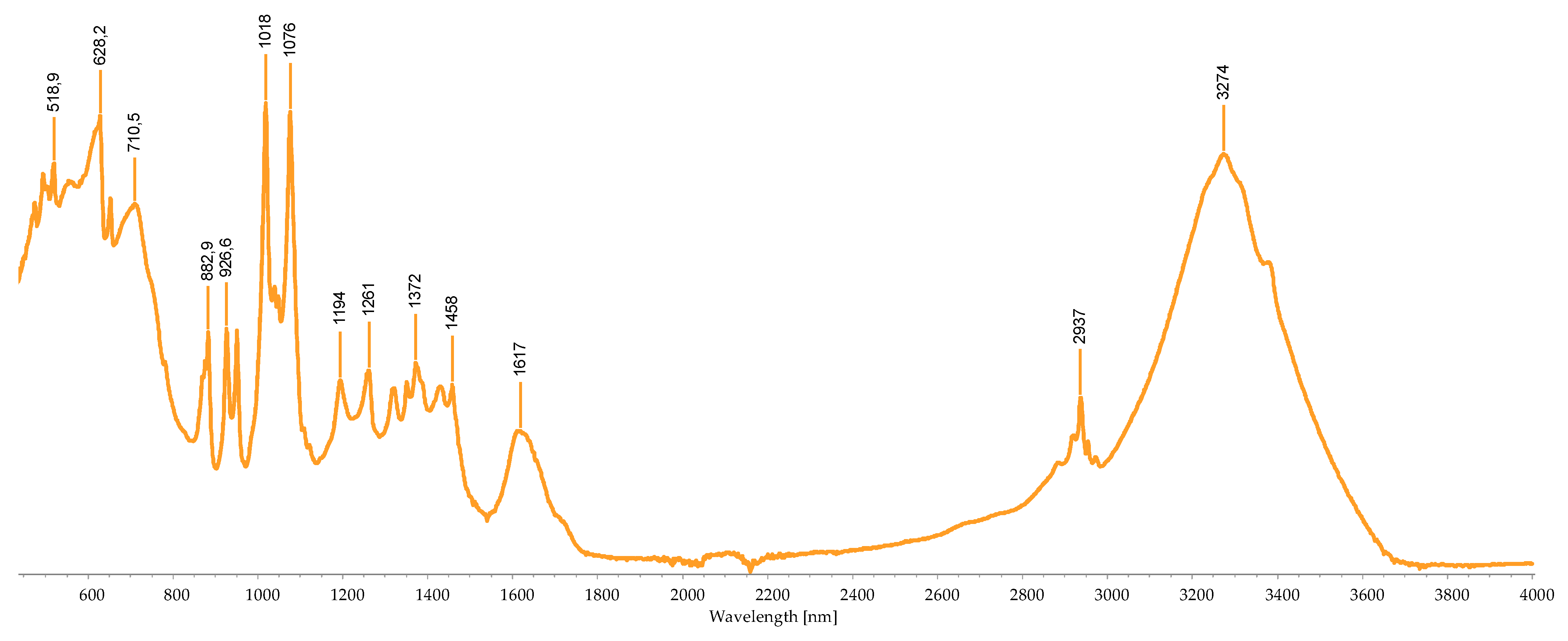
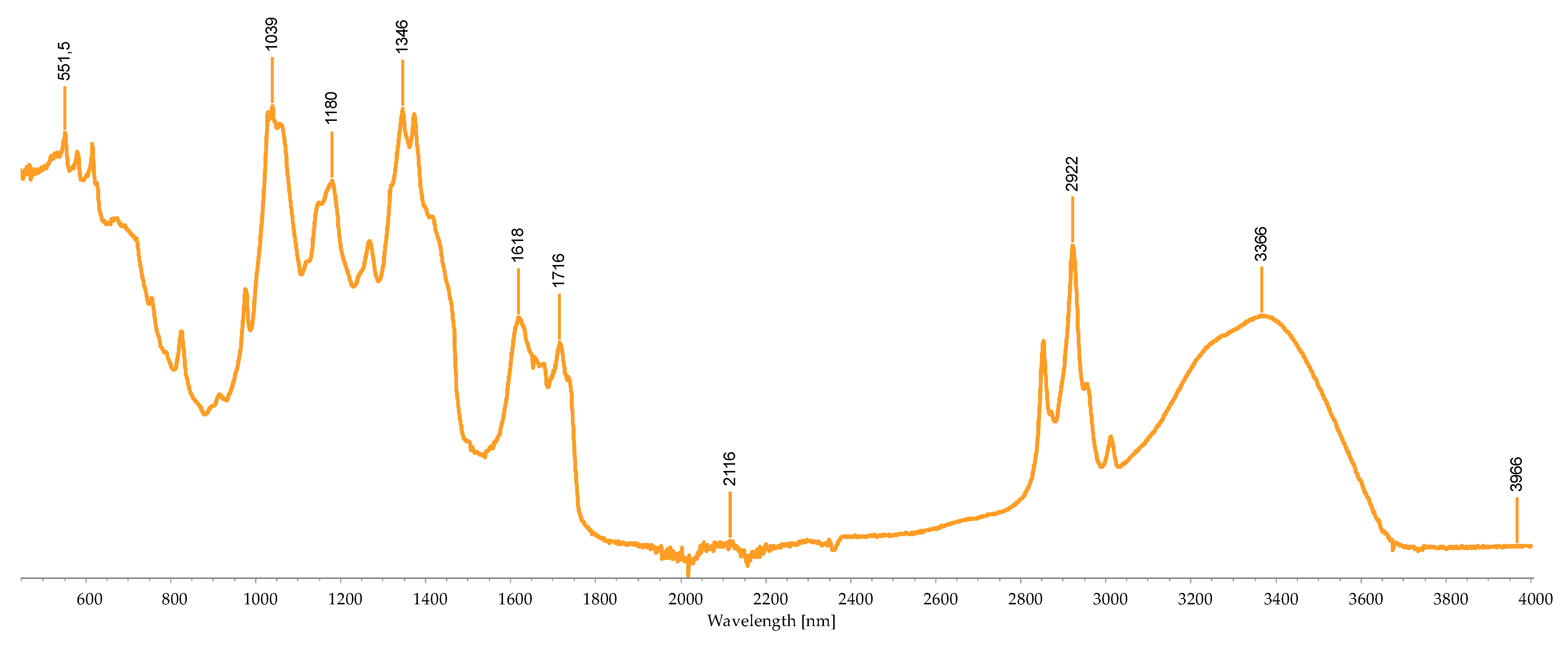
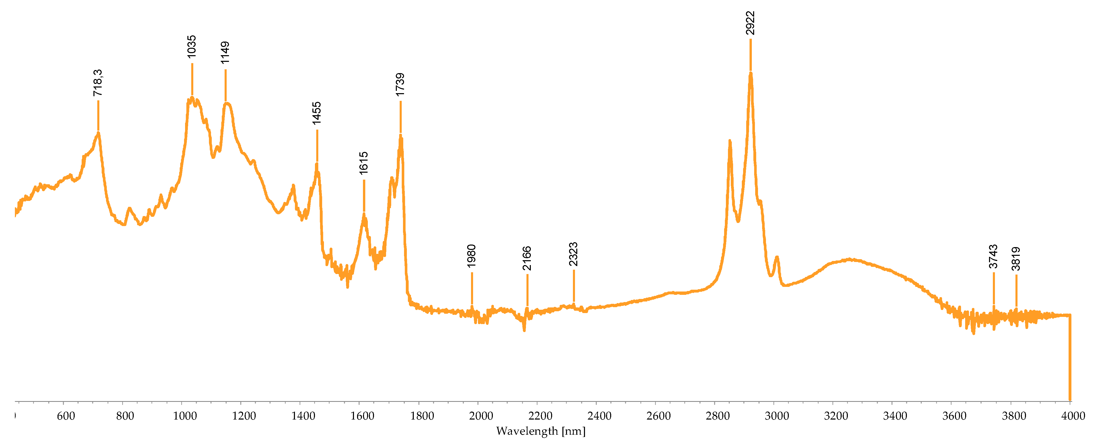
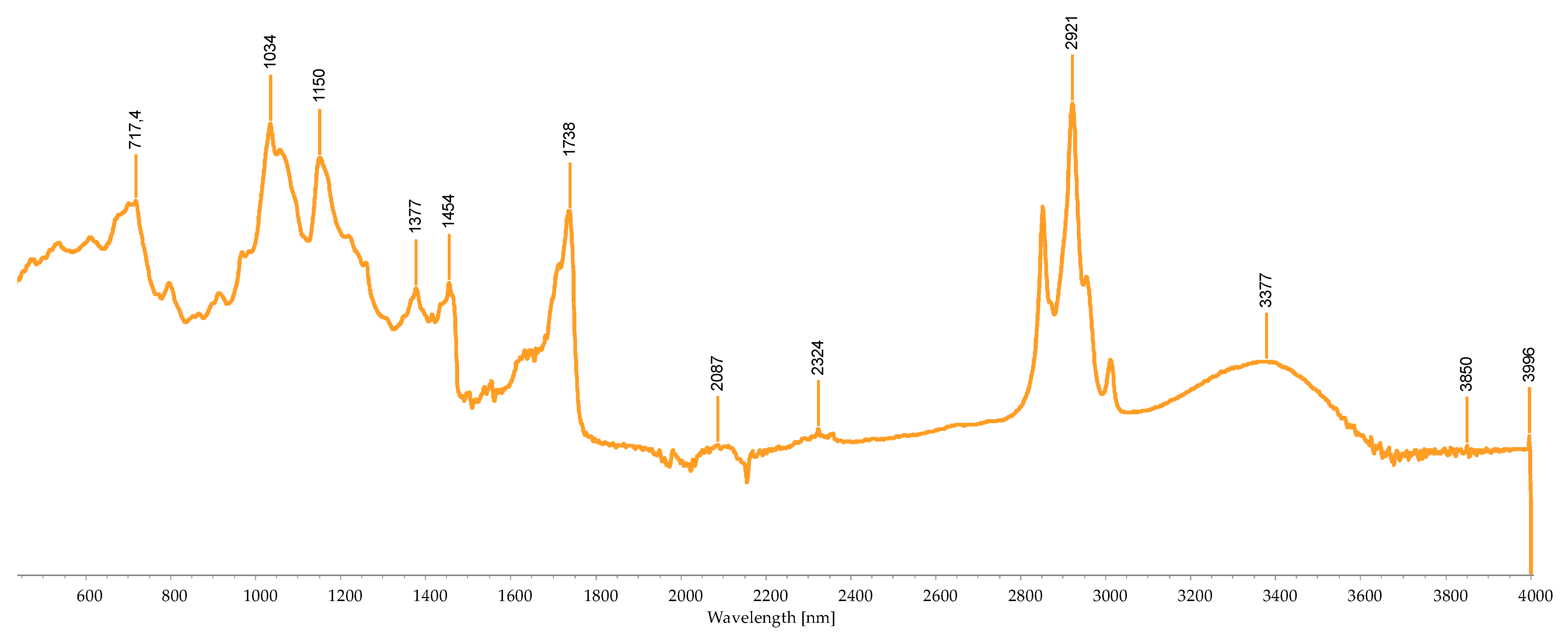
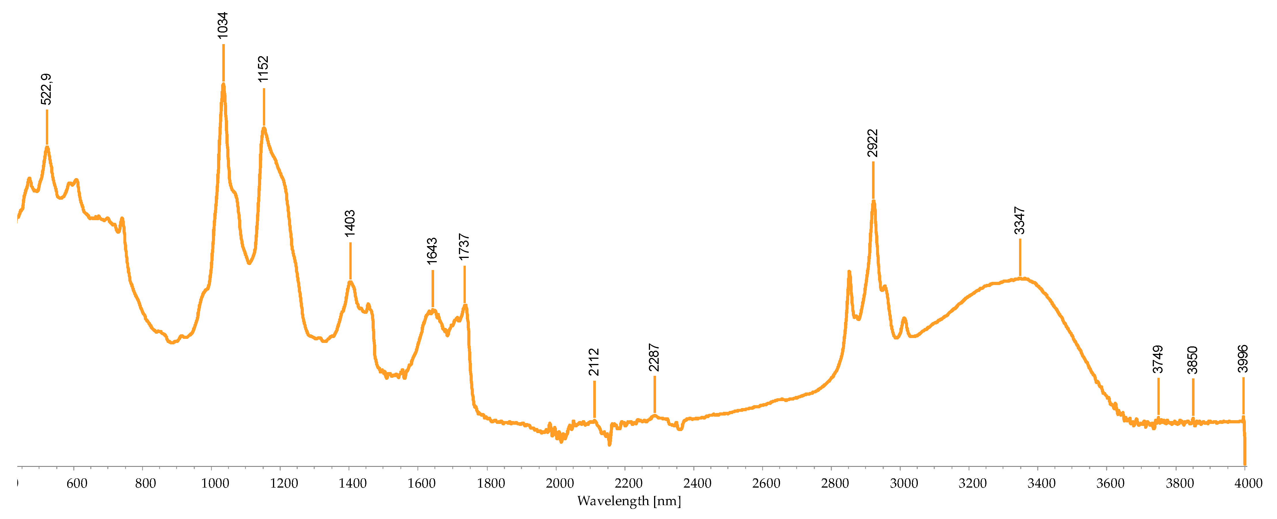
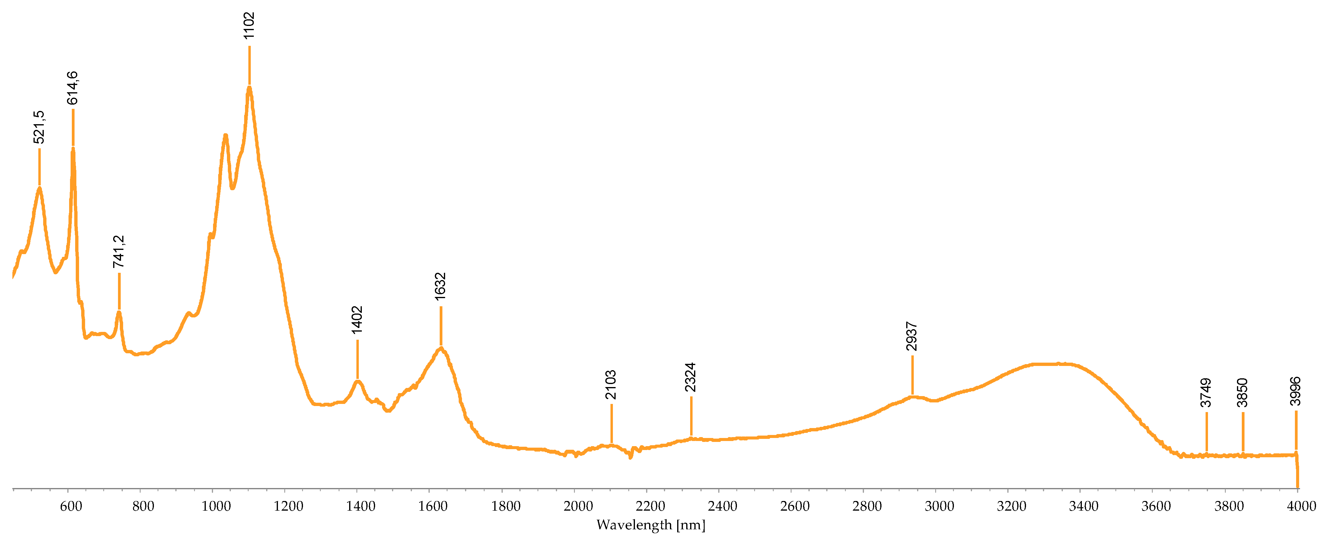
3.2.3. FTIR-ATR Analysis

The analysis of the extracts by FTIR-ATR allows for the identification of the types of chemical bonds that the compounds contain (Table 5 and Figure 3, Figure 4, Figure 5, Figure 6, Figure 7, Figure 8, Figure 9, Figure 10 and Figure 11). In this case, lyophilized samples that had visible substrate were analyzed because it was not possible to collect a sample for analysis. The analysis to follow was carried out considering the information found in the literature, although the information on pigments and this type of analysis is still very limited.
All the pigment extracts showed a “noise”, which was associated with the humidity present in the air and carbon dioxide. This disturbance was visible in greater predominance between 400 and 700 cm−1 (chemical bounds related to C-O bonds of various compounds) and 1800 and 2300 cm−1 (carbon dioxide area).
Pigments A and B present the spectra peaks substantially in the same areas. However, analyzing the main peaks present in the FTIR-ATR spectra and the respective connections allows us to reach the conclusion that it can be either a chlorophyll or a carotenoid.
By observing the graphs and the peaks present, it is possible to affirm that the extract pigments C and H only have the same peak. According to the literature, this peak is a C-N bond that is associated with chlorophyll. Most peaks, despite not being the same, represent the same chemical bonds, thus allowing the conclusion that both extracts are carotenoids, and C has a CO-C-O bond, indicating the presence of fucoxanthin.
Extract D and I are the crude extracts of F. vesiculosus, and despite the spectra not being very similar, both have bonds strongly related to the presence of chlorophylls and carotenoids.
The extracts R and M refer to the crude extract of ethanol from C. jubata. With the detected peaks of FTIR-ATR, it was possible to identify chemical bonds related to the presence of carotenoids, phycoerythrin, and chlorophylls.
Taking into account the information found and the peaks detected for extract S, its spectrum indicates an extract enriched in phycoerythrin with traces of chlorophyll.
Table 5 shows the identified peaks of the pigment’s extracts.

3.3. Concentration of the Isolated Pigments

The concentration of the purified fractions was determined from the dry weight of the extract (Table 6). The crude extract (D, I, M, R) contained, as expected, the highest concentration.

3.4. Antifungal Activity

After the confirmation of the extract’s sterility, their anti-dermatophytic effect was evaluated on Trichophyton mentagrophytes, T. rubrum, and Microsporum canis via microdilution assays following the standard methods E.DEF 9.3 EUCAST. The extracts J, L, M, and O did not show inhibition of the growth of the fungi tested.
None of the pigment extracts of F. vesiculosus showed fungal growth inhibition, not even the crude extracts.
Table 7 presents the MIC values for pigment extracts R and S of C. jubata. Only these pigment extracts showed antifungal activity against the fungi tested: T. mentagrophytes, T. rubrum, and M. canis. The antifungal activity of these pigments was tested for three weeks.
During the first week after the preparation of the extracts, the MIC100 observed for the crude ethanol extract R was 3450 μg/mL for T. mentagrophytes. The concentration that inhibits 50% of the growth, the MIC50 of the extract R was 1725 μg/mL, and the MFC was 3450 μg/mL. For T. rubrum, the MIC and MFC of the crude extract were 3450 μg/mL. For M. canis, the MIC of 862.5 μg/mL was only observed in the first week of the assay. Throughout the 3-week period of assays, the pigments seemed to lose their effect.
Extract S was able to inhibit the growth of all the fungi tested. The isolated pigments showed a MIC100 of 2783.25 μg/mL in the Trichophyton spp. tested and in M. canis during the first week after the preparation of the extract.
It was made a control with itraconazole. The results of the MIC100 and MIC50 of the itraconazole are together in the same column because they did not vary over the weeks hence the representation W = 1, 2, 3. The MIC100 and MIC50 of itraconazole against T. mentagrophytes, T. rubrum, and M. canis were 1 and 0.5 μg/mL, respectively. These data are supported by the literature in which the minimum inhibition concentration in dermatophytes normally is not higher than 1 μg/mL [62,63,64].

4. Discussion

With the continuous rise of fungal infections and the increase in fungal resistance to traditional antifungals, there is a need to find new treatment alternatives. Seaweeds are potential sources of novel therapeutic agents since they produce a wide range of secondary metabolites that, when compared to the ones produced by terrestrial plants, have an extraordinary molecular structure and pharmacological effects, including anticancer, antibacterial, antifungal, antiviral, anti-inflammatory, among others [65,66,67].
During the extraction protocols, it was observed that the extraction of pigments from seaweeds depends on several variables, including the solvent type, extraction duration, and sample state (whether fresh, dried, frozen, or milled) in accordance with previous reports [34,68]. The entire extraction process was thought up to obtain the most efficient method of extracting pigments. Due to the distance from the beach to the laboratory, it was not possible to use the fresh seaweed, so the fresh biomass was stored in the freezer until further used. After thawing, the seaweeds were washed, as described in the methods, and dried in a forced-air oven at 40 °C. The temperature of drying is important because high temperatures can compromise the process of extraction, influencing the concentration of pigments extracted. Despite some studies reporting the use of fresh seaweed, this study used dried milled seaweed because of the wider accessibility of the solvents to all the structures of the seaweed [34]. If fresh seaweed had been used, the solvent would not reach all the seaweeds’ cellular structures, and the extraction would not have been as efficient.
As previously mentioned, the efficiency of pigment extraction depends on several factors, and one of them is the solvent. It is important to test the behavior of the solvent used for the extraction in the different seaweeds because their efficiency can vary with the pigments that are tested. For example, most of the carotenoids are fat-soluble, so to monetize the extraction it must be used fat solvents like, acetone, alcohols, esters, and hexanes [69]. In this work, two solvents were used: ethanol 96% and acetone 99%. The aim was to see which one of them was more efficient in pigment extraction. To the naked eye, both seem to extract the pigment from biomass until exhaustion, but in the column chromatography, the acetone extracts showed to be better separated via the column. The number of fractions is also higher than the ones obtained with the ethanol. With this analysis, it was concluded that the best solvent to extract pigment was acetone. This is in agreement with the literature, in which other studies conclude that acetone is the best solvent to extract pigments in general [32,68,69,70]. Another detail observed over the weeks of assays was that despite being solubilized in water, the pigments extracted with acetone remained more stable than those that were extracted with ethanol. After several days, the ethanol extracts start to show some deposits in the bottom of the falcon. The explanation for this observation was not found in the literature, so more studies must be conducted to understand this finding.
In the course of this work, it was possible to perceive that the process of identification and characterization of pigments is complex, and it is not possible to use only a simple technique to accomplish it. To obtain a more consistent characterization, three techniques were used for the identification of the pigments: UV-visible spectrophotometry, FTIR, and TLC. Due to the lack of information about the characterization of seaweed pigments, these techniques complement themselves and give a result that is more trustworthy.
The PBPs have several applications in biotechnology, biomedicine, pharmacology, dye, cosmetics, and food colorants [71]. In pharmacology, it has shown a strong antioxidant effect against reactive oxygen species (ROS). Some in vitro studies showed the PBP’s anti-inflammatory, anti-viral, and anti-tumor activities [72]. The extract S was identified as a phycobiliprotein and was the one with the highest bioactivity. This extract showed antifungal activity against all the dermatophytes tested. There are already some reports of the antifungal and antimicrobial activities of the PBPs. Righini et al. [59] described the antifungal activity of PBPs against spore germination and mycelial growth of the phytopathogen fungi Botrytis cinerea. This class of pigment also showed antibacterial activity against Staphylococcus aureus, Streptococcus pyogenes, Escherichia coli, and others [73,74].
Although considered an important raw source of pigments with various bioactivities, there is no information relative to the antifungal potential of the seaweed pigments [6]. Therefore, this study demonstrates that seaweed pigments and pigment-enriched extracts can be a key to finding new antifungal natural solutions. However, further studies are needed to unravel how to maintain the stability of the pigments, preventing degradation. There is still a long road ahead; however, this study demonstrates a new pathway of an environmentally sustainable source to the nutraceutical, cosmetical, and pharmaceutical areas. This presented antifungal and fungicidal activity against T. mentagrophytes, T. rubrum, and M. canis at 3450 μg/mL. Consistently, the fraction isolated from this crude extract also exhibited antifungal and fungicidal activity against the three dermatophytes tested at a lower concentration (2783.25 μg/mL). Nevertheless, the bioactivity of the extracts R and S decreased during the 3-week period of assays, demonstrating that during this period of time, the pigments were not stable. This study demonstrates that seaweed pigments and pigment-enriched extracts can be a key to finding new antifungal natural solutions. However, further studies are needed to unravel how to maintain the stability of the pigments, preventing degradation. There is still a long road ahead; however, this study demonstrates a new pathway of an environmentally sustainable source to the nutraceutical, cosmetical, and pharmaceutical areas.
The chlorophylls are the most widely distributed pigments in every seaweed, and they are highly sensitive to heat, light, oxygen, and enzymes, leading to their easy degradation and color alteration [75,76]. The color change in the chlorophyll can occur due to the replacement of the magnesium atom in chlorophyll’s structure to hydrogen ions, causing the structure of chlorophyll to shift to pheophytin, becoming olive brown color [75,77]. In the present work, we observed a decrease in the antifungal activity of the tested pigments over time, but it is worth mentioning that a natural catabolic product of chlorophyll also has interesting potential as cytotoxic effects on cancer cell lines [78]. The main reasons for the carotenoid’s degradation could be oxidation and isomerization, which reduce the redness and yellowness of the extracts. Normally, carotenoids oxidize in the presence of oxygen. However, other factors such as light, heat, metal ions, and enzymes can also speed up the process [75,79]. Despite the lack of information about PBPs in general, they are starting to appear as applications in food colorants, and for that, it is necessary to understand the stability of these pigments [80,81]. Studies have shown that PBPs are stable under basic pH, and in this case, they remain stable from 0 to 50 °C. The color of the PBPs remains stable for 20 days after the extraction. So, from this information and from the results observed during the work the PBPs are the most stable pigments present in seaweeds [82].
Due to the properties of the pigments, they can be integrated into the formulation of cosmetic products with various purposes, one of them being antimicrobial products, like antimicrobial creams [15,21,23,25,83,84]. The antifungal activity observed in our study indicates that the pigments extracted, characterized, and selected might be used in topical applications to cure dermatophytosis.

5. Conclusions

The present study reports for the first time the isolation and characterization by several tools (UV-visible spectroscopy, FTIR, TLC) of pigments of the seaweeds F. vesiculosus and C. jubata, as well as their antifungal activity against dermatophytes. This work allowed us to understand that only an isolated pigment from C. jubata shows antifungal activity against T. rubrum, T. mentagrophytes, and M. canis, the extract S containing phycobiliproteins and the crude extract of ethanol R containing several pigments. These results demonstrate the possible application of seaweed pigments as an alternative to conventional antifungal treatments. Further assays and chemical characterization will be needed for the two best extracts that showed best results in pre-screening analysis, as cytotoxic assay, liquid chromatography, and mechanism of action.
This work demonstrates that the purity rate of pigments is not a keystone for their antifungal activity since the crude extract and phycobiliprotein extract were the best extracts in the assays. The phycobiliprotein extract is a green extraction method. FTIR-ATR and UV/V spectrophotometry characterization techniques coupled with software program (which is already applied in the industrial) can be applied to the nutraceutical and natural pharmaceutical products.

Author Contributions

Conceptualization, L.G., J.C., C.F., T.G. and L.P.; methodology, L.G., J.C., C.F. and T.G.; validation, C.F., T.G. and L.P.; investigation, L.G., J.C. and C.F.; resources, T.G. and L.P.; writing—original draft preparation, L.G. and J.C.; writing—review and editing, C.F., T.G. and L.P.; supervision, T.G. and L.P. All authors have read and agreed to the published version of the manuscript.

Funding

This study was also financed by the European Regional Development Fund (ERDF) via the Centro 2020 Regional Operational Programme: project CENTRO-01-0145-FEDER-000012- HealthyAging2020 and CENTRO-01-0145-FEDER-022095: ViraVector; the COMPETE 2020–Operational Programme for Competitiveness and Internationalisation, and the Portuguese national funds via FCT, I.P.: UIDB/04539/2020, UIDP/04539/2020 and LA/P/0058/2020 and Chantal Fernandes by 10.54499/DL57/2016/CP1448/CT0025.

Institutional Review Board Statement

Not applicable.

Informed Consent Statement

Not applicable.

Data Availability Statement

The data presented in this study are available on request from the corresponding author.

Conflicts of Interest

The authors declare no conflicts of interest.

References

  1. Jiménez, E.; Dorta, F.; Medina, C.; Ramírez, A.; Ramírez, I.; Peña-Cortés, H. Anti-Phytopathogenic Activities of Macro-Algae Extracts. Mar. Drugs 2011, 9, 739–756. [Google Scholar] [CrossRef] [PubMed]
  2. Déléris, P.; Nazih, H.; Bard, J.-M. Seaweeds in Human Health. In Seaweed in Health and Disease Prevention; Elsevier: Amsterdam, The Netherlands, 2016; pp. 319–367. [Google Scholar]
  3. Pádua, D.; Rocha, E.; Gargiulo, D.; Ramos, A.A. Bioactive Compounds from Brown Seaweeds: Phloroglucinol, Fucoxanthin and Fucoidan as Promising Therapeutic Agents against Breast Cancer. Phytochem. Lett. 2015, 14, 91–98. [Google Scholar] [CrossRef]
  4. Pinheiro, M.S.; de Aguiar, P.F.; Quaresma, C.H.; Garcia, S. Validation of Impregnation Process for Homeopathic Globules by Spectrophotometric-UV Method. Brvaz. J. Pharm. Sci. 2014, 50, 137–146. [Google Scholar] [CrossRef]
  5. Jesumani, V.; Du, H.; Aslam, M.; Pei, P.; Huang, N. Potential Use of Seaweed Bioactive Compounds in Skincare—A Review. Mar. Drugs 2019, 17, 688. [Google Scholar] [CrossRef] [PubMed]
  6. Gomes, L.; Monteiro, P.; Cotas, J.; Gonçalves, A.M.M.; Fernandes, C.; Gonçalves, T.; Pereira, L. Seaweeds’ Pigments and Phenolic Compounds with Antimicrobial Potential. Biomol. Concepts 2022, 13, 89–102. [Google Scholar] [CrossRef] [PubMed]
  7. Janarthanan, M.; Senthil Kumar, M. The Properties of Bioactive Substances Obtained from Seaweeds and Their Applications in Textile Industries. J. Ind. Text. 2018, 48, 361–401. [Google Scholar] [CrossRef]
  8. Pereira, L. Macroalgae. Encyclopedia 2021, 1, 177–188. [Google Scholar] [CrossRef]
  9. Sustainable Global Resources of Seaweeds Volume 2; Ranga Rao, A.; Ravishankar, G.A. (Eds.) Springer International Publishing: Cham, Switzerland, 2022; ISBN 978-3-030-92173-6. [Google Scholar]
  10. Aryee, A.N.; Agyei, D.; Akanbi, T.O. Recovery and Utilization of Seaweed Pigments in Food Processing. Curr. Opin. Food Sci. 2018, 19, 113–119. [Google Scholar] [CrossRef]
  11. Pereira, L. Calliblepharis Jubata. Available online: http://www.flordeutopia.pt/macoi/ (accessed on 8 July 2022).
  12. Araujo, G.S.; Cotas, J.; Morais, T.; Leandro, A.; García-Poza, S.; Gonçalves, A.M.M.; Pereira, L. Calliblepharis jubata Cultivation Potential—A Comparative Study between Controlled and Semi-Controlled Aquaculture. Appl. Sci. 2020, 10, 7553. [Google Scholar] [CrossRef]
  13. Balina, K.; Romagnoli, F.; Blumberga, D. Chemical Composition and Potential Use of Fucus vesiculosus from Gulf of Riga. Energy Procedia 2016, 95, 43–49. [Google Scholar] [CrossRef]
  14. Maehre, H.K.; Malde, M.K.; Eilertsen, K.-E.; Elvevoll, E.O. Characterization of Protein, Lipid and Mineral Contents in Common Norwegian Seaweeds and Evaluation of Their Potential as Food and Feed. J. Sci. Food Agric. 2014, 94, 3281–3290. [Google Scholar] [CrossRef]
  15. Freitas, M.V.; Pacheco, D.; Cotas, J.; Mouga, T.; Afonso, C.; Pereira, L. Red Seaweed Pigments from a Biotechnological Perspective. Phycology 2021, 2, 1–29. [Google Scholar] [CrossRef]
  16. Cserháti, T. Liquid Chromatography of Natural Pigments and Synthetic Dyes; Elsevier Science Ltd.: Amsterdam, The Netherlands, 2006. [Google Scholar]
  17. Morais, T.; Cotas, J.; Pacheco, D.; Pereira, L. Seaweeds Compounds: An Ecosustainable Source of Cosmetic Ingredients? Cosmetics 2021, 8, 8. [Google Scholar] [CrossRef]
  18. Kalasariya, H.S.; Yadav, V.K.; Yadav, K.K.; Tirth, V.; Algahtani, A.; Islam, S.; Gupta, N.; Jeon, B.-H. Seaweed-Based Molecules and Their Potential Biological Activities: An Eco-Sustainable Cosmetics. Molecules 2021, 26, 5313. [Google Scholar] [CrossRef] [PubMed]
  19. Pereira, A.G.; Otero, P.; Echave, J.; Carreira-Casais, A.; Chamorro, F.; Collazo, N.; Jaboui, A.; Lourenço-Lopes, C.; Simal-Gandara, J.; Prieto, M.A. Xanthophylls from the Sea: Algae as Source of Bioactive Carotenoids. Mar. Drugs 2021, 19, 188. [Google Scholar] [CrossRef]
  20. Pérez-Gálvez, A.; Viera, I.; Roca, M. Carotenoids and Chlorophylls as Antioxidants. Antioxidants 2020, 9, 505. [Google Scholar] [CrossRef] [PubMed]
  21. Silva, A.; Silva, S.A.; Carpena, M.; Garcia-Oliveira, P.; Gullón, P.; Barroso, M.F.; Prieto, M.A.; Simal-Gandara, J. Macroalgae as a Source of Valuable Antimicrobial Compounds: Extraction and Applications. Antibiotics 2020, 9, 642. [Google Scholar] [CrossRef]
  22. Cabral, E.M.; Oliveira, M.; Mondala, J.R.M.; Curtin, J.; Tiwari, B.K.; Garcia-Vaquero, M. Antimicrobials from Seaweeds for Food Applications. Mar. Drugs 2021, 19, 211. [Google Scholar] [CrossRef]
  23. Tamilarasu, A.; Nethaji, M.; Bharathi, S.; Lloyd, C.C.; Somu, S.L.R. Seaweeds: A Potent Source of Antimicrobial Drugs for Aquaculture Industry. Med. Plants Int. J. Phytomed. Relat. Ind. 2021, 13, 33–44. [Google Scholar] [CrossRef]
  24. Pang, R.; Tao, J.-Y.; Zhang, S.-L.; Zhao, L.; Yue, X.; Wang, Y.-F.; Ye, P.; Dong, J.-H.; Zhu, Y.; Wu, J.-G. In Vitro Antiviral Activity of Lutein against Hepatitis B Virus. Phyther. Res. 2010, 24, 1627–1630. [Google Scholar] [CrossRef]
  25. Fatima, K.; Mathew, S.; Suhail, M.; Ali, A.; Damanhouri, G.; Azhar, E.; Qadri, I. Docking Studies of Pakistani HCV NS3 Helicase: A Possible Antiviral Drug Target. PLoS ONE 2014, 9, e106339. [Google Scholar] [CrossRef]
  26. Gupta, A.K.; Venkataraman, M.; Renaud, H.J.; Summerbell, R.; Shear, N.H.; Piguet, V. The Increasing Problem of Treatment-resistant Fungal Infections: A Call for Antifungal Stewardship Programs. Int. J. Dermatol. 2021, 60, e474–e479. [Google Scholar] [CrossRef]
  27. Lopes, G.; Pinto, E.; Salgueiro, L. Natural Products: An Alternative to Conventional Therapy for Dermatophytosis? Mycopathologia 2017, 182, 143–167. [Google Scholar] [CrossRef]
  28. Dhayanithi, N.; Kumar, T.A.; Kalaiselvam, M.; Balasubramanian, T.; Sivakumar, N. Anti-Dermatophytic Activity of Marine Sponge, Sigmadocia carnosa (Dendy) on Clinically Isolated Fungi. Asian Pac. J. Trop. Biomed. 2012, 2, 635–639. [Google Scholar] [CrossRef]
  29. Fernandes, C.; Sousa-Baptista, J.; Lenha-Silva, A.F.; Calheiros, D.; Correia, E.; Figueirinha, A.; Salgueiro, L.; Gonçalves, T. Azorean Black Tea (Camellia sinensis) Antidermatophytic and Fungicidal Properties. Molecules 2023, 28, 7775. [Google Scholar] [CrossRef]
  30. Correia, E.E.M.; Figueirinha, A.; Rodrigues, L.; Pinela, J.; Calhelha, R.C.; Barros, L.; Fernandes, C.; Salgueiro, L.; Gonçalves, T. The Chemical Profile, and Antidermatophytic, Anti-Inflammatory, Antioxidant and Antitumor Activities of Withania chevalieri, AE Gonç. Ethanolic Extract. Plants 2023, 12, 2502. [Google Scholar] [CrossRef]
  31. Calheiros, D.; Dias, M.I.; Calhelha, R.C.; Barros, L.; Ferreira, I.C.F.R.; Fernandes, C.; Gonçalves, T. Antifungal Activity of Spent Coffee Ground Extracts. Microorganisms 2023, 11, 242. [Google Scholar] [CrossRef]
  32. Yang, L.; Li, P.; Fan, S. The Extraction of Pigments from Fresh Laminaria japonica. Chin. J. Oceanol. Limnol. 2008, 26, 193–196. [Google Scholar] [CrossRef]
  33. Cotas, J.; Pacheco, D.; Araujo, G.S.; Valado, A.; Critchley, A.T.; Pereira, L. On the Health Benefits vs. Risks of Seaweeds and Their Constituents: The Curious Case of the Polymer Paradigm. Mar. Drugs 2021, 19, 164. [Google Scholar] [CrossRef] [PubMed]
  34. Silva, A.; Abreu, H.; Silva, A.; Cardoso, S. Effect of Oven-Drying on the Recovery of Valuable Compounds from Ulva rigida, Gracilaria sp. and Fucus vesiculosus. Mar. Drugs 2019, 17, 90. [Google Scholar] [CrossRef] [PubMed]
  35. Cotas, J. Fucus ceranoides (Ochrophyta, Phaeophyceae): Bioatividades Dependentes Do Gradiente Salino. Master’s Thesis, University of Coimbra, Coimbra, Portugal, 2015. [Google Scholar]
  36. Cotas, J.; Figueirinha, A.; Pereira, L.; Batista, T. The Effect of Salinity on Fucus ceranoides (Ochrophyta, Phaeophyceae) in the Mondego River (Portugal). J. Oceanol. Limnol. 2019, 37, 881–891. [Google Scholar] [CrossRef]
  37. Sudhakar, M.P.; Jagatheesan, A.; Perumal, K.; Arunkumar, K. Methods of Phycobiliprotein Extraction from Gracilaria crassa and Its Applications in Food Colourants. Algal Res. 2015, 8, 115–120. [Google Scholar] [CrossRef]
  38. Uju; Dewi, N.P.S.U.K.; Santoso, J.; Setyaningsih, I.; Hardingtyas, S.D.; Yopi. Extraction of Phycoerythrin from Kappaphycus alvarezii Seaweed Using Ultrasonication. IOP Conf. Ser. Earth Environ. Sci. 2020, 414, 012028. [Google Scholar] [CrossRef]
  39. Monteiro, P.; Cotas, J.; Pacheco, D.; Figueirinha, A.; da Silva, G.J.; Pereira, L.; Gonçalves, A.M.M. Seaweed as Food: How to Guarantee Their Quality? In Sustainable Global Resources of Seaweeds Volume 2; Springer International Publishing: Cham, Switzerland, 2022; pp. 309–321. [Google Scholar]
  40. Poole, C.F. Thin-Layer Chromatography: Challenges and Opportunities. J. Chromatogr. A 2003, 1000, 963–984. [Google Scholar] [CrossRef] [PubMed]
  41. Misra, N.N.; Rai, D.K.; Hossain, M. Analytical Techniques for Bioactives from Seaweed. In Seaweed Sustainability; Elsevier: Amsterdam, The Netherlands, 2015; pp. 271–287. [Google Scholar]
  42. Merdekawati, W.; Limantara, L.; Susanto, A. TThe Content and Pigment Composition of Sargassum crassifolium J. Agardh on Several Drying Treatments. INA Pap. 2018, 1–6. [Google Scholar] [CrossRef]
  43. Ghaliaoui, H.; Mokrane, N.; Hazzit, M.; Hadjadj, M.; Otmani, F.-S.; Touati, S.; Seridi, H. Impact of Freezing and Drying Preprocessing on Pigments Extraction from the Brown Seaweed «Phyllaria reniformis» Collected in Algerian Coast. Carpathian J. Food Sci. Technol. 2020, 12, 81–94. [Google Scholar] [CrossRef]
  44. Arendrup, M.C.; Kahlmeter, G.; Guinea, J.; Meletiadis, J. How to: Perform Antifungal Susceptibility Testing of Microconidia-Forming Dermatophytes Following the New Reference EUCAST Method E.Def 11.0, Exemplified by Trichophyton. Clin. Microbiol. Infect. 2021, 27, 55–60. [Google Scholar] [CrossRef]
  45. Espinel-Ingroff, A.; Chaturvedi, V.; Fothergill, A.; Rinaldi, M.G. Optimal Testing Conditions for Determining MICs and Minimum Fungicidal Concentrations of New and Established Antifungal Agents for Uncommon Molds: NCCLS Collaborative Study. J. Clin. Microbiol. 2002, 40, 3776–3781. [Google Scholar] [CrossRef] [PubMed]
  46. Bhatia, S.; Nagpal, K.; Bera, T.; Sharma, A.; Sharma, K. Evaluation of Pharmacognostical, Phytochemical and Anti-Microbial Properties of Porphyra vietnamensis. Int. J. Green Pharm. 2015, 9, 131. [Google Scholar] [CrossRef]
  47. Forgacs, E.; Cserhati, T. Thin-Layer Chromatography of Natural Pigments: New Advances. J. Liq. Chromatogr. Relat. Technol. 2002, 25, 1521–1541. [Google Scholar] [CrossRef]
  48. Johnston, A.; Scaggs, J.; Mallory, C.; Haskett, A.; Warner, D.; Brown, E.; Hammond, K.; McCormick, M.M.; McDougal, O.M. A Green Approach To Separate Spinach Pigments by Column Chromatography. J. Chem. Educ. 2013, 90, 796–798. [Google Scholar] [CrossRef]
  49. Koizumi, J.; Takatani, N.; Kobayashi, N.; Mikami, K.; Miyashita, K.; Yamano, Y.; Wada, A.; Maoka, T.; Hosokawa, M. Carotenoid Profiling of a Red Seaweed Pyropia yezoensis: Insights into Biosynthetic Pathways in the Order Bangiales. Mar. Drugs 2018, 16, 426. [Google Scholar] [CrossRef]
  50. Robertson, D. Algal Pigments. Available online: https://wordpress.clarku.edu/debrobertson/laboratory-protocols/algal-pigments/ (accessed on 25 November 2022).
  51. Rajauria, G.; Abu-Ghannam, N. Isolation and Partial Characterization of Bioactive Fucoxanthin from Himanthalia elongata Brown Seaweed: A TLC-Based Approach. Int. J. Anal. Chem. 2013, 2013, 802573. [Google Scholar] [CrossRef] [PubMed]
  52. Savira, A.D.R.; Amin, M.N.G.; Alamsjah, M.A. The Effect of Different Type of Solvents on the Antioxidant Activity of Fucoxanthin Extract from Brown Seaweed Sargassum duplicatum. IOP Conf. Ser. Earth Environ. Sci. 2021, 718, 012010. [Google Scholar] [CrossRef]
  53. Dumay, J.; Morançais, M. Proteins and Pigments. In Seaweed in Health and Disease Prevention; Elsevier: Amsterdam, The Netherlands, 2016; pp. 275–318. [Google Scholar]
  54. Clementson, L.A.; Wojtasiewicz, B. Dataset on the Absorption Characteristics of Extracted Phytoplankton Pigments. Data Br. 2019, 24, 103875. [Google Scholar] [CrossRef]
  55. Latsos, C.; van Houcke, J.; Blommaert, L.; Verbeeke, G.P.; Kromkamp, J.; Timmermans, K.R. Effect of Light Quality and Quantity on Productivity and Phycoerythrin Concentration in the Cryptophyte Rhodomonas sp. J. Appl. Phycol. 2021, 33, 729–741. [Google Scholar] [CrossRef]
  56. Meng, D.; Zhang, L.; Wang, Q.; Zhang, Y.; Sun, Y.; Zhang, H.; Wang, Z.; Zhou, Z.; Yang, R. Self-Assembly of Phycoerythrin with Oligochitosan by Electrostatic Interaction for Stabilization of Phycoerythrin. J. Agric. Food Chem. 2021, 69, 12818–12827. [Google Scholar] [CrossRef] [PubMed]
  57. MubarakAli, D.; Gopinath, V.; Rameshbabu, N.; Thajuddin, N. Synthesis and Characterization of CdS Nanoparticles Using C-Phycoerythrin from the Marine Cyanobacteria. Mater. Lett. 2012, 74, 8–11. [Google Scholar] [CrossRef]
  58. Mudunkotuwa, I.A.; Al Minshid, A.; Grassian, V.H. ATR-FTIR Spectroscopy as a Tool to Probe Surface Adsorption on Nanoparticles at the Liquid–Solid Interface in Environmentally and Biologically Relevant Media. Analyst 2014, 139, 870–881. [Google Scholar] [CrossRef]
  59. Righini, H.; Francioso, O.; Di Foggia, M.; Quintana, A.M.; Roberti, R. Preliminary Study on the Activity of Phycobiliproteins against Botrytis Cinerea. Mar. Drugs 2020, 18, 600. [Google Scholar] [CrossRef]
  60. Setyawati, H.; Darmokoesoemo, H.; Ningtyas, A.T.A.; Kadmi, Y.; Elmsellem, H.; Kusuma, H.S. Effect of Metal Ion Fe(III) on the Performance of Chlorophyll as Photosensitizers on Dye Sensitized Solar Cell. Results Phys. 2017, 7, 2907–2918. [Google Scholar] [CrossRef]
  61. Suica-bunghez, I.R.; Sorescu, A.A.; Doncea, S.M.; Constantin, M.; Raut, I.; Ion, R.M. Phytochemical, Antioxidant and Antimicrobial Characterization of Hedera Helix l. Extract. J. Plant Dev. 2020, 27, 47–53. [Google Scholar] [CrossRef]
  62. Aneke, C.I.; Rhimi, W.; Otranto, D.; Cafarchia, C. Synergistic Effects of Efflux Pump Modulators on the Azole Antifungal Susceptibility of Microsporum Canis. Mycopathologia 2020, 185, 279–288. [Google Scholar] [CrossRef]
  63. Bhatia, V.; Sharma, P. Determination of Minimum Inhibitory Concentrations of Itraconazole, Terbinafine and Ketoconazole against Dermatophyte Species by Broth Microdilution Method. Indian J. Med. Microbiol. 2015, 33, 533–537. [Google Scholar] [CrossRef]
  64. Gupta, A.K.; Kohli, Y.; Batra, R. In Vitro Activities of Posaconazole, Ravuconazole, Terbinafine, Itraconazole and Fluconazole against Dermatophyte, Yeast and Non-Dermatophyte Species. Med. Mycol. 2005, 43, 179–185. [Google Scholar] [CrossRef]
  65. Indira, K.; Balakrishnan, S.; Srinivasan, M.; Bragadeeswaran, S.; Balasubramanian, T. Evaluation of In Vitro Antimicrobial Property of Seaweed (Halimeda tuna) from Tuticorin Coast, Tamil Nadu, Southeast Coast of India. Afr. J. Biotechnol. 2013, 12, 284–289. [Google Scholar] [CrossRef]
  66. Pérez, M.J.; Falqué, E.; Domínguez, H. Antimicrobial Action of Compounds from Marine Seaweed. Mar. Drugs 2016, 14, 52. [Google Scholar] [CrossRef]
  67. Thakur, R.; Kalsi, A.S. Outbreaks And Epidemics of Superficial Dermatophytosis Due To Trichophyton mentagrophytes Complex and Microsporum canis: Global And Indian Scenario. Clin. Cosmet. Investig. Dermatol. 2019, 12, 887–893. [Google Scholar] [CrossRef]
  68. Pardilhó, S.L.; Machado, S.; Bessada, S.M.F.; Almeida, F.; Oliveira, M.B.; Dias, J.M. Marine Macroalgae Waste from Northern Portugal: A Potential Source of Natural Pigments? Waste Biomass Valorization 2021, 12, 239–249. [Google Scholar] [CrossRef]
  69. Warkoyo, W.; Saati, E.A. The Solvent Effectiveness on Extraction Process of Seaweed Pigment. Makara J. Technol. 2011, 15, 5–8. [Google Scholar] [CrossRef]
  70. Sudhakar, M.P.; Ananthalakshmi, J.S.; Nair, B.B. Extraction, Purification and Study on Antioxidant Properties of Fucoxanthin from Brown Seaweeds. J. Chem. Pharm. Res. 2013, 5, 169–175. [Google Scholar]
  71. Kannaujiya, V.K.; Kumar, D.; Singh, V.; Sinha, R.P. Advances in Phycobiliproteins Research: Innovations and Commercialization. In Natural Bioactive Compounds; Elsevier: Amsterdam, The Netherlands, 2021; pp. 57–81. [Google Scholar]
  72. Li, W.; Su, H.-N.; Pu, Y.; Chen, J.; Liu, L.-N.; Liu, Q.; Qin, S. Phycobiliproteins: Molecular Structure, Production, Applications, and Prospects. Biotechnol. Adv. 2019, 37, 340–353. [Google Scholar] [CrossRef]
  73. Hemlata; Afreen, S.; Fatma, T. Extraction, Purification and Characterization of Phycoerythrin from Michrochaete and Its Biological Activities. Biocatal. Agric. Biotechnol. 2018, 13, 84–89. [Google Scholar] [CrossRef]
  74. Najdenski, H.M.; Gigova, L.G.; Iliev, I.I.; Pilarski, P.S.; Lukavský, J.; Tsvetkova, I.V.; Ninova, M.S.; Kussovski, V.K. Antibacterial and Antifungal Activities of Selected Microalgae and Cyanobacteria. Int. J. Food Sci. Technol. 2013, 48, 1533–1540. [Google Scholar] [CrossRef]
  75. Ngamwonglumlert, L.; Devahastin, S.; Chiewchan, N. Natural Colorants: Pigment Stability and Extraction Yield Enhancement via Utilization of Appropriate Pretreatment and Extraction Methods. Crit. Rev. Food Sci. Nutr. 2017, 57, 3243–3259. [Google Scholar] [CrossRef]
  76. Özkan, G.; Ersus Bilek, S. Enzyme-Assisted Extraction of Stabilized Chlorophyll from Spinach. Food Chem. 2015, 176, 152–157. [Google Scholar] [CrossRef]
  77. Delgado-Vargas, F.; Paredes-Lopez, O. Natural Colorants for Food and Nutraceutical Uses; CRC Press: Boca Raton, FL, USA, 2002; ISBN 9781420031713. [Google Scholar]
  78. Wang, Y.-T.; Yang, C.-H.; Huang, T.-Y.; Tai, M.; Sie, R.-H.; Shaw, J.-F. Cytotoxic Effects of Chlorophyllides in Ethanol Crude Extracts from Plant Leaves. Evid.-Based Complement. Altern. Med. 2019, 2019, 9494328. [Google Scholar] [CrossRef]
  79. Boon, C.S.; McClements, D.J.; Weiss, J.; Decker, E.A. Factors Influencing the Chemical Stability of Carotenoids in Foods. Crit. Rev. Food Sci. Nutr. 2010, 50, 515–532. [Google Scholar] [CrossRef]
  80. Galetović, A.; Seura, F.; Gallardo, V.; Graves, R.; Cortés, J.; Valdivia, C.; Núñez, J.; Tapia, C.; Neira, I.; Sanzana, S.; et al. Use of Phycobiliproteins from Atacama Cyanobacteria as Food Colorants in a Dairy Beverage Prototype. Foods 2020, 9, 244. [Google Scholar] [CrossRef]
  81. Sonani, R.R. Recent Advances in Production, Purification and Applications of Phycobiliproteins. World J. Biol. Chem. 2016, 7, 100. [Google Scholar] [CrossRef]
  82. Nowruzi, B.; Konur, O.; Anvar, S.A.A. The Stability of the Phycobiliproteins in the Adverse Environmental Conditions Relevant to the Food Storage. Food Bioprocess Technol. 2022, 15, 2646–2663. [Google Scholar] [CrossRef]
  83. Polat, S.; Trif, M.; Rusu, A.; Šimat, V.; Čagalj, M.; Alak, G.; Meral, R.; Özogul, Y.; Polat, A.; Özogul, F. Recent Advances in Industrial Applications of Seaweeds. Crit. Rev. Food Sci. Nutr. 2021, 63, 4979–5008. [Google Scholar] [CrossRef]
  84. Numan, M.; Bashir, S.; Mumtaz, R.; Tayyab, S.; Rehman, N.U.; Khan, A.L.; Shinwari, Z.K.; Al-Harrasi, A. Therapeutic Applications of Bacterial Pigments: A Review of Current Status and Future Opportunities. 3 Biotech 2018, 8, 207. [Google Scholar] [CrossRef]
Figure 1. Representation of separated pigments in the bottom of the column at two stages of the elution using n-hexane/acetone (7:3, v/v).
Applsci 14 01456 g001
Figure 2. Visible spectra from the seaweed pigments fractions.
Applsci 14 01456 g002
Figure 3. FTIR-ATR spectra of the extract A.
Applsci 14 01456 g003
Figure 4. FTIR-ATR spectra of the extract B.
Applsci 14 01456 g004
Figure 5. FTIR-ATR spectra of the extract C.
Applsci 14 01456 g005
Figure 6. FTIR-ATR spectra of the extract D.
Applsci 14 01456 g006
Figure 7. FTIR-ATR spectra of the extract H.
Applsci 14 01456 g007
Figure 8. FTIR-ATR spectra of the extract I.
Applsci 14 01456 g008
Figure 9. FTIR-ATR spectra of the extract M.
Applsci 14 01456 g009
Figure 10. FTIR-ATR spectra of the extract R.
Applsci 14 01456 g010
Figure 11. FTIR-ATR spectra of the extract S.
Applsci 14 01456 g011
Table 1. Pigments detected in seaweeds.
PhylumChlPBPsCarotenoidsReferences
Chlorophytaa, b β-Carotene, lutein, neoxanthin, violaxanthin, and zeaxanthin[5,6,7,8,10]
Ochrophytaa, c β-Carotene, fucoxanthin, and violaxanthin
RhodophytaaPhycocyanin
Phycoerythrin
β-Carotene, lutein, and zeaxanthin
Table 2. F. vesiculosus pigments characterization via the Rf value obtained in TLC.
Nomenclature of the PigmentsRetention Factor (Rf)PigmentsReferences
Ethanol extractsA0.54Chlorophyll b[46,47,48,49,50,51,52]
0.85β-carotene
B0.48Fucoxanthin
C0.48Fucoxanthin
D
Crude Extract
0.7* NA
0.13Neoxanthin
0.19* NA
0.65* NA
0.9β-carotene
0.95β-carotene
Acetone ExtractsG0.38* NA
H0.06Neoxanthin
0.11Neoxanthin
I
Crude Extract
0.38* NA
0.45Lutein
0.02Chlorophyll c
0.59Fucoxanthin
0.85* NA
* NA—Not identified.
Table 3. C. jubata pigments characterization via the Rf value obtained in TLC.
Nomenclature of the PigmentsRetention Factor (Rf)PigmentsReferences
Acetone ExtractsJ0.82* NA[46,47,48,49,50]
L0.53Chlorophyll b
M
(Crude Extract)
0.04Chlorophyll c
0.42Lutein
0.62* NA
0.68Chlorophyll a
0.74Zeaxanthin
0.81* NA
0.84* NA
0.86* NA
0.91β-carotene
Ethanol extractsO0.53Chlorophyll b
0.7Violaxanthin
R
(Crude Extract)
0.75* NA
0.86* NA
0.91β-carotene
* NA—Not identified
Table 4. Identification of the pigment through the absorbance peaks.
ExtractPeaks (nm)PigmentReference
A415, 665Chl a[36,42,50,51,52,53,54]
B410, 430, 455Fucoxanthin
C410, 430, 455, 670Fucoxanthin enriched with Chl a
D415, 670Chl a
G455, 475β-carotene
H415, 450, 500, 535, 670Enriched Chl a
I415, 670Chl a
J425, 450, 475, 670Enriched Extract
L420, 450, 480Enriched Extract
M420, 475, 675Enriched Extract
O425, 450, 475, 665Violaxanthin enriched with Chl a
R415, 435, 480, 665Enriched Extract
S495, 630Mixture of PBS
Table 5. FTIR-ATR bands identification and characterization of the pigments from the extracts.
Wave Number
(cm−1)
BondPossible CompoundABCDHIMRSReferences
1034C-NPhycoerythrin/
Chlorophyll
+[36,55,56,57,58,59,60,61]
1035C-NPhycoerythrin/
Chlorophyll
+
1037C-NPhycoerythrin/
Chlorophyll
+
1039C-NPhycoerythrin/
Chlorophyll
++
1057C-OCommon bonds present in the compounds+
1076(>P=O) stretchingPhosphate compound+
1149C-O stretchCommon bonds present in the compounds++
1152C-O stretchCommon bonds present in the compounds+
1159C-O stretchCommon bonds present in the compounds+
1176CO-C-OFucoxanthin+
1180CO-C-OFucoxanthin+
1372C-NPhycoerythrin/
Chlorophyll
+
1377C-NPhycoerythrin/
Chlorophyll
+++
1403−COO−Chlorophyll/
Phycoerythrin
+++
1455CH2 scissoringCarotenoid++
1457CH2 scissoringCarotenoid+
1458CH2 scissoringCarotenoid+
1615C=C aromaticChlorophyll+
1617C=C aromaticChlorophyll+
1618C=C aromaticChlorophyll+
1634C=OPhycoerythrin+
1636C=OCarotenoid+
1651C=OPhycoerythrin+
1733-C=OCaro/chl+
1738-C=OCaro/chl++
1739-C=OCaro/chl+
1742-C=OCaro/chl+
2852O-CH3Carotenoids++++
2853O-CH3Carotenoids++
2921CH2 stretchCarotenoids+
2922CH2 stretchCarotenoids+++++
2926CH2 stretchCarotenoids+
2936CH3 stretchCarotenoids/Phycoerythrin+
2937CH3 stretchCarotenoids/Phycoerythrin +
3010 Chlorophyll+
3011 Chlorophyll+++
Caro (=Carotenoids).
Table 6. Pigment concentration from F. vesiculosus and C. jubata.
F. vesiculosus PigmentsConcentration (µg/mL)C. jubata
Pigments
Concentration
(µg/mL)
A266.66 J133.33
B233.33L100
C166.66M766.67
D1666.7O100
G166.67R6900
H200S11,133
I1833.3
Table 7. Minimum inhibitory concentration (MIC) of extract R and S and itraconazole for T. mentagrophytes, T. rubrum, and M. canis over three weeks (W).
Concentrationμg/mL
ExtractsRSItra.
WeeksW1W2W3W1W2W3W1, 2, 3
T. mentagrophytesMIC1003450>3450>34502783.255566.55566.51
MIC501725>3450>3450>5566.52783.252783.250.5
MFC3450>3450>34502783.255566.5>5566.5>1
T. rubrumMIC1003450345034502783.252783.255566.51
MIC50172517251725Nd *Nd *2783.250.5
MFC3450>3450>34502783.252783.25>5566.5>1
M. canisMIC1003450>3450>34502783.252783.252783.251
MIC50862.5>3450>34501391.6251391.631391.630.5
MFC>3450>3450>34502783.255566.55566.5>1
* Not determined.
Disclaimer/Publisher’s Note: The statements, opinions and data contained in all publications are solely those of the individual author(s) and contributor(s) and not of MDPI and/or the editor(s). MDPI and/or the editor(s) disclaim responsibility for any injury to people or property resulting from any ideas, methods, instructions or products referred to in the content.

