Article

Adjuvant Effects of Lavandula angustifolia Oil in Experimental Carrageenan-Induced Thrombosis

by
Valeriu Mihai But
1,
Vasile Rus
2,
Tamás Ilyés
3,
Mădălina Luciana Gherman
4,
Ioana Cristina Stănescu
5,
Sorana D. Bolboacă
1,*,† and
Adriana Elena Bulboacă
6,†
1
Department of Medical Informatics and Biostatistics, “Iuliu Haţieganu” University of Medicine and Pharmacy, Louis Pasteur Street 6, 400349 Cluj-Napoca, Romania
2
Department of Cell Biology, Histology and Embryology, University of Agricultural Sciences and Veterinary Medicine, 400374 Cluj-Napoca, Romania
3
Department of Medical Biochemistry, “Iuliu Hațieganu” University of Medicine and Pharmacy, 400012 Cluj-Napoca, Romania
4
Experimental Center, “Iuliu Haţieganu” University of Medicine and Pharmacy, 400012 Cluj-Napoca, Romania
5
Department of Neurology, “Iuliu Hațieganu” University of Medicine and Pharmacy, Victor Babeş Street 43, 400012 Cluj-Napoca, Romania
6
Department of Pathophysiology, “Iuliu Haţieganu” University of Medicine and Pharmacy, Victor Babeş Street 2–4, 400012 Cluj-Napoca, Romania
*
Author to whom correspondence should be addressed.
These authors contributed equally to this work.
Appl. Sci. 2024, 14(5), 1852; https://doi.org/10.3390/app14051852
Submission received: 27 December 2023 / Revised: 22 January 2024 / Accepted: 20 February 2024 / Published: 23 February 2024

Abstract

:
Antinociceptive, sedative, anti-inflammatory, and antioxidant effects of lavender oil (LO) have been documented. The aim of our study was to evaluate the adjuvant effects of pretreatment with LO compared to standard treatment (low molecular weight heparin) in thrombosis. We evaluated the effects of two doses of LO in addition to nadroparin calcium (NC) on experimentally induced thrombosis in rats. The groups were as follows: the control (C) group received intraperitoneal (i.p.) saline and vehicle (DMSO), the thrombosis (T) group received saline plus vehicle pretreatment, nadroparin calcium (NC) was administrated subcutaneously (s.c.), TNCL1 and TNCL2 received pretreatment with LO (TNCL1—100 mg/kg body weight (b.w.) i.p. and TNCL2—200 mg/kg b.w. i.p. and NC s.c.). Thrombosis was successfully obtained in all groups, except the C group. Statistically significant differences between groups (p-values < 0.001) were found for the levels of oxidative stress biomarkers (malondialdehyde, nitric oxide, and total oxidative stress) and antioxidant parameters (total antioxidant capacity and thiols), TNF-α, MCP-1, and RANTES. Dose-dependent effects are seen on the biomarkers under evaluation, with higher LO doses producing the best outcomes. When compared to the group receiving standard treatment (NC alone), the LO pretreatment led to an increase in antioxidant levels (p-values < 0.001) and a decrease in oxidative stress and pro-inflammatory levels (p-values < 0.001). Lavender oil associated with NC treatment alleviates the inflammatory components of experimental carrageenan-induced thrombosis in rats by decreasing oxidative stress and inflammatory cytokines and improving antioxidant activity.

1. Introduction

Thrombosis involves the activation of platelets, secretory function, and the activation of both the intrinsic and extrinsic coagulation systems. Thrombosis can develop in arteries (e.g., ischemic heart disease and ischemic stroke) or veins (e.g., deep-vein thrombosis and pulmonary embolism) [1,2]. Two thromboembolic conditions, ischemic heart disease and stroke, accounted for one in four deaths worldwide in 2010 [3]. In the United States, the mortality rate from pulmonary embolism increased slightly from 2008 to 2018 with an average annual percentage change of 0.6% (95%CI 0.2 to 0.9) [4]. In Europe and the USA, venous thromboembolism affects one to two individuals per (VTE) [5], with a slight increase over time [6]. Venous thromboembolism occurs four times more in high-income countries compared to low-income [7], is associated with a risk of recurrence (up to ~40% after 10 years [8]), and has a higher fatality rate among people with pulmonary embolism (PE, 9.7%) compared to deep-venous thrombosis (DVT, 4.6%) [9].
Inflammation plays a role in the initiation and development of thrombosis. Tissue factor (TF), also called factor III, is a prothrombotic mediator found in leukocytes that activates thrombosis in inflammatory status. By activating TF expression, pro-inflammatory cytokines, and chemokines, including interleukin 1 (IL-1), IL-6, IL-8, tumor necrosis factor-alpha (TNF-α), and monocyte chemoattractant protein 1 (MCP-1), it can trigger the procoagulant state. Additionally, inflammation promotes the expression of procoagulant factors, enhancing the activation of the coagulation system. Inflammation can simultaneously inhibit the expression of natural anticoagulants. In addition, chronic inflammation can cause endothelial damage by impairing the anti-aggregate, anticoagulant, and vasodilatory properties of the endothelium and reducing the expression of anti-thrombotic factors [10].
Antiplatelet drugs act to stop or minimize the activation and aggregation of platelets and reduce the activation of coagulation. Aspirin, clopidogrel, and ticagrelor are common antiplatelet medications. Anticoagulant medications reduce the body’s production of coagulation factors, thinning the blood and reducing the risk of stroke and other diseases associated with blood clots [11]. Nadroparin calcium (Fraxiparine) is a low molecular weight heparin obtained from porcine heparin by depolymerization with nitrous acid [12]. Low molecular weight heparin (LMWH) is an anticoagulant compound, manufactured using unfractionated heparin (UFH) by chemical or enzymatic depolymerization, with an approximate molecular mass of 5000 Da. Activation of antithrombin is the primary mechanism by which LMWHs exert their anticoagulant effect. A unique pentasaccharide sequence facilitates their interaction with antithrombin. This association induces a structural alteration in antithrombin, enhancing its ability to block the activated factor X [13,14]. Given the intricate nature of inflammation and thrombosis and the interplay between these processes, the treatment of thrombosis could be enhanced by the administration of anti-inflammatory agents that do not have side effects or bleeding risks.
Lavender belongs to the Lamiaceae family and consists of around 50 species. Many of these species serve as decorative plants, culinary herbs, and precursors of essential oils. Lavender oil (LO) comprises over 100 distinct chemical components (such as monoterpenes, sesquiterpenes, and diterpenes) with linalyl acetate and linalool being the main compounds [15]. Chemical composition of Lavandula angustifolia depends on the geographical origin of cultivation [16] as well as on the timing of preparation (before or at the end of the flowing period) [17]. Lavandula angustifolia oil has antibacterial (e.g., eucalyptol, linalool, terpinen-4-ol, and α-terpineol), anti-inflammatory (e.g., 1,8-cineole, borneol, and camphor), antioxidant (e.g., phenolic compounds), and analgesic effects [18,19,20,21]. The main LO active compounds (linalyl acetate and linalool) inhibit the primary pro-inflammatory cytokines and their receptors and therefore play an anti-inflammatory and immunoregulatory role [18]. Other active compounds are 1,8-cineol, lavandulol, lavandulyl acetate, camphor, cis-β-ocimene, trans-β-ocimene, 1-terpinen-4-ol, α-terpineol, limonene, tannins, coumarins, flavonoids, phytosterols, and triterpenes [22]. We reported the effect of Lavandula angustifolia oil on shortening the length of thrombosis and reducing microscopic tissue damage in an experimental model of thrombosis, with better results obtained when administrated intraperitoneally [23]. Our current study aimed to evaluate the anti-inflammatory and antioxidant effects of Lavandula angustifolia oil in addition to nadroparin calcium (Fraxiparine) in carrageenan-induced thrombosis.

2. Materials and Methods
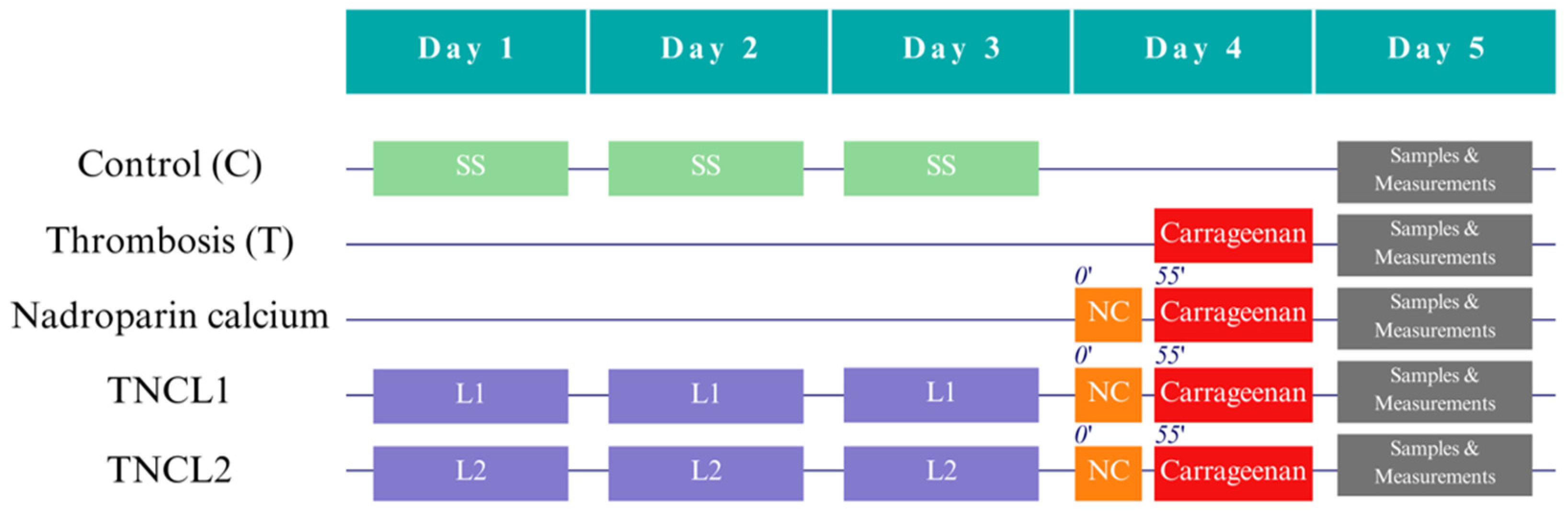
2.1. Study Design

Fifty 16-week-old male Wistar Bratislava albino rats with tails longer than 13 cm were selected for the experiment from the Animal Department of the Faculty of Medicine, “Iuliu Haţieganu” University of Medicine and Pharmacy Cluj-Napoca. The animals weighed 300–400 g and had unrestricted access to standard pellets as food (Cantacuzino Institute, Bucharest, Romania) and water ad libitum. After randomization (simple random method) of the animals into five groups of ten rats each, they were kept in polypropylene cages with a day–night regime at a constant temperature (24 ± 2 °C) and humidity (60 ± 5%) at the Department of Pathophysiology. The experiment flow, the groups, and the specific interventions are summarized in Figure 1.
Carrageenan, a pro-inflammatory sulfated polysaccharide, was used to induce local inflammation. The inflammatory response is accompanied by increased vascular permeability, infiltration of leukocytes, and other inflammatory mediators. In addition, it is known to stimulate the release of inflammatory mediators such as histamine, trypsin, and cytokines [24,25].
The animals were pretreated for three days before induction of thrombosis. We administered 86 UI/kg b.w. of nadroparin calcium subcutaneously 55 min prior to the inducement of thrombosis in the nadroparin calcium group and the lavender oil pretreatment groups. We used kappa-carrageenan (κ-carrageenan) [26] for the experimental tail thrombosis model on Wistar Bratislava rats. On the fourth day of the experiment, we administrated 4 mg/kg b.w. of κ-carrageenan in saline solution into the dorsal vein of the rat tail, followed by ligation of the tail base for 10 min.
Diethylether was used as an anesthetic during thrombosis induction. We used dimethylsulfoxide (DMSO) 5% as a vehicle for Lavandula angustiofolia oil. The substances used for biochemical determinations, ELISA kits, lavender oil, kappa-carrageenan, and DMSO, were purchased from Sigma-Aldrich (St. Louis, MO, USA).
The Ethics Committee of Iuliu Hațieganu University of Medicine and Pharmacy Cluj-Napoca (AVZ82/29.03.2022) approved this study. Our study also have the approval of the National Sanitary Veterinary and Food Safety Authority, Cluj Branch (135/24.05.2022), respecting the national and international requirements. In our experiment, all procedures followed the guidelines of the Institutional Research Committee and the Helsinki Declaration on Animal Studies. The use of animals complied with all applicable international and institutional regulations. We ensured that the animals were handled minimally and experienced minimal suffering.

2.2. Measurements

We collected blood samples from each rat in heparinized tubes from the retro-orbital plexuses 24 h after thrombosis induction. During the procedure, we kept the rats under mild anesthesia with xylazine (2 mg/kg b.w., i.p.) and ketamine (20 mg/kg b.w., i.p.). We centrifuged the blood samples at 4 °C for 20 min at 1620× g to obtain plasma samples. The plasma was placed in Eppendorf tubes and frozen at −80 °C for subsequent examination. At the end of the experiment, the animals were euthanized by an overdose of anesthetics.
Creatinine and urea values were measured to assess the renal function of the rats. Liver function was monitored by the analysis of transaminases, alanine aminotransferase (ALT), and aspartate aminotransferase (AST). Total oxidative stress (TOS), nitric oxide (NOx), and malondialdehyde (MDA) levels were analyzed to evaluate oxidative stress activities. Total antioxidant capacity (TAC) and thiol levels were used to determine the blood’s antioxidant capability. The TECAN SUNRISE spectrophotometer was used for all spectroscopic evaluations. The enzyme-linked immunosorbent assay (ELISA) approach was employed to quantify the plasma concentrations of the inflammatory cytokines, including TNF-α, RANTES, and MCP-1.
The same unblinded researcher conducted measurements of the bleeding time and clotting time using a chronometer. The same unblinded researcher measured the length of the macroscopic thrombosis at the distal end of the tails with a ruler 24 h after administration of κ-carrageenan.
The same researcher who collected all the samples and applied a specific protocol for each procedure, performed the histologic evaluation. Skin fragments were excised from the rat tail, specifically a 3 cm segment located 5 cm from the apex of the tail. The fragments underwent fixation in a 10% formalin solution for four days. Next, the specimens were embedded in paraffin and subsequently sectioned into slices with a thickness of 5 µm. The sections underwent staining using Goldner’s trichrome procedure and were thereafter inspected with an optical microscope.

2.3. Statistical Methods

The plasma levels of the evaluated biomarkers were presented as median values [Q1 to Q3] (where Q1 represents the 25th percentile and Q3 represents the 75th percentile) and min to max (with min indicating the lowest value and max indicating the highest value), to effectively capture the variability of the raw data, considering the limited number of rats in each group. The Kruskal–Wallis test was applied to assess the overall significance of the differences between groups, followed by post-hoc multiple comparisons of mean ranks of all groups whenever statistical significance was reached. The Weissgerber et al. [27] method was used to graphically display the distribution of the evaluated biomarkers. Microsoft Excel (Microsoft Office Excel 365, Redmond, WA, USA) was used to summarize the raw data. The Statistica program (v. 13.5, TIBCO Software Inc, Palo Alto, CA, USA) was used to perform exploratory inferential analysis. All tests were performed as two-tailed tests, and a p-value less than 0.05 was considered statistically significant.

3. Results

Throughout the experiment, no incidents were encountered. As a result, the statistical analysis was conducted on ten rats per group.

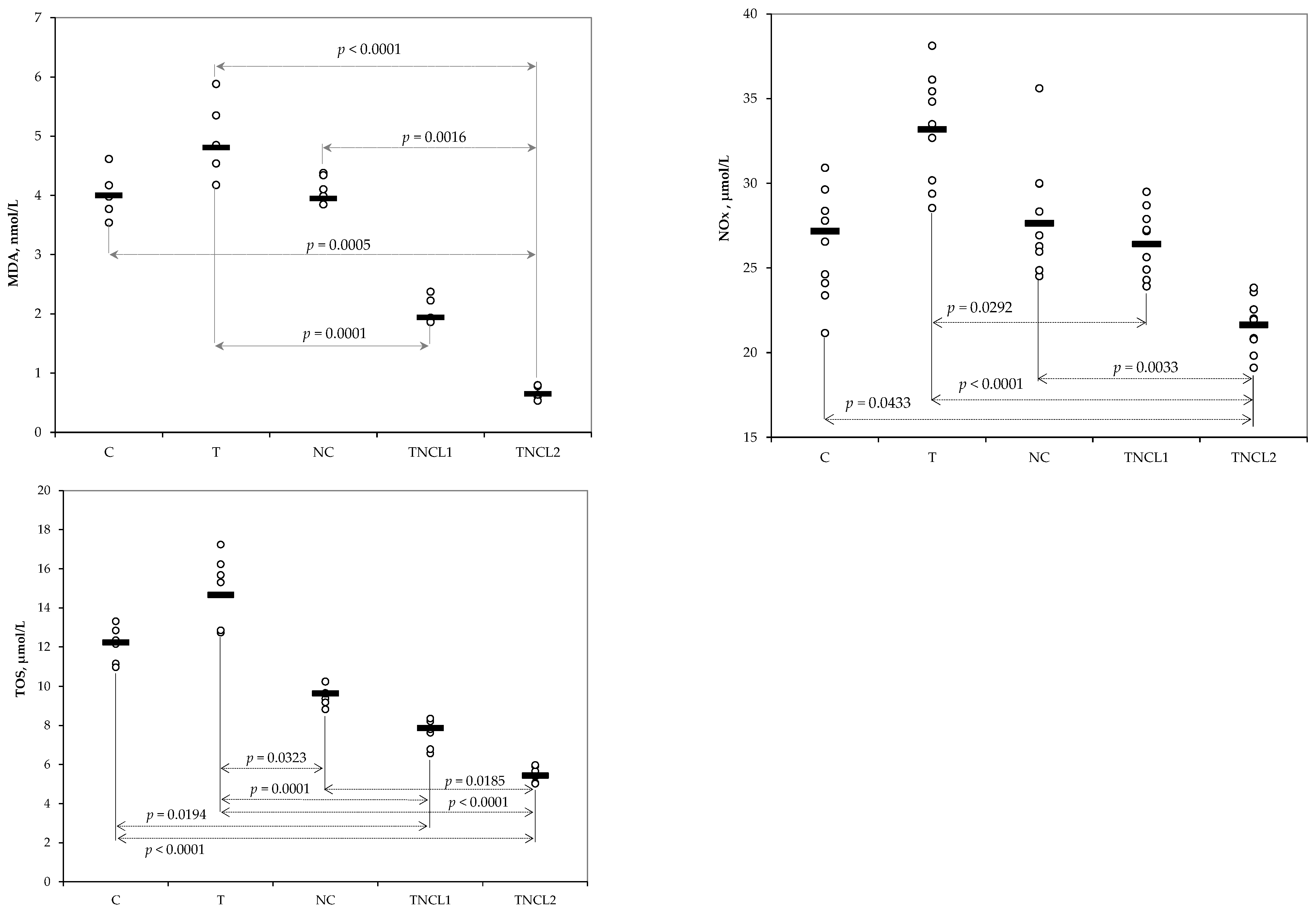
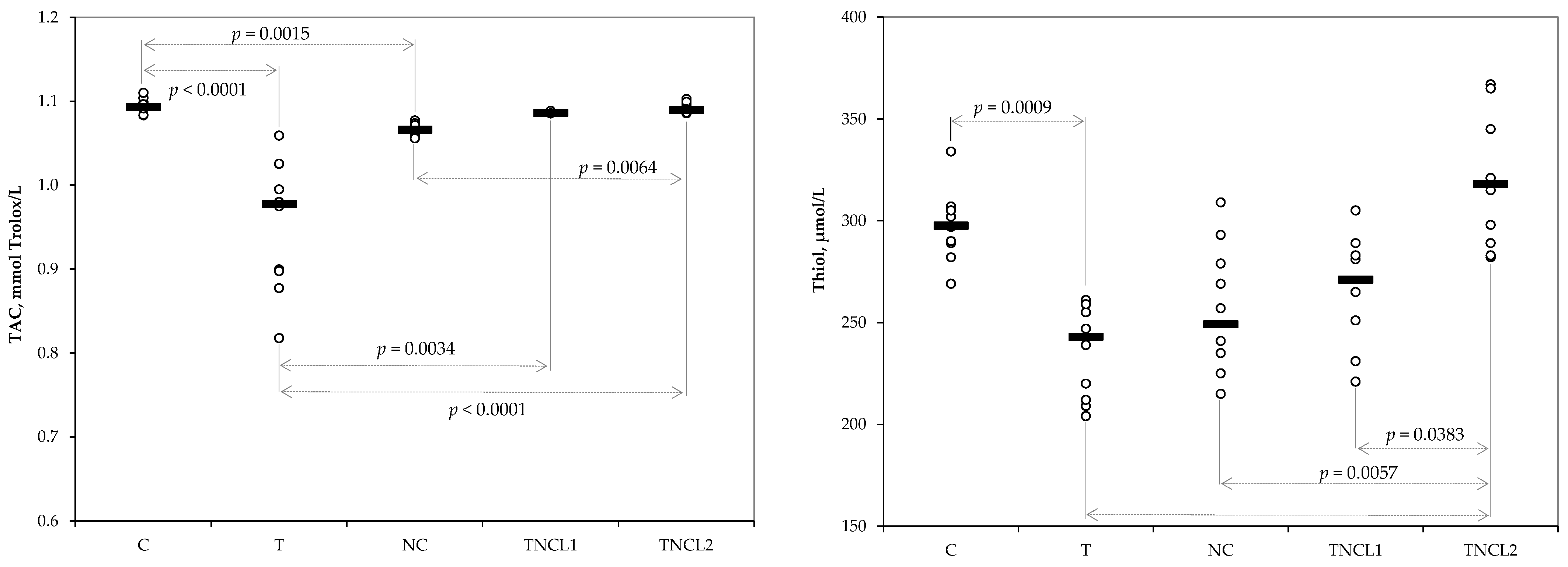
3.1. Levels of Oxidative Stress Parameters

The oxidative stress markers assessed reflected differences between the groups, with statistically significant improvements in the LO pretreated groups compared to the thrombosis group (Table 1 and Figure 2). The TNCL2 group showed significantly lower values for all oxidative stress parameters than the NC group.
The results showed no statistically significant differences between the T and NC groups in plasma levels of total plasma antioxidant capacity (TAC) and thiol. Groups pretreated with LO showed no differences compared to the control group (Table 2 and Figure 3).

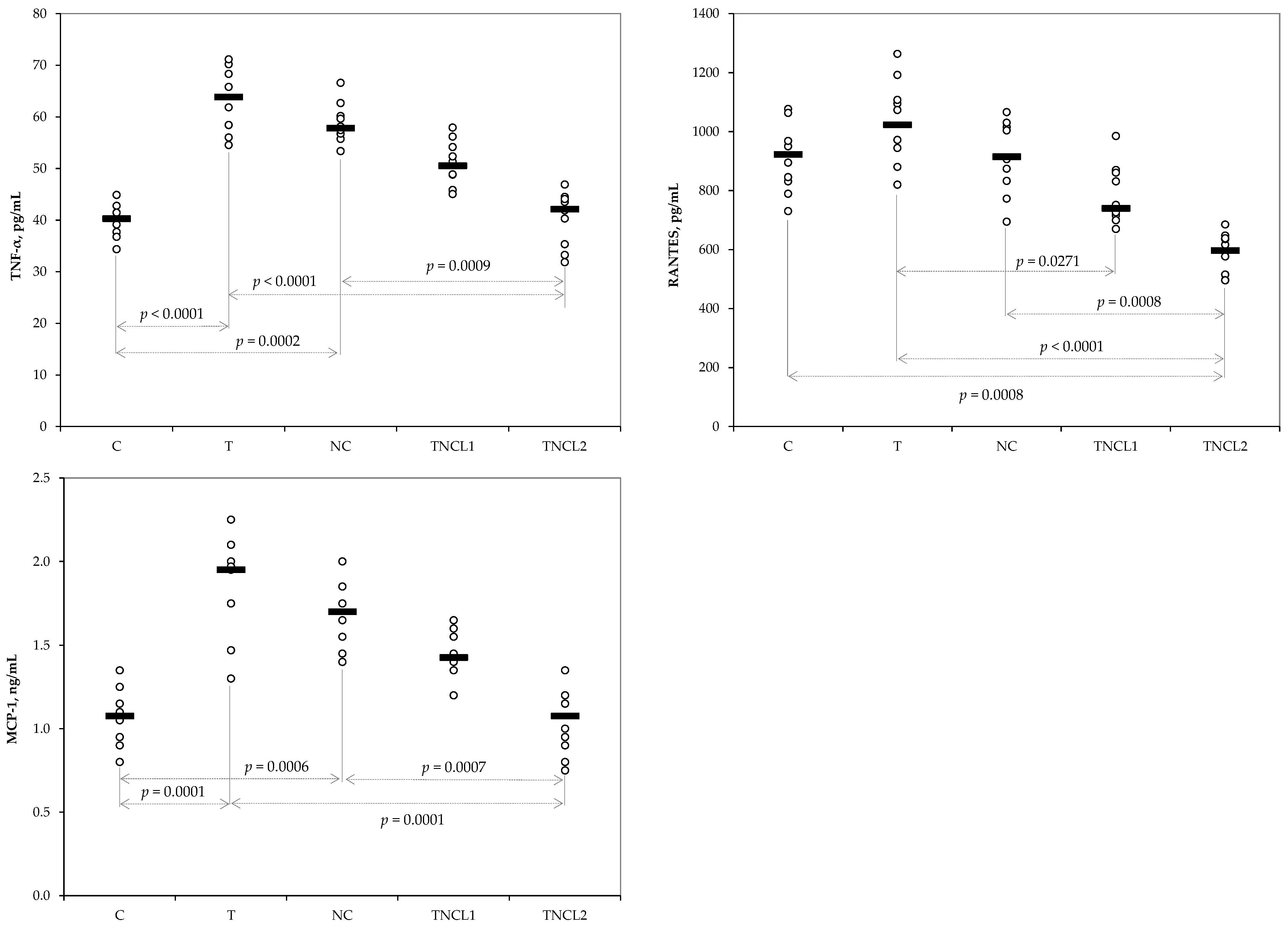
3.2. Plasma Levels of Inflammatory Cytokines

The TNF-α, RANTES, and MPC-1 levels in plasma were significantly higher in the T and NC groups than in the TNCL2 group (Table 3 and Figure 4).

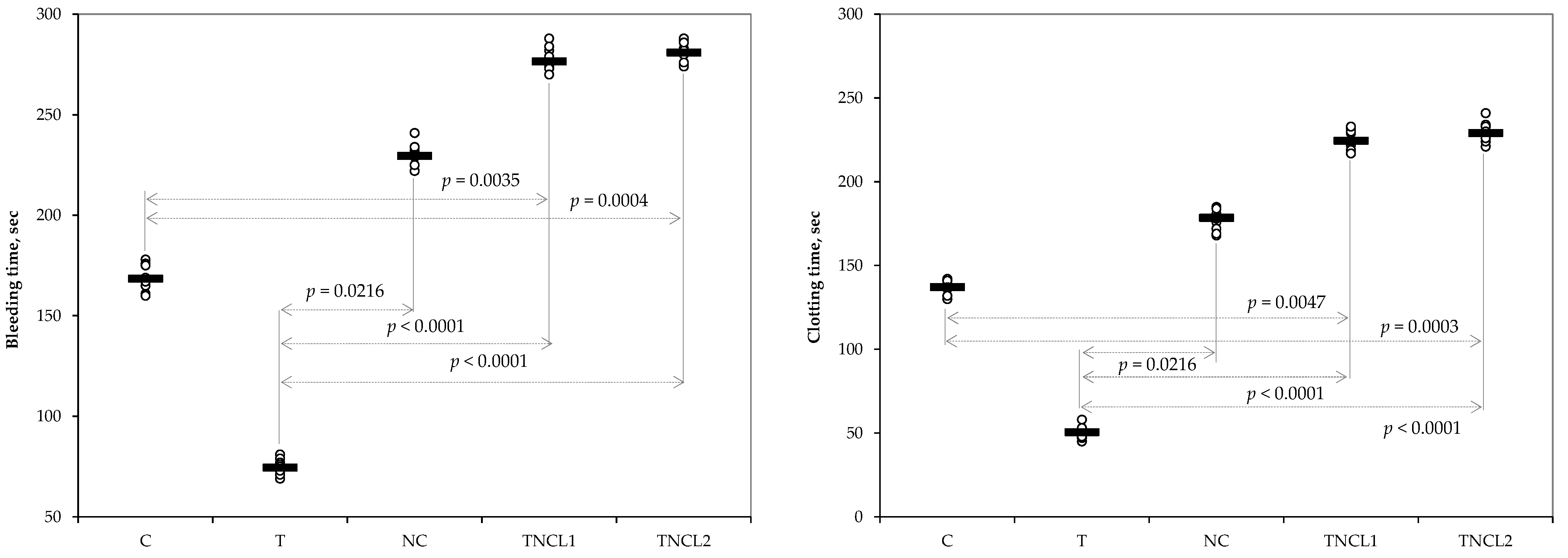
3.3. Variation of Bleeding and Clotting Time and Thrombus Length

The administration of nadroparin calcium effectively inhibits the formation of thrombi, as evidenced by the absence of macroscopic thrombosis. Compared to the T group, the NC, TNCL1, and TNCL2 groups showed significantly increased bleeding and clotting time. No statistically significant differences were observed across nadroparin calcium and lavender oil-treated groups regarding these measurements (Table 4 and Figure 5).

3.4. Renal and Liver Functions

The induction of thrombosis was associated with increased creatinine, urea, ALT, and AST. However, when comparing the control group (C) with the thrombosis group (T), a statistically significant difference was observed for creatinine levels only (Table 5). Prior to nadroparin calcium administration, both doses of lavender oil prevented increases in urea, AST, and ALT levels. The higher LO (200 mg/kg b.w.) prevented liver and kidney toxicity, with levels significantly decreasing in the TNCL1 and TNCL2 groups, except creatinine, where the decrease was not statistically significant.

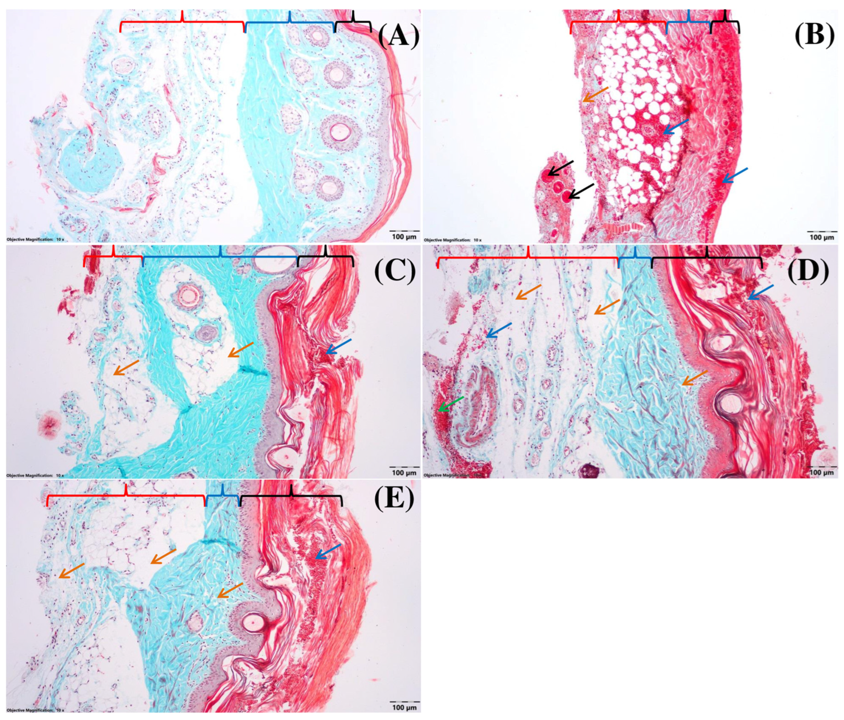
3.5. Histological Assay

The control group (C, Figure 6A) had no microscopic alterations in the epidermis, dermis, and hypodermis. In the T group, various pathological abnormalities were observed within the three skin layers, as shown in Figure 6B. Thrombi formed in the hypodermis led to complete or partial obliteration of the lumen in several veins and venules. The observation of severe diastasis resulted from the obliteration of blood circulation in the venous system within the lax connective tissue of the hypodermis, leading to significant capillary stasis. Multiple lakes of transfused plasma in different phases of coagulation were observable alongside a substantial quantity of red blood cells. Transfused red blood cells were also shown to infiltrate the adipose tissue within the hypodermis.
In the layers of the dermis, veins can be seen where phenomena such as intravascular coagulation and lysis of red blood cells are observed. The interstitial spaces inside dense connective tissue have greater dimensions due to heightened tissue fluid levels. Excessive dilatation of tiny blood vessels and the presence of many red blood cells can be noticed in the superficial dermis. Significant quantities of fluid, together with a multitude of red blood cells, collect beneath the basement membrane of the epidermis, resulting in its detachment from the superficial dermis in certain areas.
Within the epidermis, a multitude of haematocytes may be found among the keratinocytes, particularly in the outermost layer. This presence of haematocytes contributes to the separation of the keratin layers. The presence of edema and subepidermal hemorrhages results in an appearance of nuclear characteristics commonly observed in keratinocytes located in the stratum spinosum and a portion of those in the basal layer of the epidermis, indicative of cells undergoing the first stages of apoptosis.
In the NC group, discrete edema may be observed in the hypodermis (Figure 6C) and superficial dermis. There are no changes in the deep layers (basal and spinous) of the epidermis, but numerous red blood cells are observed in the keratinized cells layer.
In the TNCL1 group, numerous diffuse hemorrhages and no veins were observed in the deep hypodermis (Figure 6D), whose lumen was obliterated by thrombi. Moreover, a pronounced edema in the superficial hypodermis and at the level of the adipose panicles is observed. At the level of the dermis from place to place towards the junction with the epidermis, a discrete edema is present. Cells with pyknotic nuclei can be observed at the epidermis level, mainly in the basal layer and in the first rows of cells of the spinous layer. On the other hand, in the superficial layer between the rows of keratinized cells, there are numerous red blood cells.
In the TNCL2 group, in the hypodermis (Figure 6E), microhemorrhages and edema are present from place to place, which is pronounced in some areas. In the deep dermis, no structural changes are observed. In the superficial layer, the edema is discreet in certain areas, and from place to place, it is pronounced. In the epidermis, especially in the areas where the edema is pronounced in the dermis, numerous cells with pyknotic nuclei can be observed in the basal and spinous layers. Numerous red blood cells are seen in the keratinized layer.

4. Discussion

Our results indicate that the administration of lavender oil as an adjuvant before nadroparin calcium therapy (Fraxiparine) has dose-dependent anti-inflammatory and antioxidant effects in carrageenan-induced thrombosis.
The role of oxidative stress has been emphasized in the onset and advancement of inflammation and coagulation intricated processes [28]. Oxidative stress is a pathological state that occurs due to an imbalance between the synthesis of free radicals and the capacity of antioxidant systems to counteract their effects. The induction of oxidative stress can potentially trigger the formation of thrombi. The onset of endothelial dysfunction caused by oxidative stress may contribute to thrombus formation by decreasing the synthesis of anti-thrombotic molecules and increasing the production of prothrombotic molecules. This phenomenon promotes the recruitment and adhesion of leukocytes and platelets to the endothelial surface, as well as the activation and aggregation of these circulating cells. Oxidative stress can alter the functionality of fibrinogen, leading to defective fibrin polymerization and the subsequent development of altered fibrin networks associated with a prothrombotic phenotype [29]. Clinical data support the link between inflammation and thrombosis and show that inflammatory conditions increase the likelihood of thrombotic events [30]. In contrast, the release of thrombotic elements, including fibrinogen, factor V, and thrombin, can trigger inflammatory cascades, producing an accelerated inflammatory response. Kuijpers et al. [31] highlighted the involvement of tissue factor (TF) and other components of the extrinsic coagulation pathway, which play a crucial role in amplifying inflammatory response. Sriram and Insel highlighted, in the context of dysregulation of angiotensin signaling in patients with SARS-CoV-2, the release of pro-inflammatory (e.g., TNFα and RANTES), prothrombotic (e.g., TXA2), platelet-activating factor (PAF) molecules, and a possible increase in MCP-1 levels, which can enhance both inflammation and platelet activation [32]. Linalool and linalyl acetate are aromatic compounds derived mainly from plants, especially from the Lamiaceae family, such as Lavandula angustifolia. The results show that the compounds can inhibit the activation of signaling pathways that influence the production and maintenance of inflammatory cytokines. In particular, linalool and linalyl acetate were found to inhibit the activation of nuclear factor kappa-light-chain enhancer of activated B cells (NF-Kβ) and mitogen-activated protein kinases (MAPK), suggesting the potential for their use as anti-inflammatory agents [17,33].
Our study shows the efficacy of pretreatment with LO in the improvement of oxidative stress markers (Table 1, Figure 2). The pretreatment with LO induced an increase in total antioxidant capacity (TAC) and plasma levels of thiol in a dose-dependent manner, with the best results being achieved by the group with the highest dose (Table 2 and Figure 3). Our results are consistent with reported results in the scientific literature, namely elevated levels of MDA [34,35]. Our results are also consistent with findings reported on specific diseases. Aboutaleb et al. [36] demonstrated the efficacy of Lavandula angustifolia oil in reducing the oxidative stress on renal ischemia. Amer et al. [37] found evidence of the effectiveness of LO in decreasing malondialdehyde (MDA) levels and increasing total antioxidant capacity in broiler chickens. Linalool and linalyl acetate, the main components of Citrus myrtifolia and Lavandula angustifolia, showed significant anti-inflammatory properties by effectively reducing NOx and MCP-1 levels [38].
Our results suggest that Lavandula angustifolia oil has anti-inflammatory properties attributed to its primary active components, linalool and linalyl acetate. The anti-inflammatory effect is evident in the modulation of inflammatory cytokine levels. The administration of nadroparin calcium did not significantly reduce the plasma levels of TNF-α (Table 3 and Figure 4). The rats that received the highest LO showed the most favorable TNF-α levels, which were statistically significantly decreased compared to the C and NC groups (Table 3 and Figure 4). Fraxiparine (NC group) was inefficient in reducing the amount of the RANTES chemokine or MPC-1 in our study, while the highest dose of LO significantly reduced the plasma levels of RANTES chemokine and MPC-1 compared to the NC group (Table 3 and Figure 4). Our findings align with previous reports. Sadeghzadeh et al. [39] demonstrated that the severity of myocardial injury is reduced by attenuating pro-inflammatory cytokines, TNF-α, and oxidative stress. Ma et al. [40] reported a reduction in the infiltration of inflammatory cells and a reduction in the production of pro-inflammatory cytokines, including TNF-α, IL-6, IL-1β, IL-8, and MCP-1 in pulmonary inflammation caused by cigarette smoking.
The Low Molecular Weight Heparins (LMWH) do not show effects on inflammatory markers and cytokines [41,42]. The administration of nadroparin calcium therapy significantly increased bleeding and clotting times, but the LO did not demonstrate efficacy compared to the NC group (Table 4 and Figure 5). Our results could be explained by the absence of side effects of the LO (e.g., bleeding).
The results observed in our study on the evaluated biomarkers align with the results of histopathological analysis (Figure 6) and demonstrate the anti-thrombotic properties of LO. Our results are consistent with previous in vitro and in vivo studies documenting the anti-thrombotic and antiplatelet properties of LO’s main compounds (linalool and linalyl acetate) [23,43].
Renal and liver function varied between groups, with increased values in the T group compared to the C group (Table 5), showing the impairment induced by thrombosis. The lowest value of plasma levels of creatinine, urea, AST, and ALT were observed in the group with the highest dose of LO. The nephroprotective effect of LO on creatinine levels in an experimental model of kidney injury was reported by Aboutaleb et al. [36]. In addition, linalool is recognized for its natural properties, including anti-inflammatory, antioxidant, and hepatoprotective effects [44].
Our study demonstrates the effectiveness of lavender oil in nadroparin calcium-treated rats with experimental thrombosis. Given the different amounts of chemical compounds in Lavandula from different geographic areas, quantification of the active compounds could increase the power of evidence regarding the anti-inflammatory and oxidative stress effects on thrombosis. The tissue levels of the biomarkers could also be of interest in the demonstration of efficacy as a preliminary step in translation toward investigations on humans. It might also be of interest to investigate the synergic effect of LO in combination with other natural compounds (e.g., curcumin [45,46,47], ginger [24], etc.) added to standard thrombosis treatment to minimize the side effects of standard medications.

5. Conclusions

Our study demonstrated the beneficial anti-inflammatory and antioxidant effects of lavender oil pretreatment associated with nadroparin calcium treatment in rats with experimentally induced thrombosis. Lavandula angustifolia oil’s efficacy varies with dose, with the best effects being observed for a dose of 200 mg/kg b.w. LO and 86 IU/kg b.w. nadroparin calcium. The positive effects on plasma levels of the investigated oxidative stress and antioxidant biomarkers, the reduction in inflammatory cytokines, and the preservation of renal and hepatic functions support the added value of LO.

Author Contributions

Conceptualization, V.M.B., A.E.B., I.C.S. and S.D.B.; methodology, V.M.B., A.E.B., S.D.B., V.R. and M.L.G.; validation, S.D.B.; formal analysis, V.M.B. and S.D.B.; investigation, V.M.B., A.E.B., V.R., T.I. and M.L.G.; resources, V.M.B., A.E.B., V.R., T.I., M.L.G. and S.D.B.; data curation, S.D.B.; writing—original draft preparation, V.M.B.; writing—review and editing, A.E.B., V.R., I.C.S., T.I. and S.D.B.; visualization, V.R. and S.D.B.; supervision, A.E.B. and S.D.B.; project administration, V.M.B. and A.E.B.; funding acquisition, V.M.B. All authors have read and agreed to the published version of the manuscript.

Funding

This research was funded by the “Iuliu Hațieganu” University of Medicine and Pharmacy Cluj-Napoca grant number PCD 881/9/12.01.2022.

Institutional Review Board Statement

The animal experiments were conducted in accordance with the protocols approved by the Ethics Committee of the Iuliu Haţieganu University of Medicine and Pharmacy Cluj-Napoca (AVZ82/29.03.2022), and this study had approval from the National Sanitary Veterinary and Food Safety Authority, Cluj Branch (135/24.05.2022).

Informed Consent Statement

Not applicable.

Data Availability Statement

The raw data analyzed in this study are part of a Ph.D. study and can be obtained upon reasonable request addressed to Valeriu Mihai But ([email protected]).

Acknowledgments

The authors would like to thank Uifalean Ana for helping us with the biochemical assays and Molnar Mirel for helping handle the rats during the experiment.

Conflicts of Interest

The authors declare no conflicts of interest.

References

  1. Wendelboe, A.M.; Raskob, G.E. Global Burden of Thrombosis: Epidemiologic Aspects. Circ. Res. 2016, 118, 1340–1347. [Google Scholar] [CrossRef]
  2. ISTH Steering Committee for World Thrombosis Day. Thrombosis: A major contributor to the global disease burden. J. Thromb. Haemost. 2014, 12, 1580–1590. [Google Scholar] [CrossRef]
  3. Lozano, R.; Naghavi, M.; Foreman, K.; Lim, S.; Shibuya, K.; Aboyans, V.; Abraham, J.; Adair, T.; Aggarwal, R.; Ahn, S.J.; et al. Global and regional mortality from 235 causes of death for 20 age groups in 1990 and 2010: A systematic analysis for the Global Burden of Disease Study 2010. Lancet 2012, 380, 2095–2128. [Google Scholar] [CrossRef]
  4. Martin, K.A.; Molsberry, R.; Cuttica, M.J.; Desai, K.R.; Schimmel, D.R.; Khan, S.S. Time Trends in Pulmonary Embolism Mortality Rates in the United States, 1999 to 2018. J. Am. Heart Assoc. 2020, 9, e016784. [Google Scholar] [CrossRef] [PubMed]
  5. Lutsey, P.L.; Zakai, N.A. Epidemiology and prevention of venous thromboembolism. Nat. Rev. Cardiol. 2023, 20, 248–262. [Google Scholar] [CrossRef] [PubMed]
  6. Brækkan, S.K.; Hansen, J.B. VTE epidemiology and challenges for VTE prevention at the population level. Thromb. Update 2023, 10, 100132. [Google Scholar] [CrossRef]
  7. Siegal, D.M.; Eikelboom, J.W.; Lee, S.F.; Rangarajan, S.; Bosch, J.; Zhu, J.; Yusuf, S. Venous Thromboembolism Collaboration. Variations in incidence of venous thromboembolism in low-, middle-, and high-income countries. Cardiovasc. Res. 2021, 117, 576–584. [Google Scholar] [CrossRef] [PubMed]
  8. Prandoni, P.; Noventa, F.; Ghirarduzzi, A.; Pengo, V.; Bernardi, E.; Pesavento, R.; Iotti, M.; Tormene, D.; Simioni, P.; Pagnan, A. The risk of recurrent venous thromboembolism after discontinuing anticoagulation in patients with acute proximal deep vein thrombosis or pulmonary embolism. A prospective cohort study in 1626 patients. Haematologica 2007, 92, 199–205. [Google Scholar] [CrossRef] [PubMed]
  9. Naess, I.A.; Christiansen, S.C.; Romundstad, P.; Cannegieter, S.C.; Rosendaal, F.R.; Hammerstrøm, J. Incidence and mortality of venous thrombosis: A population-based study. J. Thromb. Haemost. 2007, 5, 692–699. [Google Scholar] [CrossRef] [PubMed]
  10. De Azeredo, E.L.; Monteiro, R.Q.; de-Oliveira Pinto, L.M. Thrombocytopenia in Dengue: Interrelationship between Virus and the Imbalance between Coagulation and Fibrinolysis and Inflammatory Mediators. Mediat. Inflamm. 2015, 2015, 313842. [Google Scholar] [CrossRef] [PubMed]
  11. Li, H.; Liu, B.; Wu, J.; Yu, H.; Huang, H.; Chen, X.; Chen, B.; Wu, S.; Ma, J.; Liu, W.; et al. The inhibitory effect of tachyplesin I on thrombosis and its mechanisms. Chem. Biol. Drug Des. 2019, 94, 1672–1679. [Google Scholar] [CrossRef] [PubMed]
  12. Barradell, L.B.; Buckley, M.M. Nadroparin Calcium1: A Review of its Pharmacology and Clinical Applications in the Prevention and Treatment of Thromboembolic Disorders. Drugs 1992, 44, 858–888. [Google Scholar] [CrossRef] [PubMed]
  13. Casu, B.; Oreste, P.; Torri, G.; Zoppetti, G.; Choay, J.; Lormeau, J.C.; Petitou, M.; Sinac, P. The structure of heparin oligosaccharide fragments with high anti-(factor Xa) activity containing the minimal antithrombin III-binding sequence. Chemical and 13C nuclear-magnetic-resonance studies. Biochem. J. 1981, 197, 599–609. [Google Scholar] [CrossRef]
  14. Zhong, G.L.; Gong, Y.; Yu, C.; Wu, S.; Ma, Q.; Wang, Y.; Ren, J.; Zhang, X.-C.; Yang, W.-H.; Zhu, W. Significantly inhibitory effects of low molecular weight heparin (Fraxiparine) on the motility of lung cancer cells and its related mechanism. Tumor Biol. 2015, 36, 4689–4697. [Google Scholar] [CrossRef]
  15. Souri, F.; Rakhshan, K.; Erfani, S.; Azizi, Y.; Nasseri Maleki, S.; Aboutaleb, N. Natural lavender oil (Lavandula angustifolia) exerts cardioprotective effects against myocardial infarction by targeting inflammation and oxidative stress. Inflammopharmacology 2019, 27, 799–807. [Google Scholar] [CrossRef]
  16. Białoń, M.; Krzyśko-Łupicka, T.; Nowakowska-Bogdan, E.; Wieczorek, P.P. Chemical Composition of Two Different Lavender Essential Oils and Their Effect on Facial Skin Microbiota. Molecules 2019, 24, 3270. [Google Scholar] [CrossRef]
  17. Pandur, E.; Balatinácz, A.; Micalizzi, G.; Mondello, L.; Horváth, A.; Sipos, K.; Horváth, G. Anti-inflammatory effect of lavender (Lavandula angustifolia Mill.) essential oil prepared during different plant phenophases on THP-1 macrophages. BMC Complement Med. Ther. 2021, 21, 287. [Google Scholar] [CrossRef]
  18. Giovannini, D.; Gismondi, A.; Basso, A.; Canuti, L.; Braglia, R.; Canini, A.; Mariani, F.; Cappeli, G. Lavandula angustifolia Mill. Essential Oil Exerts Antibacterial and Anti-Inflammatory Effect in Macrophage Mediated Immune Response to Staphylococcus aureus. Immunol. Investig. 2016, 45, 11–28. [Google Scholar] [CrossRef]
  19. Cardia, G.F.E.; Silva-Filho, S.E.; Silva, E.L.; Uchida, N.S.; Cavalcante, H.A.O.; Cassarotti, L.L.; Salvadego, V.E.C.; Spironello, R.A.; Bersani-Amado, C.A.; Cuman, R.K.N. Effect of Lavender (Lavandula angustifolia) Essential Oil on Acute Inflammatory Response. Evid. Based Complement. Altern. Med. 2018, 2018, 1413940. [Google Scholar] [CrossRef]
  20. Silva, G.L.; Luft, C.; Lunardelli, A.; Amaral, R.H.; Melo, D.A.; Donadio, M.V.; Nunes, F.B.; Azambuja, M.S.D.; Santana, J.C.; Moraes, C.M.B. Antioxidant, analgesic and anti-inflammatory effects of lavender essential oil. An. Acad. Bras. Cienc. 2015, 87, 1397–1408. [Google Scholar] [CrossRef]
  21. Hawrył, A.; Hawrył, M.; Waksmundzka-Hajnos, M. Liquid chromatography fingerprint analysis and antioxidant activity of selected lavender species with chemometric calculations. PLoS ONE 2019, 14, e0218974. [Google Scholar] [CrossRef] [PubMed]
  22. Ben Salah, M.; Abderraba, M.; Tarhouni, M.R.; Abdelmelek, H. Effects of ultraviolet radiation on the kinetics of in vitro percutaneous absorption of lavender oil. Int. J. Pharm. 2009, 382, 33–38. [Google Scholar] [CrossRef] [PubMed]
  23. But, V.M.; Bulboacă, A.E.; Rus, V.; Ilyés, T.; Gherman, M.L.; Bolboacă, S.D. Anti-inflammatory and antioxidant efficacy of lavender oil in experimentally induced thrombosis. Thromb. J. 2023, 21, 85. [Google Scholar] [CrossRef] [PubMed]
  24. Boarescu, I.; Pop, R.M.; Boarescu, P.-M.; Bocsan, I.C.; Gheban, D.; Bulboaca, A.E.; Buzoianu, A.D.; Bolboaca, S.D. Ginger (Zingiber officinale) Root Capsules Enhance Analgesic and Antioxidant Efficacy of Diclofenac Sodium in Experimental Acute Inflammation. Antioxidants 2023, 12, 745. [Google Scholar] [CrossRef] [PubMed]
  25. Hagimori, M.; Kamiya, S.; Yamaguchi, Y.; Arakawa, M. Improving frequency of thrombosis by altering blood flow in the carrageenan-induced rat tail thrombosis model. Pharmacol. Res. 2009, 60, 320–323. [Google Scholar] [CrossRef] [PubMed]
  26. Kod’ousek, R.; Jezdínský, J.; Krajcí, D. Histological and ultrastructural changes of cardiomyocytes in experimental rats with tail thrombosis following subplantar application of carrageenin. Med. Princ. Pract. Int. J. Kuwait Univ. Health Sci. Cent. 2007, 16, 360–366. [Google Scholar] [CrossRef] [PubMed]
  27. Weissgerber, T.L.; Milic, N.M.; Winham, S.J.; Garovic, V.D. Beyond bar and line graphs: Time for a new data presentation paradigm. PLoS Biol. 2015, 13, e1002128. [Google Scholar] [CrossRef] [PubMed]
  28. Lei, Y.; Wang, K.; Deng, L.; Chen, Y.; Nice, E.C.; Huang, C. Redox Regulation of Inflammation: Old Elements, a New Story: Redox Modifications in Inflammation. Med. Res. Rev. 2015, 35, 306–340. [Google Scholar] [CrossRef]
  29. Medcalf, R.L. Fibrinolysis, inflammation, and regulation of the plasminogen activating system. J. Thromb. Haemost. 2007, 5, 132–142. [Google Scholar] [CrossRef]
  30. Stark, K.; Massberg, S. Interplay between inflammation and thrombosis in cardiovascular pathology. Nat. Rev. Cardiol. 2021, 18, 666–682. [Google Scholar] [CrossRef]
  31. Kuijpers, M.J.E.; Heemskerk, J.W.M.; Jurk, K. Molecular Mechanisms of Hemostasis, Thrombosis and Thrombo-Inflammation. Int. J. Mol. Sci. 2022, 23, 5825. [Google Scholar] [CrossRef]
  32. Sriram, K.; Insel, P.A. Inflammation and thrombosis in COVID-19 pathophysiology: Proteinase-activated and purinergic receptors as drivers and candidate therapeutic targets. Physiol. Rev. 2021, 101, 545–567. [Google Scholar] [CrossRef]
  33. Rai, V.K.; Sinha, P.; Yadav, K.S.; Shukla, A.; Saxena, A.; Bawankule, D.U.; Tandon, S.; Khan, F.; Chanotiya, C.S.; Yadav, N.P. Anti-psoriatic effect of Lavandula angustifolia essential oil and its major components linalool and linalyl acetate. J. Ethnopharmacol. 2020, 261, 113127. [Google Scholar] [CrossRef]
  34. Aykal, G.; Güven, R.; Yeğin, A.; Ellidağ, H.Y.; Bayindir, A.; Yilmaz, N. The Diagnostic Value of Oxidative/Antioxidative Balance Parameters in Venous Thromboembolism. Clin. Lab. 2015, 61, 769–775. [Google Scholar] [CrossRef]
  35. Türker, F.S.; Malbora, A.; Erisir, M. Oxidative status and antioxidant enzyme levels in deep venous thrombosis patients. Am. J. Cardiovasc. Dis. 2021, 11, 176–183. [Google Scholar]
  36. Aboutaleb, N.; Jamali, H.; Abolhasani, M.; Pazoki Toroudi, H. Lavender oil (Lavandula angustifolia) attenuates renal ischemia/reperfusion injury in rats through suppression of inflammation, oxidative stress and apoptosis. Biomed. Pharmacother. 2019, 110, 9–19. [Google Scholar] [CrossRef]
  37. Amer, S.A.; Abdel-Wareth, A.A.A.; Gouda, A.; Saleh, G.K.; Nassar, A.H.; Sherief, W.R.I.A.; Albogami, S.; Shalaby, S.I.; Abdelazim, A.M.; Abomughaid, M.M.I. Impact of Dietary Lavender Essential Oil on the Growth and Fatty Acid Profile of Breast Muscles, Antioxidant Activity, and Inflammatory Responses in Broiler Chickens. Antioxidants 2022, 11, 1798. [Google Scholar] [CrossRef]
  38. Plastina, P.; Apriantini, A.; Meijerink, J.; Witkamp, R.; Gabriele, B.; Fazio, A. In Vitro Anti-Inflammatory and Radical Scavenging Properties of Chinotto (Citrus myrtifolia Raf.) Essential Oils. Nutrients 2018, 10, 783. [Google Scholar] [CrossRef] [PubMed]
  39. Sadeghzadeh, J.; Vakili, A.; Bandegi, A.R.; Sameni, H.R.; Zahedi Khorasani, M.; Darabian, M. Lavandula Reduces Heart Injury via Attenuating Tumor Necrosis Factor-Alpha and Oxidative Stress in A Rat Model of Infarct-Like Myocardial Injury. Cell J. 2017, 19, 84–93. [Google Scholar] [CrossRef] [PubMed]
  40. Ma, J.; Xu, H.; Wu, J.; Qu, C.; Sun, F.; Xu, S. Linalool inhibits cigarette smoke-induced lung inflammation by inhibiting NF-κB activation. Int. Immunopharmacol. 2015, 29, 708–713. [Google Scholar] [CrossRef] [PubMed]
  41. Kursat Poyrazoglu, O.; Dogukan, A.; Yalnız, M.; Seckin, D.; lhsan Gunal, A. Acute Effect of Standard Heparin Versus Low Molecular Weight Heparin on Oxidative Stress and Inflammation in Hemodialysis Patients. Ren. Fail. 2006, 28, 723–727. [Google Scholar] [CrossRef]
  42. Romano, L.G.R.; Hunfeld, N.G.M.; Kruip, M.J.H.A.; Endeman, H.; Preijers, T. Population pharmacokinetics of nadroparin for thromboprophylaxis in COVID-19 intensive care unit patients. Br. J. Clin. Pharmacol. 2023, 89, 1617–1628. [Google Scholar] [CrossRef]
  43. Ballabeni, V.; Tognolini, M.; Chiavarini, M.; Impicciatore, M.; Bruni, R.; Bianchi, A.; Barocelli, E. Novel antiplatelet and antithrombotic activities of essential oil from Lavandula hybrida Reverchon “grosso”. Phytomedicine 2004, 11, 596–601. [Google Scholar] [CrossRef] [PubMed]
  44. Venmathi Maran, B.A.; Iqbal, M.; Gangadaran, P.; Ahn, B.C.; Rao, P.V.; Shah, M.D. Hepatoprotective Potential of Malaysian Medicinal Plants: A Review on Phytochemicals, Oxidative Stress, and Antioxidant Mechanisms. Molecules 2022, 27, 1533. [Google Scholar] [CrossRef] [PubMed]
  45. Dogaru, G.; Bulboaca, A.E.; Gheban, D.; Boarescu, P.M.; Rus, V.; Festila, D.; Sitar-Taut, A.-V.; Stanescu, I. Effect of Liposomal Curcumin on Acetaminophen Hepatotoxicity by Down-regulation of Oxidative Stress and Matrix Metalloproteinases. In Vivo 2020, 34, 569–582. [Google Scholar] [CrossRef] [PubMed]
  46. Bulboaca, A.E.; Porfire, A.; Bolboaca, S.D.; Nicula, C.A.; Festila, D.G.; Roman, A.; Râjnoveanu, R.M.; Râjnoveanu, A.; Dogaru, G.; Boarescu, P.-M.; et al. Protective Effects of Liposomal Curcumin on Oxidative Stress/Antioxidant Imbalance, Metalloproteinases 2 and -9, Histological Changes and Renal Function in Experimental Nephrotoxicity Induced by Gentamicin. Antioxidants 2021, 10, 325. [Google Scholar] [CrossRef] [PubMed]
  47. Boarescu, I.; Pop, R.M.; Boarescu, P.-M.; Bocșan, I.C.; Gheban, D.; Râjnoveanu, R.-M.; Râjnoveanu, A.; Bulboacă, A.E.; Buzoianu, A.D.; Bolboacă, S.D. Anti-Inflammatory and Analgesic Effects of Curcumin Nanoparticles Associated with Diclofenac Sodium in Experimental Acute Inflammation. Int. J. Mol. Sci. 2022, 23, 11737. [Google Scholar] [CrossRef]
Figure 1. Group and treatment flow. The control group (C) was injected with saline solution (SS) 0.9% 1 mL i.p. (intraperitoneal) and DMSO (dimethyl sulfoxide) as a vehicle; thrombosis (T) was induced on the fourth day of the experiment with k-carrageenan 4 mg/kg b.w. (b.w. = body weight), 1 mL i.v. (intravenous) in T; the nadroparin calcium (NC) group was pretreated with nadroparin calcium 86 UI/kg b.w. subcutaneously (s.c.); the TNCL1 group was pretreated with lavender oil i.p. (100 mg/kg b.w.) and vehicle, and the TNCL2 group was pretreated with lavender oil i.p. (200 mg/kg b.w.) and vehicle.
Applsci 14 01852 g001
Figure 2. Plasma levels of oxidative stress markers by groups. (C = control group; T = thrombosis group; NC = subcutaneous nadroparin calcium group; TNCL1 = lavender oil 100 mg/kg b.w. and nadroparin calcium; TNCL2 = lavender oil 200 mg/kg b.w. and nadroparin calcium).
Applsci 14 01852 g002
Figure 3. Plasma levels of antioxidant capacity and thiol by groups. (C = control group; T =thrombosis group; NC = subcutaneous nadroparin calcium group; TNCL1 = lavender oil 100 mg and nadroparin calcium group; TNCL2 = lavender oil 200 mg and nadroparin calcium group).
Applsci 14 01852 g003
Figure 4. Plasma levels of measured inflammatory cytokines by groups. (C = control group; T = thrombosis group; NC = subcutaneous nadroparin calcium group; TNCL1 = lavender oil 100 mg/kg b.w. and nadroparin calcium; TNCL2 = lavender oil 200 mg/kg b.w. and nadroparin calcium).
Applsci 14 01852 g004
Figure 5. Bleeding and clotting time by group. (C = control group; T = thrombosis group; NC = subcutaneous nadroparin calcium group; TNCL1 = lavender oil 100 mg/kg b.w. and nadroparin calcium; TNCL2 = lavender oil 200 mg/kg b.w. and nadroparin calcium groups).
Applsci 14 01852 g005
Figure 6. Histopathological examination of the tails. (A)—control (C) group; (B)—thrombosis (T) group; (C)—nadroparin calcium (NC) group; (D)—lavender oil 100 mg (TNCL1) group; (E)—lavender oil 200 mg (TNCL2) group. Goldner’s trichrome stain; red brace—hypodermis; blue brace—dermis; black brace—epidermis; black arrow—thrombosis; blue arrow—hemorrhage; orange arrow—edema; green arrow—congestion.
Applsci 14 01852 g006
Table 1. Levels of oxidative stress markers in plasma by groups.
GroupMDA, nmol/LNOx, μmol/LTOS, μmol H2O2 equiv./L
C4 [3.77 to 4.34]27.18 [24.23 to 29.32]12.23 [11.13 to 12.72]
{3.42 to 4.73}{21.17 to 30.91}{10.39 to 13.68}
T4.81 [4.56 to 5.3]33.18 [30.8 to 35.28]14.66 [13.88 to 16.09]
{4.18 to 5.88}{28.54 to 38.12}{12.73 to 17.23}
NC3.95 [3.80 to 4.08]27.63 [26.05 to 30.00]9.64 [9.23 to 10.23]
{3.44 to 4.38}{24.52 to 35.61}{8.5 to 10.98}
TNCL11.94 [1.91 to 2.18]26.41 [24.67 to 27.73]7.87 [7.51 to 8.18]
{1.86 to 2.38}{23.91 to 29.50}{6.56 to 8.35}
TNCL20.64 [0.61 to 0.76]21.64 [20.80 to 22.42]5.43 [5.03 to 5.64]
{0.52 to 0.82}{19.11 to 23.84}{4.80 to 5.97}
* stat. (p-value)43.4 (<0.0001)32.8 (<0.0001)46.3 (<0.0001)
Data are summarized as median [Q1 to Q3] and min to max, where Q1 and Q3 are the first and third quartiles, min is the minimum, and max is the maximum value. * Kruskal–Wallis test; C = control group; T = thrombosis group; NC = subcutaneous nadroparin calcium group; TNCL1 = lavender oil 100 mg/kg b.w. and nadroparin calcium; TNCL2 = lavender oil 200 mg/kg b.w. and nadroparin calcium; MDA = malondialdehyde; NOx = nitric oxide; TOS = total oxidative stress.
Table 2. Levels of antioxidant capacity and thiol in plasma by groups.
GroupTAC, mmol Trolox/LTHIOL, μmol/L
C1.093 [1.091 to 1.098]297.5 [289.25 to 304.25]
{1.08 to 1.11}{269 to 334}
T0.98 [0.9 to 0.99]243 [214 to 254.5]
{0.82 to 1.06}{204 to 261}
NC1.07 [1.06 to 1.07]249 [236 to 276.5]
{1.05 to 1.08}{215 to 309}
TNCL11.086 [1.086 to 1.088]271 [254.5 to 282.5]
{1.085 to 1.088}{221 to 305}
TNCL21.089 [1.086 to 1.094]318 [291.3 to 342.8]
{1.09 to 1.10}{282 to 367}
* stat. (p-value)40.4 (<0.0001)29.9 (<0.0001)
* Kruskal–Wallis test. C = control group; T = thrombosis group; NC = subcutaneous nadroparin calcium group; TNCL1 = lavender oil 100 mg and nadroparin calcium group; TNCL2 = lavender oil 200 mg and nadroparin calcium group. TAC = total antioxidant capacity.
Table 3. Levels of inflammatory cytokines in plasma by groups.
GroupTNF-α, pg/mLRANTES, pg/mLMCP-1, ng/mL
C40.28 [38.08 to 41.21]922.68 [835.29 to 985.39]1.08 [0.91 to 1.23]
{34.38 to 44.92}{730.45 to 1077.15}{0.8 to 1.35}
T63.85 [58.42 to 69.75]1022.75 [949.84 to 1104.55]1.95 [1.54 to 1.99]
{54.56 to 77.05}{820.15 to 1263.8}{1.3 to 2.25}
NC57.82 [55.06 to 60.06]914.5 [843.34 to 1012.98]1.7 [1.58 to 1.85]
{52.81 to 66.63}{694.9 to 1066.55}{1.4 to 2}
TNCL150.52 [48.85 to 53.72]739.2 [703.98 to 853.64]1.43 [1.31 to 1.55]
{45.06 to 57.93}{670.5 to 985.4}{1.2 to 1.65}
TNCL242.13 [36.57 to 43.96]596.85 [518.06 to 637.93]1.08 [0.91 to 1.20]
{31.86 to 46.92}{496.45 to 685.5}{0.75 to 1.35}
* stat. (p-value)41.0 (<0.0001)32.4 (<0.0001)35.6 (<0.0001)
Data are summarized as the median [Q1 to Q3] and min to max, where Q1 is the 25th percentile, Q3 is the 75th percentile, min is the minimum, and max is the maximum value. * Kruskal–Wallis test. C = control group; T = thrombosis group; NC = subcutaneous nadroparin calcium group; TNCL1 = lavender oil 100 mg/kg b.w. and nadroparin calcium; TNCL2 = lavender oil 200 mg/kg b.w. and nadroparin calcium; TNF-α = tumor necrosis factor α; RANTES = regulated on activation, normal T cell expressed and secreted; MCP-1 = monocyte chemoattractant protein 1.
Table 4. Variation in thrombus length, bleeding, and clotting time by groups.
GroupLength, cmBleeding Time, sClotting Time, s
C0168.5 [165.5 to 175.75]137 [133 to 140.5]
{160 to 178}{130 to 142}
T9.25 [8.95 to 9.58]74.5 [71.5 to 76.75]50.5 [47.25 to 52.75]
{8.3 to 9.9}{69 to 81}{45 to 58}
NC0229.5 [228.25 to 231.75]178.5 [173 to 181.75]
{222 to 241}{168 to 185}
TNCL10276.5 [274 to 281.25]224.5 [221.25 to 229.75]
{270 to 288}{217 to 233}
TNCL20281 [276.25 to 285.25]229 [226.25 to 233.75]
{274 to 288}{221 to 241}
* stat. (p-value)n.a.45.0 (<0.0001)45.2 (<0.0001)
Data are summarized as the median [Q1 to Q3] and min to max, where Q1 is the 25th percentile, Q3 is the 75th percentile, min is the minimum, and max is the maximum value. * Kruskal–Wallis test. C = control group; T = thrombosis group; NC = subcutaneous nadroparin calcium group; TNCL1 = lavender oil 100 mg/kg b.w. and nadroparin calcium; TNCL2 = lavender oil 200 mg/kg b.w. and nadroparin calcium.
Table 5. Levels of creatinine, urea, ALT, and AST in plasma by groups.
GroupCreatinine, mg/dLUrea, mg/dLALT, UIAST, UI
C0.86 [0.79 to 0.92]49.15 [46.42 to 50.99]45.47 [42.61 to 48.24]41.93 [38.19 to 43.79]
{0.73 to 1.01}{43.05 to 54.35}{37.88 to 49.99}{31.75 to 47.24}
T0.98 [0.96 to 1.01] a154.39 [50.37 to 57.1]58.02 [54.69 to 61.45]50.64 [45.7 to 53.53]
{0.9 to 1.22}{46.31 to 61.03}{53.01 to 67.52}{43.48 to 56.88}
NC0.90 [0.88 to 0.96]46.63 [45.67 to 49.69]42.7 [39.68 to 45.45]44.96 [42.85 to 49.95]
{0.75 to 0.98}{44.38 to 52.1}{38.21 to 52.52}{39.93 to 52.11}
TNCL10.90 [0.82 to 0.92]45.99 [45.19 to 46.79] b135.84 [32.95 to 38.97] c135.95 [33.4 to 38.51] d1,d2
{0.79 to 0.99}{42.45 to 48.24}{29.48 to 42.7}{31.38 to 40.92}
TNCL20.79 [0.76 to 0.85] a240.85 [38.06 to 41.93] b2,b3,b431.03 [29.7 to 33.72] c2,c3,c429.02 [26.08 to 30.4] d3,d4,d5
{0.71 to 0.87}{35.03 to 44.38}{26.43 to 35.95}{24.4 to 32.4}
* stat. (p-value)22.1 (0.0001)32.7 (<0.0001)40.5 (<0.0001)39.5 (<0.0001)
Data are summarized as the median [Q1 to Q3] and min to max, where Q1 is the 25th percentile, Q3 is the 75th percentile, min is the minimum, and max is the maximum value. * Kruskal–Wallis test. C = control group; T = thrombosis group; NC = subcutaneous nadroparin calcium group; TNCL1 = lavender oil 100 mg/kg b.w. and nadroparin calcium; TNCL2 = lavender oil 200 mg/kg b.w. and nadroparin calcium; ALT = alanine aminotransferase; AST = aspartate aminotransferase. Post-hoc analysis: a1 0.029 C vs. T; a2 <0.0001 T vs. TNCL2; b1 0.0232 T vs. TNCL1; b2 0.0008 C vs. TNCL2; b3 <0.0001 T vs. TNCL2; b4 0.0124 NC vs. TNCL2; c1 <0.0001 T vs. TNCL1; c2 0.0029 C vs. TNCL2; c3 <0.0001 T vs. TNCL2; c4 0.01751 NC vs. TNCL2; d1 0.0010 T vs. TNCL1; d2 0.0454 NC vs. TNCL1; d3 0.0175 C vs. TNCL2; d4 <0.0001 T vs. TNCL2; d5 <0.0001 NC vs. TNCL2.
Disclaimer/Publisher’s Note: The statements, opinions and data contained in all publications are solely those of the individual author(s) and contributor(s) and not of MDPI and/or the editor(s). MDPI and/or the editor(s) disclaim responsibility for any injury to people or property resulting from any ideas, methods, instructions or products referred to in the content.

