


The Effect of a Magnetic Field on the Transport of Functionalized Magnetite Nanoparticles into Yeast Cells

 ,
,  and
and
Abstract
1. Introduction
2. Materials and Methods
2.1. Materials
2.1.1. Synthesis Procedure
2.1.2. Yeast Cells Preparation
2.2. Methods
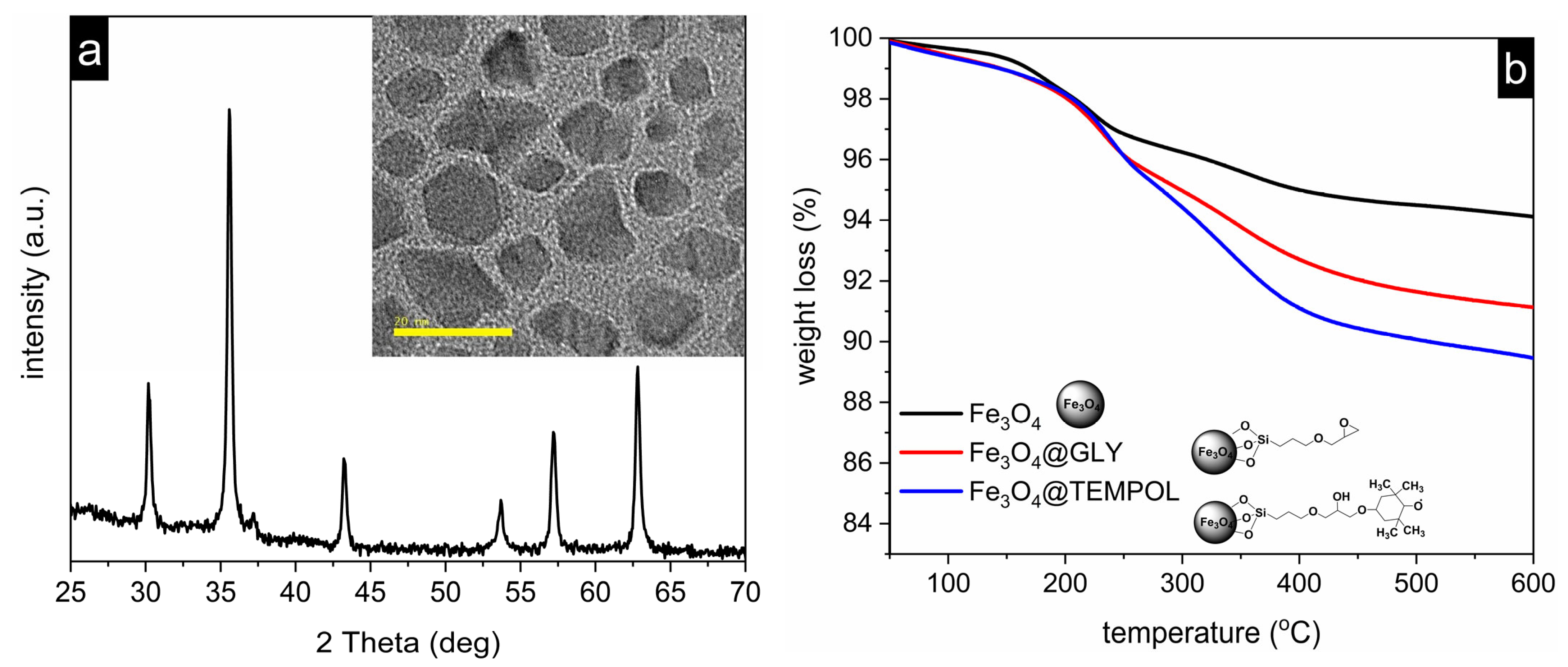
2.2.1. Nanoparticles Characterization
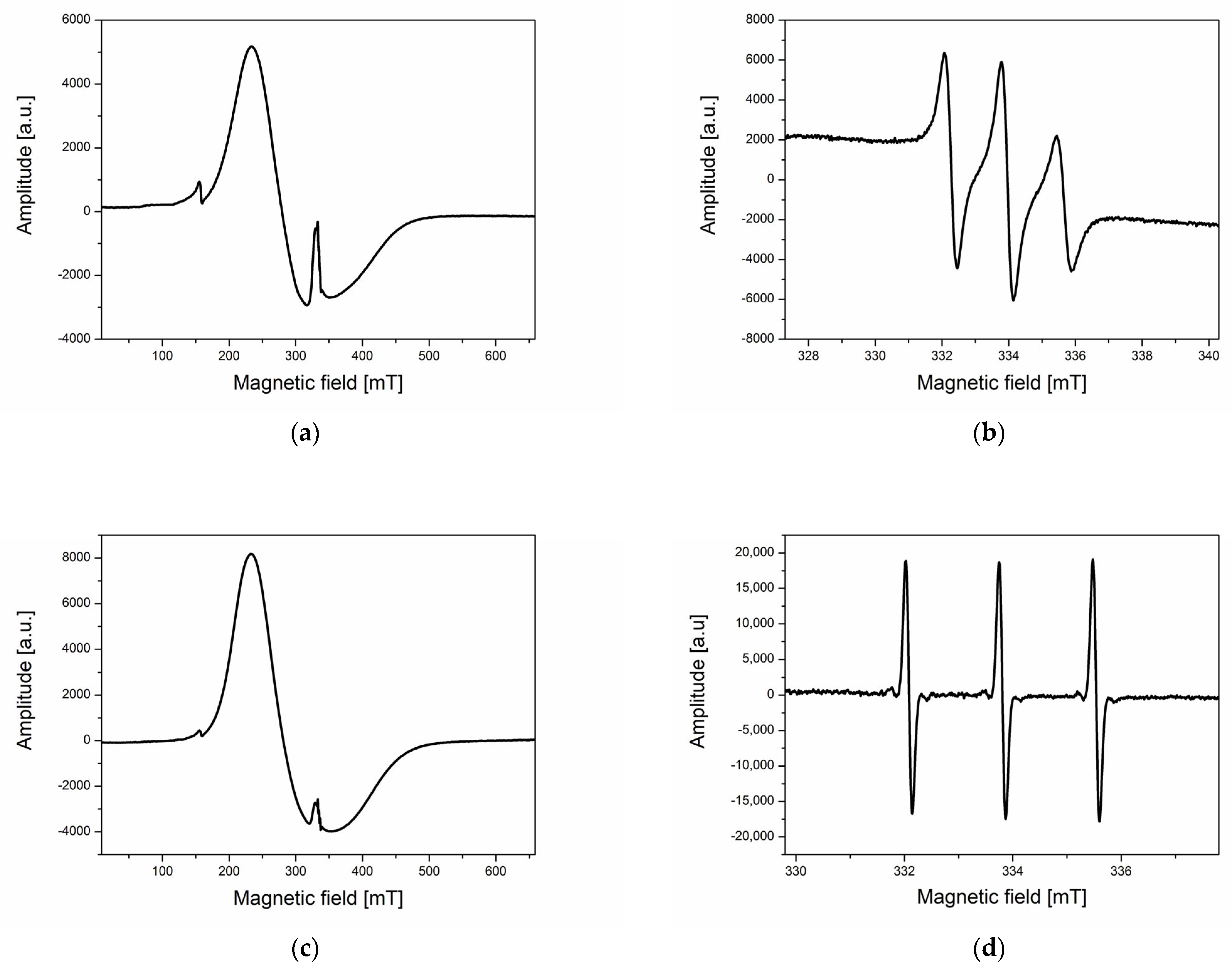
2.2.2. ESR Measurements
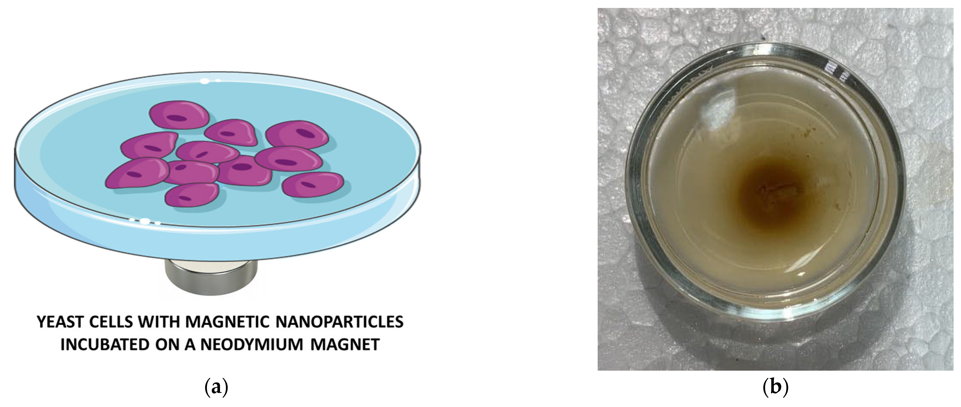
2.2.3. The Impact of a Stable Magnetic Field on Nanoparticle Uptake by Yeast Cells
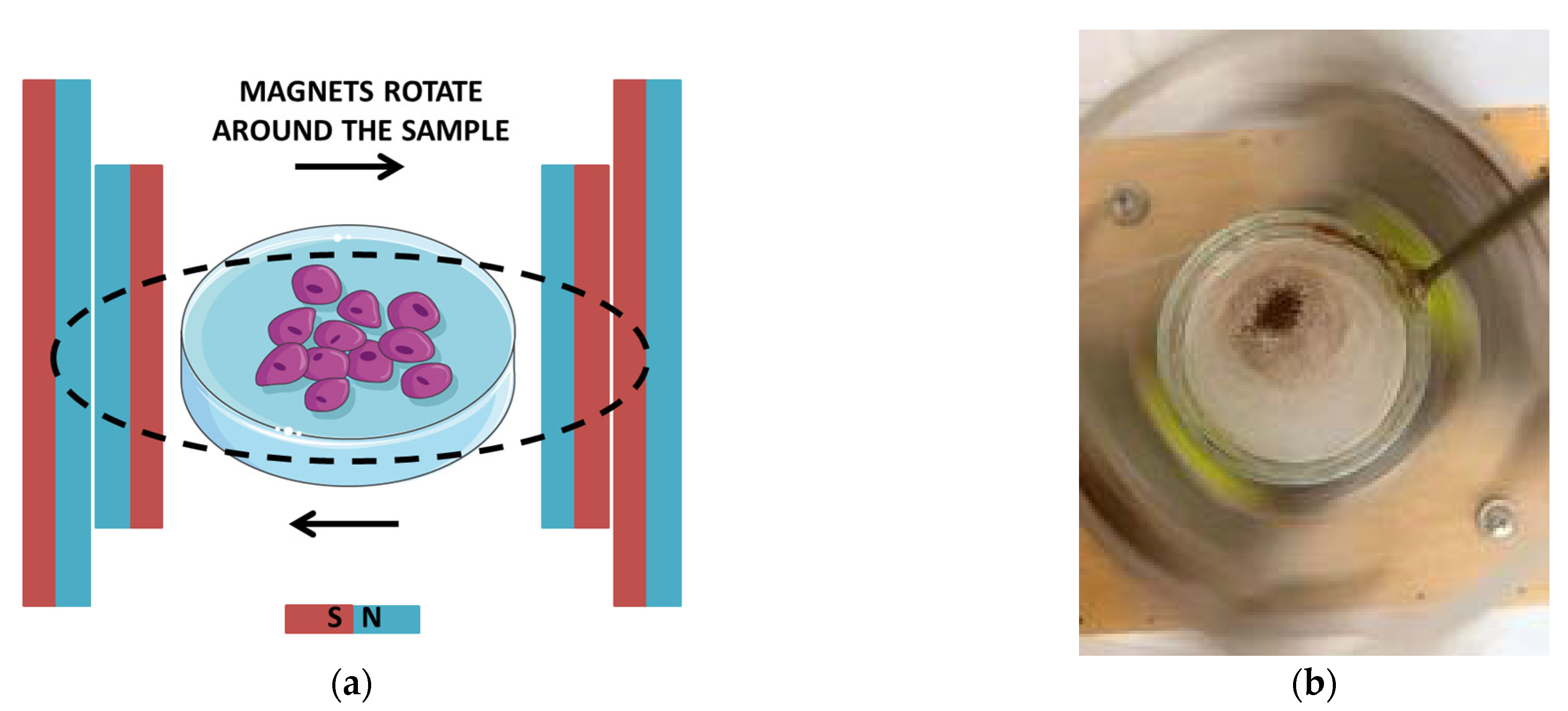
2.2.4. The Impact of a Rotating Magnetic Field on Nanoparticle Uptake by Yeast Cells
2.2.5. Microscopic Observation of Yeast Cell Proliferation
2.2.6. SEM and EDS of Yeast Cells with Fe3O4@TEMPOL Nanoparticles
2.2.7. Statistical Analysis
3. Results and Discussion
4. Conclusions
Author Contributions
Funding
Institutional Review Board Statement
Informed Consent Statement
Data Availability Statement
Conflicts of Interest
References
- Benguettat-El Mokhtari, I.; Schmool, D.S. Ferromagnetic Resonance in Magnetic Oxide Nanoparticules: A Short Review of Theory and Experiment. Magnetochemistry 2023, 9, 191. [Google Scholar] [CrossRef]
- Kianfar, E. Magnetic Nanoparticles in Targeted Drug Delivery: A Review. J. Supercond. Nov. Magn. 2021, 34, 1709–1735. [Google Scholar] [CrossRef]
- Avasthi, A.; Caro, C.; Pozo-Torres, E.; Leal, M.P.; García-Martín, M.L. Magnetic Nanoparticles as MRI Contrast Agents. Top. Curr. Chem. 2020, 378, 40. [Google Scholar] [CrossRef]
- Caspani, S.; Magalhães, R.; Araújo, J.P.; Tavares Sousa, C. Magnetic Nanomaterials as Contrast Agents for MRI. Materials 2020, 13, 2586. [Google Scholar] [CrossRef]
- Stueber, D.D.; Villanova, J.; Aponte, I.; Xiao, Z.; Colvin, V.L. Magnetic Nanoparticles in Biology and Medicine: Past, Present, and Future Trends. Pharmaceutics 2021, 13, 943. [Google Scholar] [CrossRef]
- Kianfar, E. Magnetic Nanoparticles in Medical Imaging. Imaging Med. 2022, 14, 1–16. [Google Scholar]
- Liu, X.; Zhang, Y.; Wang, Y.; Zhu, W.; Li, G.; Ma, X.; Zhang, Y.; Chen, S.; Tiwari, S.; Shi, K.; et al. Comprehensive understanding of magnetic hyperthermia for improving antitumor therapeutic efficacy. Theranostics 2020, 10, 3793–3815. [Google Scholar] [CrossRef]
- Peiravi, M.; Eslami, H.; Ansari, M.; Zare-Zardini, H. Magnetic hyperthermia: Potentials and limitations. J. Indian Chem. Soc. 2022, 99, 100269. [Google Scholar] [CrossRef]
- Aslam, H.; Shukrullah, S.; Naz, M.Y.; Fatima, H.; Hussain, H.; Ullah, S.; Assiri, M.A. Current and future perspectives of multifunctional magnetic nanoparticles based controlled drug delivery systems. J. Drug Deliv. Sci. Technol. 2022, 67, 102946. [Google Scholar] [CrossRef]
- Spoială, A.; Ilie, C.-I.; Motelica, L.; Ficai, D.; Semenescu, A.; Oprea, O.-C.; Ficai, A. Smart Magnetic Drug Delivery Systems for the Treatment of Cancer. Nanomaterials 2023, 13, 876. [Google Scholar] [CrossRef] [PubMed]
- Montiel Schneider, M.G.; Martín, M.J.; Otarola, J.; Vakarelska, E.; Simeonov, V.; Lassalle, V.; Nedyalkova, M. Biomedical Applications of Iron Oxide Nanoparticles: Current Insights Progress and Perspectives. Pharmaceutics 2022, 14, 204. [Google Scholar] [CrossRef] [PubMed]
- Sousa de Almeida, M.; Susnik, E.; Drasler, B.; Taladriz-Blanco, P.; Petri-Fink, A.; Rothen-Rutishauser, B. Understanding nanoparticle endocytosis to improve targeting strategies in nanomedicine. Chem. Soc. Rev. 2021, 50, 5397. [Google Scholar] [CrossRef] [PubMed]
- Haddad, M.; Frickenstein, A.N.; Wilhelm, S. High-throughput single-cell analysis of nanoparticle-cell interactions. Trac-Trend. Anal. Chem. 2023, 166, 117172. [Google Scholar] [CrossRef] [PubMed]
- Ostrowski, A.; Nordmeyer, D.; Boreham, A.; Holzhausen, C.; Mundhenk, L.; Graf, C.; Meinke, M.C.; Vogt, A.; Hadam, S.; Lademann, J.; et al. Overview about the localization of nanoparticles in tissue and cellular context by different imaging techniques. Beilstein J. Nanotech. 2015, 23, 263–280. [Google Scholar] [CrossRef] [PubMed]
- FitzGerald, L.I.; Johnston, A.P.R. It’s what’s on the inside that counts: Techniques for investigating the uptake and recycling of nanoparticles and proteins in cells. J. Colloid. Interf. Sci. 2021, 587, 64–78. [Google Scholar] [CrossRef]
- Krzyminiewski, R.; Dobosz, B.; Schroeder, G.; Kurczewska, J. ESR as a monitoring method of the interactions between TEMPO-functionalized magnetic nanoparticles and yeast cells. Sci. Rep. 2019, 9, 18733. [Google Scholar] [CrossRef]
- Krzyminiewski, R.; Dobosz, B.; Krist, B.; Schroeder, G.; Kurczewska, J.; Bluyssen, H.A.R. ESR method in monitoring of nanoparticle endocytosis in cancer cells. Int. J. Mol. Sci. 2020, 21, 4388. [Google Scholar] [CrossRef]
- Shashni, B.; Nagasaki, Y. Newly Developed Self-Assembling Antioxidants as Potential Therapeutics for the Cancers. J. Pers. Med. 2021, 11, 92. [Google Scholar] [CrossRef]
- He, W.; Liu, Y.; Wamer, W.G.; Yin, J.-J. Electron spin resonance spectroscopy for the study of nanomaterial-mediated generation of reactive oxygen species. J. Food Drug Anal. 2014, 22, 49–63. [Google Scholar] [CrossRef]
- Suzen, S.; Gurer-Orhan, H.; Saso, L. Detection of Reactive Oxygen and Nitrogen Species by Electron Paramagnetic Resonance (EPR) Technique. Molecules 2017, 22, 181. [Google Scholar] [CrossRef]
- Roessler, M.M.; Salvadori, E. Principles and applications of EPR spectroscopy in the chemical sciences. Chem. Soc. Rev. 2018, 47, 2534. [Google Scholar] [CrossRef]
- Jiang, J.; Tian, S.; Wang, K.; Wang, Y.; Zang, S.; Yu, A.; Zhang, Z. Electron spin resonance spectroscopy for immunoassay using iron oxide nanoparticles as probe. Anal. Bioanal. Chem. 2018, 410, 1817–1824. [Google Scholar] [CrossRef] [PubMed]
- Dogan, N.; Ozel, F.; Koten, H. Structural, Morphological, and Magnetic Characterization of Iron Oxide Nanoparticles Synthesized at Different Reaction Times via Thermal Decomposition Method. Curr. Nanosci. 2023, 19, 33–38. [Google Scholar] [CrossRef]
- Suryawanshi, P.L.; Sonawane, S.H.; Bhanvase, B.A.; Ashokkumar, M.; Pimplapure, M.S.; Gogate, P.R. Synthesis of iron oxide nanoparticles in a continuous flow spiral microreactor and Corning® advanced flow™ reactor. Green Process. Synth. 2018, 7, 1–11. [Google Scholar] [CrossRef]
- Bakker, M.G.; Fowler, B.; Bowman, M.K.; Patience, G.S. Experimental methods in chemical engineering: Electron paramagnetic resonance spectroscopy-EPR/ESR. Can. J. Chem. Eng. 2020, 98, 1668–1681. [Google Scholar] [CrossRef]
- Vasić, K.; Knez, Ž.; Konstantinova, E.A.; Kokorin, A.I.; Gyergyek, S.; Leitgeb, M. Structural and magnetic characteristics of carboxymethyl dextran coated magnetic nanoparticles: From characterization to immobilization application. React. Funct. Polym. 2020, 148, 104481. [Google Scholar] [CrossRef]
- Elamin, N.Y.; Modwi, A.; El-Fattah, W.A.; Rajeh, A. Synthesis and structural of Fe3O4 magnetic nanoparticles and its effect on the structural optical, and magnetic properties of novel Poly(methyl methacrylate)/Polyaniline composite for electromagnetic and optical applications. Opt. Mater. 2023, 135, 113323. [Google Scholar] [CrossRef]
- Abele, N.; Münz, F.; Zink, F.; Gröger, M.; Hoffmann, A.; Wolfschmitt, E.-M.; Hogg, M.; Calzia, E.; Waller, C.; Radermacher, P.; et al. Relation of Plasma Catecholamine Concentrations and Myocardial Mitochondrial Respiratory Activity in Anesthetized and Mechanically Ventilated, Cardiovascular Healthy Swine. Int. J. Mol. Sci. 2023, 24, 17293. [Google Scholar] [CrossRef]
- Vanreusel, I.; Vermeulen, D.; Goovaerts, I.; Stoop, T.; Ectors, B.; Cornelis, J.; Hens, W.; de Bliek, E.; Heuten, H.; Van Craenenbroeck, E.M.; et al. Circulating Reactive Oxygen Species in Adults with Congenital Heart Disease. Antioxidants 2022, 11, 2369. [Google Scholar] [CrossRef]
- Velayutham, M.; Poncelet, M.; Eubank, T.D.; Driesschaert, B.; Khramtsov, V.V. Biological Applications of Electron Paramagnetic Resonance Viscometry Using a 13C-Labeled Trityl Spin Probe. Molecules 2021, 26, 2781. [Google Scholar] [CrossRef]
- Gertsenshteyn, I.; Giurcanu, M.; Vaupel, P.; Halpern, H. Biological validation of electron paramagnetic resonance (EPR) image oxygen thresholds in tissue. J. Physiol. 2021, 599, 1759–1767. [Google Scholar] [CrossRef]
- Dobosz, B.; Krzyminiewski, R.; Kucińska, M.; Murias, M.; Schroeder, G.; Kurczewska, J. The spin probes as scavengers of free radicals in cells. Appl. Sci. 2022, 12, 7999. [Google Scholar] [CrossRef]
- Nibbe, P.; Schleusener, J.; Siebert, S.; Borgart, R.; Brandt, D.; Westphalen, R.; Schüler, N.; Berger, B.; Peters, E.M.J.; Meinke, M.C.; et al. Oxidative stress coping capacity (OSC) value: Development and validation of an in vitro measurement method for blood plasma using electron paramagnetic resonance spectroscopy (EPR) and vitamin C. Free Radic. Biol. Med. 2023, 194, 230–244. [Google Scholar] [CrossRef]
- Vesković, A.; Nakarada, Đ.; Pavićević, A.; Prokić, B.; Perović, M.; Kanazir, S.; Popović-Bijelić, A.; Mojović, M. In Vivo/Ex Vivo EPR Investigation of the Brain Redox Status and Blood-Brain Barrier Integrity in the 5xFAD Mouse Model of Alzheimer’s Disease. Curr. Alzheimer Res. 2021, 18, 25–34. [Google Scholar] [CrossRef]
- Jakubowska, M.A.; Pyka, J.; Michalczyk-Wetula, D.; Baczyński, K.; Cieśla, M.; Susz, A.; Ferdek, P.E.; Płonka, B.K.; Fiedor, L.; Płonka, P.M. Electron paramagnetic resonance spectroscopy reveals alterations in the redox state of endogenous copper and iron complexes in photodynamic stress-induced ischemic mouse liver. Redox Biol. 2020, 34, 101566. [Google Scholar] [CrossRef]
- Tzivaki, M.; Hassan, A.; Waller, E. Electron paramagnetic resonance spectroscopy for the detection of radiation exposure in dreissenid mussels. Radiat. Prot. Dosim. 2023, 199, 1626–1631. [Google Scholar] [CrossRef]
- Karmakar, P.; Mishra, L.; Mishra, M. Electron spin resonance spectroscopy: A tool for dating mollusc shells, corals, and other materials. In Spectroscopic and Microscopy Techniques for Archaeological and Cultural Heritage Research, 2nd ed.; IOP Publishing Ltd.: Bristol, UK, 2023; pp. 9-1–9-11. [Google Scholar]
- Timar-Gabor, A.; Kabacińska, Z.; Constantin, D.; Dave, A.K.; Buylaert, J.-P. Reconstructing dust provenance from quartz optically stimulated luminescence (OSL) and electron spin resonance (ESR) signals: Preliminary results on loess from around the world. Radiat. Phys. Chem. 2023, 212, 111138. [Google Scholar] [CrossRef]
- Ghimire, L.; Waller, E. Electron Paramagnetic Resonance Measurements of Lifetime Doses in Teeth of Durham Region Residents, Ontario. Health Phys. 2023, 124, 175–191. [Google Scholar] [CrossRef]
- Cui, X.; Zhang, Z.; Yang, Y.; Li, S.; Lee, C.-S. Organic radical materials in biomedical applications: State of the art and perspectives. Exploration 2022, 2, 20210264. [Google Scholar] [CrossRef]
- Feliciano, C.P.; Nagasaki, Y. Antioxidant Nanomedicine Protects against Ionizing Radiation-Induced Life-Shortening in C57BL/6J Mice. ACS Biomater. Sci. Eng. 2019, 5, 5631–5636. [Google Scholar] [CrossRef]
- Mołoń, M.; Szlachcikowska, D.; Stępień, K.; Kielar, P.; Galiniak, S. Two faces of TEMPO (2,2,6,6-tetramethylpiperidinyl-1-oxyl)—An antioxidant or a toxin? BBA–Mol. Cell Res. 2023, 1870, 119412. [Google Scholar] [CrossRef]
- Feliciano, C.P.; Cammas-Marion, S.; Nagasaki, Y. Recent advances in self-assembling redox nanoparticles as a radiation protective agent. AIMS Mol. Sci. 2023, 10, 52–69. [Google Scholar] [CrossRef]
- Matsumoto, K.-I.; Nakanishi, I.; Zhelev, Z.; Bakalova, R.; Aoki, I. Nitroxyl Radical as a Theranostic Contrast Agent in Magnetic Resonance Redox Imaging. Antioxid. Redox Signal. 2022, 36, 95–121. [Google Scholar] [CrossRef]
- Azuma, R.; Yamasaki, T.; Emoto, M.C.; Sato-Akaba, H.; Sano, K.; Munekane, M.; Fujii, H.G.; Mukai, T. Effect of relative configuration of TEMPO-type nitroxides on ascorbate reduction. Free Radic. Biol. Med. 2023, 194, 114–122. [Google Scholar] [CrossRef]
- Nakamura, H.; Watano, S. Direct Permeation of Nanoparticles Across Cell Membrane: A Review. Kona Powder Part. J. 2018, 35, 49–65. [Google Scholar] [CrossRef]
- Escobar, J.F.; Vaca-González, J.J.; Guevara, J.M.; Garzón-Alvarado, D.A. Effect of magnetic and electric fields on plasma membrane of single cells: A computational approach. Eng. Rep. 2020, 2, e12125. [Google Scholar] [CrossRef]
- Beddoes, C.M.; Case, C.P.; Briscoe, W.H. Understanding nanoparticle cellular entry: A physicochemical perspective. Adv. Colloid Interfac. 2015, 218, 48–68. [Google Scholar] [CrossRef]
- Wen, Z.; Liu, C.; Teng, Z.; Jin, Q.; Liao, Z.; Zhu, X.; Huo, S. Ultrasound meets the cell membrane: For enhanced endocytosis and drug delivery. Nanoscale 2023, 15, 13532. [Google Scholar] [CrossRef]
- Zablotskii, V.; Syrovets, T.; Schmidt, Z.W.; Dejneka, A.; Simmet, T. Modulation of monocytic leukemia cell function and survival by high gradient magnetic fields and mathematical modeling studies. Biomaterials 2014, 35, 3164–3171. [Google Scholar] [CrossRef]
- Min, K.A.; Shin, M.C.; Yu, F.; Yang, M.; David, A.E.; Yang, V.C.; Rosania, G.R. Pulsed magnetic field improves the transport of iron oxide nanoparticles through cell barriers. ACS Nano 2013, 7, 2161–2171. [Google Scholar] [CrossRef]
- Soheilian, R.; Choi, Y.S.; David, A.E.; Abdi, H.; Maloney, C.E.; Erb, R.M. Toward Accumulation of Magnetic Nanoparticles into Tissues of Small Porosity. Langmuir 2015, 31, 8267–8274. [Google Scholar] [CrossRef]
- Zablotskii, V.; Polyakova, T.; Lunov, O.; Dejneka, A. How a High-Gradient Magnetic Field Could Affect Cell Life. Sci. Rep. 2016, 6, 37407. [Google Scholar] [CrossRef]
- Zablotskii, V.; Lunov, O.; Kubinova, S.; Polyakova, T.; Sykova, E.; Dejneka, A. Effects of high-gradient magnetic fields on living cell machinery. J. Phys. D Appl. Phys. 2016, 49, 493003. [Google Scholar] [CrossRef]
- Zablotskii, V.; Lunov, O.; Dejneka, A.; Jastrabik, L.; Polyakova, T.; Syrovets, T.; Simmet, T. Nanomechanics of magnetically driven cellular endocytosis. Appl. Phys. Lett. 2011, 99, 183701. [Google Scholar] [CrossRef]
- Uzhytchak, M.; Lynnyk, A.; Zablotskii, V.; Dempsey, N.M.; Dias, A.L.; Bonfim, M.; Lunova, M.; Jirsa, M.; Kubinová, Š.; Lunov, O.; et al. The use of pulsed magnetic fields to increase the uptake of iron oxide nanoparticles by living cells. Appl. Phys. Lett. 2017, 111, 243703. [Google Scholar] [CrossRef]
- Wang, H.; Zhang, X. Magnetic Fields and Reactive Oxygen Species. Int. J. Mol. Sci. 2017, 18, 2175. [Google Scholar] [CrossRef]
- van der Laan, K.J.; Morita, A.; Perona-Martinez, F.P.; Schirhagl, R. Evaluation of the Oxidative Stress Response of Aging Yeast Cells in Response to Internalization of Fluorescent Nanodiamond Biosensors. Nanomaterials 2020, 10, 372. [Google Scholar] [CrossRef]
- Postaru, M.; Tucaliuc, A.; Cascaval, D.; Galaction, A.-I. Cellular Stress Impact on Yeast Activity in Biotechnological Processes—A Short Overview. Microorganisms 2023, 11, 2522. [Google Scholar] [CrossRef]
- Eigenfeld, M.; Wittmann, L.; Kerpes, R.; Schwaminger, S.; Becker, T. Quantifcation methods of determining brewer’s and pharmaceutical yeast cell viability: Accuracy and impact of nanoparticles. Anal. Bioanal. Chem. 2023, 415, 3201–3213. [Google Scholar] [CrossRef]
- Pawlaczyk, M.; Pasieczna-Patkowska, S.; Schroeder, G. Photoacoustic Spectroscopy of Surface-Functionalized Fe3O4-SiO2 Nanoparticles. Appl. Spectrosc. 2020, 74, 712–719. [Google Scholar] [CrossRef]
- Pawlaczyk, M.; Frański, R.; Cegłowski, M.; Schroeder, G. Mass spectrometric investigation of organo-functionalized magnetic nanoparticles binding properties toward chalcones. Materials 2021, 14, 4705. [Google Scholar] [CrossRef]
- Baghdadi, Y.N.; Youssef, L.; Bouhadir, K.; Harb, M.; Mustapha, S.; Patra, D.; Tehrani-Bagha, A.R. Thermal and mechanical properties of epoxy resin reinforced with modified iron oxide nanoparticles. J. Appl. Polym. Sci. 2021, 138, 50533. [Google Scholar] [CrossRef]
- Krzyminiewski, R.; Dobosz, B.; Schroeder, G.; Kurczewska, J. The principles of a new method, MNF-3D, for concentration of magnetic particles in three-dimensional space. Measurement 2017, 112, 137–140. [Google Scholar] [CrossRef]
- Krzyminiewski, R.; Dobosz, B.; Schroeder, G.; Kurczewska, J. Focusing of Fe3O4 nanoparticles using a rotating magnetic field in various environments. Phys. Lett. A 2018, 382, 3192–3196. [Google Scholar] [CrossRef]
- Dobosz, B.; Schroeder, G.; Kurczewska, J. Comments on “The principles of a new method, MNF-3D, for concentration of magnetic particles in three-dimensional space”. Measurement 2023, 118, 113146. [Google Scholar] [CrossRef]
- Sharpe, M.A.; Baskin, D.S.; Pichumani, K.; Ijare, O.B.; Helekar, S.A. Rotating Magnetic Fields Inhibit Mitochondrial Respiration, Promote Oxidative Stress and Produce Loss of Mitochondrial Integrity in Cancer Cells. Front. Oncol. 2021, 10, 768758. [Google Scholar] [CrossRef]
- Sládičeková, K.H.; Bereta, M.; Misek, J.; Parizek, D.; Jakuš, J. Biological Effects of a Low-Frequency Electromagnetic Field on Yeast Cells of the Genus Saccharomyces Cerevisiae. Acta Med. Martiniana 2021, 21, 34–41. [Google Scholar] [CrossRef]
- Peng, Q.; Huo, D.; Li, H.; Zhang, B.; Li, Y.; Liang, A.; Wang, H.; Yu, Q.; Li, M. ROS independent toxicity of Fe3O4 nanoparticles to yeast cells: Involvement of mitochondrial dysfunction. Chem. Biol. Interact. 2018, 1, 20–26. [Google Scholar] [CrossRef]
- Pahlevan, M.; Toivakka, M.; Alam, P. Mechanical properties of TEMPO-oxidised bacterial cellulose-amino acid biomaterials. Eur. Polym. J. 2018, 101, 29–36. [Google Scholar] [CrossRef]
- Chen, L.; Chen, C.; Wang, P.; Song, T. Mechanisms of Cellular Effects Directly Induced by Magnetic Nanoparticles under Magnetic Fields. Hindawi J. Nanomater. 2017, 2017, 1564634. [Google Scholar] [CrossRef]


| Time (h) | Control Sample (×106/mL) | YC + MNPs 1 (×106/mL) | YC + MNPs + NM 2 (×106/mL) |
|---|---|---|---|
| 0 N | 305 ± 46 | 255 ± 38 | 282 ± 42 |
| 0.75 N | 349 ± 52 | 327 ± 49 | 262 ± 39 |
| 1.5 N | 355 ± 53 | 274 ± 41 | 373 ± 56 |
| 2.25 N | 269 ± 40 | 390 ± 59 | 383 ± 58 |
| 3 N | 343 ± 52 | 380 ± 57 | 363 ± 55 |
| 3.75 N | 326 ± 49 | 385 ± 58 | 339 ± 51 |
| Time (h) | Control Sample (×106/mL) | YC + MNPs 1 (×106/mL) | YC + MNPs + 15 2 (×106/mL) | YC + MNPs + 30 3 (×106/mL) |
|---|---|---|---|---|
| 0 N | 359 ± 54 | 332 ± 49 | 336 ± 50 | 270 ± 40 |
| 0.75 Y | 354 ± 53 | 424 ± 64 | 271 ± 41 | 273 ± 40 |
| 1.5 Y | 391 ± 59 | 396 ± 59 | 396 ± 61 | 260 ± 39 |
| 2.25 N | 332 ± 50 | 360 ± 54 | 332 ± 50 | 322 ± 48 |
| 3 N | 400 ± 60 | 352 ± 53 | 357 ± 54 | 347 ± 52 |
| 3.75 N | 376 ± 56 | 321 ± 48 | 452 ± 68 | 340 ± 51 |
Disclaimer/Publisher’s Note: The statements, opinions and data contained in all publications are solely those of the individual author(s) and contributor(s) and not of MDPI and/or the editor(s). MDPI and/or the editor(s) disclaim responsibility for any injury to people or property resulting from any ideas, methods, instructions or products referred to in the content. |
© 2024 by the authors. Licensee MDPI, Basel, Switzerland. This article is an open access article distributed under the terms and conditions of the Creative Commons Attribution (CC BY) license (https://creativecommons.org/licenses/by/4.0/).
Share and Cite
Dobosz, B.; Gunia, E.; Kotarska, K.; Schroeder, G.; Kurczewska, J. The Effect of a Magnetic Field on the Transport of Functionalized Magnetite Nanoparticles into Yeast Cells. Appl. Sci. 2024, 14, 1343. https://doi.org/10.3390/app14041343
Dobosz B, Gunia E, Kotarska K, Schroeder G, Kurczewska J. The Effect of a Magnetic Field on the Transport of Functionalized Magnetite Nanoparticles into Yeast Cells. Applied Sciences. 2024; 14(4):1343. https://doi.org/10.3390/app14041343
Chicago/Turabian StyleDobosz, Bernadeta, Eliza Gunia, Klaudia Kotarska, Grzegorz Schroeder, and Joanna Kurczewska. 2024. "The Effect of a Magnetic Field on the Transport of Functionalized Magnetite Nanoparticles into Yeast Cells" Applied Sciences 14, no. 4: 1343. https://doi.org/10.3390/app14041343
APA StyleDobosz, B., Gunia, E., Kotarska, K., Schroeder, G., & Kurczewska, J. (2024). The Effect of a Magnetic Field on the Transport of Functionalized Magnetite Nanoparticles into Yeast Cells. Applied Sciences, 14(4), 1343. https://doi.org/10.3390/app14041343