


Analysis of the Fatty Acid Profile in Cream, Buttermilk Fractions, and Anhydrous Milk Fat: Influence of Physicochemical and Microbiological Parameters on the Fatty Acid Profile

 ,
, 
Abstract
1. Introduction
2. Materials and Methods
2.1. Reagents and Chemicals
2.2. Sample Details
2.3. Microbiology Analysis
2.4. Extraction of Fatty Acids
2.5. Characterization of Milk Fatty Acids Using GC-MS Analysis
2.6. Identification of the Obtained Microorganisms by MALDI-ToF MS
2.7. Statistical Analysis
3. Results and Discussion
Supplementary Materials
Author Contributions
Funding
Data Availability Statement
Acknowledgments
Conflicts of Interest
References
- Chandan, R.C. Dairy ingredients for food processing: An overview. In Dairy Ingredients for Food Processing; Blackwell Publishing: Hoboken, NJ, USA, 2011; pp. 3–33. [Google Scholar] [CrossRef]
- Kailasapathy, K. Chemical composition, physical, and functional properties of milk and milk ingredients. In Dairy Processing and Quality Assurance; Blackwell Publishing: Hoboken, NJ, USA, 2015; pp. 77–105. [Google Scholar] [CrossRef]
- NIIR Board. Modern Technology of Milk Processing & Dairy Products; NIIR Project Consultancy Services: Delhi, India, 2013. [Google Scholar]
- Achaw, O.W.; Danso-Boateng, E. Milk and dairy products manufacture. In Chemical and Process Industries: With Examples of Industries in Ghana; Springer International Publishing: Cham, Switzerland, 2021; pp. 293–374. [Google Scholar] [CrossRef]
- Widyastuti, Y.; Febrisiantosa, A. The role of lactic acid bacteria in milk fermentation. Food Nutr. Sci. 2014, 5, 435–442. [Google Scholar] [CrossRef]
- Illingworth, D.; Patil, G.R.; Tamime, A.Y. Anhydrous Milk Fat Manufacture and Fractionation. In Dairy Fats and Related Products; Blackwell Publishing: Hoboken, NJ, USA, 2009; pp. 108–166. [Google Scholar] [CrossRef]
- Gauvin, M.-P.; Pouliot, Y.; Britten, M. Characterization of buttermilk serum fractions and their effect on rennet-induced coagulation of casein micelle dispersions. Int. Dairy J. 2017, 76, 10–17. [Google Scholar] [CrossRef]
- Barłowska, J.; Litwińczuk, Z.; Król, J.; Kędzierska-Matysek, M. Fatty acid profile and mineral content in milk from cows of various breeds over spring-summer feeding period. Pol. J. Food Nutr. Sci. 2006, 15, 13–16. [Google Scholar]
- Palmquist, D.L.; Beaulieu, A.D.; Barbano, D.M. Feed and animal factors influencing milk fat composition. J. Dairy Sci. 1993, 76, 1753–1771. [Google Scholar] [CrossRef]
- Bazmi, A.; Relkin, P. Effects of processing conditions on structural and functional parameters of whipped dairy emulsions containing various fatty acid compositions. J. Dairy Sci. 2009, 92, 3566–3574. [Google Scholar] [CrossRef]
- Kelly, A.L.; Fox, P.F. Biochemistry of milk processing. In Food Biochemistry and Food Processing; John Wiley & Sons, Inc.: Hoboken, NJ, USA, 2012; pp. 465–490. [Google Scholar] [CrossRef]
- Santin Junior, I.A.; Silva, K.C.C.; Cucco, D.C. Milk Fatty Acids Profile and the Impact on Human Health. Dairy Vet. Sci. J. 2019, 10, 555779. [Google Scholar] [CrossRef]
- Calder, P.C. Functional Roles of Fatty Acids and Their Effects on Human Health. J. Parenter. Enter. Nutr. 2015, 39, 18–32. [Google Scholar] [CrossRef] [PubMed]
- Lindmark, M.H. Fatty acids in bovine milk fat. Food Nutr. Res. 2008, 52, 1821. [Google Scholar] [CrossRef] [PubMed]
- Sharma, G.; Prakash, D.; Gupta, C.; Prakash, D.; Sharma, G. Phytochemicals of nutraceutical importance: Do they defend against diseases. In Phytochemicals of Nutraceutical Importance; CABI: Oxon, UK, 2014; pp. 1–9. [Google Scholar] [CrossRef]
- Gabbi, A.M.; McManus, C.M.; Marques, L.T.; Abreu, A.S.; Machado, S.C.; Zanela, M.B.; Barbosa, R.S.; Fischer, V. Different levels of supplied energy for lactating cows affect physicochemical attributes of milk. J. Anim. Feed Sci. 2018, 27, 11–17. [Google Scholar] [CrossRef]
- Reklewska, B.; Bernatowicz, E.; Reklewski, Z.; Kuczyńska, B.; Zdziarski, K.; Sakowski, T.; Słoniewski, K. Functional Components of Milk Produced by Polish Black-And-White, Polish Red and Simmental Cows. Electron. J. Pol. Agric. Univ. 2005, 8, 25. [Google Scholar]
- Christie, W.W. Handbook of Chromatography; MH, K., Ed.; CRC Press: Boca Raton, FL, USA, 1984; Volume 1, pp. 33–46. [Google Scholar]
- Martínez, B.; Miranda, J.M.; Franco, C.M.; Cepeda, A.; Rodríguez, J.L. Development of a simple method for the quantitative determination of fatty acids in milk with special emphasis on long-chain fatty acids. J. Food 2012, 10, 27–35. [Google Scholar] [CrossRef]
- Walczak-Skierska, J.; Monedeiro, F.; Rudnicka, J.; Pomastowski, P. Optimizing Milk Quality and Shelf Life: Investigating Refrigeration Effects on Fatty Acid and Protein Profiles. ACS Food Sci. Technol. 2024, 4, 382–391. [Google Scholar] [CrossRef]
- Bruker Daltonics. MALDI Biotyper 3.1 User Manual; Bruker Daltonics: Billerica, MA, USA, 2012; Volume 1, pp. 1–212. [Google Scholar]
- Czeszewska-Rosiak, G.; Złoch, M.; Radosińska, M.; Florkiewicz, A.B.; Tretyn, A.; Pomastowski, P. The usefulness of the MALDI–TOF MS technique in the determination of dairy samples’ microbial composition: Comparison of the new EXS 2600 system with MALDI Biotyper platform. Arch. Microbiol. 2024, 206, 172. [Google Scholar] [CrossRef] [PubMed]
- Sert, D.; Mercan, E. Characterisation of physicochemical, microbiological, thermal, oxidation properties and fatty acid composition of butter produced from thermosonicated cream. Int. Dairy J. 2020, 109, 104777. [Google Scholar] [CrossRef]
- Brożek, O.; Kiełczewska, K.; Bohdziewicz, K. Characterisation of selected emulsion phase parameters in milk, cream and buttermilk. Pol. J. Food Nutr. Sci. 2022, 72, 5–15. [Google Scholar] [CrossRef]
- Bumbadiya, M.R.; Maji, S.; Sao, K.; Ranvir, S.G. Butter Oil (Ghee): Composition, Processing, and Physicochemical Changes during Storage. In The Chemistry of Milk and Milk Products; Apple Academic Press: Palm Bay, FL, USA, 2023; pp. 159–184. [Google Scholar]
- Khan, I.T.; Nadeem, M.; Imran, M.; Khalique, A. Impact of post fermentation cooling patterns on fatty acid profile, lipid oxidation and antioxidant features of cow and buffalo milk set yoghurt. Lipids Health Dis. 2020, 19, 74. [Google Scholar] [CrossRef] [PubMed]
- O’Connell, A.; Ruegg, P.L.; Jordan, K.; O’Brien, B.; Gleeson, D. The effect of storage temperature and duration on the microbial quality of bulk tank milk. J. Dairy Sci. 2016, 99, 3367–3374. [Google Scholar] [CrossRef]
- Soni, R.; Jain, N.K.; Shah, V.; Soni, J.; Suthar, D.; Gohel, P. Development of probiotic yogurt: Effect of strain combination on nutritional, rheological, organoleptic and probiotic properties. J. Food Sci. Technol. 2020, 57, 2038–2050. [Google Scholar] [CrossRef] [PubMed]
- Pasvolsky, R.; Zakin, V.; Ostrova, I.; Shemesh, M. Butyric acid released during milk lipolysis triggers biofilm formation of Bacillus species. Int. J. Food Microbiol. 2014, 181, 19–27. [Google Scholar] [CrossRef]
- Sanchez-Juanes, F.; Alonso, J.M.; Zancada, L.; Hueso, P. Glycosphingolipids from bovine milk and milk fat globule membranes: A comparative study. Adhesion to enterotoxigenic Escherichia coli strains. Biol. Chem. 2009, 390, 31–40. [Google Scholar] [CrossRef]
- Ali-Vehmas, T.E.R.H.I.; Westphalen, P.; Myllys, V.; Sandholm, M. Binding of Staphylococcus aureus to milk fat globules increases resistance to penicillin-G. J. Dairy Res. 1997, 64, 253–260. [Google Scholar] [CrossRef] [PubMed]
- Patton, S.; Keenan, T.W. The milk fat globule membrane. Biochim. Biophys. Acta (BBA)-Rev. Biomembr. 1975, 415, 273–309. [Google Scholar] [CrossRef]
- Teh, K.H.; Flint, S.; Brooks, J.; Knight, G. (Eds.) Biofilms in the Dairy Industry; John Wiley & Sons: Hoboken, NJ, USA, 2015. [Google Scholar]
- Thierry, A.; Collins, Y.F.; Mukdsi, M.A.; McSweeney, P.L.; Wilkinson, M.G.; Spinnler, H.E. Lipolysis and metabolism of fatty acids in cheese. In Cheese; Academic Press: Cambridge, MA, USA, 2017; pp. 423–444. [Google Scholar] [CrossRef]
- Deeth, H.C. Lipoprotein lipase and lipolysis in milk. Int. Dairy J. 2006, 16, 555–562. [Google Scholar] [CrossRef]
- Klungel, G.H.; Slaghuis, B.A.; Hogeveen, H. The effect of the introduction of automatic milking systems on milk quality. J. Dairy Sci. 2000, 83, 1998–2003. [Google Scholar] [CrossRef] [PubMed]
- Chen, L.; Coolbear, T.; Daniel, R.M. Characteristics of proteinases and lipases produced by seven Bacillus sp. isolated from milk powder production lines. Int. Dairy J. 2004, 14, 495–504. [Google Scholar] [CrossRef]
- Montanhini, M.T.M.; dos Santos Bersot, L. Evaluation of psychrotrophic behavior and lipolytic and proteolytic activity of Bacillus cereus isolated from refrigerated dairy products. Acta Sci. Technol. 2013, 35, 163–167. [Google Scholar] [CrossRef]
- Parkash, M.; Rajasekar, K.; Karmegam, N. Bacterial Population of Raw Milk and Their Proteolytic and Lipolytic Activities. Res. J. Appl. Sci. 2007, 3, 848–851. [Google Scholar]
- Baur, C.; Krewinkel, M.; Kranz, B.; von Neubeck, M.; Wenning, M.; Scherer, S.; Stoeckel, M.; Hinrichs, J.; Stressler, T.; Lutz Fischer, L. Quantification of the proteolytic and lipolytic activity of microorganisms isolated from raw milk. Int. Dairy. J. 2015, 49, 23–29. [Google Scholar] [CrossRef]
- Javed, S.; Azeem, F.; Hussain, S.; Rasul, I.; Siddique, M.H.; Riaz, M.; Afzal, M.; Kouser, A.; Nadeem, H. Bacterial lipases: A review on purification and characterization. Prog. Biophys. Mol. Biol. 2018, 132, 23–34. [Google Scholar] [CrossRef]
- Chakraborty, K.; Raj, R.P. An extra-cellular alkaline metallolipase from Bacillus licheniformis MTCC 6824: Purification and biochemical characterization. Food Chem. 2008, 109, 727–736. [Google Scholar] [CrossRef]
- Chakraborty, K.; Raj, R.P. Selective enrichment of n−3 polyunsaturated fatty acids with C18–C20 acyl chain length from sardine oil using Pseudomonas fluorescens MTCC 2421 lipase. Food Chem. 2009, 114, 142–150. [Google Scholar] [CrossRef]
- Gururaj, P.; Ramalingam, S.; Devi, G.N.; Gautam, P. Process optimization for production and purification of a thermostable, organic solvent tolerant lipase from Acinetobacter sp. AU07. Braz. J. Microbiol. 2016, 47, 647–657. [Google Scholar] [CrossRef] [PubMed]
- Chen, L.D.R.M.; Daniel, R.M.; Coolbear, T. Detection and impact of protease and lipase activities in milk and milk powders. Int. Dairy J. 2003, 13, 255–275. [Google Scholar] [CrossRef]
- Vithanage, N.R.; Dissanayake, M.; Bolge, G.; Palombo, E.A.; Yeager, T.R.; Datta, N. Biodiversity of culturable psychrotrophic microbiota in raw milk attributable to refrigeration conditions, seasonality and their spoilage potential. Int. Dairy J. 2016, 57, 80–90. [Google Scholar] [CrossRef]
- Salgado, C.A.; Baglinière, F.; Vanetti, M.C.D. Spoilage potential of a heat-stable lipase produced by Serratia liquefaciens isolated from cold raw milk. Lwt 2020, 126, 109289. [Google Scholar] [CrossRef]
- Hantsis-Zacharov, E. and Halpern, M. Culturable psychrotrophic bacterial communities in raw milk and their proteolytic and lipolytic traits. Appl. Environ. Microbiol. 2007, 73, 7162–7168. [Google Scholar] [CrossRef] [PubMed]
- Svendsen, A.; Borch, K.; Barfoed, M.; Nielsen, T.B.; Gormsen, E.; Patkar, S.A. Biochemical properties of cloned lipases from the Pseudomonas family. Biochim. Biophys. Acta (BBA)-Lipids Lipid Metab. 1995, 1259, 9–17. [Google Scholar] [CrossRef]
- Zhang, S.; Lv, J. Purification and properties of heat-stable extracellular protease from Pseudomonads fluorescens BJ-10. J. Food Sci. Technol. 2014, 51, 1185–1190. [Google Scholar] [CrossRef]

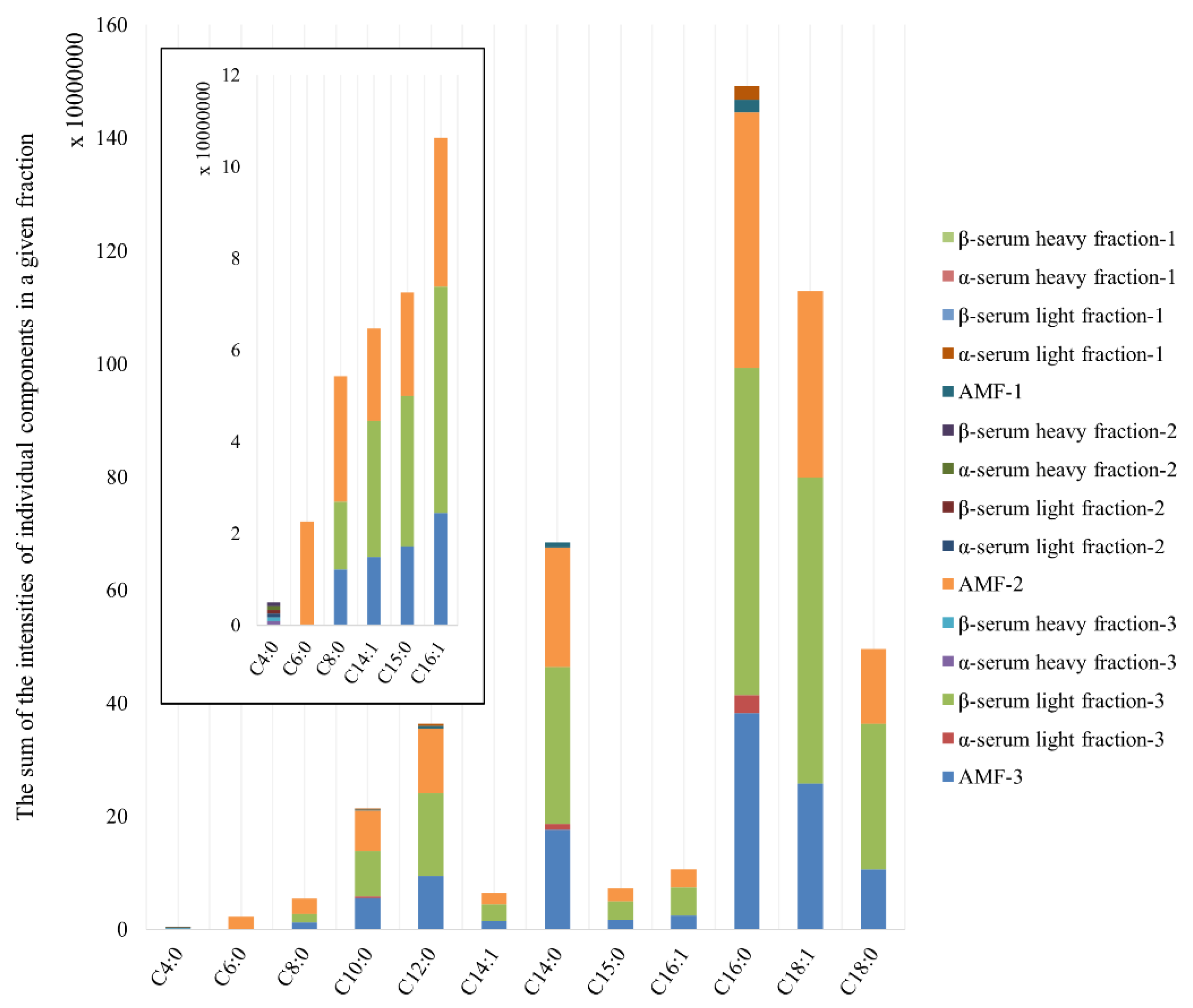
| Retention Time (min) | Volatile Organic Compound | AMF-3 | α-Serum Light Fraction-3 | β-Serum Light Fraction-3 | AMF-2 | β-Serum Heavy Fraction-2 | AMF-1 | α-Serum Light Fraction-1 |
|---|---|---|---|---|---|---|---|---|
| 6.211 | Butanoic acid (C4:0) | 0 | 0 | 0 | 0 | X | 0 | 0 |
| 9.328 | Hexanoic acid (C6:0) | 0 | 0 | 0 | X | 0 | 0 | 0 |
| 12.475 | Octanoic acid (C8:0) | X | 0 | X | X | 0 | 0 | 0 |
| 16.363 | Decanoic acid (C10:0) | X | X | X | X | 0 | X | X |
| 20.692 | Dodecanoic acid (C12:0) | X | 0 | X | X | 0 | X | X |
| 24.91 | (Z)-9-Tetradecenoic acid (C14:1) | X | 0 | X | X | 0 | 0 | 0 |
| 25.01 | Tetradecanoic acid (C14:0) | X | X | X | X | 0 | X | 0 |
| 27.086 | Pentadecanoic acid (C15:0) | X | 0 | X | X | 0 | 0 | 0 |
| 28.85 | (Z)-9-Hexadecenoic acid (C16:1) | X | 0 | X | X | 0 | 0 | 0 |
| 29.109 | Hexadecanoic acid (C16:0) | X | X | X | X | 0 | X | X |
| 33.144 | (E)-9-octadecenoic acid (C18:1) | X | 0 | X | X | 0 | 0 | 0 |
| 33.58 | Octadecanoic acid (C18:0) | X | 0 | X | X | 0 | 0 | 0 |
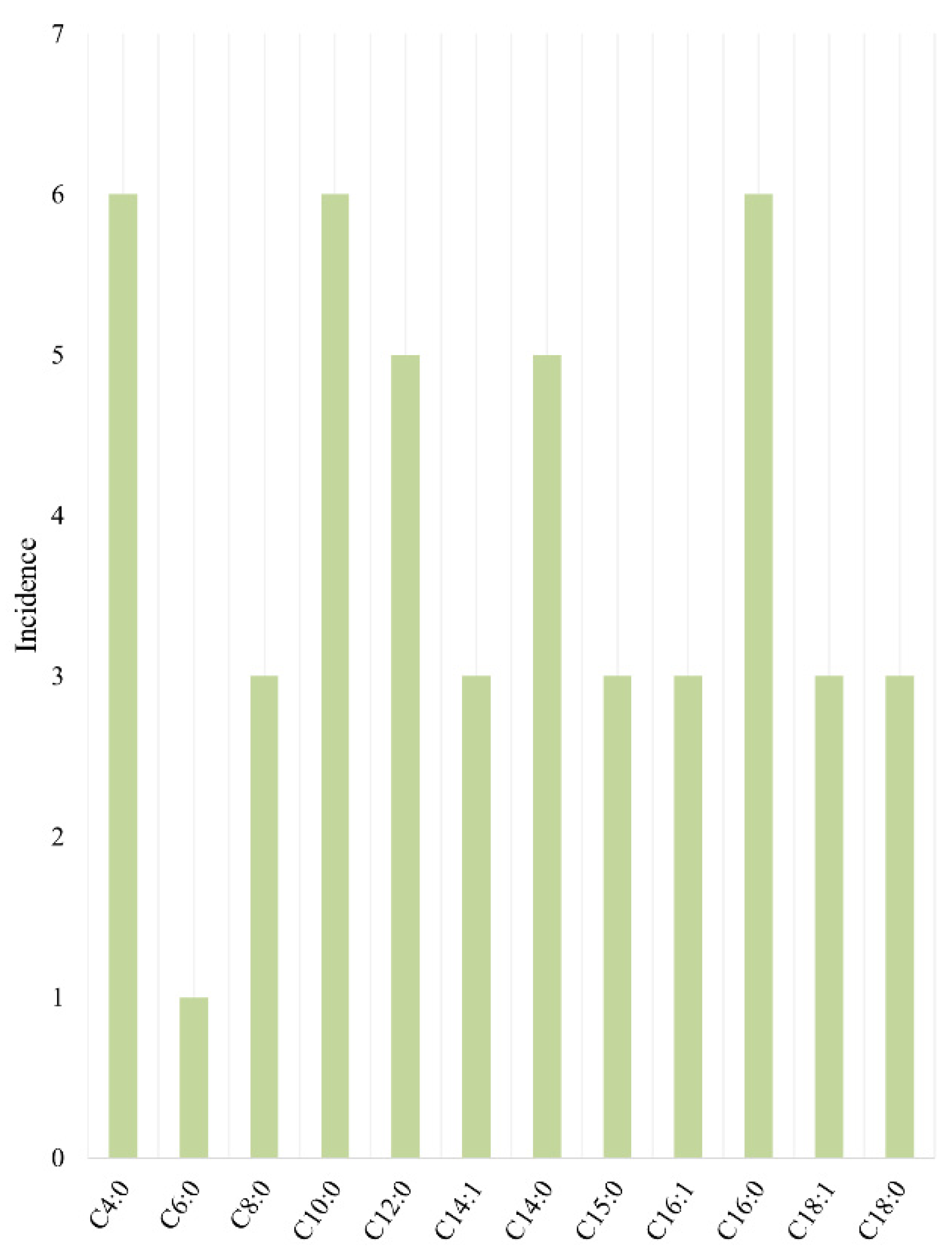
| Nr. | Name Sample | Microorganisms | MSP Score * |
|---|---|---|---|
| 1 | Part 1 | ||
| α–serum light fraction | Pseudomonas fluorescens Bacillus cereus Serratia marcescens Escherichia coli Micrococcus luteus Staphylococcus aureus |
2.21 2.18 2.32 2.08 2.36 2.19 | |
| α–serum heavy fraction | Micrococcus luteus | 2.03 | |
| β-serum light fraction | Pseudomonas fluorescens Lactococcus raffinolactis Serratia marcescens Micrococcus luteus Escherichia coli |
2.14 2.23 2.45 2.07 2.06 | |
| β-serum heavy fraction | Micrococcus luteus Staphylococcus aureus |
2.29 2.32 | |
| AMF ** | no identified microorganisms | ||
| Part 2 | |||
| 2 | α–serum light fraction | Micrococcus luteus Serratia marcescens |
2.20 2.37 |
| α–serum heavy fraction | Micrococcus luteus Staphylococcus aureus Lactococcus raffinolactis Escherichia coli |
2.12 2.21 2.35 2.41 | |
| β-serum light fraction | Serratia marcescens Micrococcus luteus |
2.40 2.27 | |
| β-serum heavy fraction | Pseudomonas fluorescens Staphylococcus aureus |
2.26 2.19 | |
| AMF ** | no identified microorganisms | ||
| Part 3 | |||
| 3 | α–serum light fraction | Micrococcus luteus Escherichia coli |
2.22 2.36 |
| α–serum heavy fraction | Pseudomonas fluorescens Lactococcus raffinolactis |
2.20 2.16 | |
| β-serum light fraction | Pseudomonas fluorescens Lactococcus raffinolactis |
2.36 2.14 | |
| β-serum heavy fraction | Lactococcus raffinolactis Micrococcus luteus |
2.02 2.26 | |
| AMF ** | no identified microorganisms | ||
Disclaimer/Publisher’s Note: The statements, opinions and data contained in all publications are solely those of the individual author(s) and contributor(s) and not of MDPI and/or the editor(s). MDPI and/or the editor(s) disclaim responsibility for any injury to people or property resulting from any ideas, methods, instructions or products referred to in the content. |
© 2024 by the authors. Licensee MDPI, Basel, Switzerland. This article is an open access article distributed under the terms and conditions of the Creative Commons Attribution (CC BY) license (https://creativecommons.org/licenses/by/4.0/).
Share and Cite
Gużewska, G.; Monedeiro-Milanowski, M.; Florkiewicz, A.B.; Arendowska, I.; Walczak-Skierska, J.; Białczak, D.; Pomastowski, P.P. Analysis of the Fatty Acid Profile in Cream, Buttermilk Fractions, and Anhydrous Milk Fat: Influence of Physicochemical and Microbiological Parameters on the Fatty Acid Profile. Appl. Sci. 2024, 14, 6117. https://doi.org/10.3390/app14146117
Gużewska G, Monedeiro-Milanowski M, Florkiewicz AB, Arendowska I, Walczak-Skierska J, Białczak D, Pomastowski PP. Analysis of the Fatty Acid Profile in Cream, Buttermilk Fractions, and Anhydrous Milk Fat: Influence of Physicochemical and Microbiological Parameters on the Fatty Acid Profile. Applied Sciences. 2024; 14(14):6117. https://doi.org/10.3390/app14146117
Chicago/Turabian StyleGużewska, Gaja, Maciej Monedeiro-Milanowski, Aleksandra Bogumiła Florkiewicz, Izabela Arendowska, Justyna Walczak-Skierska, Dorota Białczak, and Paweł Piotr Pomastowski. 2024. "Analysis of the Fatty Acid Profile in Cream, Buttermilk Fractions, and Anhydrous Milk Fat: Influence of Physicochemical and Microbiological Parameters on the Fatty Acid Profile" Applied Sciences 14, no. 14: 6117. https://doi.org/10.3390/app14146117
APA StyleGużewska, G., Monedeiro-Milanowski, M., Florkiewicz, A. B., Arendowska, I., Walczak-Skierska, J., Białczak, D., & Pomastowski, P. P. (2024). Analysis of the Fatty Acid Profile in Cream, Buttermilk Fractions, and Anhydrous Milk Fat: Influence of Physicochemical and Microbiological Parameters on the Fatty Acid Profile. Applied Sciences, 14(14), 6117. https://doi.org/10.3390/app14146117