Article

Effects of Protein Structure Changes on Texture of Scallop Adductor Muscles under Ultra-High Pressure

1
School of Light Industry, Harbin University of Commerce, Harbin 150028, China
2
School of Food Engineering, Harbin University of Commerce, Harbin 150080, China
3
College of Engineering, Northeast Agricultural University, Harbin 150038, China
4
College of Food Science, Northeast Agricultural University, Harbin 150038, China
*
Author to whom correspondence should be addressed.
Appl. Sci. 2023, 13(24), 13247; https://doi.org/10.3390/app132413247
Submission received: 18 August 2023 / Revised: 5 October 2023 / Accepted: 6 October 2023 / Published: 14 December 2023
(This article belongs to the Section Food Science and Technology)

Abstract

:
In order to investigate the effect of protein structure changes on the texture of scallop adductor muscles under ultra-high pressure, the protein structure, hardness, elasticity, cohesion, and chewing properties of untreated scallops maintained at 200 MPa for 60 s, 120 s, and 180 s were measured and compared. At the same time, sensory indicators were evaluated to verify the effect of ultra-high pressure treatment on the quality of scallop adductor muscles. The results indicated that the mass fraction of the α-helix was decreased by 13.70% and the mass fraction of β–folding was 2.72 times higher in the adductor muscle of scallops compared to the fresh adducts without ultra-high pressure treatment, maintained for 180 s at an ultra-high pressure of 200 MPa. At the same time, the value of I850/I830 of protein tyrosine residues was 1.094, which weakened the water retention ability of the protein, the elasticity of the scallop adduct was reduced from the original 7.16 N to 6.17 N, the cohesion was reduced by 3.76%, and the hardness was increased by 17.45%. This improved the cooking quality of scallops, which was consistent with the sensory evaluation results. Therefore, under ultra-high pressure treatment, changes in the protein structure of the adductor muscle of scallops had a certain impact on their texture, which was able to provide support for in-depth research on the mechanism of ultra-high pressure action.

1. Introduction

Food texture is the sensory expression of food structure and its response to external forces, which refers to the comprehensive sensing of the rheological properties of food via mechanical, tactile, visual, and auditory methods. Food texture is a physical property related to the internal structure and state of food [1,2]. As one of the various classification methods on food texture from different perspectives, Szczesniak divides the texture of food into three categories, namely mechanical properties, geometric properties, and other properties, where mechanical properties include the hardness, elasticity, cohesion, adhesion, and chewability of food [3]. The geometric characteristics mainly represent the shape, size, and direction of food particles. At the same time, the texture of food also includes other properties, such as juiciness, greasiness, and wetness.
Scallops, which are rich in protein and trace elements with low levels of fat, have been popular with the population’s increasing demand for healthy food. Subsequently, how to protect the quality of scallops in the circulation process in a way that can be objectively and accurately evaluated has become a concern of many researchers. Scallops are a typical mollusk composed of sarcoplasmic protein and myofibrillar protein macro-molecules, and the changes in the characteristics of myofibrillar fibers are closely related to the qualitative characteristics of the adductor muscle of scallops [4]. The application of ultra-high pressure will destroy the non-covalent bonds in the protein, and the activity of protease changes with the increase in pressure, which hydrolyzes the protein in the adductor muscle of scallops and loosens the fibrous tissue. The effect of ultra-high pressure also destroys the mesh structure of proteins [5,6,7], thus affecting the cohesion, hardness, elasticity, and other structural indicators of scallop adductor muscles [8]. Therefore, there might be a correlation between protein structure and textural changes in closed-shell scallop muscles.
Ultra high-pressure packaging technology could maintain the composition of macro-molecules, such as proteins, while water molecules enter the macro-molecular groups and would adhere to the amino acids of proteins through filling and infiltration, thus breaking the non-covalent bonds in the tertiary structure of proteins [9]. The molecular chain of the denatured protein molecules would be stretched out so that its original three-dimensional structure is changed during pressure relief. The muscle fibers and adhesive tissues would be relaxed. In particular, treatment with ultra-high pressure would only change the non-covalent bond structure of the molecules, as it maintains the covalent bond structure of small molecules to a great extent and also protects the natural quality and nutritional value of food [10,11].
Ultra high-pressure packaging could improve textural characteristics and protein stability and extend the shelf life of Chinese tubular whip shrimp and black tiger shrimp [12,13]. Furthermore, the influence of ultra-high-pressure technology on the quality of scallops was studied [14]. The treatment of scallops through pressure at 250 MPa for 5 min could improve the texture of the shell muscle of scallops. Ultra-high pressure was also used to package different kinds of scallops and significantly improved water-holding capacity, reduced the total number of colonies, and extended the shelf-life of the scallops [15,16]. This article links the changes in protein structure and texture of scallop adductor muscle under ultra-high pressure, which could lay a foundation for the further exploration of the mechanism of ultra-high pressure action.

2. Experimental Materials and Test Methods

2.1. Experimental Materials

The samples used in the experiment were gulf scallops, which are abundant along the Liaoning coast, that were purchased from the seafood market of Bohai University in Jinzhou, Liaoning Province. The average mass of the 24 scallops required for the experiment was 50 g ± 5 g, and the longest axis diameter was about 95 ± 5 mm.

2.2. Sample Processing and Preparation

The sample processing process is shown in Figure 1.
During the sample processing, the ultra-high pressure was set to 200 MPa and divided into three groups (60 s, 120 s, and 180 s) based on different holding times. Two scallops were used in each group (for parallel testing), and then compared with untreated scallops. All experiments were performed at room temperature at around 20 °C.

2.3. Experimental Reagents and Main Equipment

2.3.1. Experimental Reagents

The main reagents included concentrated hydrochloric acid analysis pure, pure boric acid analysis pure, perchloric acid analysis pure, pure sodium hydroxide, anhydrous ethanol, glycerol, distilled water, 2.5% glutaraldehyde solution, 0.2 mol/L phosphate buffer and other auxiliary experiments for determination.

2.3.2. Main Instruments and Equipment of the Experiment

HPP.L2-600 Ultra high pressure processing equipment (Huatengmiao Biological Engineering Technology Co., Ltd., Tianjin, China); S-4800 Field emission scanning electron microscopy (Hitachi, Tokyo, Japan); LabRAM HR Evolution Raman spectrum analyzer (HORIBA Jobin Yvon, Paris, France); ALPHA1-2 LD plus lyophilizer(Martin Christ Company, Martin Christ Company, Osterode, German); H1850 High speed centrifuge (Hunan Xiangyi Laboratory Instrument development Co., Ltd., Changsha, China); PL602-L Analytical balance (METTLER TOLEDO Corporation, Zurich, Switzerland); TA-XT PLUS Texture analyzer (Stable Micro Systems, London, UK).

2.4. Experimental Methods

2.4.1. Determination of Musculature of Adductor of Scallops (Texture Profile Analysis, TPA)

The adductor muscle of scallops treated with ultra-high pressure was removed and placed directly under the probe of the texture analyzer. Three different test points were taken to determine the hardness, elasticity, cohesive, and chewiness of the adductor muscle of scallops, and three parallel experiments were conducted in each group. During the experiment, the probe of P/0.5 was used, the trigger force was 5 g, the detection speed and test speed were 2 mm/s and 3 mm/s respectively, the test acceleration was 5 mm/m2, the depth was 50%, and the time was 5 s [17].

2.4.2. Raman Spectrum Analysis

The protein in the adductor muscle of scallops undergoes denaturation under ultra-high pressure. Therefore, the changes in structural indicators such as elasticity, hardness, chewiness, and cohesion of the adductor muscle can be obtained based on Raman spectroscopy, the protein structure of the ultra-high pressure treated scallop adductor muscle can be analyzed, which can lay the foundation for in-depth analysis of the ultra-high pressure shelling mechanism of scallops [18,19].
The adductor muscle of the ultra-high pressure package was cut along the longitudinal axis and divided into 0.5 × 0.5 × 0.2 cm pieces. The Raman spectroscopy analyzer LabRAM HR Evolution produced by HORIBA Jobin Yvon of France was used for analysis at following condition: wavelength of the Raman spectroscopy analyzer at 515.4 nm, the gap at 200 μm, the power at 129 mW, the exposure time at 60 s and the wavenumber range of detection is 400~4000 cm−1. At last, the average value of the results of the three times is taken to draw the Raman spectrum. After the data is processed by Labspec5.0 software, secondary structure of the protein was calculated by using Alix software.

2.4.3. (Scanning Electron Microscopy) SEM Analysis

The shell muscle sample of the contact surface of scallops treated with ultra-high pressure and separated from the shell was removed and fixed with 2.5% glutaraldehyde solution for 24 h. After that, it was rinsed with 0.2 mol/L phosphate buffer three times for 10 min each time, and then gradient elution with 50%, 60%, 70%, 80%, 90%, and 100% ethanol respectively for 10 min. The samples were subjected to ion gradient gold plating, and the images were observed under S-4800 field emission scanning electron microscopy (SEM) after drying.

2.4.4. Analysis of Sensory Evaluation

The adductor muscle of scallops treated with ultra-high pressure and separated from the shells placed in a porcelain plate, then it is steamed with boiling water for 3 min and cooled for 1 min at room temperature, and cool it for 1 min. The sensory evaluation is based on the sensory evaluation table to score the sample. According to the method of literature [20], six people were selected to form a sensory evaluation group, and the appearance, odor, taste, and texture of the sample were evaluated. The evaluation criteria are shown in the Table 1.

2.4.5. Statistic Analysis

The Duncan method was used for significance analysis of experimental data, the experimental data of variance analysis is represented as mean + standard deviation, and the significance level of 0.05 is tested between data groups. Figures was prepared by Origin 8.0.

3. Influence of Ultra-High Pressure Packaging Technology on Protein Structure of Scallop Adductor Muscle

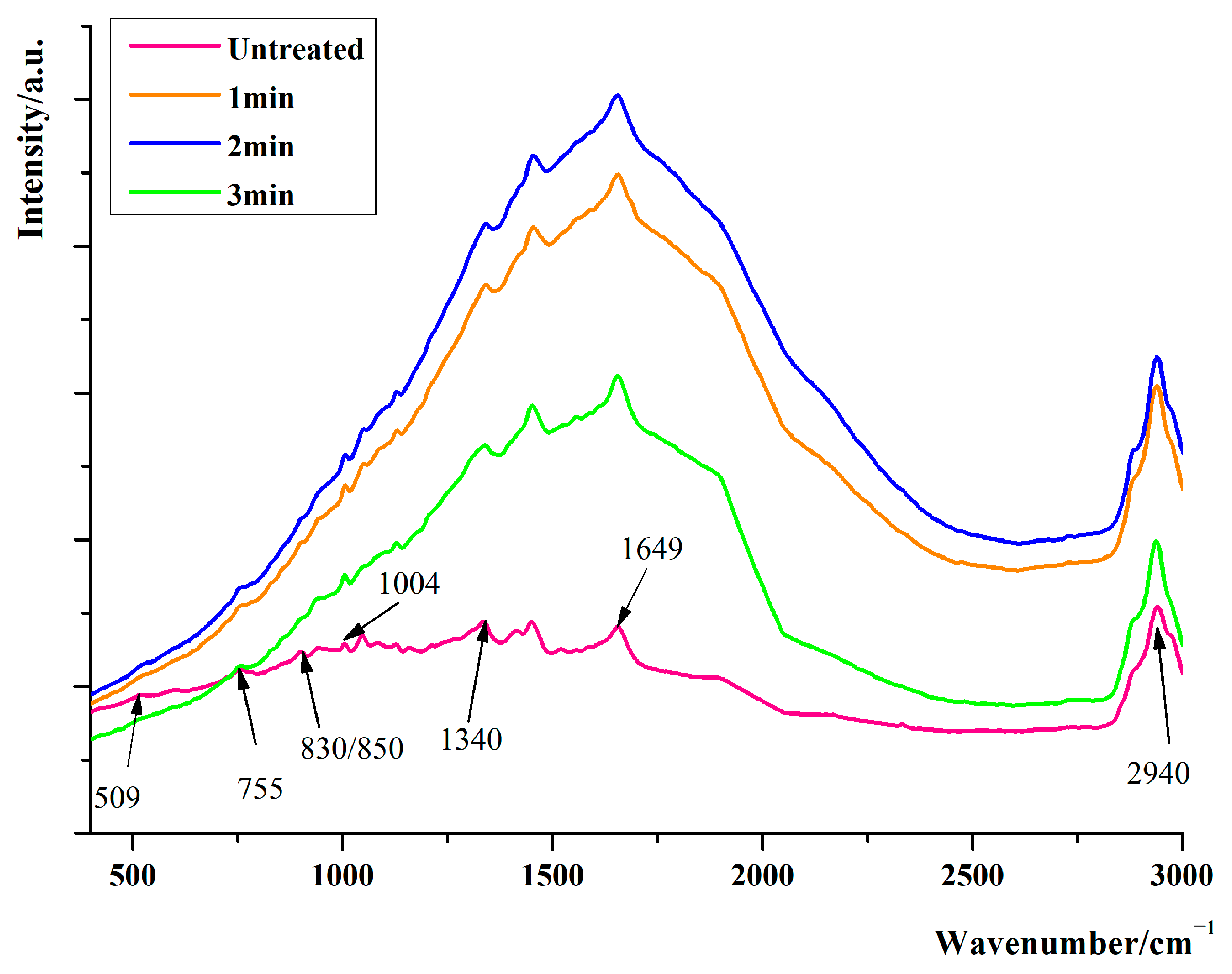
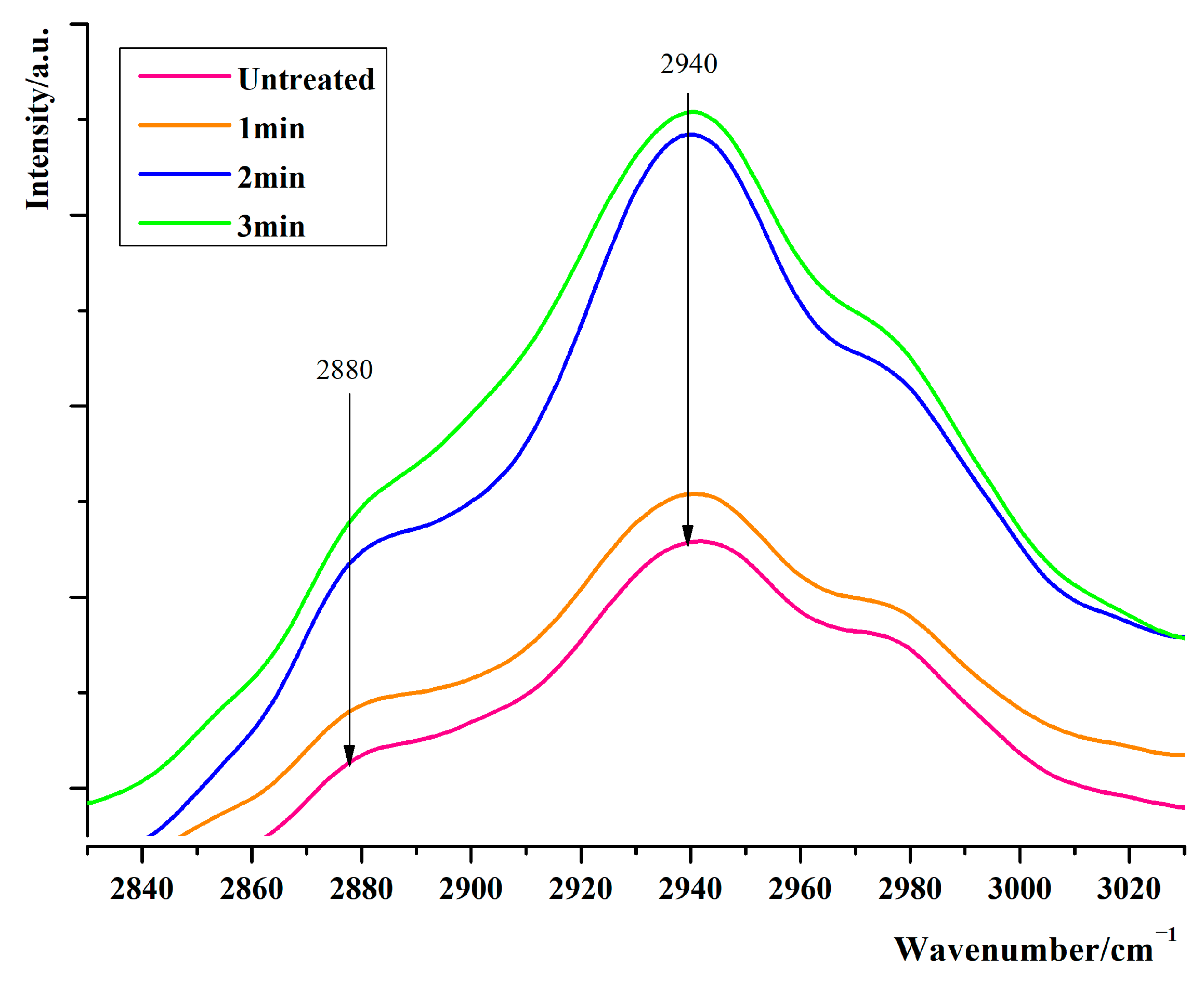
3.1. Raman Spectroscopic Analysis of Proteins in Adductor Muscle of Scallops under Ultra-High Pressure with Different Holding Times

According to Figure 2., there were 7 characteristic peaks of the adductor muscle of scallops under ultra-high pressure mainly concentrated in two bands. One band concentrated between 500 cm−1 and 1800 cm−1 especially, which is usually referred to as the fingerprint region, which mainly reflects the microenvironment changes of amino acid residues and the spatial conformation of proteins in the adductor muscle of scallops. In this region of the Raman spectrum, 6 characteristic peaks appeared; other is the C-H stretching vibration region between 2800 cm−1 and 3050 cm−1. By analyzing the changes in parameters such as peak shape and peak intensity of the characteristic peaks in these two Raman spectral bands of the adductor muscle at the scallop interface position, the effect of different ultra-high pressure treatment conditions on the structural changes of the adductor muscle at the scallop interface position is explained. The attribution of Raman spectral characteristic bands of the adductor muscle of scallops [21,22,23] is shown in Table 2.

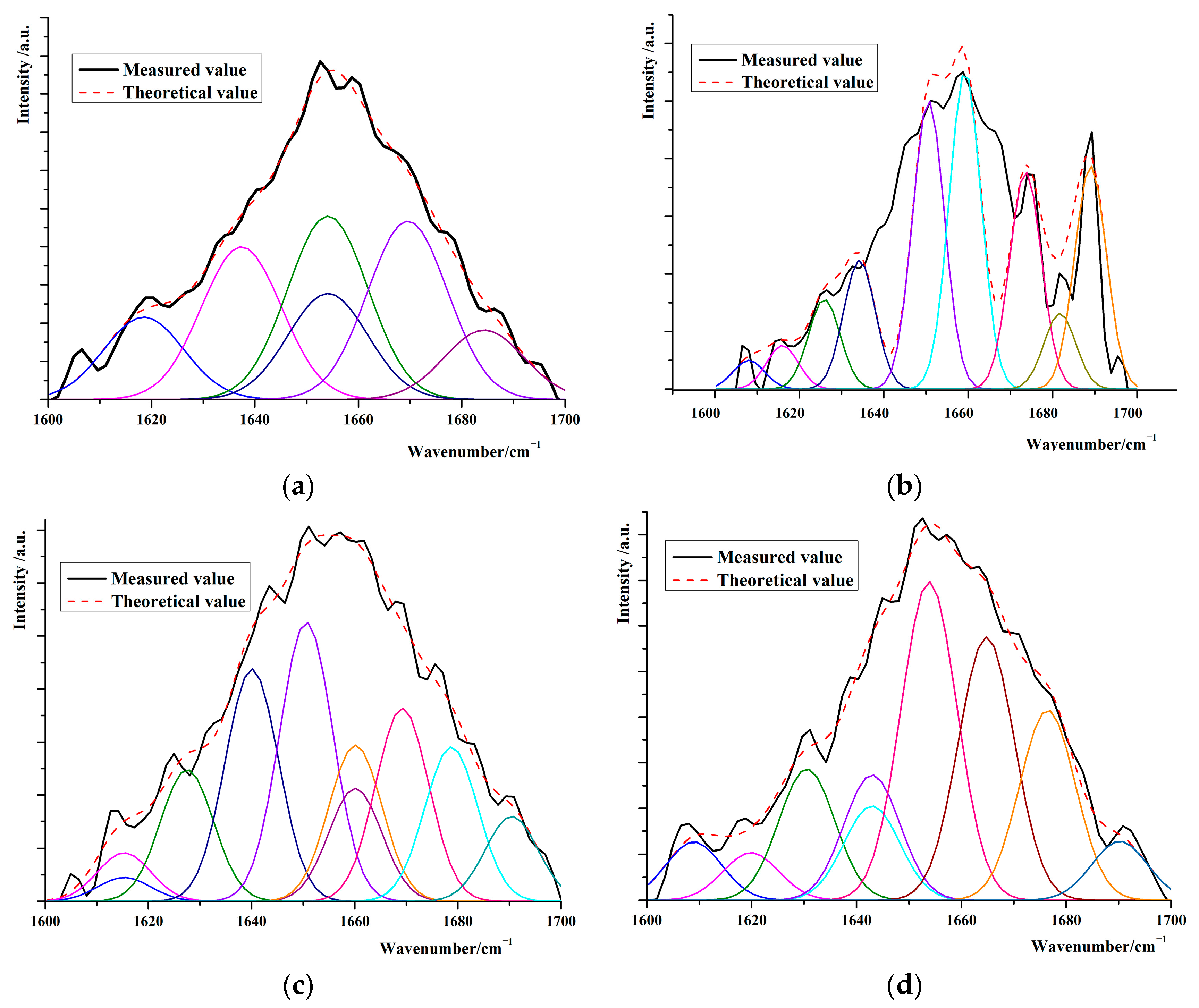
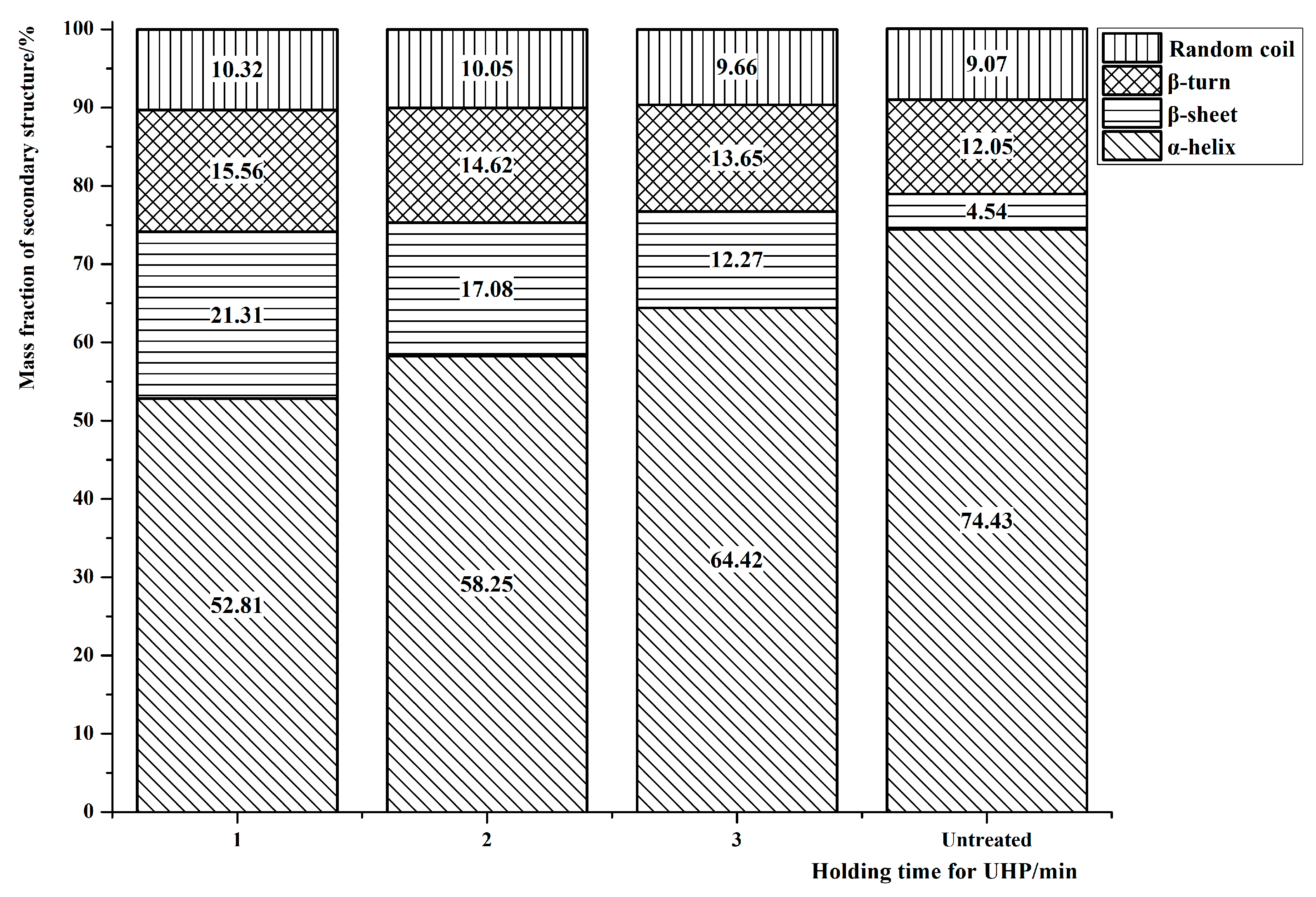
3.2. Changes in the Secondary Structure of Protein in the Interfacial Adductor Muscle of Scallops

There is a phenylalanine ring at 1004 cm−1, whose structure is not affected by protein structure, was used as the internal standard to normalize the data. After processing, Alix computing software was used to carry out deconvolution, second-order derivation and curve fitting for the processed Raman spectrum data, and then the Raman spectrum was divided into peaks. According to the obtained peak area, the secondary structure of the protein was calculated, and the influence of different ultra-high pressure treatment conditions on the secondary structure of the protein in the adductor muscle at the interface of the scallop was analyzed, as shown in Figure 3.
By comparing the peak areas corresponding to α-helix, β-sheet, β-turn and random coil in proteins, according to the peak segmentation and iterative fitting curve of the adductor muscle on the scallop interface in the amide I region as shown in Figure 3, the relative contents of each secondary structure contained in the protein of the adductor muscle at the interface of the scallop were obtained. The relative contents of the main secondary structure of the protein were shown in Figure 4 under the condition of different holding time.
As can be seen in Figure 3, the mass fraction of α-helix structure decreased, and the mass fractions of β-sheet, β-turn, and random coil increased in the protein secondary structure of the adductor muscle of scallops under UHP force compared with that of the adductor muscle of scallops untreated. However, as far as the UHP treatment was concerned, the mass fraction of α-helix increased with the increase of the holding time; the mass fractions of the other three structures gradually decreased. According to the results obtained from the split-peak fitting calculation, the mass fraction of α-helix decreased by 29.29% when the holding time was 1 min, while the mass fraction of α-helix only decreased by 13.70% compared with the untreated ones when the holding time was 3 min. The α-helix structure is connected by hydrogen bonding, and the increase in its mass fraction indicates less damage to the hydrogen bonding by the action of UHP; the trend in the mass fraction of the α-helix versus the β-sheet is shown in the results calculated using split-peak fitting. The mass fractions of β-sheet of the adductor muscle of scallops increased by 447.14%, 276.87%, and 173.23%, respectively, under the treatments with different holding pressure times; the mass fractions of β-turn increased by 21.97%, 21.15% and 13.17%, respectively; and the mass fraction of the random coil also increased by 14.76%, 11.01% and 6.94%, respectively, under different holding times. In conclusion, the protein structure of the adductor muscle changed significantly under different holding times, which lays a foundation for the further elaboration of the reasons for the changes in the texture of the adductor muscle of scallops.

3.3. Influence of Pressure Holding Time on Side Chain Conformation of Protein in Adductor Muscle at the Interface of Scallops

3.3.1. Influence of Pressure Holding Time on Tyrosine Residues in Proteins

The changes of tyrosine residues in proteins are usually measured by the loudness ratio of the absorption peak near 830 cm−1 and 850 cm−1. According to the conditions of different holding times, the Raman spectra of adductor muscle proteins at the scallop interface can obtain the Raman characteristics of tyrosine residues, as shown in Figure 5.
From Figure 5, it can be seen that the absorption peaks near 830 cm−1 and 850 cm−1 become more pronounced compared to untreated scallops. With the prolongation of holding time, this indicates that the effect of ultra-high pressure has caused certain changes in the conformation of tyrosine residues. The absorption peak areas of each absorption peak are calculated based on the absorption peak morphology shown in the Raman spectrum in Figure 5, the values of I850/I830 absorption peak intensities are 1.037, 1.019, 1.028, and 1.094 among untreated scallops, holding time of 1 min, 2 min and 3 min. It can be seen that the ratio decreases first and then increases with the prolongation of pressure holding time, but the values are all more than 1. This indicates that under experimental pressure of 200 MPa, pressure was holded for 1 min and 2 min can cause some tyrosine residues originally exposed on the surface of the polypeptide chain to be re-embedded inside the shell protein molecules at the interface position of the scallop, the hydrophobic interaction in the polar microenvironment weakens, increasing the stability of the protein’s tertiary structure. However, when the holding time is extended to 3 min, the tyrosine residues embedded in the protein molecule were re exposed, and the tyrosine residues transform into hydrogen bonded receptors or the interaction between the donor and the polar microenvironment results in an increase in I850/I830, leading to changes in the biological characteristics and solubility of the protein.

3.3.2. Changes of Tryptophan Residues in Adductor Muscle Proteins at the Interface of Scallops

In the Raman profile, the absorption peak near 755 cm−1 represents the microenvironmental state of tryptophan residues, and the change in the absorption peak intensity reflects the exposure state of tryptophan residues.
As can be seen in Figure 5, the tryptophan residues in the protein molecules of the closed shell muscle of scallops gradually transition from an embedded state to an exposed state as the insulation time prolongs based on the Raman spectroscopy of the closed shell muscle of scallops. This is consistent with the Fermi resonance line analysis of tyrosine. This exposure of amino acid side chains may be caused by the unfolding of the protein spatial structure, leading to an increase in the hydrophobicity of the protein.

3.3.3. Influence of Pressure Holding Time on the Vibration of C-H Bond of Protein in Adductor Muscle at the Interface of Scallops

A very strong and sharp absorption peak appears near 2900 cm−1 in the Raman spectrum of the scallop adductor muscle shown in Figure 2. It can be seen from Table 1 that this absorption peak represents the stretching vibration of the C-H bond in the protein, including the asymmetric stretching vibration of CH2 or the symmetric stretching vibration of CH3, that is, the intuitive reflection of the structure and characteristics of Aromatic amino acid of the protein. In addition, a relatively weak sub peak was found near 2875 cm−1 in the Raman spectrum, which characterizes the asymmetric stretching vibration of CH2. Changes in the intensity of absorption peaks of aromatic amino acids of scallop interfacial position closed-shell muscle proteins reflected in Raman spectrograms were related to changes in the hydrophobic interactions between the C-H of aromatic amino acids caused by stretching of the α-helical structure in the secondary structure of proteins, and if the intensity of Raman characteristic peaks corresponding to C-H stretching vibrations characterizing the C-H stretching vibrations in Raman spectra increased, this indicated that the hydroxyl group became more exposed in the polar microenvironment. The Raman feature profiles of aromatic amino acids under different holding time conditions are shown in Figure 6.
As can be seen from Figure 6, the absorption peaks were gradually enhanced with the prolongation of the holding time, which was attributed to the fact that in the process of the α-helix structure in the protein molecule being continuously stretched into the β-sheet, the aromatic amino acids in the originally embedded state were continuously exposed to the polar environment, which resulted in the increasing intensity of the absorption peaks.

3.3.4. Influence of Pressure Holding Time on Protein Disulfide Bond

The corresponding characteristic absorption spectrum of a disulfide bond in the Raman spectrum is in the region of 500 cm−1~550 cm−1 as showed in Table 1 and Figure 2. The influence of different pressure holding times on the conformation of protein disulfide bonds in adductor muscle at the interface position of scallops is shown in Figure 7.
The absorption peak of the disulfide bond undergoes a significant shift as the holding time prolongs was shown in Figure 7. The absorption peak of disulfide bonds in the protein of the adductor muscle of scallops without ultra-high pressure treatment appears around 516 cm−1, and the absorption peak of disulfide bonds shifts towards the high-frequency direction by 12 cm when the holding time is 1 min. The absorption peak continues to move towards the high-frequency direction and appears around 532 cm−1 when the holding time is 2 min, the absorption peak of disulfide bonds appears around 541 cm−1 when the holding time increases to 3 min. The position where the peak appears can characterize the spatial conformation of the disulfide bond. When the disulfide bond absorption peak is located near 515 cm−1 to 525 cm−1, the conformation is twisted trans (g-g-t); while its conformation is twisted trans (t-g-t) when it is between 535 cm−1 and 545 cm−1. The spatial conformation of the protein disulfide bond in the closed shell muscle of the scallop changes from the initial g-g-t conformation to the t-g-t conformation When the pressure holding time is extended, which is indicated by Raman spectroscopy.

4. Scanning Electron Microscope Analysis of the Effect of Ultra-High Pressure Treatment on the Protein of the Adductor Muscle at the Interface of Scallops

The microscopic morphology of the scallop adductor muscle was obtained, as shown in Figure 8.
From Figure 8, it can be seen that the adductor muscle fibers of scallops without ultra-high pressure treatment are continuous, complete, parallel and evenly distributed, with regularity. A small amount of spherical and ordered loose structures appeared on the surface of protein fibers in the adductor muscle of scallops when the holding time was 1 min; the number of non fibrous protein structures on the surface of adductor muscle protein molecules significantly increases when the holding time is 2 min, which may be due to the exposure of amino acid side chains in the protein to the surface of polypeptide chains under experimental pressure; the 180° flipped structure replacing the original uniformly distributed fibers appeared in the protein structure of the adductor muscle, which was consistent with the results of Raman spectroscopy analysis when the holding time was extended to 3 min.

5. Effect of Pressure Holding Time on Musculature of Adductor of Scallops

The hardness, elasticity, cohesiveness, and chewiness of scallops treated with ultra-high pressure of 200 MPa for 1 min, 2 min and 3 min were measured. The experimental data were analyzed for differences using the Waller Duncan method in Origin, and the experimental results are shown in Table 3.
As shown in Table 3, the hardness of the adductor muscle of scallops gradually increases with the prolongation of holding time; When the holding time was 3 min, the hardness of the adductor muscle of scallops increased by 38.21%. This is due to the change in the conformation of disulfide bond between protein molecules of scallop adductor muscle (Figure 7), which makes them cross-linked with sulfhydryl group [24,25], improves the stability of molecular structure, makes the fiber of scallop adductor muscle firmer, increases hardness, and alleviates the speed of structural change and mass decline of scallop adductor muscle tissue [26,27].
As can be seen from Table 3, the elasticity of the ultrahigh-pressure-treated and untreated scallop closed-shell muscle was reduced compared with that of the untreated scallop closed-shell muscle, and the shorter the holding time was, the greater the degree of reduction in elasticity was; the elasticity of the scallop closed-shell muscle decreased by 29.33% when the holding time was 1 min, and the elasticity of the scallop closed-shell muscle decreased by 13.83% when the holding time was 3 min. Based on the results of the analysis of the secondary structure of the protein of the scallop closed-shell muscle According to the results of the secondary structure of scallop closed-shell muscle protein (Figure 4), although the UHP treatment decreased the elasticity of scallop closed-shell muscle, the loss of elasticity gradually decreased with the extension of holding time, which was related to the increase of the mass fraction of α-helical structure in the secondary structure of scallop closed-shell muscle protein [28,29,30]; at the same time, the UHP treatment enhanced the hydrophobicity of protein residues, which also enhanced the elasticity of the closed-shell muscle, and the result was in agreement with that of the results of the study in Section 3.3.
As can be seen from Table 4, the scallop closed-shell muscle cohesion increased with the prolongation of the holding time, when the holding time was 180 s, the scallop closed-shell muscle cohesion was 20.21%, which was elevated by 83.73% compared with that of the untreated scallop closed-shell muscle, as can be seen in part 3.3.4 of the text, the action of the ultrahigh pressure altered the spatial conformation of the disulfide bond of the scallop closed-shell muscle protein, which caused the myosin to cross-link and increased the intermolecular forces and increased the cohesion of scallop closed-shell muscle [31,32].
As shown in Table 4, the chewiness of scallop closed-shell muscle was enhanced by the ultrahigh pressure treatment, but with the prolongation of the holding time, the chewiness gradually decreased, and the chewiness of scallop closed-shell muscle was increased by 596.30% and 194.44% when the holding time was 1 min and 3 min, respectively, compared with the initial value, and the hardness of the scallop closed-shell muscle was significantly and positively correlated with the hardness of the scallop closed-shell muscle [33,34].
In conclusion, the holding pressure time had a very significant effect on the texture of scallop closed-shell muscle. Volume compression of scallop closed-shell muscle under ultrahigh pressure increased the interprotein forces and the degree of exposure of hydrophobic groups, which led to an increase in hardness, cohesion and masticatory force, and a decrease in elasticity of scallop closed-shell muscle. Therefore, the UHP treatment changed the protein structure of scallop closed-shell muscle, which altered the texture of closed-shell muscle and improved the initial quality of closed-shell muscle.

6. Sensory Evaluation of the Adductor Muscle of Scallops under Ultra-High Pressure

The sensory evaluation scores of the adductor muscle of scallops under different holding times are shown in Table 3. From Table 3, it can be seen that the comprehensive score of the adductor muscle with a holding time of 2 min is relatively high, indicating that maintaining an ultra-high pressure of 200 MPa for 2 min can effectively improve the appearance, flavor, and cooking quality of scallops. This is consistent with the results of texture analysis and protein structure change analysis.

7. Conclusions

The scallops in this study were subjected to ultra-high pressure of 200 MPa for 1 min, 2 min, and 3 min, respectively. The protein structure of scallop adductor muscle was analyzed using Raman spectroscopy and compared with the untreated protein structure of scallop adductor muscle. The results indicate that the mass fraction of theα-helix structure was decreased, while the mass percentage of β- folder and regular curl structures were increased after ultra-high pressure treatment; however the mass fraction of the α-helix structure was increased by 41.34% as the holding time increased from 1 min to 3 min. Although the elasticity of the adductor muscle of the scallop decreased compared to the untreated one, the elasticity of the adductor muscle of the scallop increased only in terms of the extension of the pressure holding time. At the same time, the exposed state of amino acid residues and changes in disulfide bond conformation weaken the water locking ability of the adductor muscle protein, resulting in an increase in the hardness, cohesion, and chewing ability of the adductor muscle in scallops. Based on the sensory evaluation results, it can be concluded that the changes in protein structure of the adductor muscle of scallops under ultra-high pressure treatment have a significant impact on their texture. This result lays the foundation for further exploration of the mechanism of ultra-high pressure action.

Author Contributions

Conceptualization, X.G.; methodology, J.C.; software, N.X.; formal analysis, J.W.; resources, J.C.; data curation, C.L.; writing—original draft preparation, X.G.; writing—review and editing, Y.Z.; supervision, L.H.; project administration, J.C.; funding acquisition, D.L. All authors have read and agreed to the published version of the manuscript.

Funding

This research work was supported by the National Science and Technology Support Plan Fund (2016 YFD0400301); The Youth Innovation Talent Project of Harbin Commercial University (Research on Protein Molecular Mechanics Effect and Texture Control Technology in Ultrahigh Pressure Processing of Aquatic Products); The PhD Research Initiation Support Program of Harbin University of Commerce (22BQ01); the discipline team of food based functional active packaging of the Northeast Agricultural University (54941112).

Data Availability Statement

The data presented in this study are available in this article.

Conflicts of Interest

The authors declare no conflict of interest.

References

  1. Naciye, K.; Ravi, P.; Irem, S.; Aybike, K.; Aybike, S.; Anjineyulu, K. Impact of different microwave treatments on food texture. J. Texture Stud. 2021, 53, 710–736. [Google Scholar]
  2. Muhammad, A.K.; Sher, A.; Yang, H.J.; Asghar, A.K.; Zulfiqar, A.; Ronald, K.T.; Zhou, G.H. Improvement of color, texture and food safety of ready-to-eat high pressure-heat treated duck breast. Food Chem. 2019, 277, 646–654. [Google Scholar]
  3. Szczeshiak, A.S. Texture: Is it still an overlooked food attribute. Food Technol. 1990, 44, 86–95. [Google Scholar]
  4. Tong, S.N.; Liu, R.; Wang, W.; Lv, X.R.; Xu, Y.X.; Mi, H.B.; Li, J.R.; Yi, S.M.; Li, X.P. Synergistic effect of ultrahigh pressure and allicin on gel properties, flavor characteristics, and myosin structure of the obturator muscle of the scallop (Patinopecten yessoensis). Food Sci. 2023, 88, 3007–3021. [Google Scholar] [CrossRef] [PubMed]
  5. Chen, W.X.; Ou, Y.J.; Xu, W.Q.; Ni, J.; Tan, J.Y.; Shen, J. Effects of storage conditions on nutrition and quality of live Patinopecten yessoensis. Fish. Mod. 2019, 46, 83–88. [Google Scholar]
  6. Xuan, X.T.; Cui, Y.; Lin, X.D.; Yu, J.F.; Liao, X.J.; Ling, J.G.; Shang, H.T. Impact of high hydrostatic pressure on the shelling efficacy, physicochemical properties, and microstructure of fresh razor clam (Sinonovacula constricta). J. Food Sci. 2018, 83, 284–293. [Google Scholar] [CrossRef] [PubMed]
  7. Yu, Z.C.; Liu, C.; Fu, Q. The differences of bacterial communities in the tissues between healthy and diseased Yesso scallop (Patinopecten yessoensis). AMB Express 2019, 9, 148. [Google Scholar] [CrossRef] [PubMed]
  8. Ma, Y.X.; Li, M.; Sun, J.X. Characterization of bacterial community associated with four organs of the yesso scallop (Patinopecten yessoensis) by high-throughput sequencing. J. Ocean. Univ. China 2019, 18, 493–500. [Google Scholar] [CrossRef]
  9. Wang, Z.Y.; Wu, Z.X.; Zhao, G.H.; Li, D.Y.; Liu, X.Y.; Qin, L.J.; Jiang, P.F.; Zhou, D.Y. Effect of air frying and baking on physicochemical properties and digestive properties of scallop (Patinopecten yessoensis) adductor muscle. Food Biosci. 2023, 52, 102460. [Google Scholar] [CrossRef]
  10. Cui, Y.; Lin, X.D.; Kang, M.L.; Yu, J.F.; Guo, R.X.; Ling, J.G. Advances in application of ultra high pressure for preservation and processing of aquatic products. Food Sci. 2016, 37, 291–299. [Google Scholar]
  11. Zhang, B.; Fang, C.D.; Hao, G.J. Effect of kappa-carrageen an oligosaccharides on myofibrillar protein oxidation in peeled shrimp (Litopenaeus vannamei) during long-term frozen storage. Food Chem. 2018, 245, 254–261. [Google Scholar] [CrossRef] [PubMed]
  12. Wang, Z.Y.; Yang, W.G.; Zhou, G.; Xu, D.L.; Lou, Q.M.; Zhang, J.J.; Cui, Y. Shelling of Solenocera melantho Using Ultra High Pressure and Its Effect on the Quality of Muscle. Food Sci. 2017, 38, 43–48. [Google Scholar]
  13. Kong, N.; Zhao, J.Y.; Zhao, B.; Liu, J.Y.; Li, F.Z.; Wang, L.L.; Song, L.S. Effects of high temperature stress on the intestinal histology and microbiota in Yesso scallop Patinopecten yessoensis. Mar. Environ. Res. 2023, 185, 105881. [Google Scholar] [CrossRef] [PubMed]
  14. Wen, L.H.; Chen, Q.H.; Xuan, X.T.; Cui, Y.; Shang, H.T.; Ling, J.G.; Chen, X.E.; Fang, X.B. Effects of ultra-high pressure treatment on shucking and quality of Patinopecten yessoensis. J. Zhejiang Ocean Univ. 2020, 39, 117–122. [Google Scholar]
  15. Yue, J.; Zhang, Y.F.; Jin, Y.F.; Deng, Y.; Zhao, Y.Y. Impact of high hydrostatic pressure on non-volatile and volatile compounds of squid muscles. Food Chem. 2016, 194, 12–19. [Google Scholar] [CrossRef] [PubMed]
  16. Lai, K.M.; Chi, H.Y.; Hsu, K.C. High-pressure treatment for shelf-life extension and quality improvement of oysters cooked in a traditional Taiwanese oyster omelet. J. Food Prot. 2010, 73, 53–61. [Google Scholar] [CrossRef]
  17. Shi, Y.M.; Li, Y.Y.; Wu, D.D.; Wang, P.; Zhang, D.K.; Lou, Y.H. Effects of different freezing methods on quality of Penaeus vannameiv. Food Ferment. Ind. 2019, 45, 94–100. [Google Scholar]
  18. Dong, X.H.; Zhao, M.M.; Jiang, Y.M. Application of ultra—High pressure technique in protein food industry. Food Ind. Technol. 2012, 33, 451–454. [Google Scholar]
  19. Chen, Q.M.; Xie, Y.F.; Guo, Y.F.; Cheng, Y.L.; Yao, W.R. Application of Raman spectroscopy in a correlation study between protein oxidation/denaturation and conformational changes in beef after repeated freeze–thaw. Int. J. Food Sci. Technol. 2021, 57, 719–727. [Google Scholar] [CrossRef]
  20. Yan, L.X.; Tian, Y.Y.; Jiang, M.H.; Liu, J.R.; Xu, T.Y. Characteristics changes in activity and taste properties in Yesso Scallop Patinopecten yessoensis: From waterless transportation to wet storage selling. Fish. Sci. 2022, 41, 44–51. [Google Scholar]
  21. Bryant, R.N.; Pasteris, J.D.; Fike, D.A. Express: Variability in the Raman spectrum of unpolished growth and fracture surfaces of pyrite due to laser heating and crystal orientation. Appl. Spectrosc. 2018, 72, 37–47. [Google Scholar] [CrossRef]
  22. Liang, Z.W.; Du, Z.F.; Li, C.L.; Wang, M.X.; Wang, B.; Zhang, X. Confocal raman micro-spectroscopy analysis of mussel foot. Spectrosc. Spectr. Anal. 2020, 40, 755–759. [Google Scholar]
  23. Sun, W.Z.; Zhao, Q.Z.; Zhao, M.M. Structural evaluation of myofibrillar proteins during processing of Cantonese sausage by Raman spectroscopy. J. Agric. Food Chem. 2011, 59, 11070–11077. [Google Scholar] [CrossRef] [PubMed]
  24. Jia, Y.H.; Yang, X.S.; Xu, Z.; Guo, Q.Y.; Li, X.Y. Effect of moisture content on the texture and chroma of lightly baked scallop. Food Mach. 2010, 26, 47–50. [Google Scholar]
  25. Lu, B.; Li, M.D.; Zhang, Y.F.; Niu, X.C.; Wang, Z.J.; Jing, L.Z.; Liu, J. Raman spectroscopic characterization of the structural changes in soybean protein isolate induced by low pressure homogenization. Mod. Food Sci. Technol. 2018, 34, 58–63. [Google Scholar]
  26. Ramirez-Suarez, J.C.; Morrissey, M.T. Effect of high pressure processing (HPP) on shelf life of albacoretuna (Thunnus alalunga) minced muscle. Innov. Food Sci. Emerg. Technol. 2006, 7, 19–27. [Google Scholar] [CrossRef]
  27. Zhang, L.; Lu, H.X.; Li, J.R. Effects of ultra-high pressure treatments on the qualities of Litopenaeus vannamei. Food Res. Dev. 2010, 31, 1–6. [Google Scholar]
  28. Ranman, M.S.; Al-farsi, S.A. Instru mental texture profile analysis (TPA) of date flesh as a function of moisture content. J. Food Eng. 2005, 66, 505–511. [Google Scholar] [CrossRef]
  29. Zhang, G.N.; Hu, b.; Wu, F.R. Effects of Angelica sinensis on the microstructure of myogenic fibronectin in snakehead mullet. J. Huazhong Agric. Univ. 2018, 37, 117–122. [Google Scholar]
  30. Jiang, L.T. The relation of chemical component to texture of white sufu. Mod. Food Sci. Technol. 2010, 26, 797–800. [Google Scholar]
  31. Jia, Y.; Hu, Z.H.; Wang, X.L.; Chen, S.H.; Liu, X.J.; Xia, L. Effect of ultra-High Pressure Treatment on Shucking and Processing Properties of Squilla. Food Sci. 2015, 36, 47–52. [Google Scholar]
  32. Gong, X.; Chang, J.; Zhang, Y.L.; Li, D.T.; Xia, N.; Wang, J.; Sun, Z.H. Structural Changes of the Interface Material of Scallop Adductor under Ultra-High Pressure. Processes 2023, 11, 521. [Google Scholar] [CrossRef]
  33. Zeng, X.Y.; Jiao, D.X.; Yu, X.N.; Chen, L.H.; Chen, L.H.; Sun, Y.; Guo, A.R.; Zhu, C.; Wu, J.S.; Liu, J.S.; et al. Effect of ultra-high pressure on the relationship between endogenous proteases and protein degradation of Yesso scallop (Mizuhopecten yessoensis) adductor muscle during iced storage. Food Chem. 2022, 15, 100438. [Google Scholar] [CrossRef] [PubMed]
  34. Chang, J.; Gong, X.; Zhang, Y.L.; Sun, Z.L.; Xia, N.; Zhang, H.J.; Wang, J.; Zhang, X. Simulation analysis of organic-inorganic interface failure of scallop under ultra-high pressure. Coatings 2022, 12, 963. [Google Scholar] [CrossRef]
Figure 1. Flow chart of sample processing.
Applsci 13 13247 g001
Figure 2. Raman spectra of adductor muscle of scallop under different holding times.
Applsci 13 13247 g002
Figure 3. Distinct peaks and iterative fitting curves of scallop closed-shell muscle protein amide I with different holding times (a) 1 min (b) 2 min (c) 3 min (d) 4 min.
Applsci 13 13247 g003
Figure 4. Effect of holding time on secondary structure of protein in adductor muscle of scallops.
Applsci 13 13247 g004
Figure 5. Structural changes of tyrosine residues under different holding times.
Applsci 13 13247 g005
Figure 6. Structural changes of aromatic amino acids under different holding time conditions.
Applsci 13 13247 g006
Figure 7. Changes of disulfide bonds under different holding times.
Applsci 13 13247 g007
Figure 8. Microstructure of scallop closed-shell muscle fibers under different holding times. (a) Untreated (b) 100 MPa (c) 200 MPa (d) 400 MPa.
Applsci 13 13247 g008
Table 1. Standard for sensory evaluation of scallops.
Sensory AttributesStateScore
AppearanceThe shell meat is milky white with a bright luster3
The shell meat is light yellow and shiny in water2
The shell meat is dark yellow and lacks luster1
OdorFresh and sweet without obvious fishy taste3
average and slightly fishy2
Mild freshness and strong fishy taste1
StateFresh and sweet3
Moderate sweetness2
Mild sweetness1
TextureGood elasticity without obvious fiber or stickiness3
Moderate elasticity and slight stickiness2
Poor elasticity and obvious fiber sensation1
Table 2. Identification of Raman Spectral Characteristic Bands in the adductor muscle of Scallops.
Wave Number/cm−1Band Attribution Information
509Stretching vibration of S-S
755Stretching vibration of Tryptophan
830/853The fundamental and overtone Fermi resonances of tyrosine
1004Respiratory vibration of Phenylalanine ring
1333III band of amide
1657I band of amide
2945Telescopic vibration of C-H
Table 3. Effects of different holding time on musculature of adductor of scallops.
NumberHolding
Time
/min
Texture Analyzer
Hardness/NElasticity/NCohesiveness/%Chewiness/N
1untreated1.23 ± 0.01 d7.16 ± 0.02 a11.00 ± 0.04 d1.08 ± 0.01 d
211.49 ± 0.03 c5.06 ± 0.07 d11.16 ± 0.19 c7.52 ± 0.12 a
321.53 ± 0.02 b5.58 ± 0.24 c19.71 ± 0.07 b6.30 ± 0.16 b
431.70 ± 0.02 a6.17 ± 0.36 b20.21 ± 0.10 a3.18 ± 0.08 c
Note: The data is x ± SD of three parallel samples, and the values marked with different letters in the same column indicate significant differences (p < 0.05).
Table 4. Sensory evaluation results of the adductor muscle of scallops under different holding times.
Sensory
Attributes
Holding
Time
/min
AppearanceOdorStateTexture
Number
1untreated2.38 ± 0.032.91 ± 0.012.48 ± 0.112.24 ± 0.04
212.64 ± 0.022.89 ± 0.032.65 ± 0.022.47 ± 0.07
322.73 ± 0.052.84 ± 0.062.94 ± 0.022.89 ± 0.02
432.81 ± 0.082.72 ± 0.052.83 ± 0.082.51 ± 0.03
Disclaimer/Publisher’s Note: The statements, opinions and data contained in all publications are solely those of the individual author(s) and contributor(s) and not of MDPI and/or the editor(s). MDPI and/or the editor(s) disclaim responsibility for any injury to people or property resulting from any ideas, methods, instructions or products referred to in the content.

