Review

Radiomic Analysis for Human Papillomavirus Assessment in Oropharyngeal Carcinoma: Lessons and Pitfalls for the Next Future

1
Department of Experimental and Clinical Biomedical Sciences “Mario Serio”, University of Florence, 50121 Florence, Italy
2
Radiation Oncology Unit, Azienda Ospedaliero-Universitaria Careggi, 50134 Florence, Italy
*
Author to whom correspondence should be addressed.
Appl. Sci. 2023, 13(23), 12942; https://doi.org/10.3390/app132312942
Submission received: 8 September 2023 / Revised: 27 November 2023 / Accepted: 30 November 2023 / Published: 4 December 2023
(This article belongs to the Special Issue Advances in Radiation Therapy for Tumor Treatment)

Abstract

:

Featured Application

This review reports the current evidence available regarding the role of radiomic features extracted from conventional imaging (MRI, CT and 18F-FDG PET scans) in assessing the status of Human Papillomavirus (HPV) in Oropharyngeal Squamous Cell Carcinoma (OPSCC) patients. The paper evaluates the possible future adoption of radiomic models in determining viral status in a non-invasive manner, especially for those not tolerating biopsy, when biopsy is not feasible or in case of discrepancies between immunohistochemistry and Polymerase Chain Reaction. This could also lead to more tailored treatment strategies.

Abstract

Background: Oropharyngeal Squamous Cell Carcinoma (OPSCC) is rapidly increasing due to the spread of Human Papillomavirus (HPV) infection. HPV-positive disease has unique characteristics, with better response to treatment and consequent better prognosis. HPV status is routinely assessed via p16 immunohistochemistry or HPV DNA Polymerase Chain Reaction. Radiomics is a quantitative approach to medical imaging which can overcome limitations due to its subjective interpretation and correlation with clinical data. The aim of this narrative review is to evaluate the impact of radiomic features on assessing HPV status in OPSCC patients. Methods: A narrative review was performed by synthesizing literature results from PUBMED. In the search strategy, Medical Subject Headings (MeSH) terms were used. Retrospective mono- or multicentric works assessing the correlation between radiomic features and HPV status prediction in OPSCC were included. Selected papers were in English and included studies on humans. The range of publication date was July 2015–April 2023. Results: Our research returned 23 published papers; the accuracy of radiomic models was evaluated by ROC curves and AUC values. MRI- and CT-based radiomic models proved of comparable efficacy. Also, metabolic imaging showed crucial importance in the determination of HPV status, albeit with lower AUC values. Conclusions: Radiomic features from conventional imaging can play a complementary role in the assessment of HPV status in OPSCC. Both primary tumor- and nodal-related features and multisequencing-based models demonstrated higher accuracy.

1. Introduction

Head and Neck Squamous Cell Carcinoma (HNSCC) is the seventh most common malignancy worldwide, with 890,000 new cases and 450,000 deaths [1]. Among HNSCC, Oropharyngeal Squamous Cell Carcinoma (OPSCC) is a specific subtype which arises most frequently from the tonsils, followed by the base-tongue, the soft palate and the uvula. Human Papillomavirus (HPV) infection is strictly related to the development of OPSCC and is responsible for its growing incidence [2], despite a reduction in tobacco smoking and alcohol consumption in Western countries. HPV-positive (HPV+) and HPV-negative (HPV−) OPSCC present as different entities, with peculiar clinical presentation, pathogenetic mechanisms and prognosis. HPV+ patients are usually younger, with no smoking history and fewer comorbidities [3]; HPV+ disease is typically related to nodal spread with cystic neck masses at onset, whereas HPV− cancer symptoms are usually associated with local primary growth. Hence, HPV+ OPSCC is characterized by earlier detection, better prognosis and response rate to Chemoradiotherapy (CRT) [4], which is classified in the 8th edition of TNM staging according to the American Joint Committee on Cancer (AJCC).
Five-year overall survival rates account for 80% in HPV-positive patients versus 50% in HPV-negative ones [5]. Therefore, de-escalation strategies have been investigated to minimize treatment-related adverse events in the former group, even if any modification in treatment regimens is not supported by current clinical data yet [6].
Regardless of HPV status, in early-stage disease, conservative surgery or radiotherapy (either external beam or brachytherapy for selected stage I) give similar rates of locoregional control. Post-operative (chemo-)radiation can be required to decrease the risk of local recurrence in case of pT3-4 (UICC TNM 8th edition), positive margin (tumor at 1 mm from the margin), close resection margin (between 1 and 5 mm), perineural infiltration, lymphovascular spread, >1 invaded lymph node and the presence of extracapsular nodal infiltration. For locally advanced disease (cT3–4 cN0 cM0, cT1–4 cN1–3 cM0), the standard of care is represented by concomitant platinum-based chemoradiotherapy, whereas surgery followed by (C)RT may be an alternative option [7].
HPV infection is most frequently assessed by p16 Immunohistochemistry (IHC) on biopsied tissue or by testing for the presence of HPV DNA or mRNA in the tumor by using Polymerase Chain Reaction (PCR) or in situ hybridization [8]. p16 is routinely used as a surrogate marker; however, in approximately 17% of p16-positive patients, PCR results are negative, with almost the same inferior survival rates of those with p16-negative disease [9]. In addition, surgical biopsy may expose patients to complications such as bleeding, especially in those with hemorrhagic diathesis. Also, immunostaining sensitivity can be influenced by inflammatory changes in the specimen [10]. Moreover, IHC and PCR are time-consuming, expensive and not always available [11].
Therefore, the development of a non-invasive tool to determine HPV status could prove useful.
For what concerns diagnostic imaging, HPV-related disease has peculiar growth patterns and nodal involvement which differ from HPV-negative OPSCC; for example, Cantrell et al. showed that poorly defined borders of the primary tumor and invasion of adjacent muscles are characteristic for HPV-negative disease [12]. Other studies reported a substantial difference in intra-tumoral heterogeneity between HPV-positive and HPV-negative OPSCC primary sites in radiological imaging [13,14,15].
Unfortunately, overlapping radiological characteristics do not allow one to precisely predict HPV status [16].
Radiomics is a non-invasive technique which implies the extraction of different quantitative features from routine medical images (e.g., Magnetic Resonance Imaging (MRI) or Computed Tomography (CT) scans), their selection and subsequent analysis for correlation with clinical data [17,18]. The extracted features describe characteristics such as tumor signal intensity, shape and texture patterns and provide reader-independent data for predictive modeling [19].
The feature-based radiomic workflow firstly relies on the determination of imaging modality, followed by image processing, which represents the attempt at homogenizing imaging for feature extraction according to pixel spacing or gray-level intensities, and then by the Region of Interest (ROI) or Volume of Interest (VOI) segmentation, for the definition of those regions where features are calculated. Then, quantitative features can be extracted with a possible large number (even more than 1000) from a single ROI, since the process is based on a huge number of mathematical operations.
Features can be divided into many subgroups [20]:
-
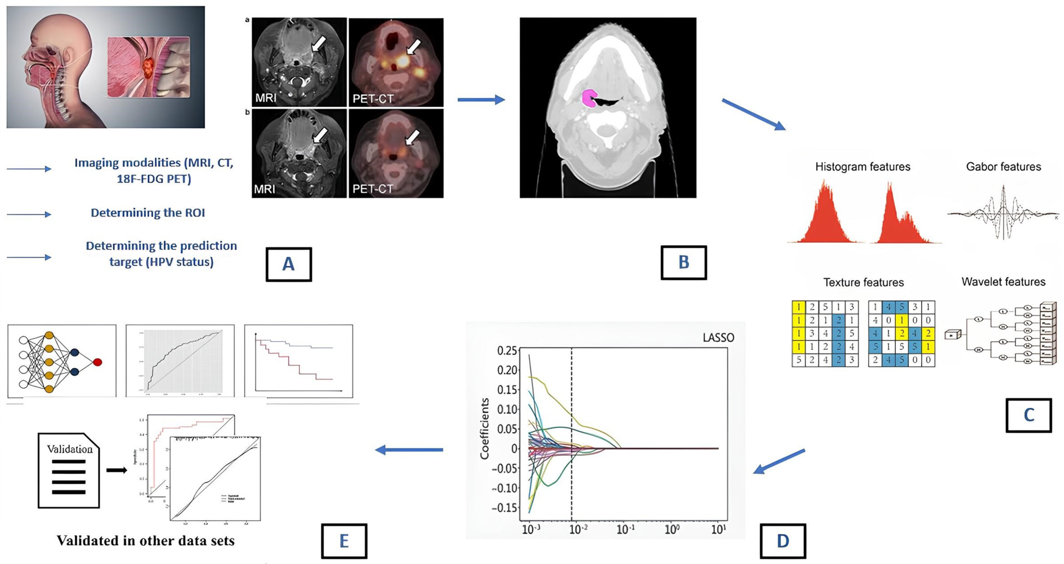
Shape features: these are related to the geometric properties of the ROI/VOI (volume, diameter, sphericity and compacity);
-
First order (histogram) features: these describe the distribution of voxel intensities within the image region (mean, median, skewness, kurtosis);
-
Second order texture features: these consider the statistical relationship between neighboring voxels or groups of voxels within the segmented lesion. Many matrices, such as the Gray-Level Co-occurrence Matrix (GLCM), Gray-Level Run Length Matrix (GLRLM) and Neighborhood Gray-Level Different Matrix (NGLDM), can be used for feature extraction [21]. GLCM provides information about pixel pair distribution within the image; GLRLM identifies the length of consecutive voxels with the same intensity in a pre-set direction in the image; NGLDM analyzes the difference between a gray value and the average gray value of its neighbors within a certain distance [22].
-
Higher-order texture features: additional mathematical transformations are used to highlight specific aspects of the ROI. This can lead to a virtually endless number of features among which fractals, SUV metric for PET specific applications [23,24], Minkowski functionals, wavelet transform and Laplacian transforms of Gaussian-filtered images are included [25,26].
After feature extraction, the most relevant features must be selected among thousands available; redundant or non-reproducible features are generally excluded and feature selection or dimensionality reduction algorithms are usually adopted. Common examples involve hierarchical clustering, principal component analysis, Least Absolute Shrinkage and Selection Operator (LASSO) regularized logistic regression and maximum relevance minimum redundancy filtering [27]. The modeling process then follows in the radiomic workflow. In this phase, reproducible radiomic features can be used to build radiomic models for clinical selected endpoints.
In this context, machine learning is the most common approach to radiomic-based prediction; it is based on training a model by showing it examples of input–output behavior so that the system learns the desired relationship without explicit manual programming. Numerous machine learning classifiers have therefore been developed, all utilizing many algorithms to find the optimal performance across repeated iterations of training and testing [27].
After model building, model independent validation is usually performed. In this step, the radiomic-based model is applied to datasets on which it was not trained. Internal validation refers to the performance in patients from a similar population to where the sample originated from. Internal validation contrasts with external validation, due to differences between the populations used to develop and test the model. Many internal validation approaches are available: the split-sample (where training data are divided into two parts: one to develop the model and another to measure its performance), the cross-validation (which uses different portions of the data to test and train a model on different iterations) and bootstrapping (it resamples a single dataset to create many simulated samples, of the same size as the original dataset) [28]. If the results of internal test performance are satisfactory, then the model is tested on an external validation dataset to assess generalizability in routine clinical practice [29].
Recently, the application of radiomics has been investigated in OPSCC in terms of HPV status prediction, survival prediction and risk stratification [30] to overcome limitations due to subjective interpretation of medical imaging. Since HPV status can be relevant for the determination of a tailored treatment strategy, radiomics might indirectly influence clinical practice and decision-making in the near future.
In this regard, the aim of the present narrative review is to discuss the available evidence regarding the role of quantitative features extracted from CT, MRI or Positron-Emission Tomography (PET) scans in assessing HPV status.

2. Materials and Methods

A narrative review was performed that synthesized literature results from PUBMED. In the search strategy, Medical Subject Headings (MeSH) terms were used. Search terms included in all possible combinations were Human Papillomavirus, HPV, Radiomics, Texture Analysis, Deep Learning, Machine Learning, Oropharyngeal Squamous Cell Carcinoma, OPSCC, Head and Neck Cancer. Identified records were first screened by title and abstract, and those judged to be not pertinent were excluded. Then, full-text articles were assessed for eligibility. In this step, systematic and narrative reviews and meta-analysis were excluded, as well as papers investigating the relationship between radiomic features and other outcomes than HPV assessment (e.g., tumor staging, risk stratification, treatment response assessment, gene expression). In the end, only retrospective mono- or multicentric series assessing the correlation between MRI, CT and 18F-FDG PET radiomic features and HPV status prediction in oropharyngeal cancer patients were included (Figure 1). Selected papers were in English and included studies on humans. The range of publication date was July 2015–April 2023. A PRISMA flow-chart illustrating the various phases of the review search and the study selection process is reported in Figure 2.

3. Results

Our initial search returned 107 published papers; after a first screening by title and abstract, 44 works were excluded as not relevant to our purpose. Among the 63 full texts available for eligibility, 40 were further excluded (reviews n = 7; assessing other outcomes than HPV n = 33). In the end, 23 works were included and their main findings are summarized in Table 1, Table 2 and Table 3.
A total of 4778 patients (n = 2565 HPV-positive; n = 1586 HPV-negative) treated between 2003 and 2020 were retrospectively included in the present analysis. Most radiomic features were extracted from the primary tumor; in one work, nodes were also used for feature extraction.
Selected papers were divided according to referred imaging technique (CT, MRI or PET). For each paper, first author, publication year, number of enrolled patients, enrollment period and HPV status (mostly assessed via IHC) were recorded. All included studies were retrospective and 4/23 were multicentric. In most series, the predictive performance of radiomic models was based on Receiver Operating Characteristics (ROC) curves and ROC Area Under the Curve (AUC) scores (range: 0–1), with higher AUC values indicating better performance in determining HPV status.

4. Discussion

In this narrative review, we summarize the main findings about the application of radiomics in HPV status prediction according to different imaging techniques.
A growing body of evidence suggests that radiomics could help in the development of tailored therapy in HPV+ OPSCC, due to its contribution in determining HPV status, along with the standard assessment methods routinely adopted in clinical practice. The use of radiomic features through the analysis of conventional imaging may be helpful in non-invasively stratifying patients according to their HPV status, thus improving the development of more personalized therapies. In most series, the effectiveness of radiomic models is evaluated using ROC curves and calculation of the AUC.
Our conclusions are in line with those previously published by Caprini et al. [30]. In their review, they summarized the most recent advances in the application of radiomics in OPSCC for assessing HPV status, predicting survival outcomes and identifying relevant pathological features (such as lymphovascular invasion or extracapsular nodal spread or metastatic lymph node involvement).
On the other hand, in 2022, Spadarella et al. [10] conducted a systematic literature review with Radiomic Score Quality (RSQ) assessment focusing on the application of radiomics in OPSCC for HPV infection prediction. They selected 19 articles with a median RSQ of 33% and found that radiomics and machine learning studies for the assessment of HPV have shown low overall quality. They therefore recommended higher methodological quality, appropriate standardization and greater attention to model validation before clinical adoption.
Although no direct comparison was made, MRI-based predictive radiomic models showed similar performance when compared to CT-based ones [11]. This suggests that Contrast-Enhanced (CE) T1-weighted MRI and CT might reveal similar textural properties relevant for the discrimination of HPV-positive and HPV-negative disease, even if which technique is the most appropriate is still under investigation. MRI could outperform CT imaging for staging and radiomic analysis due to its better soft tissue contrast; however, the choice will depend on the confidence and experience of the radiologists within the center. On the other hand, the 18F-FDG PET-based radiomic model slightly underperforms compared to MRI and CT. This can be attributed to the non-morphological nature of this imaging technique, which is less able to provide textural detail of tumor tissue.

4.1. MRI-Based Radiomic Features (Table 1)

MRI features have been widely investigated in many retrospective clinical studies with good performance results in assessing HPV+ status [31,32,33]. In this context Li et al. [34] recently analyzed 141 patients and 2092 radiomic features extracted from both Primary Tumor (PT) and Largest Pathological Nodes (LN) on Contrast-Enhanced (CE) T1-weighted and T2-weighted MRI. They found that a model built upon PT and LN-fused imaging features outperformed the sole use of PT or LN features in the prediction of p16 status and that a fusion model based on multisequencing yielded better performance than single-sequence models, with an AUC of 0.91. In the same year, a Dutch study [35] supported the role of MRI-radiomics as a potential imaging biomarker with an AUC of 0.79 in a population of 249 patients. Among selected features (n = 498) in pre-treatment T1-weighted MRI scans were tumor intensity variation, sphericity, diameter and compactness. The combination of clinical and radiomic models provided an even higher performance, with an AUC value of 0.89. The combined model also outperformed the only radiomic one (AUC 0.871 vs. AUC 0.764) in Bos et al. [11] in a population of 153 patients whose features were extracted from pre-treatment T1-weighted MRI scans. Clinical variables included gender, age, tobacco and alcohol exposure.
Suh et al. [36] investigated three machine-learning classifiers when using multisequencing features (Apparent Diffusion Coefficient (ADC), T1-weighted imaging, fat-suppressed T2-weighted imaging, fat-suppressed CE T1-weighted imaging). Logistic regression and random forest classifier yielded higher accuracy than XG boost classifier for the determination of HPV infection status (AUC 0.77 and 0.76 vs. 0.71).
In Ravanelli et al.’s work [37], texture analysis was performed on T2-weighted, contrast-enhanced T1-weighted and ADC maps obtained from Diffusion-Weighted Imaging (DWI) series. They found that ADC was significantly lower in HPV-positive oropharyngeal squamous cell carcinoma compared with HPV-oropharyngeal squamous cell carcinoma. The reason for this finding remains unknown, but some hypotheses may be conceived. The first one is the small stromal volume found in HPV-positive disease [38]; also, the nests of lymphoid cells surrounding cancer cells in HPV+ disease could explain the increased tissue density and the consequent lower ADC [39].
Despite the favorable data in this context, clinical application of radiomics is hampered by laborious and time-consuming tumor segmentation. Alternative delineation strategies could overcome these obstacles. As a matter of fact, Bos et al. [40] in 2022, investigated different delineation methods for detecting HPV status in OPSCC with the result that 2D simple delineation of the largest tumor axial diameter outperformed 3D delineation (AUC 0.84 vs. AUC 0.76). Faster delineation could improve adoption of radiomics in clinical practice.
Table 1. Retrospective studies of HPV status prediction according to MRI-radiomic features.
StudyYearImaging ModalityNo. of PatientsHPV StatusEnrollment PeriodProspectiveMulticenterPredictive PerformancesMain Findings
Giannitto C et al.
[31]
2020MRI32HPV+: 20
HPV−: 9
NA: 3
2008–2016NoNoHigher values of GOH/10Percentile (p = 0.03) and lower values of GOH/90Percentile (p = 0.03) for HPV+MRI-based radiomics is a feasible and promising approach for the prediction of tumor phenotype
Park YM et al.
[32]
2022MRI155HPV+: 136
HPV−: 19
November 2005–December 2015NoNoAUC 0.792MRI radiomics showed satisfactory performance in predicting HPV status
Sohn B et al.
[33]
2021MRI62HPV+: 52
HPV−: 10
July 2012–June 2018NoNoAUC 0.982 (training set); AUC 0.744 (test set)Radiomics-based MRI regarded as a potential imaging biomarker for the assessment of HPV status
Li Q et al. [34]2023MRI141HPV+: 78
HPV−: 63
January 2011–December 2020NoYesAUC 0.91PT-LN fusion model based on multisequence MRI could serve as a noninvasive method for assessing HPV status in OPSCC
Boot PA et al.
[35]
2023MRI249HPV+: 91
HPV−: 158
2008–2018NoNoAUC 0.79 (radiomic model only); AUC 0.89 (radiomic and clinical model combination)MR-radiomic features can predict HPV status with sufficient accuracy, supporting the role of MRI-based radiomics as a potential imaging biomarker
Bos P et al. [11]2021MRI153HPV+: 76
HPV−: 77
January 2010–December 2015NoNoAUC 0.764 (radiomic only); AUC 0.871 (radiomic and clinical model combination)Models based on clinical variables and/or radiomic tumor features can predict HPV status in OPSCC patients
Suh CH et al.
[36]
2020MRI60HPV+: 48
HPV−: 12
April 2012–November 2017NoNoAUC 0.77 (logistic regression); AUC 0.76 (random forest); AUC 0.71 (XG boost)MRI radiomic signature can guide classification of HPV status
Ravanelli M et al. [37]2018MRI59HPV+: 28
HPV−: 31
March 2010–April 2017NoNoSensitivity of 83.3% and specificity of 92.6%ADC is significantly lower in OPSCC HPV+ compared with HPV− OPC
Bos P et al. [40]2022MRI153HPV+: 76
HPV−: 77
January 2010–December 2015NoNoAUC 0.84Labor- and time-consuming full tumor delineations may be substituted from alternative delineations in a model that predicts HPV status in OPSCC
MRI, Magnetic Resonance Imaging; HPV, Human Papillomavirus; NA, Not Assessed; GOH, Gradient Orient Histogram; AUC, Area Under the Curve; OPSCC, Oropharyngeal Squamous Cell Carcinoma; ADC, Apparent Diffusion Coefficient.

4.2. CT-Based Radiomic Features (Table 2)

Radiomic features primarily associated with the spatial arrangement and morphological appearance of the tumor on Contrast-Enhanced (CE) diagnostic CT datasets may be potentially used for prediction of HPV status, beyond what is simply apparent to the trained human eye [15,16,41]. Much evidence has shown that pre-treatment CT-based radiomics could potentially also aid prognostication for patients with OPSCC. Choi et al. [34] retrospectively evaluated 86 OPSCC patients and provided preliminary evidence that CT-based radiomics could aid in HPV status assessment (with an AUC of 0.834 in the external validation cohort) and in survival prediction (with shape feature being the most significant). Data of Ou et al. [35] provided the same correlation between radiomic signature and survival with a better prognostic performance when p16 status was combined with radiomic features. The main differences are represented by the fact that Ou et al. analyzed a treated population of locally advanced head and neck cancer patients, whereas Choi et al.’s target population consisted of pre-treated OPSCC patients.
Peritumoral features were also evaluated in CT scans for the same prognostications with good performance results. Song et al. [42] firstly applied CT-based radiomics to the area around the tumor, which proved able to suggest discriminable differences between HPV+ and HPV− disease. The former is indeed characterized by less overall stroma, smoother borders to the nests and leading edges and by more homogeneous cellularity usually without keratin production, which suggests less intensity disorder and microscale heterogeneity in gradient orientation outside the tumor.
In 2020, Bagher-Ebadian [43] analyzed 187 OPSCC patients and 172 radiomic features extracted from pre-treatment CE-CT of the Gross Tumor Volume (GTV) with the result that HPV+ disease correlates with smaller lesion size, higher intensities, higher heterogeneity and greater sphericity/roundness. In 2022, the same author [44] investigated both clinical (T status, smoking habit, age) and radiomic (tumor morphology and intensity contrast) features for the prediction of HPV status. This pilot study on 128 OPSCC patients showed encouraging results in the characterization of HPV-positive disease, with an AUC of 0.895 for combined clinical and radiomic models.
In 2017, CT radiomic features were evaluated after radical chemoradiation in HNSCC patients for the assessment of HPV status, along with Local Control (LC). Extracted features were based on intensity, shape, texture and wavelet transform; the results show that more heterogeneous tumor density was associated with better LC and HPV prediction (AUC of 0.85 in the training set and AUC of 0.78 in the validation set) [45].
In 2018, Leijenaar et al. [46] investigated a radiomic approach for HPV status prediction in 778 OPSCC patients randomly assigned to the training (n = 628) and validation (n = 150) sets. In total, 902 radiomic features were extracted on CT scans from the GTV and used to build a multivariable logistic regression model. The AUC was between 0.70 and 0.80 for all training sets and for a subset of artefact-free training data and did not differ significantly for the validation datasets.
In this regard, radiomics could prove to be a cost-effective and complementary method for HPV screening, as well as in other non-oropharyngeal squamous cell carcinomas. A radiomic biomarker could also play an important role in retrospective HPV analyses when no tissue samples are available or in countries where it is not routinely performed.
Table 2. Retrospective studies of HPV status prediction according to CT-radiomic features.
StudyYearImaging ModalityNo. of PatientsHPV StatusEnrollment PeriodProspectiveMulticenterPredictive PerformancesMain Findings
Buch K et al.
[15]
2015CT40HPV+: 29
HPV−: 11
December 2009–October 2013NoNoStatistically significant differences between HPV-positive and HPV-negative tumors (p = 0.006)Texture analysis may be considered for the evaluation of HPV status
Mungai F et al.
[16]
2019CT50HPV+: 35
HPV−: 15
October 2014–October 2017NoNoDecrease in NGLDM contrast and busyness associated with increased likelihood of HPV+ statusTexture analysis of CT images of the primary OPSCC can distinguish between HPV-related and HPV-negative lesions and predict the HPV status of the tumor
Ranjbar S et al.
[41]
2018CT107HPV+: 92
HPV−: 15
01 January 2010–31 December 2014NoNoAccuracy of 75.7%HPV infection can be inferred from the CT appearance of OPSCC beyond what is apparent to the trained human eye
Choi Y et al.
[47]
2020CT86HPV+: 53
HPV−: 33
January 2009–September 2019NoNoAUC 0.865 (training set); AUC 0.747 (test set)CT-based radiomics may be useful in predicting HPV status in OPSCC
Ou D et al. [48]2017CT120HPV+: 27
HPV−: 74
NA: 19
June 2006–October 2012NoNoAUC 0.78 (radiomics) vs. AUC 0.64 (p16)Radiomics signature provides additional information to HPV/p16 status
Song B et al.
[42]
2021CT582HPV+: 457
HPV−: 125
2005–2010NoNoAUC 0.70 (validation cohort); AUC 0.89 (training cohort)Intratumoral and peritumoral radiomic features can predict HPV status of OPSCC patients
Bagher-Ebadian H et al.
[43]
2020CT187HPV+: 116
HPV−: 71
NANoNoAUC 0.878Radiomic features associated with spatial arrangement and morphological appearance of the tumor CT datasets may be exploited for classification of HPV status
Bagher-Ebadian H et al.
[44]
2022CT128HPV+: 60
HPV−: 68
NANoNoAUC 0.789 (radiomic model); AUC 0.895 (radiomic and clinical model combination)Radiomics-based classifier enables better prediction of HPV than clinical factors; the combination of both yields even higher accuracy
Bogowicz M et al.
[45]
2017CT149HPV+: 62
HPV−: 87
2003–2013NoNoAUC 0.85 (training set); AUC 0.78 (validation set)Heterogeneity of HNSCC tumor density is associated with HPV status and local control after radical chemoradiation
Leijenar RT et al. [46]2018CT778HPV+: 426
HPV−: 352
NANoYesAUC 0.76 (all training data); AUC 0.73 (training pts without artifacts)Standard medical images can provide molecular information; radiomics can serve as an imaging biomarker of HPV status
CT, Computed Tomography; HPV, Human Papillomavirus; NGLDM, Neighborhood Gray-Level Different Matrix; NA, Not Assessed; OPSCC, Oropharyngeal Squamous Cell Carcinoma; AUC, Area Under the Curve.

4.3. 18F-Fluorodeoxyglucose Positron-Emission Tomography (18F-FDG PET)-Based Radiomic Features (Table 3)

18F-FDG PET/CT has a high tumor detection rate and can better define a tumor’s outline from the background. It also has a higher performance in detecting nodal and distant metastases, if compared to single-modality MRI or CT, and it allows direct imaging of metabolism. 18F-FDG PET/CT metabolic parameters can differ between HPV-positive and HPV-negative disease, possibly due to the different distribution of hypoxic areas within the primary tumor [49].
Radiomic models based on features extracted from 18F-FDG PET/CT scans proved useful in the determination of HPV status in many series.
Fujima et al. [14] retrospectively analyzed 171 OPSCC patients who underwent pretreatment 18F-FDG PET and who were divided into a training (n = 90) and a validation (n = 30) cohort. In the training session, 2160 18F-FDG-PET images were analyzed to create a diagnostic model to distinguish between HPV-positive and HPV-negative OPSCC. Confirmation of diagnostic accuracy was reached through the analysis of validation cohort data, with a sensitivity of 0.83, specificity of 0.83, positive predictive value of 0.88, negative predictive value of 0.77 and diagnostic accuracy of 0.83; the results of visual evaluation of images by two independent radiologists were 0.78, 0.5, 0.7, 0.6 and 0.67 (reader 1) and 0.56, 0.67, 0.71, 0.5 and 0.6 (reader 2), respectively. A significant difference was then outlined between deep-learning- and radiologist-based diagnostic accuracy.
Haider et al. [50] explored PET and non-contrast-enhanced CT radiomic features both in primary tumors and in metastatic cervical lymph nodes from OPSCC. In total, 435 primary tumors and 741 metastatic adenopathies were analyzed and divided into training and validation cohorts. Single-modality PET and CT final models had similar classification performance in independent validation; nonetheless, PET and CT combined models outperformed single-modality PET- or CT-based models, with AUC of 0.78 and 0.77 for prediction of HPV in cross-validation and independent validation, respectively. For all lymph nodes, AUC was 0.73, whereas final models achieved an AUC of 0.83 for a virtual VOI combining primary tumor and lymph nodes.
Context-aware saliency-guided radiomics was also applied for prediction of survival outcomes and HPV status in a retrospective multicenter study from Lv et al. [51]. Six models were constructed after feature extraction; in the OPC HPV testing cohort, the model FusedImg (fused PET/CT imaging) showed higher AUC for HPV status prediction compared with the Origin model (0.653 vs. 0.484). In the OPC testing cohort, also, radiomic score for the prediction of both survival outcomes and HPV (Rad_Ocm_HPV) performed the best for OS and DFS predictions, compared with radiomic score for outcome prediction (Rad_Ocm) or HPV detection (Rad_HPV) alone.
Radiomics is time-consuming because of the need for Region-Of-Interest (ROI) mapping, complex preprocessing and feature extraction. This extensive process can represent a disadvantage for its clinical application. Therefore, a recent Korean study [52] tried to develop an HPV status classifier model based on metabolic parameters that were simple and easy to measure. Indeed, only the maximum standard uptake volume (SUVmax) and SUVmax- tumor-to-liver ratio (TLR) were required.
It found that the model based on metabolic parameters and clinical data showed higher performance than models using either PET or clinical parameters alone.
Table 3. Retrospective studies of HPV status prediction according to 18F-FDG PET/CT-radiomic features.
StudyYearImaging ModalityNo. of PatientsHPV StatusEnrollment PeriodProspectiveMulticenterPredictive PerformancesMain Findings
Fujima N et al.
[14]
202018F-FDG PET/CT120HPV+: 70
HPV−: 50
January 2010–June 2019NoNoAUC 0.83 (deep learning diagnostic model)Deep learning diagnostic model with FDG-PET imaging data can be useful for determining the HPV status in patients with OPSCC
Haider SP et al.
[50]
202018F-FDG PET/CT435HPV+: 315
HPV−: 120
2009–2019NoNoAUC 0.78 (cross-validation); AUC 0.77 (independent validation)Potential added value from combining PET- and CT-based radiomics for prediction of HPV status
Lv W et al.
[51]
202218F-FDG PET/CT806HPV+: 115
HPV−: 86
NANoYesAUC 0.653 (FusedImg model)Radiomics score can be used as a surrogate for HPV status
Woo C et al.
[52]
202318F-FDG PET/CT126HPV+: 103
HPV−: 23
January 2012–February 2020NoYesAUC (PET+ clinical) 0.78An HPV status classifier was developed by combining metabolic parameters derived from 18F-FDG PET/CT and clinical parameters in OPSCC
18F-FDG PET/CT, 18F-Fluorodeoxyglucose Positron-Emission Tomography; HPV, Human Papillomavirus; AUC, Area Under the Curve; OPSCC, Oropharyngeal Squamous Cell Carcinoma; NA, Not Assessed.
In the context of metabolic imaging, 18F-FDG PET/MRI scan is a new hybrid technology that combines functional uptake information from PET and anatomical and soft tissue details provided by MRI with the advantages of more accurate diagnosis and therefore treatment options, reduced radiation exposure and convenience of two scans in one. In terms of head and neck cancer, 18F-FDG PET/MRI could be useful for evaluating cancer stages in OPSCC patients based on the new American Joint Committee on Cancer (AJCC) staging system due to the fusion of high-resolution and multiplanar MRI images with 18F-FDG avidity [53]. In regard to the prediction of HPV status, Freihat et al. [54] retrospectively analyzed the feasibility of PET/MRI parameters in assessing HPV status in OPSCC patients and found that primary tumor pre-treatment ADC could discriminate HPV status with 76.9% sensitivity and 72.2% specificity. Both ADC and metabolic PET parameters could predict tumor response to treatment. In Samolyk-Kogaczewska’s work [55] investigating the role of PET/MRI in pre-operative stages of head and neck cancer, a correlation could not be demonstrated between maximal SUV and HPV status, and maximal tumor diameter determined with CT or PET/MRI did not correlate with the presence of p16 or HPV.
Combined PET/MRI parameters (metabolo-volumetric parameters corrected by tumor cellularity on simultaneous 18F-FDG PET/MRI) have shown the capability of predicting treatment failure in surgically resected head and neck cancer [56]. Combined PET/MRI parameters could then be exploited as a prognostic imaging modality for the assessment of HPV status as well. Further studies are warranted in this regard.

5. Major Limitations of Radiomic Analysis in Clinical Practice and New Future Perspectives

Our search is limited by the small number of retrospective evidence available regarding only the relationship between radiomic features and the assessment of HPV infection status in selected oropharyngeal squamous cell carcinoma patients. Radiomics’ impact on survival outcomes or on response prediction to treatment has not been assessed in the present analysis.
Radiomics has shown promising results when used to assess HPV status in OPSCC patients; however, its practical application in daily routine still presents some critical issues, mainly represented by lack of validation. As a matter of fact, lack of image and feature extraction standardization, data sharing-related problems and clinicians’ lack of confidence in the radiomic setting are the major current pitfalls. Furthermore, tumor segmentation and features extraction raise concerns regarding the reproducibility and the repeatability of this procedure [20]. Also, the “black-box” nature of deep-learning may raise concerns. Generated models are indeed not (or barely) interpretable and this is currently one of the major ethical challenges of the application of artificial intelligence (AI) in medical image analysis [57].
Further cooperation is then required from radiomics and other disciplines; in this regard, radio-genomics, which relates to imaging features and gene signatures, has recently achieved good results [19].
Finally, an increasing number of prospective, multicenter, large-sample studies with external validation should be encouraged. Most reported studies are indeed limited by their retrospective nature, which inevitably lead to biases, and by monocentric design.
Song et al. conducted interesting research focusing on characteristics and trends in the emerging field of radiomics. They found that studies related to lung, breast and prostate cancers are more developed than studies on head and neck disease, which showed lower inter-class and intra-class correlation and a consequent need of integration and fusion of methods and topics from other fields [58]. Head and neck tumors represent a real challenge for both clinicians and radiologists due to their complex anatomy, often small size, microenvironment variability and changes after treatment. Feature-based and deep-learning-based radiomics may overcome current pitfalls in imaging in head and neck cancers, and further efforts must be directed to standardize, refine and finally implement software in current clinical practice [59].

6. Conclusions

Our descriptive analysis of the available literature demonstrated that radiomic features extracted from conventional imaging can be complementary to Immunohistochemistry and PCR in the prediction of HPV status in OPSCC patients, with the result that molecular information can be inferred from standard medical imaging. The prospect of determining this information non-invasively is enticing both for patients and clinicians. Some patients can simply not tolerate a biopsy, whereas others present with tumors that cannot be reached with conventional surgical techniques. A radiomic model should consider both the primary tumor and pathologic lymph nodes (especially in HPV-positive disease) and should be integrated with clinical evaluation to provide additional details. Also, multisequencing radiomic models, MRI- or CT-based, can achieve higher prognostic accuracy compared with single-sequence-based models. These achievements can prove useful in those situations of inconsistency between p16 and HPV DNA status or whenever a tissue sample is not available, thus leading to the best tailored treatment strategy. New prospective studies and larger population samples are needed to validate current results.

Author Contributions

I.M. (Ilaria Morelli), I.D., C.B., V.S., P.B., I.M. (Icro Meattini), G.F., M.L. and L.L. created the concept, wrote the main manuscript text and established the method of the critical review. I.M. (Ilaria Morelli), M.B., M.V. and N.B. reviewed references and prepared the table. All authors have read and agreed to the published version of the manuscript.

Funding

This research received no external funding.

Institutional Review Board Statement

Not applicable.

Informed Consent Statement

Not applicable.

Data Availability Statement

Not applicable.

Conflicts of Interest

The authors declare no conflict of interest.

References

  1. Bray, F.; Ferlay, J.; Soerjomataram, I.; Siegel, R.L.; Torre, L.A.; Jemal, A. Global cancer statistics 2018: GLOBOCAN estimates of incidence and mortality worldwide for 36 cancers in 185 countries. CA Cancer J. Clin. 2018, 68, 394–424. [Google Scholar] [CrossRef] [PubMed]
  2. Chaturvedi, A.K.; Engels, E.A.; Pfeiffer, R.M.; Hernandez, B.Y.; Xiao, W.; Kim, E.; Jiang, B.; Goodman, M.T.; Sibug-Saber, M.; Cozen, W.; et al. Human papillomavirus and rising oropharyngeal cancer incidence in the United States. J. Clin. Oncol. Off. J. Am. Soc. Clin. Oncol. 2011, 29, 4294–4301. [Google Scholar] [CrossRef] [PubMed]
  3. Elrefaey, S.; Massaro, M.A.; Chiocca, S.; Chiesa, F.; Ansarin, M. HPV in oropharyngeal cancer: The basics to know in clinical practice. Acta Otorhinolaryngol. Ital. 2014, 34, 299–309. [Google Scholar] [PubMed]
  4. Ragin, C.C.R.; Taioli, E. Survival of squamous cell carcinoma of the head and neck in relation to human papillomavirus infection: Review and meta-analysis. Int. J. Cancer 2007, 121, 1813–1820. [Google Scholar] [CrossRef] [PubMed]
  5. Ang, K.K.; Harris, J.; Wheeler, R.; Weber, R.; Rosenthal, D.I.; Nguyen-Tân, P.F.; Westra, W.H.; Chung, C.H.; Jordan, R.C.; Lu, C.; et al. Human papillomavirus and survival of patients with oropharyngeal cancer. N. Engl. J. Med. 2010, 363, 24–35. [Google Scholar] [CrossRef] [PubMed]
  6. Golusinski, P.; Corry, J.; Poorten, V.V.; Simo, R.; Sjögren, E.; Mäkitie, A.; Kowalski, L.P.; Langendijk, J.; Braakhuis, B.J.M.; Takes, R.P.; et al. De-escalation studies in HPV-positive oropharyngeal cancer: How should we proceed? Oral Oncol. 2021, 123, 105620. [Google Scholar] [CrossRef]
  7. Machiels, J.-P.; René Leemans, C.; Golusinski, W.; Grau, C.; Licitra, L.; Gregoire, V. Squamous cell carcinoma of the oral cavity, larynx, oropharynx and hypopharynx: EHNS-ESMO-ESTRO Clinical Practice Guidelines for diagnosis, treatment and follow-up. Ann. Oncol. Off. J. Eur. Soc. Med. Oncol. 2020, 31, 1462–1475. [Google Scholar] [CrossRef]
  8. Rahimi, S.; Akaev, I.; Brennan, P.A.; Virgo, A.; Marani, C.; Gomez, R.S.; Yeoh, C.C. A proposal for classification of oropharyngeal squamous cell carcinoma: Morphology and status of HPV by immunohistochemistry and molecular biology. J. Oral Pathol. Med. 2020, 49, 110–116. [Google Scholar] [CrossRef]
  9. Prigge, E.-S.; Arbyn, M.; von Knebel Doeberitz, M.; Reuschenbach, M. Diagnostic accuracy of p16INK4a immunohistochemistry in oropharyngeal squamous cell carcinomas: A systematic review and meta-analysis. Int. J. Cancer 2017, 140, 1186–1198. [Google Scholar] [CrossRef]
  10. Spadarella, G.; Ugga, L.; Calareso, G.; Villa, R.; D’Aniello, S.; Cuocolo, R. The impact of radiomics for human papillomavirus status prediction in oropharyngeal cancer: Systematic review and radiomics quality score assessment. Neuroradiology 2022, 64, 1639–1647. [Google Scholar] [CrossRef]
  11. Bos, P.; van den Brekel, M.W.M.; Gouw, Z.A.R.; Al-Mamgani, A.; Waktola, S.; Aerts, H.J.W.L.; Beets-Tan, R.G.H.; Castelijns, J.A.; Jasperse, B. Clinical variables and magnetic resonance imaging-based radiomics predict human papillomavirus status of oropharyngeal cancer. Head Neck 2021, 43, 485–495. [Google Scholar] [CrossRef] [PubMed]
  12. Cantrell, S.C.; Peck, B.W.; Li, G.; Wei, Q.; Sturgis, E.M.; Ginsberg, L.E. Differences in imaging characteristics of HPV-positive and HPV-Negative oropharyngeal cancers: A blinded matched-pair analysis. AJNR Am. J. Neuroradiol. 2013, 34, 2005–2009. [Google Scholar] [CrossRef] [PubMed]
  13. Tahari, A.K.; Alluri, K.C.; Quon, H.; Koch, W.; Wahl, R.L.; Subramaniam, R.M. FDG PET/CT imaging of oropharyngeal squamous cell carcinoma: Characteristics of human papillomavirus-positive and -negative tumors. Clin. Nucl. Med. 2014, 39, 225–231. [Google Scholar] [CrossRef]
  14. Fujima, N.; Homma, A.; Harada, T.; Shimizu, Y.; Tha, K.K.; Kano, S.; Mizumachi, T.; Li, R.; Kudo, K.; Shirato, H. The utility of MRI histogram and texture analysis for the prediction of histological diagnosis in head and neck malignancies. Cancer Imaging Off. Publ. Int. Cancer Imaging Soc. 2019, 19, 5. [Google Scholar] [CrossRef] [PubMed]
  15. Buch, K.; Fujita, A.; Li, B.; Kawashima, Y.; Qureshi, M.M.; Sakai, O. Using Texture Analysis to Determine Human Papillomavirus Status of Oropharyngeal Squamous Cell Carcinomas on CT. AJNR Am. J. Neuroradiol. 2015, 36, 1343–1348. [Google Scholar] [CrossRef] [PubMed]
  16. Mungai, F.; Verrone, G.B.; Pietragalla, M.; Berti, V.; Addeo, G.; Desideri, I.; Bonasera, L.; Miele, V. CT assessment of tumor heterogeneity and the potential for the prediction of human papillomavirus status in oropharyngeal squamous cell carcinoma. Radiol. Med. 2019, 124, 804–811. [Google Scholar] [CrossRef] [PubMed]
  17. Lambin, P.; Rios-Velazquez, E.; Leijenaar, R.; Carvalho, S.; van Stiphout, R.G.P.M.; Granton, P.; Zegers, C.M.L.; Gillies, R.; Boellard, R.; Dekker, A.; et al. Radiomics: Extracting more information from medical images using advanced feature analysis. Eur. J. Cancer Oxf. Engl. 1990 2012, 48, 441–446. [Google Scholar] [CrossRef] [PubMed]
  18. Bibault, J.-E.; Xing, L.; Giraud, P.; El Ayachy, R.; Giraud, N.; Decazes, P.; Burgun, A.; Giraud, P. Radiomics: A primer for the radiation oncologist. Cancer Radiother. J. Soc. Fr. Radiother. Oncol. 2020, 24, 403–410. [Google Scholar] [CrossRef]
  19. Lambin, P.; Leijenaar, R.T.H.; Deist, T.M.; Peerlings, J.; de Jong, E.E.C.; van Timmeren, J.; Sanduleanu, S.; Larue, R.T.H.M.; Even, A.J.G.; Jochems, A.; et al. Radiomics: The bridge between medical imaging and personalized medicine. Nat. Rev. Clin. Oncol. 2017, 14, 749–762. [Google Scholar] [CrossRef]
  20. Nardone, V.; Boldrini, L.; Grassi, R.; Franceschini, D.; Morelli, I.; Becherini, C.; Loi, M.; Greto, D.; Desideri, I. Radiomics in the Setting of Neoadjuvant Radiotherapy: A New Approach for Tailored Treatment. Cancers 2021, 13, 3590. [Google Scholar] [CrossRef]
  21. Scapicchio, C.; Gabelloni, M.; Barucci, A.; Cioni, D.; Saba, L.; Neri, E. A deep look into radiomics. Radiol. Med. 2021, 126, 1296–1311. [Google Scholar] [CrossRef] [PubMed]
  22. Oliva, J.T.; Lee, H.D.; Spolaôr, N.; Coy, C.S.R.; Wu, F.C. Prototype system for feature extraction, classification and study of medical images. Expert Syst. Appl. 2016, 63, 267–283. [Google Scholar] [CrossRef]
  23. Cusumano, D.; Dinapoli, N.; Boldrini, L.; Chiloiro, G.; Gatta, R.; Masciocchi, C.; Lenkowicz, J.; Casà, C.; Damiani, A.; Azario, L.; et al. Fractal-based radiomic approach to predict complete pathological response after chemo-radiotherapy in rectal cancer. Radiol. Med. 2018, 123, 286–295. [Google Scholar] [CrossRef] [PubMed]
  24. Barucci, A.; Farnesi, D.; Ratto, F.; Pelli, S.; Pini, R.; Carpi, R.; Esposito, M.; Olmastroni, M.; Romei, C.; Taliani, A.; et al. Fractal-Radiomics as Complexity Analysis of CT and MRI Cancer Images. In Proceedings of the IEEE Workshop on Complexity in Engineering (COMPENG), Florence, Italy, 10–12 October 2018; pp. 1–5. [Google Scholar] [CrossRef]
  25. Rizzo, S.; Botta, F.; Raimondi, S.; Origgi, D.; Fanciullo, C.; Morganti, A.G.; Bellomi, M. Radiomics: The facts and the challenges of image analysis. Eur. Radiol. Exp. 2018, 2, 36. [Google Scholar] [CrossRef] [PubMed]
  26. Van Timmeren, J.E.; Cester, D.; Tanadini-Lang, S.; Alkadhi, H.; Baessler, B. Radiomics in medical imaging—“how-to” guide and critical reflection. Insights Imaging 2020, 11, 91. [Google Scholar] [CrossRef] [PubMed]
  27. Avery, E.; Sanelli, P.C.; Aboian, M.; Payabvash, S. Radiomics: A Primer on Processing Workflow and Analysis. Semin. Ultrasound CT MRI 2022, 43, 142–146. [Google Scholar] [CrossRef] [PubMed]
  28. Steyerberg, E.W.; Harrell, F.E.; Borsboom, G.J.; Eijkemans, M.J.; Vergouwe, Y.; Habbema, J.D. Internal validation of predictive models: Efficiency of some procedures for logistic regression analysis. J. Clin. Epidemiol. 2001, 54, 774–781. [Google Scholar] [CrossRef] [PubMed]
  29. Elmahdy, M.; Sebro, R. Radiomics analysis in medical imaging research. J. Med. Radiat. Sci. 2023, 70, 3–7. [Google Scholar] [CrossRef]
  30. Caprini, E.; D’Agnese, G.; Brennan, P.A.; Rahimi, S. Human papilomaviru-related oropharyngeal squamous cell carcinoma and radiomics: A new era? J. Oral Pathol. Med. 2023, 52, 300–304. [Google Scholar] [CrossRef]
  31. Giannitto, C.; Marvaso, G.; Botta, F.; Raimondi, S.; Alterio, D.; Ciardo, D.; Volpe, S.; De Piano, F.; Ancona, E.; Tagliabue, M.; et al. Association of quantitative MRI-based radiomic features with prognostic factors and recurrence rate in oropharyngeal squamous cell carcinoma. Neoplasma 2020, 67, 1437–1446. [Google Scholar] [CrossRef]
  32. Park, Y.M.; Lim, J.-Y.; Koh, Y.W.; Kim, S.-H.; Choi, E.C. Machine learning and magnetic resonance imaging radiomics for predicting human papilloma virus status and prognostic factors in oropharyngeal squamous cell carcinoma. Head Neck 2022, 44, 897–903. [Google Scholar] [CrossRef]
  33. Sohn, B.; Choi, Y.S.; Ahn, S.S.; Kim, H.; Han, K.; Lee, S.-K.; Kim, J. Machine Learning Based Radiomic HPV Phenotyping of Oropharyngeal SCC: A Feasibility Study Using MRI. Laryngoscope 2021, 131, E851–E856. [Google Scholar] [CrossRef] [PubMed]
  34. Li, Q.; Xu, T.; Gong, J.; Xiang, S.; Shen, C.; Zhou, X.; Hu, C.; Wu, B.; Lu, X. Applying multisequence MRI radiomics of the primary tumor and lymph node to predict HPV-related p16 status in patients with oropharyngeal squamous cell carcinoma. Quant. Imaging Med. Surg. 2023, 13, 2234–2247. [Google Scholar] [CrossRef] [PubMed]
  35. Boot, P.A.; Mes, S.W.; de Bloeme, C.M.; Martens, R.M.; Leemans, C.R.; Boellaard, R.; van de Wiel, M.A.; de Graaf, P. Magnetic resonance imaging based radiomics prediction of Human Papillomavirus infection status and overall survival in oropharyngeal squamous cell carcinoma. Oral Oncol. 2023, 137, 106307. [Google Scholar] [CrossRef]
  36. Suh, C.H.; Lee, K.H.; Choi, Y.J.; Chung, S.R.; Baek, J.H.; Lee, J.H.; Yun, J.; Ham, S.; Kim, N. Oropharyngeal squamous cell carcinoma: Radiomic machine-learning classifiers from multiparametric MR images for determination of HPV infection status. Sci. Rep. 2020, 10, 17525. [Google Scholar] [CrossRef] [PubMed]
  37. Ravanelli, M.; Grammatica, A.; Tononcelli, E.; Morello, R.; Leali, M.; Battocchio, S.; Agazzi, G.M.; Buglione di Monale E Bastia, M.; Maroldi, R.; Nicolai, P.; et al. Correlation between Human Papillomavirus Status and Quantitative MR Imaging Parameters including Diffusion-Weighted Imaging and Texture Features in Oropharyngeal Carcinoma. AJNR Am. J. Neuroradiol. 2018, 39, 1878–1883. [Google Scholar] [CrossRef] [PubMed]
  38. Driessen, J.P.; Caldas-Magalhaes, J.; Janssen, L.M.; Pameijer, F.A.; Kooij, N.; Terhaard, C.H.J.; Grolman, W.; Philippens, M.E.P. Diffusion-weighted MR imaging in laryngeal and hypopharyngeal carcinoma: Association between apparent diffusion coefficient and histologic findings. Radiology 2014, 272, 456–463. [Google Scholar] [CrossRef] [PubMed]
  39. Westra, W.H. The morphologic profile of HPV-related head and neck squamous carcinoma: Implications for diagnosis, prognosis, and clinical management. Head Neck Pathol. 2012, 6 (Suppl. S1), 48–54. [Google Scholar] [CrossRef]
  40. Bos, P.; van den Brekel, M.W.M.; Taghavi, M.; Gouw, Z.A.R.; Al-Mamgani, A.; Waktola, S.; J W L Aerts, H.; Beets-Tan, R.G.H.; Castelijns, J.A.; Jasperse, B. Largest diameter delineations can substitute 3D tumor volume delineations for radiomics prediction of human papillomavirus status on MRI’s of oropharyngeal cancer. Phys. Medica 2022, 101, 36–43. [Google Scholar] [CrossRef]
  41. Ranjbar, S.; Ning, S.; Zwart, C.M.; Wood, C.P.; Weindling, S.M.; Wu, T.; Mitchell, J.R.; Li, J.; Hoxworth, J.M. Computed Tomography-Based Texture Analysis to Determine Human Papillomavirus Status of Oropharyngeal Squamous Cell Carcinoma. J. Comput. Assist. Tomogr. 2018, 42, 299–305. [Google Scholar] [CrossRef]
  42. Song, B.; Yang, K.; Garneau, J.; Lu, C.; Li, L.; Lee, J.; Stock, S.; Braman, N.M.; Koyuncu, C.F.; Toro, P.; et al. Radiomic Features Associated With HPV Status on Pretreatment Computed Tomography in Oropharyngeal Squamous Cell Carcinoma Inform Clinical Prognosis. Front. Oncol. 2021, 11, 744250. [Google Scholar] [CrossRef] [PubMed]
  43. Bagher-Ebadian, H.; Lu, M.; Siddiqui, F.; Ghanem, A.I.; Wen, N.; Wu, Q.; Liu, C.; Movsas, B.; Chetty, I.J. Application of radiomics for the prediction of HPV status for patients with head and neck cancers. Med. Phys. 2020, 47, 563–575. [Google Scholar] [CrossRef] [PubMed]
  44. Bagher-Ebadian, H.; Siddiqui, F.; Ghanem, A.I.; Zhu, S.; Lu, M.; Movsas, B.; Chetty, I.J. Radiomics outperforms clinical factors in characterizing human papilloma virus (HPV) for patients with oropharyngeal squamous cell carcinomas. Biomed. Phys. Eng. Express 2022, 8, 045010. [Google Scholar] [CrossRef]
  45. Bogowicz, M.; Riesterer, O.; Ikenberg, K.; Stieb, S.; Moch, H.; Studer, G.; Guckenberger, M.; Tanadini-Lang, S. Computed Tomography Radiomics Predicts HPV Status and Local Tumor Control After Definitive Radiochemotherapy in Head and Neck Squamous Cell Carcinoma. Int. J. Radiat. Oncol. Biol. Phys. 2017, 99, 921–928. [Google Scholar] [CrossRef] [PubMed]
  46. Leijenaar, R.T.; Bogowicz, M.; Jochems, A.; Hoebers, F.J.; Wesseling, F.W.; Huang, S.H.; Chan, B.; Waldron, J.N.; O’Sullivan, B.; Rietveld, D.; et al. Development and validation of a radiomic signature to predict HPV (p16) status from standard CT imaging: A multicenter study. Br. J. Radiol. 2018, 91, 20170498. [Google Scholar] [CrossRef] [PubMed]
  47. Choi, Y.; Nam, Y.; Jang, J.; Shin, N.-Y.; Ahn, K.-J.; Kim, B.-S.; Lee, Y.-S.; Kim, M.-S. Prediction of Human Papillomavirus Status and Overall Survival in Patients with Untreated Oropharyngeal Squamous Cell Carcinoma: Development and Validation of CT-Based Radiomics. AJNR Am. J. Neuroradiol. 2020, 41, 1897–1904. [Google Scholar] [CrossRef] [PubMed]
  48. Ou, D.; Blanchard, P.; Rosellini, S.; Levy, A.; Nguyen, F.; Leijenaar, R.T.H.; Garberis, I.; Gorphe, P.; Bidault, F.; Ferté, C.; et al. Predictive and prognostic value of CT based radiomics signature in locally advanced head and neck cancers patients treated with concurrent chemoradiotherapy or bioradiotherapy and its added value to Human Papillomavirus status. Oral Oncol. 2017, 71, 150–155. [Google Scholar] [CrossRef] [PubMed]
  49. Wijsman, R.; Kaanders, J.H.; Oyen, W.J.; Bussink, J. Hypoxia and tumor metabolism in radiation oncology: Targets visualized by positron emission tomography. Q. J. Nucl. Med. Mol. Imaging 2013, 57, 244–256. [Google Scholar]
  50. Haider, S.P.; Mahajan, A.; Zeevi, T.; Baumeister, P.; Reichel, C.; Sharaf, K.; Forghani, R.; Kucukkaya, A.S.; Kann, B.H.; Judson, B.L.; et al. PET/CT radiomics signature of human papilloma virus association in oropharyngeal squamous cell carcinoma. Eur. J. Nucl. Med. Mol. Imaging 2020, 47, 2978–2991. [Google Scholar] [CrossRef]
  51. Lv, W.; Xu, H.; Han, X.; Zhang, H.; Ma, J.; Rahmim, A.; Lu, L. Context-Aware Saliency Guided Radiomics: Application to Prediction of Outcome and HPV-Status from Multi-Center PET/CT Images of Head and Neck Cancer. Cancers 2022, 14, 1674. [Google Scholar] [CrossRef]
  52. Woo, C.; Jo, K.H.; Sohn, B.; Park, K.; Cho, H.; Kang, W.J.; Kim, J.; Lee, S.-K. Development and Testing of a Machine Learning Model Using 18F-Fluorodeoxyglucose PET/CT-Derived Metabolic Parameters to Classify Human Papillomavirus Status in Oropharyngeal Squamous Carcinoma. Korean J. Radiol. 2023, 24, 51–61. [Google Scholar] [CrossRef] [PubMed]
  53. Tsujikawa, T.; Narita, N.; Kanno, M.; Takabayashi, T.; Fujieda, S.; Okazawa, H. Role of PET/MRI in oral cavity and oropharyngeal cancers based on the 8th edition of the AJCC cancer staging system: A pictorial essay. Ann. Nucl. Med. 2018, 32, 239–249. [Google Scholar] [CrossRef] [PubMed]
  54. Freihat, O.; Tóth, Z.; Pintér, T.; Kedves, A.; Sipos, D.; Cselik, Z.; Lippai, N.; Repa, I.; Kovács, Á. Pre-treatment PET/MRI based FDG and DWI imaging parameters for predicting HPV status and tumor response to chemoradiotherapy in primary oropharyngeal squamous cell carcinoma (OPSCC). Oral Oncol. 2021, 116, 105239. [Google Scholar] [CrossRef] [PubMed]
  55. Samolyk-Kogaczewska, N.; Sierko, E.; Dziemianczyk-Pakiela, D.; Nowaszewska, K.B.; Lukasik, M.; Reszec, J. Usefulness of Hybrid PET/MRI in Clinical Evaluation of Head and Neck Cancer Patients. Cancers 2020, 12, 511. [Google Scholar] [CrossRef] [PubMed]
  56. Kim, Y.-I.; Cheon, G.J.; Kang, S.Y.; Paeng, J.C.; Kang, K.W.; Lee, D.S.; Chung, J.-K. Prognostic value of simultaneous 18F-FDG PET/MRI using a combination of metabolo-volumetric parameters and apparent diffusion coefficient in treated head and neck cancer. EJNMMI Res. 2018, 8, 2. [Google Scholar] [CrossRef]
  57. Ibrahim, A.; Primakov, S.; Beuque, M.; Woodruff, H.C.; Halilaj, I.; Wu, G.; Refaee, T.; Granzier, R.; Widaatalla, Y.; Hustinx, R.; et al. Radiomics for precision medicine: Current challenges, future prospects, and the proposal of a new framework. Methods 2021, 188, 20–29. [Google Scholar] [CrossRef]
  58. Song, J.; Yin, Y.; Wang, H.; Chang, Z.; Liu, Z.; Cui, L. A review of original articles published in the emerging field of radiomics. Eur. J. Radiol. 2020, 127, 108991. [Google Scholar] [CrossRef]
  59. Tortora, M.; Gemini, L.; Scaravilli, A.; Ugga, L.; Ponsiglione, A.; Stanzione, A.; D’Arco, F.; D’Anna, G.; Cuocolo, R. Radiomics Applications in Head and Neck Tumor Imaging: A Narrative Review. Cancers 2023, 15, 1174. [Google Scholar] [CrossRef]
Figure 1. Workflow of radiomics in the assessment of HPV status. (A) Data selection process: determination of imaging modalities, ROIs, target prediction (HPV status); (B) Segmentation: definition of target area in medical images in which radiomic features are calculated; (C) Extraction: automatic extraction of quantitative features through software package from ROIs; (D) Selection: selection of the extracted features with exclusion of unrelated or useless ones by reducing the number of variables; (E) Modeling and validation: modeling of the selected radiomic features by specific methods, followed by discrimination and calibration. HPV, Human Papillomavirus; ROI, Region of Interest.
Applsci 13 12942 g001
Figure 2. PRISMA flow-chart illustrating the various phases of the review search and the study selection process.
Applsci 13 12942 g002
Disclaimer/Publisher’s Note: The statements, opinions and data contained in all publications are solely those of the individual author(s) and contributor(s) and not of MDPI and/or the editor(s). MDPI and/or the editor(s) disclaim responsibility for any injury to people or property resulting from any ideas, methods, instructions or products referred to in the content.

