Abstract
With the developments in digital dentistry in recent years, subtractive and additive manufacturing and materials have emerged for the production of dental restoration. Novel permanent composite-based restorative materials are also among these materials. Because of their variety and increased use by clinicians, it is also important to know the properties, such as surface roughness and color stability, that are necessary for the longevity of these new materials. This study aimed to investigate the color stability, stainability, and surface roughness (Ra) of additively and subtractively manufactured permanent composite-based restorative materials and compare them with a feldspathic glass ceramic. Two different subtractively manufactured composite-based blocks (Cerasmart 270, Vita Enamic), two different additively manufactured permanent composite-based resins (Crowntec and Permanent Crown Resin), and one feldspathic glass ceramic (Vita Mark II) as a control were compared. A total of 150 specimens were prepared (10 per material for surface roughness and 20 per material for color stability and stainability). The Ra values and the color parameters were measured before and after thermocycling. The specimens of the staining test were then divided into two subgroups and stored for 7 days in distilled water (control) or coffee. The color parameters were remeasured after the storage period. The color differences (∆E00) were evaluated for each measuring range, and these values are interpreted in terms of clinical perceptibility (ΔE00 ≤ 1.30) and clinical acceptability (ΔE00 ≤ 2.25) thresholds. All data were statistically analyzed (α = 0.05). While Vita Enamic exhibited the highest mean Ra, Crowntec showed the lowest mean Ra after thermocycling. Crowntec had the highest mean ΔE00 both after thermocycling and storage in solutions. While Vita Mark II displayed the lowest mean ΔE00 after thermocycling. Cerasmart 270 showed the lowest stainability in coffee. All tested materials showed acceptable surface roughness after thermocycling that was equal to or below the plaque accumulation threshold of 0.2 µm. However, the mean ΔE00 of tested materials were lower than clinical acceptability thresholds, except for Crowntec in all measuring ranges and Vita Enamic immersed in coffee.
1. Introduction
Restorative dentistry aims to reconstruct natural tooth structures both aesthetically and functionally. Increasing demand for restorations that have excellent aesthetics and higher durability has led to the development of alternative material solutions for indirect restorations such as veneer, inlay, onlay, and crown. Thanks to the developments in digital technology and the integration of these developments into dentistry, computer-aided design and computer-aided manufacturing (CAD/CAM) systems have been commonly used in the fabrication of dental restorations [1]. The advantages of CAD/CAM systems include free design, customized manufacture, quick chair time, infection control by removing laboratory processes, and excellent accuracy [2]. For dental accessories, including surgical, restorative endodontic dentistry, prosthetics, and orthodontics, digital technology has been increasingly used in clinical applications [3]. Improvements in technology have led to the emergence of novel materials for CAD/CAM restorations, including glass ceramics, zirconia, and composites, offering clinicians a variety of mechanical and optical features [4].
While CAD/CAM technology was until recently associated with subtractive manufacturing or milling, restorations can now be produced digitally with additive manufacturing or three-dimensional (3D) printing technology [5]. Both CAD/CAM technologies are based on the basic principle that the restorations are digitally designed with the information received from the patient using a CAD software program, and this information is saved as standard Tessellation language (STL) files [6]. In the milling technique, this document is transferred to a computer-controlled machine and restorations are produced from blocks using milling tools [7,8]. The processing principle of the 3D printing method can be expressed as the opposite of milling production [9]. The document file data are transferred to the 3D printer and the restorations are printed layer by layer. Various 3D printing technologies are applied for the production of dental restorations. Stereolithography (SLA) and direct light processing (DLP) technology are popularly used [10,11]. An SLA 3D printer uses an ultraviolet laser light source to polymerize the photosensitive resin. By repeating the curing process on each printed layer, layer-by-layer polymerization is performed, and the 3D model is completed [10]. Although DLP technology is similar to SLA, a DLP printer has digital micro-mirrors under the resin tank, and all layers of the resin are polymerized in a single exposure, which makes DLP faster than SLA. In this way, the production time of the DLP technique is significantly shorter than that of SLA [12,13].
Composite resins or glass ceramics, such as feldspathic ceramics, lithium disilicate glass ceramics, or leucite-reinforced glass ceramics, are used as CAD/CAM milled materials in the manufacturing of indirect restorations [14,15]. In recent years, novel resin-matrix milled indirect restorative materials containing varying amounts of composite resin and ceramic components in the same material have been developed to integrate the beneficial characteristics of composite resin and glass ceramic [16]. Vita Enamic, also called PICN, is the first resin-ceramic material developed for a CAD/CAM application. Vita Enamic, also known as polymer-infiltrated ceramic, consists of a feldspar ceramic structure of 86% that has been enhanced with Al2O3 particles, SiO2 particles, Na2O particles, and K2O particles in a urethane dimethacrylate (UDMA) and tri-ethylene-glycol-dimethacrylate (TEGDMA) polymer matrix (14%). This material’s partially sintered ceramic particles are infiltrated with a low-viscosity polymer under high pressure and high temperature [17]. Cerasmart 270 resin is a nanoceramic block that consists of nanometer-sized ceramic fillers dispersed into the polymer matrix [18]. These novel materials offer advantages over all ceramic materials in terms of low brittleness, chipping resistance, wear resistance, and repairability [19]. However, the milled technique generates large amounts of waste material and requires a lot of raw materials. In addition, this technique can also lead to additional costs for milling tools [20]. The cost-effectiveness of 3D printing technology, the absence of resin material waste, and the production of more complex structures compared to the milling system are its advantages over the milling method [21]. Nowadays, the development of 3D printing technologies and materials has resulted in the emergence of new printable permanent composite resins that have been proposed for the production of indirect restorations [22,23].
Color stability and surface roughness are key criteria in the dynamic oral environment for patient satisfaction and the aesthetic appearance of composite resins [24,25]. However, various factors that have unfavorable effects on the roughness and color of dental materials, such as water sorption, inadequate polymerization, absorption of food or beverages, poor oral hygiene, and thermal processes in the oral environment, are limitations of composite materials [26,27]. While color change requires the replacement of composite restorations, rough surfaces can lead to plaque adhesion, increasing the rate of gingivitis and secondary caries [28]. Thermocycling is a common artificial aging technique used to simulate the reaction of restorative material in the oral environment. The color and roughness of composite-based materials are affected by thermocycling [29,30]. Additionally, it has been demonstrated that thermocycling followed by storage in staining solutions considerably alters the composite’s optical characteristics which, in turn, influences the final aesthetic product [31,32].
Today, studies compare the color stability and surface roughness of composite-based restorative materials produced by the subtractive manufacturing technique [33,34,35]. Three-dimensional printing technology and materials have also recently been frequently used in the production of permanent composite restorations. However, these materials are new, and laboratory and clinical data are needed [36,37]. Furthermore, studies investigating the color stability and surface roughness of 3D printed permanent materials subject to thermocycling procedures and comparing them with milled materials are limited. Therefore, this study aimed to compare the color stability and surface roughness of 3D printed permanent composite resin with those of milled permanent composite materials and feldspathic glass ceramic after thermocycling, and the stainability of these materials was examined after immersion in coffee/distilled water. The first null (H0) hypothesis was that the color stability and surface roughness of the tested CAD/CAM restorative materials would not be affected by the type of material after thermocycling. The second null (H0) hypothesis was that the stainability of the tested CAD/CAM restorative materials would not be affected by the type of material after storage in coffee or distilled water.
2. Materials and Methods
A summary of the current study is presented in Figure 1. Two different subtractively manufactured composite-based CAD/CAM blocks, Vita Enamic (VE) and Cerasmart 270 (CS), and two different additively manufactured permanent composite-based resins used for two different vat polymerization methods—DLP, Crowntec (SC); SLA, Permanent Crown Resin (FP)—and one feldspathic glass ceramic, Vita Mark II block (VM), as a control group were tested. The properties of the materials and manufacturers are presented in Table 1. A total of 150 samples were prepared, n = 50/10 per material for surface roughness and n = 100/20 per material for color stability and stainability. All samples were in the A2 shade.
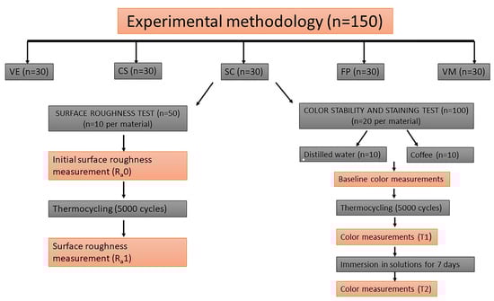
Figure 1.
Summary of the study design. VE; Vita Enamic, CS; Cerasmart 270, SC; Crowntec, FP; Permanent Crown Resin, VM; Vita Mark II.

Table 1.
Types, compositions, and manufacturing techniques of CAD/CAM materials tested and their manufacturers.
2.1. Preparation of Experimental Specimens
CAD/CAM milled specimens were produced by slicing from CAD/CAM blocks with a slow-speed precision cutting device (Isomet 1000 Precision Saw, Buehler, Lake Bluff, IL, USA) using a diamond saw under water cooling. Thirty specimens were sliced for each CAD/CAM milled material with the dimensions of a 14 mm × 12 mm and 2.0 mm thickness (Figure 2). The 3D models were designed in the Fusion 360 CAD software program (Autodesk, Mill Valley, CA, USA) for 3D printed rectangular specimens (14 mm × 12 mm × 2.0 mm) (Figure 3). These digital designs were exported to produce the specimens in STL files. Thirty specimens were produced for each 3D printed material. The SC specimens were printed using a DLP-based 3D printer (Asiga MAX U, Asiga, Sydney, Australia) with a layer thickness of 50 µm and a build orientation of 90 degrees. Following the printing process, the specimens were cleaned with an alcohol-soaked (96%) cloth and then subjected to a post-polymerization process with 4000 lighting exposures using a polymerization device (Otoflash G171, NK Optik, Baierbrunn, Germany). The FP samples were printed using the SLA-based 3D printer (Form 3, Formlabs, Somerville, MA, USA) with a layer thickness of 50 µm and a build orientation of 90 degrees. The printed specimens were washed with 99% isopropyl alcohol for 3 min using ultrasonic cleaning to remove any excess resin (Form Wash, Formlabs, Somerville, MA, USA), and the dried specimens were then subjected to a post-polymerization process using FormCure (Formlabs, Somerville, MA, USA) for 30 min at 60 °C according to the manufacturer’s recommendations. After cooling, the supporting structures were removed using low-speed rotary instruments from the specimens. All specimens were then kept in distilled water at 37 °C for 24 h.
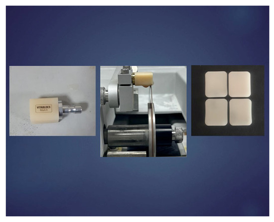
Figure 2.
Preparation of test specimens from subtractively manufactured CAD/CAM blocks.
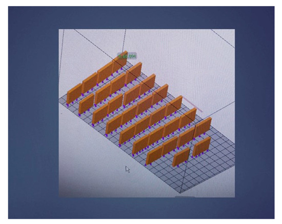
Figure 3.
3D model designed with dimensions of 14 mm × 12 mm and 2.0 mm thickness for additive manufacturing composite specimens.
The one surface of all of the specimens was then grounded with 600-, 1000-, and 1200-grit silicon carbide abrasive paper using a sanding device (Phoenix Beta, Buehler Ltd., Lake Bluff, IL, USA) for 15 s at 100 rpm for each paper, followed by the polishing procedure. All specimens were subsequently polished using a polishing system from medium to superfine (SofLEX, 3M), and the specimens’ final dimensions were then measured using a digital caliper (Digimatic CD-15DCX; Mitutoyo Inc., Tokyo, Japan). Finally, all specimens were ultrasonically cleaned using distilled water for 10 min and dried. The first surface roughness and color measurements of the test groups were made.
2.2. Measurement of Surface Roughness
For the surface roughness (Ra) test, the initial surface roughness (Ra0) of the specimens was measured using a 3D noncontact optical profilometer (ZYGO, ZeGage, Middlefield, CT, USA) before the thermocycling procedure. The surface of each specimen placed horizontally under the lens of the profilometer (20× magnification) was scanned (speed: 0.5 mm/s cutoff: 0.8 mm) at three locations to determine the mean surface roughness value. The 3D images were visualized, and roughness was evaluated with a software package (ZYGO, MetroPro software v.9.1.8, Middlefield, CT, USA) (Figure 4). Three measurements were made from the surface of each specimen at three locations: the first measurement from the top surface of the specimen, the second measurement from the middle surface of the specimen, and the third measurement from the bottom surface of the specimen. The mean of these measurements was calculated for the mean Ra value of the specimen.
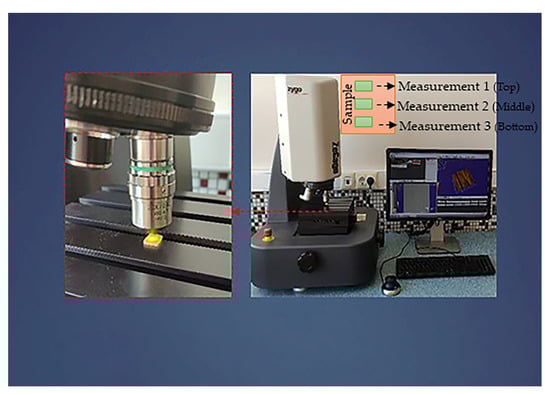
Figure 4.
Surface roughness measurement (Ra) of the specimens with a 3D noncontact optical profilometer.
2.3. Measurement of Color Parameters
For the color stability and staining test, specimens of each material were randomly divided into two subgroups depending on the immersion solutions (coffee or distilled water) (n = 10). The base color parameters of the specimens in both subgroups were measured using a spectrophotometer (VITA Easyshade V, VITA Zahnfabrik, KG, Germany) [38] according to the CIELAB system as L*, a*, b*. The color measurement of each specimen was performed three times, and the average L0*, a0*, and b0* values were recorded. The spectrophotometer’s tip was set directly on the specimen surfaces. The white background [39] was used for color measurements by a single operator (Figure 5).
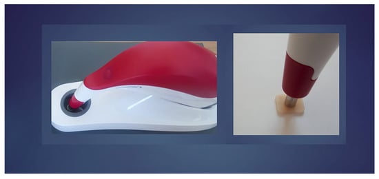
Figure 5.
Color parameter measurement of the specimens using a spectrophotometer.
After Ra0 and L0*, a0*, b0* measurements, all test groups were subjected to the thermocycling procedure for 5000 cycles in a distilled water bath ranging from 5 °C to 55 °C. Each cycle lasted 60 s and involved the following steps: 20 s in a 5 °C bath, 10 s for transferring the samples to another bath, 20 s in a 55 °C bath, and 10 s for transferring the samples back to the 5 °C bath. After the thermocycling procedure, the specimens were rinsed and dried. Surface roughness and color parameters of material groups were remeasured as Ra1 and L1*, a1*, b1* with the same measurement methods. The specimens of the staining groups were then stored in distilled water as a control or coffee solution for the staining procedure. The content of a 3 g coffee was dissolved in 300 mL of boiling distilled water (Nescafѐ Gold, Nestle, İstanbul, Turkey) [38]. The specimens were stored in containers with 300 mL of either distilled water or 300 mL of coffee solution for 7 days at 37 °C [40]. The solutions were refreshed daily during the test. The specimens were rinsed with water after the staining process and dried. The same protocol was utilized for color measurements of each specimen, and color parameter data were recorded as L2*, a2*, and b2*. The color changes (ΔE00) of each specimen were calculated in relation to the baseline color parameter value and expressed as T1 (after 5000 thermocycling) and T2 (immersion in distilled water or coffee) using the CIEDE2000 (ΔE00) formula [41].
For this study, the parametric factors of the ΔE00 were set to 1. Likewise, the threshold of clinical perceptibility was set at ΔE00 ≤ 1.30, and the threshold of clinical acceptability was set at ΔE00 ≤ 2.25 units [42].
2.4. Statistical Analysis
Statistical analyses were applied using SPSS (IBM SPSS 20.0 software; SPSS Inc., Chicago, IL, USA). The Kolmogorov–Smirnov test was applied to determine the normality of the data distributions for both analyses (Ra and ΔE00). Because the data were normally distributed, a one-way ANOVA followed by the Tukey post hoc test was used to analyze the Ra values. Kruskal–Wallis and Mann–Whitney U tests were used to analyze the non-normally distributed ΔE00 values. Ra0 and Ra1 values were compared with the paired sample t-test and the Wilcoxon test was used to compare ΔE00 values at different intervals. A value of p < 0.05 was used as the statistical significance level.
3. Results
The results of the one-way ANOVA test showed that the type of material had a statistically significant effect on Ra0 and Ra1 values (p < 0.001) (Table 2 and Table 3, respectively). The mean Ra0, Ra1, and standard deviation (SD) values of test groups, the multiple comparisons of these values according to Tukey’s post hoc tests, and the pairwise comparisons according to the paired sample t-tests are shown in Table 4.
Table 2.
One-way ANOVA results for Ra0 values.
Table 3.
One-way ANOVA results for Ra1 values.
Table 4.
The test groups’ mean Ra0 and Ra1 values (μm) and standard deviations (SD) with Tukey’s post-hoc test for multiple comparisons and paired samples t-tests.
While the highest mean Ra0 value was observed for the VE group (0.174 ± 0.043), the lowest mean Ra0 value was observed for the CS (0.037 ± 0.008). After the thermocycling procedure, the highest mean Ra1 value was examined for the VE group (0.201 ± 0.041), and the lowest mean Ra1 value was examined for the SC group (0.038 ± 0.007). All material groups’ mean surface roughness values were below or equal to the plaque accumulation threshold (0.20 µm) [43]. No statistical difference was examined among the SC, FP, and CS groups for the Ra0 and Ra1 values. The difference between the VE and VM groups and other groups was statistically significant in both mean Ra0 and mean Ra1 values. According to the pairwise comparisons of Ra0 and Ra1 values, the thermocycling increased the roughness of the materials in general. However, this increase was not statistically significant. Figure 6 displays 3D roughness images of the specimens tested before and after thermocycling.
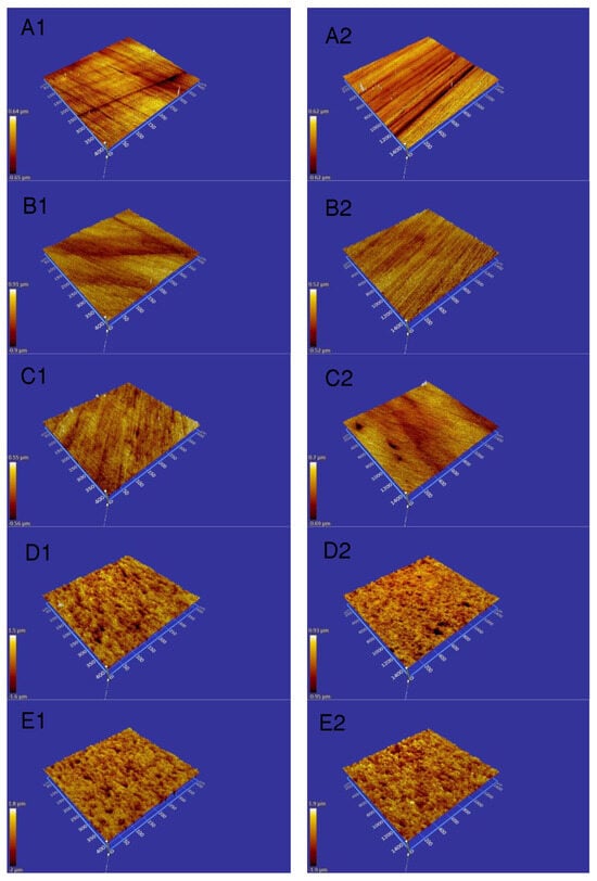
Figure 6.
3D noncontact optic profilometer images of investigated materials before and after thermocycling. (A1) Crowntec before thermocycling; (A2) Crowntec after thermocycling; (B1) Cerasmart 270 before thermocycling; (B2) Cerasmart 270 after thermocycling; (C1) Permanent Crown Resin before thermocycling; (C2) Permanent Crown Resin after thermocycling; (D1) Vita Enamic before thermocycling; (D2) Vita Enamic after thermocycling; (E1) Vita Mark II before thermocycling; (E2) Vita Mark II after thermocycling.
According to the Kruskal–Wallis statistical analysis, the comparison between tested materials at both the T1 interval and the T2 interval exhibited significant differences on the mean ΔE00 values (p < 0.001). The mean ± standard deviation values of the mean ΔE00 of each material after thermocycling (T1) and in immersed distilled water or coffee solutions (T2) are presented in Table 5.
Table 5.
Mean, standard deviations, and significance level of the color changes (ΔE00) among tested materials at different.
The SC group showed the highest mean ΔE00 values at T1 (2.57 ± 0.56) followed by VE (1.20 ± 0.44), CS (1.18 ± 0.37), FP (0.93 ± 0.25), and VM (0.76 ± 0.10). The VM group exhibited the lowest mean ΔE00 values at T1 (Figure 7). According to the Mann–Whitney U test results after thermocycling, no statistically significant difference was observed between the mean ΔE00 values of FP-VM and CS-VE. While there was a statistically significant difference between VM-CS and VM-VE, a statistically significant difference was observed between SC and all other materials. The mean ΔE00 values for all the materials were lower than the perceptibility threshold (ΔE00 ≤ 1.30) at T1, except for SC. Also, the mean ΔE00 values of the SC were above the acceptability threshold (ΔE00 ≤ 2.25) at T1.
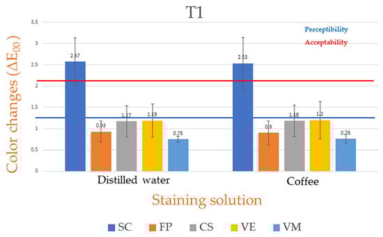
Figure 7.
Color changes of investigated materials after thermocycling at T1. SC: Crowntec; FP: Permanent Crown Resin; CS: Cerasmart 270; VE: Vita Enamic; VM: Vita Mark II.
All tested materials had higher mean ΔE00 values in coffee for 7 days compared to distilled water at T2. These high mean ΔE00 values were statistically significant for SC, FP, and VM but not for CS and VE compared to their distilled water. After immersion in coffee or distilled water, the SC group showed the highest mean ΔE00 values in either coffee solution (5.36 ± 0.58) or distilled water (3.62 ± 0.48), and the VM groups exhibited the lowest mean ΔE00 values (0.77 ± 0.04) in distilled water at T2. The CS group also showed the lowest mean ΔE00 value (1.44 ± 0.33) in coffee (Figure 8). As per the Mann-–Whitney U test results, while there was no statistically significant difference among FP, VE, and VM in the coffee solution, there was a statistically significant difference among SC and other groups and between VE and VM in distilled water. There was no statistically significant difference among FP, CS, and VE in distilled water at T2.
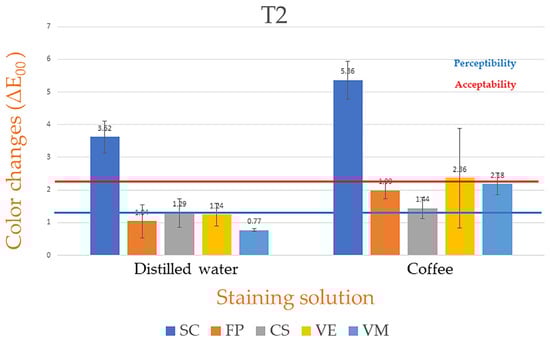
Figure 8.
Color changes of the investigated materials after storage in staining solutions at T2. SC: Crowntec; FP: Permanent Crown Resin; CS: Cerasmart 270; VE: Vita Enamic, VM: Vita Mark II.
As per the Wilcoxon signed-rank test, while the increase in the mean ΔE00 values of SC and VM immersed in distilled water was statistically significant, the increase in the mean ΔE00 values of all material groups immersed in coffee was statistically significant, except for the CS.
4. Discussion
The current study evaluated the color stability and surface roughness of additively and subtractively manufactured permanent composite-based restorative materials and compared them to those of a feldspathic glass–ceramic after thermocycling. In addition, the stainability of these materials was examined after immersion in coffee/distilled water. Based on the current study’s data, both null hypotheses of the study were rejected. Since the type of material significantly affected the color stability and surface roughness of tested restorative materials after thermocycling. In addition, the type of tested CAD/CAM material had a statistically significant effect on the stainability of materials after storage in coffee or distilled water.
Currently, CAD/CAM composite-based milled and glass ceramic restorative materials are frequently used in the construction of indirect restorations such as inlay, onlay, veneer, and crown [44]. However, the popularity of 3D printed tooth-colored restorations among clinicians has recently increased [45]. It may be considered that 3D printed materials can be replaced by CAD/CAM milled materials due to their advantages. They can be an alternative to feldspathic ceramics, which are preferred due to their aesthetic properties but are fragile. Despite the increase in variety and frequency of use of 3D printed materials, the lack of literature on properties such as surface roughness and color stability required for the longevity of the material has been the reason for the present study. Therefore, this present study aimed to investigate the surface roughness and color stability of 3D printed permanent resin and CAD/CAM milled composite-based materials and compare them to those of a feldspathic glass-ceramic. The feldspathic ceramic VM, which has been on the market for several years, was chosen as the control group due to its excellent aesthetic superiority and the required stable properties for indirect restorative materials [46,47].
The Ra value, which is the roughness average, is the universally accepted and commonly used roughness parameter. Recent studies have specified Ra = 0.2 µm as the threshold value of surface roughness for bacterial retention and plaque accumulation [42]. In this study, the mean Ra0 values of tested materials ranged between 0.037 ± 0.008 μm and 0.174 ± 0.043 μm and the mean Ra1 values of the same materials ranged between 0.038 ± 0.007 μm and 0.201 ± 0.041 μm. The mean Ra0 and Ra1 values of each material tested were equal to or below the plaque accumulation threshold. However, none of the groups’ mean surface roughness values reached the clinically unacceptable level of 10 µm [48]. A statistical difference was found in the comparison of both the Ra0 and Ra1 values of the tested materials. While the VE group had significantly the highest mean Ra0 and Ra1 values, no statistical difference existed among SC, FP, and CS. Furthermore, SC, FP, and CS had lower mean Ra values than the VE. The difference in microstructural compositions of the composite-based materials tested could explain these results. CAD/CAM composite-based resins have been introduced in different compositions and with various percentages of the polymeric matrix and inorganic filler components depending on the type of material, such as polymer-infiltrated ceramics and resin composite blocks [49,50]. Vita Enamic has two interconnected networks made of ceramic and polymer [51]. Despite the high filler content percentage of Vita Enamic, infiltration of the sintered porous ceramic matrix with low-viscosity resin presents a challenging interfacial adhesion problem. The higher surface roughness of VE compared to other resin composite materials could be due to the adhesion between the ceramic structure and the resin [52]. Alharbi et al. [53] compared the surface roughness of polymer-infiltrated ceramic materials (PICN) and CAD/CAM composite resins. They found that CAD/CAM composite resins had significantly lower roughness values than PICN materials. Likewise, Fasbinder et al. [54] examined the surface roughness of resin nanoceramic, hybrid ceramic, and leucite-reinforced ceramic and reported that hybrid ceramics, also called PICN, have higher roughness values. Koizumi et al. [55] investigated the surface roughness of current CAD/CAM resin composites and found that the baseline roughness value of VE was the highest among other resin materials compared. Similar findings have been found in the current study. CS had the lowest mean Ra0 value among the tested materials. However, there was no statistically significant difference between additively manufactured materials (FP and SC) and CS. Çakmak et al. [36] evaluated the color stability and surface roughness of additively (composite resins; Saremco Crowntec and VarseoSmile Crown Plus) and subtractively (resin nanoceramic; Ceasmart) manufactured resins used for definitive restorations. They found that Cerasmart either had Ra results similar to or lower compared to those of additively manufactured composite resins. Cerasmart 270 is a prepolymerized resin composite CAD/CAM block that has been manufactured under high pressure and temperature [17,56], and contains nanoceramic particles (silica and zirconia filler/cluster filler) embedded in a composite matrix, as is typical for resin composites. It can be assumed that the filler particles in CS are better connected to the resin matrix [17]. Structural stability and fluid consistency during printing and storage should be characteristics of 3D printed resins. Therefore, it is suggested that 3D printed composite resin materials used in the manufacture of permanent restorations, just like flowable dental composites, should contain fewer inorganic fillers than CAD/CAM composite blocks to maintain a stable liquid consistency. However, a low filler content also affects the mechanical properties of the composite material [57]. Kumari et al. [58] reported that the increase in the filler percentage may cause a decrease in the surface roughness. But, in the present study, although the 3D printed composite resins tested had lower filler content compared to CAD/CAM milled composites, the values of the surface roughness show that these composite resins have roughness similar to that of the resin nanoceramic CAD/CAM block and are better than the polymer-infiltrated ceramic and feldspathic ceramic (control) tested in the present study.
Thermocycling, a popular artificial aging method, is utilized to simulate the thermal changes in the oral cavity during eating and drinking in in-vitro studies. When composite resins are subjected to water and thermal changes as a result of thermocycling, deterioration occurs in the organic matrix structure or at the matrix-filler interface [59]. Furthermore, thermocycling has a significant impact on the surface roughness of composite resins, according to Dos Santos et al. [29]. For this reason, thermocycling was chosen as the aging method to simulate oral environmental conditions in the current study. According to the results of the current study, although thermocycling slightly increased the mean Ra values of the tested materials, this increase was not statistically significant.
It is assumed that the color changes of the resin-based restorative materials will increase at a high rate due to the degradation of the polymer matrix, unreacted monomers of the polymerization agents after aging, and staining agents [60]. The present study’s ΔE00 results show that SC had the highest mean ΔE00 values after the thermocycling and this mean ΔE00 value was above the clinically acceptable threshold value of 2.25. However, in the same interval, the mean ΔE00 values of other tested material groups were observed below the perceptibility threshold value of 1.30. While VM had the significantly lowest mean ΔE00 values, followed by FP. The mean ΔE00 value of VE was statistically similar to the FP and CS, and greater than the VM after thermocycling at T1. The color stability of resin-based composites is affected by the hydrophobic or hydrophilic features of the resin matrix. This feature determines the water absorption capacity of the composite material [61]. Vita Mark II, a feldspathic ceramic, consists of a glass-ceramic matrix that is more resistant to discoloration than resin composites, as this matrix is not subject to water absorption like polymers [62]. The lowest color change of the VM after thermocycling can be attributed to its microstructure. The color change of the VE was statistically similar to that of the FP and CS and greater than VM at T1. Although VE is composed of a feldspathic glass ceramic of 86%, urethane dimethacrylate (UDMA) and tri-ethylene-glycol-dimethacrylate (TEGDMA)-containing polymer matrix of 14% in its structure may be the reason for its lower color stability compared to VM. Moreover, TEGDMA is hydrophilic and TEGDMA-based composites are more susceptible to color change due to their high water absorption capacity [63]. The organic matrix of CS contains UDMA and Bis-MEEP. There is a Bis-EMA matrix monomer in the structure of FP and SC. These monomers have lower hydrophilic properties compared to TEGDMA [22,64]. In the present study, the hydrophobic monomer content of the CS and FP composite resins may be the reason for the lower color change.
The interesting finding was that SC showed a significantly higher mean ΔE00 value than FP, although both FP and SC were additively manufactured composite resins with similar compositions according to manufacturers’ statements (Table 1) at T1. Similarly, SC had a higher mean ΔE00 value than FP and other materials tested after immersion in coffee. Unlike the present study, Çakmak et al. [36] found that the color change of Saremco Crowntec was similar to that of milled composite (CS) and was lower than that of other 3D printed resins tested (Varseo Smile) after 10,000 coffee thermocyclings. However, they found that the mean ΔE00 values of both 3D printed materials were within the clinically acceptable threshold. Whereas, Alharbi et al. [22] reported that the color change values of another 3D printed composite resin were quite high compared to milled composite material after immersion in coffee solution. In the present study, 3D print group specimens were produced with different techniques. There is no published study comparing the effect of aging and storage in different beverages on the color stability of 3D printed permanent composite resins produced with DLP and SLA techniques. A previous study on the color stability of 3D printed interim resin materials produced with different techniques reported that the production technology of 3D printed materials may affect color stability. According to the results of that study, the 3D printing SLA group showed higher color stability than the DLP group [23]. Similarly, the differences in the production technology of 3D printing materials may have affected the color stability of the printing materials tested in the present study. In addition, the unreacted residual monomer is another factor that will cause color change even before the material is exposed to colorants. The post-polymerization process, which is a stage in the production of 3D printed materials, improves the material’s properties by polymerizing unreacted monomers [65,66]. In the present study, the different equipment used for the post-curing process and the different duration of the post-curing for each type of additively manufactured material may be the cause of the results [67,68]. These conditions should be taken into consideration in further research.
Immersion in a coffee solution is a common method for staining material. Because of the colorants’ absorption into the organic matrix of the composite resin, coffee has significant staining potential [69]. In this study, composite specimens were immersed in a coffee solution for one week. Since a previous study on the effects of different beverages on the color change of composite materials found that significant color change occurred in the first week [70], a one-week immersion period was used in this study to determine the long-term color stability of the composite materials. CS had the lowest mean ΔE00 value after immersion in coffee. However, the mean ΔE00 values of CS between the T1 and T2 intervals were not statistically significant. This may be attributed to its microstructural properties. On the contrary, the mean ΔE00 values of other tested materials showed a statistically significant increase in the coffee solution at T2. The roughness and water absorption capacity of these materials may be the reason for these results.
This in vitro study has some limitations. Some variables that composite materials will be exposed to in the oral environment, such as occlusal forces, saliva that contains some proteins and enzymes, and mouth rinsing or brushing, were not reflected in the present study. In addition, the specimens’ flat surfaces and lack of anatomical pits and grooves did not fully mimic clinical reality. In addition, other optical properties, such as translucency, should be further investigated. Only a coffee-staining medium was selected because of its popularity. Furthermore, the VITA Easyshade V spectrophotometer was used for color parameter measurement. VITA Easyshade is a clinical device that is not meant for laboratory evaluations. Based on the data of the present study, tested 3D printed permanent composite resins showed similar or lower roughness values than tested milled CAD/CAM materials, which were clinically acceptable values. However, color stability is also an important factor in the long-term success of indirect restorations. Therefore, future research is needed to improve the color stability required for the long-term use of 3D printed permanent composite resins produced with different techniques for indirect restorations.
5. Conclusions
Within the limitations of this study, it was concluded that:
- (1)
- Among the investigated materials, while subtractively manufactured Vita Enamic exhibited the highest Ra value, additively manufactured Crowntec had the lowest Ra value after thermocycling. In addition, the tested additively manufactured composite resins and the subtractively manufactured Cerasmart 270 had similar mean Ra values. However, all investigated materials showed acceptable surface roughness after thermocycling that was equal to or below the plaque accumulation threshold of 0.2 µm.
- (2)
- The thermocycling increased the roughness of all tested materials. However, this increase was not significant.
- (3)
- Crowntec exhibited clinically unacceptable color changes at all intervals. On the contrary, Permanent Crown Resin exhibited a similar or lower color change compared to tested subtractively manufactured materials and glass ceramic. In addition, the color change of these materials was clinically acceptable, except for Vita Enamic in coffee.
- (4)
- Cerasmart 270 displayed the lowest color change in coffee, followed by the Permanent Resin Crown.
- (5)
- Finally, all tested permanent composite-based CAD-CAM restorative materials may provide acceptable surface roughness over long-term use. However, since Crowntec and Vita Enamic show unacceptable color changes, these materials may not be suitable for long-term use in clinical practice in areas where aesthetics are important.
Author Contributions
Conceptualization, H.N.B. and T.T.; methodology, H.N.B. and T.T.; software, H.N.B.; validation, H.N.B. and T.T.; formal analysis, H.N.B.; investigation, H.N.B.; resources, H.N.B. and T.T.; data curation, H.N.B. and T.T.; writing—original draft preparation, H.N.B.; writing—review and editing, H.N.B. and T.T.; visualization, H.N.B.; supervision, H.N.B. and T.T.; project administration, H.N.B.; funding acquisition, H.N.B. and T.T. All authors have read and agreed to the published version of the manuscript.
Funding
This research received no external funding.
Institutional Review Board Statement
Not applicable.
Informed Consent Statement
Not applicable.
Data Availability Statement
Not applicable.
Conflicts of Interest
The authors declare no conflict of interest.
References
- Vichi, A.; Balestra, D.; Scotti, N.; Louca, C.; Paolone, G. Translucency of CAD/CAM and 3D printable composite materials for permanent dental restorations. Polymers 2023, 15, 1443. [Google Scholar] [CrossRef] [PubMed]
- Tsolakis, I.A.; Gizani, S.; Panayi, N.; Antonopoulos, G.; Tsolakis, A.I. Three-dimensional printing technology in orthodontics for dental models: A systematic review. Children 2022, 9, 1106. [Google Scholar] [CrossRef] [PubMed]
- Dimitrova, M.; Chuchulska, B.; Zlatev, S.; Kazakova, R. Colour stability of 3D-printed and prefabricated denture teeth after immersion in different colouring agents—An in vitro study. Polymers 2022, 14, 3125. [Google Scholar] [CrossRef] [PubMed]
- Baldi, A.; Comba, A.; Tempesta, R.M.; Carossa, M.; Pereira, G.K.R.; Valandro, L.F.; Paolone, G.; Vichi, A.; Goracci, C.; Scotti, N. External marginal gap variation and residual fracture resistance of composite and lithium-silicate CAD/CAM overlays after cyclic fatigue over endodontically-treated molars. Polymers 2021, 13, 3002. [Google Scholar] [CrossRef] [PubMed]
- Kessler, A.; Hickel, R.; Reymus, M. 3D printing in dentistry-state of the art. Oper. Dent. 2020, 45, 30–40. [Google Scholar] [CrossRef]
- Infante, L.; Yilmaz, B.; McGlumphy, E.; Finger, I. Fabricating complete dentures with CAD/CAM technology. J. Prosthet. Dent. 2014, 111, 351–355. [Google Scholar] [CrossRef] [PubMed]
- Schweiger, J.; Stumbaum, J.; Edelhoff, D.; Güth, J.F. Systematics and concepts for the digital production of complete dentures: Risks and opportunities. Int. J. Comput. Dent. 2018, 21, 41–56. [Google Scholar] [PubMed]
- Al-Qarni, F.D.; Goodacre, C.J.; Kattadiyil, M.T.; Baba, N.Z.; Paravina, R.D. Stainability of acrylic resin materials used in CAD-CAM and conventional complete dentures. J. Prosthet. Dent. 2020, 123, 880–887. [Google Scholar] [CrossRef]
- Ngo, T.D.; Kashani, A.; Imbalzano, G.; Nguyen, K.T.; Hui, D. Additive manufacturing (3D printing): A review of materials, methods, applications and challenges. Compos. B Eng. 2018, 143, 172–196. [Google Scholar] [CrossRef]
- Yu, B.Y.; Son, K.; Lee, K.B. Evaluation of intaglio surface trueness and margin quality of interim crowns in accordance with the build angle of stereolithography apparatus 3-dimensional printing. J. Prosthet. Dent. 2022, 126, 231–237. [Google Scholar] [CrossRef]
- Lee, B.I.; You, S.G.; You, S.M.; Kang, S.Y.; Kim, J.H. Effect of rinsing time on the accuracy of interim crowns fabricated by digital light processing: An in vitro study. J. Adv. Prosthodont. 2021, 13, 24–35. [Google Scholar] [CrossRef] [PubMed]
- Zhang, Z.C.; Li, P.L.; Chu, F.T.; Shen, G. Influence of the three-dimensional printing technique and printing layer thickness on model accuracy. J. Orofac. Orthop. 2019, 80, 194–204. [Google Scholar] [CrossRef] [PubMed]
- You, S.G.; You, S.M.; Kang, S.Y.; Bae, S.Y.; Kim, J.H. Evaluation of the adaptation of complete denture metal bases fabricated with dental CAD-CAM systems: An in vitro study. J. Prosthet. Dent. 2021, 125, 479–485. [Google Scholar] [CrossRef] [PubMed]
- Comba, A.; Paolone, G.; Baldi, A.; Vichi, A.; Goracci, C.; Bertozzi, G.; Scotti, N. Effects of substrate and cement shade on the translucency and color of CAD/CAM lithium-disilicate and zirconia ceramic materials. Polymers 2022, 14, 1778. [Google Scholar] [CrossRef] [PubMed]
- Paolone, G.; Mandurino, M.; De Palma, F.; Mazzitelli, C.; Scotti, N.; Breschi, L.; Gherlone, E.; Cantatore, G.; Vichi, A. Color stability of polymer-based composite CAD/CAM blocks: A systematic review. Polymers 2023, 15, 464. [Google Scholar] [CrossRef] [PubMed]
- Flury, S.; Diebold, E.; Peutzfeldt, A.; Lussi, A. Effect of artificial toothbrushing and water storage on the surface roughness and micromechanical properties of tooth-colored CAD-CAM materials. J. Prosthet. Dent. 2017, 117, 767–774. [Google Scholar] [CrossRef] [PubMed]
- Dayan, S.Ç.; Emre, M. Effect of different storage media on the microhardness and wear resistance of resin-matrix ceramics. Int. J. Appl. Ceram. Technol. 2019, 16, 2467–2473. [Google Scholar] [CrossRef]
- Goujat, A.; Abouelleil, H.; Colon, P.; Jeannin, C.; Pradelle, N.; Seux, D.; Grosgogeat, B. Mechanical properties and internal fit of 4 CAD-CAM block materials. J. Prosthet. Dent. 2018, 119, 384–389. [Google Scholar] [CrossRef]
- Awada, A.; Nathanson, D. Mechanical properties of resin-ceramic CAD/CAM restorative materials. J. Prosthet. Dent. 2015, 114, 587–593. [Google Scholar] [CrossRef]
- Al-Qahtani, A.S.; Tulbah, H.I.; Binhasan, M.; Abbasi, M.S.; Ahmed, N.; Shabib, S.; Farooq, I.; Aldahian, N.; Nisar, S.S.; Tanveer, S.A.; et al. Surface properties of polymer resins fabricated with subtractive and additive manufacturing techniques. Polymers 2021, 13, 4077. [Google Scholar] [CrossRef]
- Della Bona, A.; Cantelli, V.; Britto, V.T.; Collares, K.F.; Stansbury, J.W. 3D printing restorative materials using a stereolithographic technique: A systematic review. Dent. Mater. 2021, 37, 336–350. [Google Scholar] [CrossRef] [PubMed]
- Alharbi, N.; Alharbi, A.; Osman, R. Stain susceptibility of 3D-printed nanohybrid composite restorative material and the efficacy of different stain removal techniques: An in vitro study. Materials 2021, 27, 5621. [Google Scholar] [CrossRef] [PubMed]
- Ellakany, P.; Fouda, S.M.; AlGhamdi, M.A.; Aly, N.M. Comparison of the color stability and surface roughness of 3-unit provisional fixed partial dentures fabricated by milling, conventional and different 3D printing fabrication techniques. J. Dent. 2023, 131, 104458. [Google Scholar] [CrossRef] [PubMed]
- Ardu, S.; Duc, O.; Krejci, I.; Bétrisey, E.; Di Bella, E.; Daher, R. Gloss retention of direct composites and corresponding CAD/CAM composite blocks. Clin. Exp. Dent. Res. 2022, 8, 282–286. [Google Scholar] [CrossRef]
- Egilmez, F.; Ergun, G.; Cekic-Nagas, I.; Vallittu, P.K.; Lassila, L.V.J. Comparative color and surface parameters of current esthetic restorative CAD/CAM materials. J. Adv. Prosthodont. 2018, 10, 32–42. [Google Scholar] [CrossRef] [PubMed]
- Alkhadim, Y.K.; Hulbah, M.J.; Nassar, H.M. Color shift, color stability, and post-polishing surface roughness of esthetic resin composites. Materials 2020, 13, 1376. [Google Scholar] [CrossRef] [PubMed]
- Al Amri, M.D.; Labban, N.; Alhijji, S.; Alamri, H.; Iskandar, M.; Platt, J.A. In vitro evaluation of translucency and color stability of CAD/CAM polymer-infiltrated ceramic materials after accelerated aging. J. Prosthodont. 2021, 30, 318–328. [Google Scholar] [CrossRef]
- Pettini, F.; Corsalini, M.; Savino, M.G.; Stefanachi, G.; Di Venere, D.; Pappalettere, C.; Monno, G.; Boccaccio, A. Roughness analysis on composite materials (microfilled, nanofilled and silorane) after different finishing and polishing procedures. Open Dent. J. 2015, 9, 357–367. [Google Scholar] [CrossRef]
- Dos Santos, P.H.; Catelan, A.; Albuquerque Guedes, A.P.; Umeda Suzuki, T.Y.; de Lima Godas, A.G.; Fraga Briso, A.L.; Bedran-Russo, A.K. Effect of thermocycling on roughness of nanofill, microfill and microhybrid composites. Acta Odontol. Scand. 2015, 73, 176–181. [Google Scholar] [CrossRef]
- Blackburn, C.; Rask, H.; Awada, A. Mechanical properties of resin-ceramic CAD-CAM materials after accelerated aging. J. Prosthet. Dent. 2018, 119, 954–958. [Google Scholar] [CrossRef]
- Subaşı, M.G.; Alp, G.; Johnston, W.M.; Yılmaz, B. Effects of fabrication and shading technique on the color and translucency of new-generation translucent zirconia after coffee thermocycling. J. Prosthet. Dent. 2018, 120, 603–608. [Google Scholar] [CrossRef] [PubMed]
- Yuan, J.C.; Barão, V.A.R.; Wee, A.G.; Alfaro, M.F.; Afshari, F.S.; Sukotjo, C. Effect of brushing and thermocycling on the shade and surface roughness of CAD-CAM ceramic restorations. J. Prosthet. Dent. 2018, 119, 1000–1006. [Google Scholar] [CrossRef] [PubMed]
- Mahrous, A.I.; Salama, A.A.; Shabaan, A.A.; Abdou, A.; Radwan, M.M. Color stability of two different resin matrix ceramics: Randomized clinical trial. BMC Oral Health 2023, 23, 665. [Google Scholar] [CrossRef] [PubMed]
- Stamenković, D.D.; Tango, R.N.; Todorović, A.; Karasan, D.; Sailer, I.; Paravina, R.D. Staining and aging-dependent changes in color of CAD-CAM materials. J. Prosthet. Dent. 2021, 126, 672–678. [Google Scholar] [CrossRef] [PubMed]
- Labban, N.; Al Amri, M.D.; Alnafaiy, S.M.; Alhijji, S.M.; Alenizy, M.A.; Iskandar, M.; Feitosa, S. Influence of toothbrush abrasion and surface treatments on roughness and gloss of polymer-infiltrated ceramics. Polymers 2021, 13, 3694. [Google Scholar] [CrossRef] [PubMed]
- Çakmak, G.; Oosterveen-Rüegsegger, A.L.; Akay, C.; Schimmel, M.; Yılmaz, B.; Dönmez, M.B. Influence of polishing technique and coffee thermal cycling on the surface roughness and color stability of additively and subtractively manufactured resins used for definitive restorations. J. Prosthodont. 2023, in press. [Google Scholar] [CrossRef] [PubMed]
- Donmez, M.B.; Okutan, Y. Marginal gap and fracture resistance of implant-supported 3D-printed definitive composite crowns: An in vitro study. J. Dent. 2022, 124, 104216. [Google Scholar] [CrossRef] [PubMed]
- Bozoğulları, H.N.; Büyükerkmen, E.B.; Büyüközer Özkan, H. Comparison of surface roughness and color stability of different denture characterizing composite resins: The effect of different surface treatments. J. Prosthodont. 2023, 32, 53–60. [Google Scholar] [CrossRef]
- Ardu, S.; Braut, V.; Gutemberg, D.; Krejci, I.; Dietschi, D.; Feilzer, A.J. A long-term laboratory test on staining susceptibility of esthetic composite resin materials. Quintessence Int. 2010, 41, 695–702. [Google Scholar]
- Dede, D.Ö.; Şahin, O.; Köroğlu, A.; Yılmaz, B. Effect of sealant agents on the color stability and surface roughness of nanohybrid composite resins. J. Prosthet. Dent. 2016, 116, 119–128. [Google Scholar] [CrossRef]
- Commission Internationale de l’Eclairage (CIE). CIE Technical Report: Colorimetry, 3rd ed.; CIE Central Bureau: Vienna, Austria, CIE Pub No.15; 2004. [Google Scholar]
- Shin, J.W.; Kim, J.E.; Choi, Y.J.; Shin, S.H.; Nam, N.E.; Shim, J.S.; Lee, K.W. Evaluation of the color stability of 3D-printed crown and bridge materials against various sources of discoloration: An in vitro study. Materials 2020, 26, 5359. [Google Scholar] [CrossRef]
- Quirynen, M.; Bollen, C.M.; Papaioannou, W.; Van Eldere, J.; van Steenberghe, D. The influence of titanium abutment surface roughness on plaque accumulation and gingivitis: Short-term observations. Int. J. Oral. Maxillofac. Implant. 1996, 11, 169–178. [Google Scholar]
- Zhi, L.; Bortolotto, T.; Krejci, I. Comparative in vitro wear resistance of CAD/CAM composite resin and ceramic materials. J. Prosthet. Dent. 2016, 115, 199–202. [Google Scholar] [CrossRef] [PubMed]
- Kim, J.E.; Choi, W.H.; Lee, D.; Shin, Y.; Park, S.H.; Roh, B.D.; Kim, D. Color and translucency stability of three-dimensional printable dental materials for crown and bridge restorations. Materials 2021, 14, 650. [Google Scholar] [CrossRef] [PubMed]
- Giordano, R. Materials for chairside CAD/CAM-produced restorations. J. Am. Dent. Assoc. 2006, 137 (Suppl. S1), 14S–21S. [Google Scholar] [CrossRef] [PubMed]
- Bellan, M.C.; Cunha, P.F.J.S.D.; Tavares, J.G.; Spohr, A.M.; Mota, E.G. Microtensile bond strength of CAD/CAM materials to dentin under different adhesive strategies. Braz. Oral. Res. 2017, 18, e109. [Google Scholar] [CrossRef] [PubMed]
- Bollen, C.M.; Papaioanno, W.; Van Eldere, J.; Schepers, E.; Quirynen, M.; van Steenberghe, D. The influence of abutment surface roughness on plaque accumulation and peri-implant mucositis. Clin. Oral. Implant. Res. 1996, 7, 201–211. [Google Scholar] [CrossRef] [PubMed]
- Stawarczyk, B.; Özcan, M.; Trottmann, A.; Schmutz, F.; Roos, M.; Hämmerle, C. Two-body wear rate of CAD/CAM resin blocks and their enamel antagonists. J. Prosthet. Dent. 2013, 109, 325–332. [Google Scholar] [CrossRef] [PubMed]
- Stawarczyk, B.; Sener, B.; Trottmann, A.; Roos, M.; Ozcan, M.; Hämmerle, C.H. Discoloration of manually fabricated resins and industrially fabricated CAD/CAM blocks versus glass-ceramic: Effect of storage media, duration, and subsequent polishing. Dent. Mater. J. 2012, 31, 377–383. [Google Scholar] [CrossRef] [PubMed]
- Della Bona, A.; Corazza, P.H.; Zhang, Y. Characterization of a polymer-infiltrated ceramic-network material. Dent. Mater. 2014, 30, 564–569. [Google Scholar] [CrossRef]
- Grzebieluch, W.; Kowalewski, P.; Grygier, D.; Rutkowska-Gorczyca, M.; Kozakiewicz, M.; Jurczyszyn, K. Printable and machinable dental restorative composites for CAD/CAM application-comparison of mechanical properties, fractographic, texture and fractal dimension analysis. Materials 2021, 14, 4919. [Google Scholar] [CrossRef] [PubMed]
- Alharbi, N.; Teerakanok, S.; Satterthwaite, J.D.; Giordano, R.; Silikas, N. Quantitative nano-mechanical mapping AFM-based method for elastic modulus and surface roughness measurements of model polymer infiltrated ceramics. Dent. Mater. 2022, 38, 935–945. [Google Scholar] [CrossRef] [PubMed]
- Fasbinder, D.J.; Neiva, G.F. Surface evaluation of polishing techniques for new resilient CAD/CAM restorative materials. J. Esthet. Restor. Dent. 2016, 28, 56–66. [Google Scholar] [CrossRef] [PubMed]
- Koizumi, H.; Saiki, O.; Nogawa, H.; Hiraba, H.; Okazaki, T.; Matsumura, H. Surface roughness and gloss of current CAD/CAM resin composites before and after toothbrush abrasion. Dent. Mater. J. 2015, 34, 881–887. [Google Scholar] [CrossRef] [PubMed]
- Grzebieluch, W.; Mikulewicz, M.; Kaczmarek, U. Resin composite materials for chairside cad/cam restorations: A comparison of selected mechanical properties. J. Healthc. Eng. 2021, 28, 8828954. [Google Scholar] [CrossRef] [PubMed]
- Ang, S.F.; Scholz, T.; Klocke, A.; Schneider, G.A. Determination of the elastic/plastic transition of human enamel by nanoindentation. Dent. Mater. 2009, 25, 1403–1410. [Google Scholar] [CrossRef] [PubMed]
- Kumari, C.M.; Bhat, K.M.; Bansal, R. Evaluation of surface roughness of different restorative composites after polishing using atomic force microscopy. J. Conserv. Dent. 2016, 19, 56–62. [Google Scholar] [CrossRef]
- Rinastiti, M.; Ozcan, M.; Siswomihardjo, W.; Busscher, H.J. Effects of surface conditioning on repair bond strengths of non-aged and aged microhybrid, nanohybrid, and nanofilled composite resins. Clin. Oral Investig. 2011, 15, 625–633. [Google Scholar] [CrossRef]
- Awad, D.; Stawarczyk, B.; Liebermann, A.; Ilie, N. Translucency of esthetic dental restorative CAD/CAM materials and composite resins with respect to thickness and surface roughness. J. Prosthet. Dent. 2015, 113, 534–540. [Google Scholar] [CrossRef]
- Ardu, S.; Gutemberg, D.; Krejci, I.; Feilzer, A.J.; Di Bella, E.; Dietschi, D. Influence of water sorption on resin composite color and color variation amongst various composite brands with identical shade code: An in vitro evaluation. J. Dent. 2011, 39 (Suppl. S1), e37–e44. [Google Scholar] [CrossRef]
- Dellazzana, F.Z.; Rodrigues, C.S.; Mallmann, A.; Jacques, L.B. Optical properties of a glass ceramic and an indirect resin composite: Effects of polishing and staining. J. Health Sci. 2019, 21, 512–517. [Google Scholar] [CrossRef]
- Mansouri, S.A.; Zidan, A.Z. Effect of water sorption and solubility on color stability of bulk-fill resin composite. J. Contemp. Dent. Pract. 2018, 9, 1129–1134. [Google Scholar]
- Gajewski, V.E.; Pfeifer, C.S.; Fróes-Salgado, N.R.; Boaro, L.C.; Braga, R.R. Monomers used in resin composites: Degree of conversion, mechanical properties and water sorption/solubility. Braz. Dent. J. 2012, 23, 508–514. [Google Scholar] [CrossRef]
- Van Noort, R. The future of dental devices is digital. Dent. Mater. 2012, 28, 3–12. [Google Scholar] [CrossRef] [PubMed]
- Kim, D.; Shim, J.S.; Lee, D.; Shin, S.H.; Nam, N.E.; Park, K.H.; Shim, J.S.; Kim, J.E. Effects of post-curing time on the mechanical and color properties of three-dimensional printed crown and bridge materials. Polymers 2020, 23, 2762. [Google Scholar] [CrossRef] [PubMed]
- Reymus, M.; Fabritius, R.; Keßler, A.; Hickel, R.; Edelhoff, D.; Stawarczyk, B. Fracture load of 3D-printed fixed dental prostheses compared with milled and conventionally fabricated ones: The impact of resin material, build direction, post-curing, and artificial aging-an in vitro study. Clin. Oral. Investig. 2020, 24, 701–710. [Google Scholar] [CrossRef] [PubMed]
- Reymus, M.; Lümkemann, N.; Stawarczyk, B. 3D-printed material for temporary restorations: Impact of print layer thickness and post-curing method on degree of conversion. Int. J. Comput. Dent. 2019, 22, 231–237. [Google Scholar] [PubMed]
- Lima, A.F.; Soares, G.P.; Vasconcellos, P.H.; Ambrosano, G.M.; Marchi, G.M.; Lovadino, J.R.; Aguiar, F.H. Effect of surface sealants on microleakage of Class II restorations after thermocycling and long-term water storage. J. Adhes. Dent. 2011, 13, 249–254. [Google Scholar]
- Chan, K.C.; Fuller, J.L.; Hormati, A.A. The ability of foods to stain two composite resins. J. Prosthet. Dent. 1980, 43, 542–545. [Google Scholar] [CrossRef]


Disclaimer/Publisher’s Note: The statements, opinions and data contained in all publications are solely those of the individual author(s) and contributor(s) and not of MDPI and/or the editor(s). MDPI and/or the editor(s) disclaim responsibility for any injury to people or property resulting from any ideas, methods, instructions or products referred to in the content. |
© 2023 by the authors. Licensee MDPI, Basel, Switzerland. This article is an open access article distributed under the terms and conditions of the Creative Commons Attribution (CC BY) license (https://creativecommons.org/licenses/by/4.0/).