Article

Evaluation of Dentin Tubule Occlusion Using Pre-Treatment with No-Ozone Cold Plasma: An In Vitro Study

1
Corporate Affiliated Research Institute, Feagle Co., Ltd., Yangsan 50561, Republic of Korea
2
Department of Dental Hygiene, Kyungnam College of Information & Technology, Busan 47011, Republic of Korea
3
Department of Oral Anatomy, School of Dentistry, Pusan National University, Yangsan 50612, Republic of Korea
*
Author to whom correspondence should be addressed.
The two authors have contributed equally and share the first authorship.
Appl. Sci. 2023, 13(21), 11728; https://doi.org/10.3390/app132111728
Submission received: 21 September 2023 / Revised: 23 October 2023 / Accepted: 25 October 2023 / Published: 26 October 2023
(This article belongs to the Special Issue Advanced Dental Materials and Its Applications)

Abstract

:
Dentin hypersensitivity is a common disease of the oral cavity, which renders the tooth extremely sensitive to stimuli. These symptoms usually result from the exposure of the dentinal tubules to the external environment. There is a need for a treatment for dentinal hypersensitivity that can overcome the shortcomings of the existing agents. This study thus aimed to assess the therapeutic efficacy of no-ozone cold plasma (NCP), which was developed for safe use in the oral cavity, in conjunction with 1.23% acidulated phosphate fluoride (APF) and hydroxyapatite (HA), which are widely used conventional treatments of hypersensitivity. The fluoride content was evaluated using electron probe micro-analyzer (EPMA) analysis. Moreover, we confirmed the effect of NCP pre-treatment on the dentinal tubule occlusion by APF and HA as follows: scanning electron microscopy and energy dispersive X-ray spectrometry were employed to analyze the exposed dentinal tubules, and the calcium and phosphorus content were measured. Furthermore, an additional experiment was conducted using a metal mesh to analyze the working elements of NCP. All experimental results were analyzed by one-way analysis of variance and then by using the Turkey test as a post hoc test. EPMA analysis confirmed that the fluoride content of the APF and NCP group was significantly higher than that of the APF group (p < 0.001). The fluoride content of the group treated with APF and NCP equipped with a metal mesh was significantly lower than that in the group treated with APF and NCP and the group treated with APF and NCP equipped with a cotton mesh (p < 0.01). Moreover, the group treated with NCP pre-treated with HA and APF exhibited significantly greater dentinal tubule occlusion than the other groups (p < 0.05). The same result was confirmed by calculating the calcium/phosphorus ratio (p < 0.05). Pre-treatment of the enamel and dentin surfaces with plasma improved hypersensitivity by enhancing fluoride deposition with APF and dentinal tubule occlusion with HA.

1. Introduction

Dentin hypersensitivity is a dental condition which is defined as a condition with sharp pain caused by extrinsic stimuli on the abnormally exposed dentin surface [1,2]. The etiology of dentin hypersensitivity includes dental caries, gingival recession, chronic trauma from improper brushing, and damage to the gingival tissue after periodontal treatment [3,4,5]. Dentin hypersensitivity reportedly occurs in 8–35% of individuals, mainly in adolescents and adults, and is predominantly more common in women than in men [6]. According to the hydrodynamic theory, which is the most widely accepted explanation for dentin hypersensitivity, pain results from liquid moving through dentinal tubules as a result of external stimulation of exposed dentin [7,8]. The number and diameter of the dentinal tubules are known to be the most important factors affecting dentin hypersensitivity [9,10,11,12]. In addition, the number of dentinal tubules per area is reportedly higher in teeth with hypersensitivity and the diameter is twice that of normal teeth [13]. One study also reported that the diameter of the dentinal tubule has a tremendous effect on permeability, as the latter increases 16-fold when the diameter of the dentinal tubule is increased by approximately 2-fold [14].
Therefore, it is important to increase the concentration of potassium ions that induce nerve depolarization to block sensory nerve excitation in the pulp, reduce permeability of the dentinal tubule, and occlude the exposed dentinal tubule for the treatment of dentin hypersensitivity [15]. Inhibiting flow within dentin by physically closing the dentinal tubules is a widely used method for treating dentin hypersensitivity.
Kijsamanmith et al. reported the effect of sodium fluoride (NaF) on hypersensitivity; NaF-containing toothpaste and high-concentration NaF solutions are mainly used to relieve dentin hypersensitivity [16]. Because F has an affinity for Ca, it creates a barrier via the formation of calcium fluoride (CaF2) crystals on the tooth surface, conferring resistance to external stimuli. However, since CaF2 crystals are extremely small (about 0.05 μm) and easily soluble in saliva, F undergoes rapid dissipation in the oral cavity, limiting its therapeutic effect for dentin hypersensitivity [17]. In addition, topical application of high concentrations of F evinces a temporary effect within a short period of time; however, it may be toxic to odontoblasts [18].
Hydroxyapatite (HA) is another widely used material for occluding the dentinal tubules. The chemical formula of HA is Ca10(PO4)6(OH)2, which accounts for 97% of the tooth enamel component [19]. HA, a representative biomaterial, has widespread application as a coating material for titanium implants and as a bone graft material, owing to its bone conduction properties in vivo and promotion of bone formation without toxicity [20]. According to Braun et al. [21], postoperative hypersensitivity was reduced when sub-gingival calculus removal was performed using an ultrasonic device containing polishing fluid with HA granules. Toothpastes containing nano-HA can yield a considerably higher re-mineralization effect than amine fluoride-based toothpastes [22]. However, HA can be distributed to any location in the body, such as the lungs and liver, via blood circulation when it is not properly attached to the dentinal tubules in the teeth [23]. There is a need for a treatment that can overcome the shortcomings of the existing agents, reduce their side effects, and act synergistically when used in conjunction with other agents. Therefore, this study investigated the utility of plasma for the effective treatment of dentin hypersensitivity.
Plasma is a fourth-phase material generated when additional energy is applied to a gas, which is the third phase of the material, and refers to an ionized gas state. Recently, the development of a technology for generating plasma even at a very low temperature has led to various studies on the use of low-temperature plasma in the medical and dental fields. Various studies have reported the effectiveness of low-temperature plasma in tooth whitening [24], dentinal hypersensitivity reduction [25], sterilizing effect on oral microorganisms [26], cancer cell death [27], and osteoblast differentiation [28]. In our previous study, we reported that a low-temperature helium plasma jet enhanced the fluoridation effect of teeth using 1.23% acidulated phosphate fluoride (APF). However, it could not be directly applied to the oral cavity, which is located in close proximity to the respiratory system, because the danger of ozone could not be eliminated. Additionally, the specific mechanism of plasma treatment has not been elucidated.
This study, therefore, involved two kinds of experiments. While our previous study examined the effects of APF and HA, which are often used to treat dental hypersensitivity, to the best of our knowledge, this is the first study employing mesh to confirm the mechanism of action of no-ozone cold plasma (NCP) [29,30]. The tooth surface was pre-treated with the newly developed NCP (which suppress the release of ozone as much as possible) to confirm whether it would enhance the hypersensitivity amelioration effect of APF and HA and to identify the mechanism of action of NCP using mesh.

2. Materials and Methods
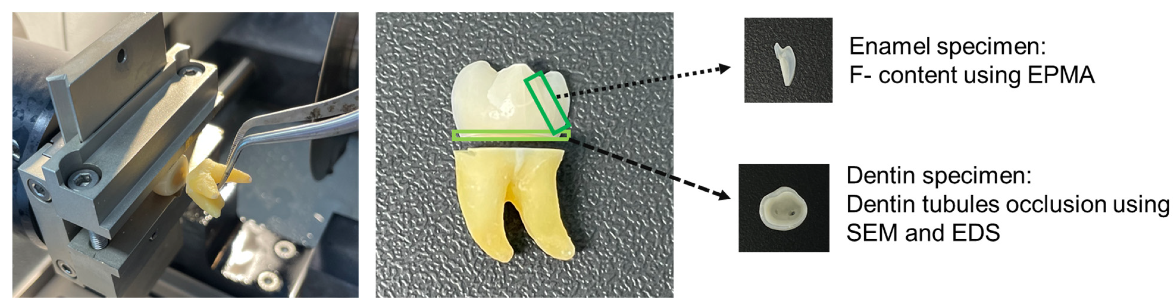
2.1. Materials

In this experiment, 1.23% APF gel (3D Dental, Houston, TX, USA) was used for topical application. The HA slurry was prepared using 60 nm nano-HA particles (MKnano, Mississauga, ON, Canada) and sodium carboxymethyl cellulose (CMC; Sigma-Aldrich, St Louis, MO, USA).

2.2. Tooth Preparation
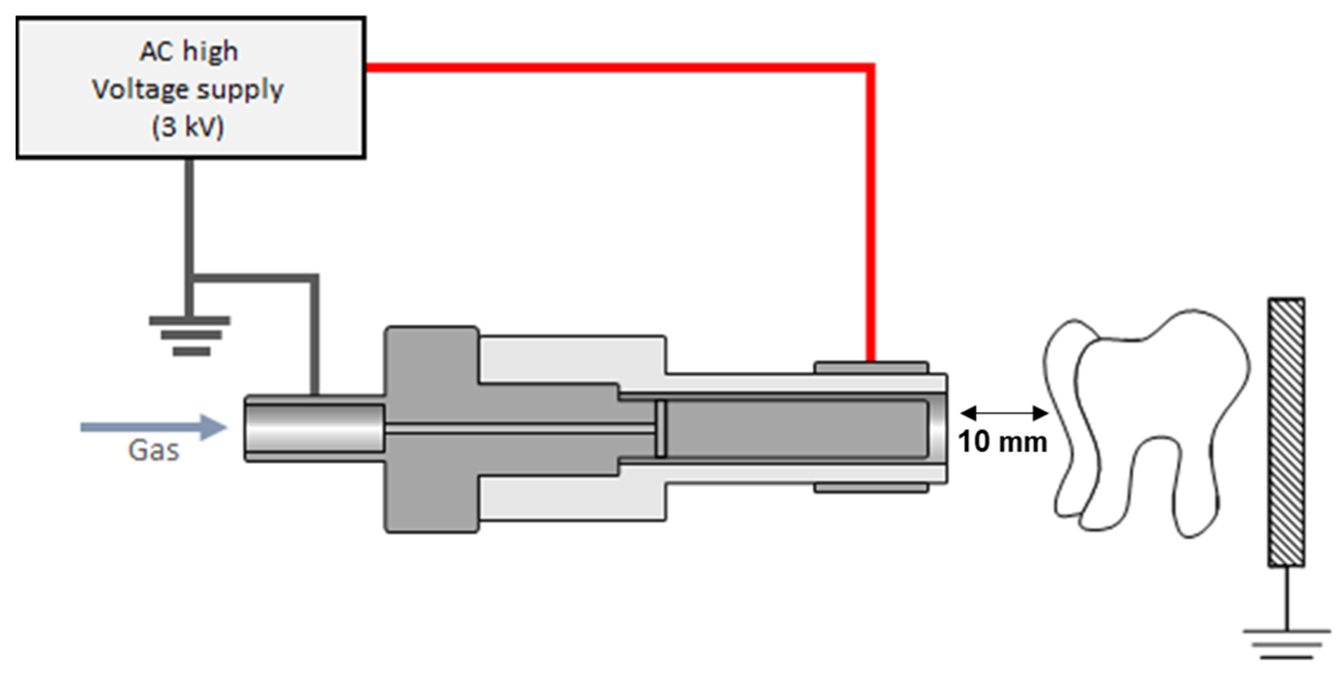
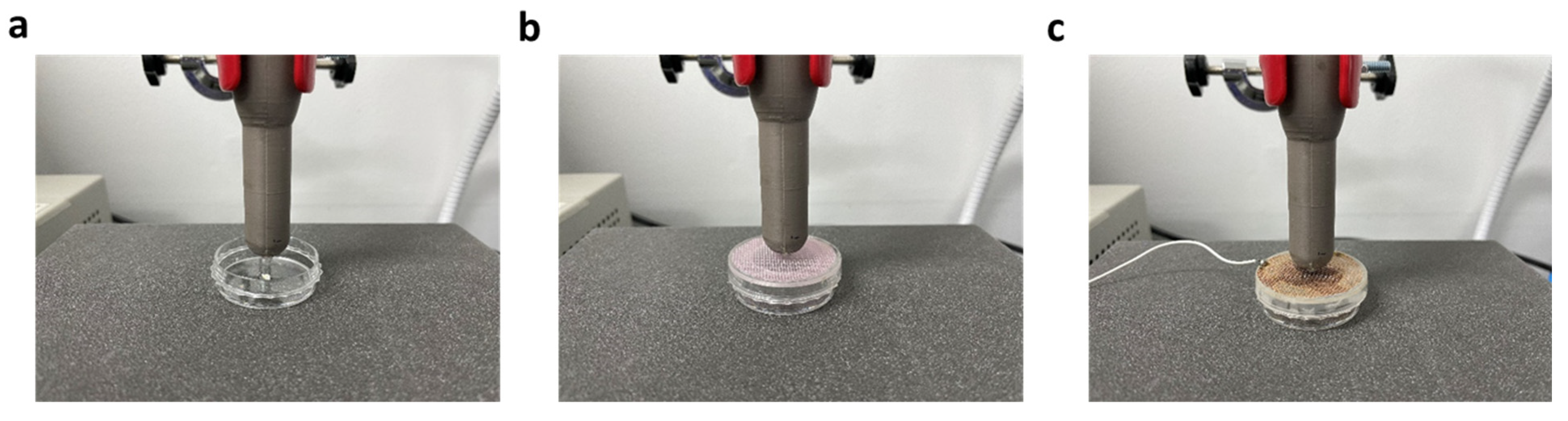
A total of 54 human mandibular third molars without dental caries or restoration were used in the study. The tooth, which is a wisdom tooth that is mostly incapable of mastication, was extracted by an oral and maxillofacial surgeon at Pusan National University Dental Hospital in Republic of Korea. The tooth specimens used in the experiment were sectioned by separating the crown and root areas using a water-cooled diamond saw (Minitom; Struers, Copenhagen, Denmark). Thereafter, the crown portion was flat polished and cut to a thickness of 1 mm to prepare an enamel specimen measuring 1 × 3 mm. The enamel was removed from the dentin sample and cut to a thickness of 0.8 mm with a diamond wheel saw to prepare a circular dentin specimen (Figure 1). To prevent contamination with bacteria until usage, they were kept in a solution of 0.4% sodium azide (Sigma-Aldrich, St Louis, MO, USA) and maintained at a temperature of 4 °C. This study was approved by the Institutional Review Board of Pusan National University (PNU IRB/2021_81_ BR).

2.3. Plasma Device

The plasma generator used in this study is a research device developed by Feagle Co., Ltd. (Yangsan, Republic of Korea). NCP, a plasma generation technology developed to reduce ozone release during plasma generation, was used in this device. The ozone level of the NCP device used was 0.006 ppm, which is approximately 10 times lower than the FDA recommended level of 0.05 ppm [29]. The device consists of a main body composed of a switched-mode power supply, solenoid valve, gas flow rate controller, high-voltage circuit, and hand-piece that generates plasma (Figure 2). A coaxial dielectric barrier discharge-type plasma source is installed within the handpiece, which consists of an inner electrode of stainless steel and an outer electrode surrounding the outer diameter of the ceramic nozzle. A Luer Lock-type fastening component is placed at the end of the hand-piece plasma-generating nozzle to permit the attachment of various tips. When the start button is pressed, argon gas flows between the inner electrode and ceramic nozzle at a rate of 2 standard L per min, and an output voltage of 3 kVp with a frequency of 20 kHz is applied to the electrodes at both ends to generate plasma. The mesh used to investigate the mechanism (Feagle Co., Ltd.) is fabricated using cotton or metal and equipped with the same opening/closing rate and can be installed in the plasma outlet of the NCP device. In the case of the metal mesh, the ground wire is connected such that the charged particles of the NCP are not transferred to the sample.

2.4. Electron Probe Microanalyzer Analysis

The impact of NCP pre-treatment on the effect of dental fluoride, i.e., APF application was determined by pre-treating the prepared enamel specimens with NCP for 3 min, followed by APF alone. The distance between the enamel specimen and NCP was 10 mm. Each specimen (n = 9) was treated with APF and NCP once daily for 4 days. Subsequently, the teeth were brushed with distilled water, and treated with artificial saliva at room temperature for 30 min, and dried. The mesh experiment was conducted to confirm the working element of plasma, which is important for the effect of NCP on F application.
The dielectric mesh was made from cotton mesh, so that most of the NCP working elements can pass through it, though the flow rate of NCP can be limited. The electric grounded mesh, on the other hand, was created utilizing a copper mesh that was directly connected to the ground panel of the power supply device, therefore eliminating the impacts of charged particles from NCP (Figure 3) [30]. Each prepared enamel sample was divided into four experimental groups as follows (n = 9): APF treatment alone, APF+ NCP, and treatment with NCP equipped with a cloth mesh at the end of the NCP device (APF+ NCP dielectric mesh (DE)), and treatment with NCP equipped with an electric grounded mesh at the end of the NCP device (APF+ NCP grounded electric mesh (EG)). After APF and NCP treatment, the teeth were brushed with distilled water, treated with artificial saliva at room temperature for 30 min, and dried. After all treatments were completed, the F content of each sample was analyzed using field emission electron probe micro-analyzer (FE-EPMA) equipment (JXA-8530F, JEOL, Tokyo, Japan) capable of qualitative and quantitative analysis of the microelements. The measurements were conducted by randomly designating the site of each specimen to ensure the accuracy and objectivity of the results [25].

2.5. Scanning Electron Microscopy and Energy Dispersive X-ray Spectrometry Analysis

Each specimen was treated with 18% EDTA (Sigma-Aldrich, St Louis, MO, USA) for 30 s to remove the smear layer from the prepared dentin specimen to confirm the efficacy of NCP pre-treatment in occluding the dentin tubules. The prepared specimens were divided into five experimental groups (n = 9): non-treated group (NT) after EDTA treatment, HA treatment group, APF treatment group, HA and APF treatment group, and a group treated with a mixture of HA and APF after NCP pre-treatment. In total, 0.1 g powdered HA was converted into gel form using 0.5 g CMC and distilled water for ease of handling. The treatment time for all experiments was 3 min. The samples were brushed with distilled water, followed by treatment with artificial saliva for 30 min and drying in a dry oven. To confirm the occlusion of the dentinal tubules, images were captured using a SUPRA 25 FE-SEM (ZEISS, Oberkochen, Germany). All dried dentin specimens were fixed on an aluminum plate coated with platinum using a vacuum, and scanning electron microscopy (SEM) was performed at an accelerated voltage of 10 kV. The photographed image was obtained by randomly selecting the area of the specimen at a magnification of 1000×. Closure of the dentinal tubules in each sample was evaluated by counting the number of exposed dentin tubules, and the average value was determined. Energy dispersive X-ray spectrometry (EDS) (Supra 25 VP, Carl Zeiss, Germany) was performed to quantify the elements formed on the dentin specimens. Area scan component analysis was performed using EDS at 15 kV and 200×, and the content of Ca and P was measured by designating an area of the same size by dividing the dentin specimen into the upper, lower, left, and right quadrants [31]. The measured Ca and P values were used to calculate the Ca/P ratio and displayed on a graph.

2.6. Statistical Analysis

All experimental results were analyzed using SPSS Version 24 statistical software package (IBM, Chicago, IL, USA). Tukey’s post hoc test was used to verify significant differences between the control and experimental groups after the one-way analysis of variance. Significance levels were set at p < 0.05, 0.01, and 0.001.

3. Results

3.1. F Level Measurement Using EPMA Analysis

The F content of each sample was quantified using EPMA analysis to confirm the effect of NCP on APF application, as shown in Figure 4a. It confirmed that the F value of the APF-treated group after only one application of NCP for 3 min was significantly higher by approximately two times than that of the APF-only group (p < 0.001). Moreover, in the group treated with APF after NCP pre-treatment, the F content of the enamel specimen increased significantly with the increase in the number of treatments (approximately 0.18% for one time, 0.36% for two times, 0.53% for three times, and 0.56% for four times). In contrast, in the case of enamel specimens treated with APF alone, F content was maintained at approximately 0.9 to 0.1% for one to three applications, but a slight increase in the F content to approximately 0.2% was observed at four applications.

3.2. Effect of Two Types of Meshes on the Improvement of APF Application via NCP Pre-Treatment

Among the various working elements of NCP, two types of meshes were investigated to identify the most important factor responsible for enhancing the APF effect due to NCP pre-treatment (Figure 4b). The F content increased approximately fourfold in the group treated with APF after NCP pre-treatment compared to that in the group treated with APF alone. The F content increased fourfold after NCP pre-treatment with a cotton mesh (DE). However, pre-treatment with NCP equipped with an electric grounded (EG) mesh significantly reduced the F content compared to the other NCP treatment groups (p < 0.01).

3.3. Dentinal Tubule Occlusion: SEM Analysis

SEM imaging and analysis were conducted to verify occlusion of the dentinal tubules by HA, APF, and NCP. All dentinal tubules were exposed in the control group, while the dentinal tubules were not occluded in the HA-alone and APF-alone groups. The group treated with a mixture of HA and APF also did not show any occlusive effect on the dentinal tubule. In contrast, the dentin tubules were significantly occluded in the group treated with a mixture of HA and APF after plasma pre-treatment with NCP (Figure 5a,b). The percentage of dentinal tubule occlusion calculated using SEM was as follows: control group, 0%; HA-only treatment, 0.5%; APF-only treatment, 2%; combination of HA and APF, 2%; and HA and APF treatment after pre-treatment with NCP, approximately 45%. Dentinal tubule occlusion was approximately 22.5-fold higher in the group treated with HA and APF after pre-treatment with NCP (NCP+HA/APF) than that in the group treated with a combination of HA and APF (HA/APF) (p < 0.05).

3.4. Evaluation of the Dentin Re-Mineralization Effect by NCP Pre-Treatment

The wt% for Ca and P in each group of specimens was analyzed using EDS. The Ca/P ratio, which was calculated from the wt% values of Ca and P (Figure 5c), was as follows: control group, 1.92; HA-only group, 2; APF-only group, 2.02; HA and APF combination group, 2.10; and group treated with HA and APF after NCP pre-treatment, 2.27. Interestingly, no statistically significant difference was observed among the groups not treated with NCP, whereas the Ca/P value of the group treated with HA/APF after NCP pre-treatment was significantly higher than that of the other groups (p < 0.05).

4. Discussion

This study investigated the possibility of increasing the efficacy of APF or HA using NCP technology developed to safely deliver low-temperature plasma to the oral cavity, which is located in close proximity to the respiratory system. The dentin is exposed to various stimuli, resulting in hypersensitivity [32,33]. Although dentin hypersensitivity is frequently encountered in clinical practice, no effective and reliable treatment method is available. The treatment methods used so far are divided into self-administered methods, such as toothpaste or rinses, and professional treatment methods that entail application of high-concentration fluoride to the tooth surface or resin restorations [34]. To date, preparations composed of various ingredients have been developed; concoctions containing biocompatible ingredients such as F, HA, and carbonate apatite have found widespread application owing to the ease of use. Several materials have been developed to reduce or eliminate the pain caused by hypersensitivity to dentin, which generally act by blocking the dentinal tubule or interfering with nerve impulse transmission.
F, which is commonly used in clinical practice, forms a protective film by the formation and deposition of CaF2 on the exposed tooth surface. APF used in the experiment is known to induce a higher CaF2 concentration in dentin than in enamel, releasing a greater amount of Ca [18]. However, it does not remain permanently on the tooth surface and is easily washed off by the presence of saliva or food, making long-term retention in the oral cavity difficult. In this study, we examined the utility of low-temperature plasma to increase the efficacy of APF and HA, which are the most frequently used conventional materials for the treatment of hypersensitivity. We found that the F content on the tooth surface was higher in the experimental group treated with APF after NCP treatment than that in the group treated with APF alone (Figure 4a). Our previous research found that the plasma jet generated using helium dramatically increased the F content of extracted teeth [25].
The results of this study are consistent with those of previous studies using a helium plasma jet: the F coating effect of APF is maintained with NCP pre-treatment, and the amount of ozone generated is negligible [29] and the plasma plume does not directly contact the teeth. Two types of meshes that can be mounted on the NCP device were fabricated and the change in the F coating effect by NCP was examined in order to determine the most important working element of NCP (which is little influenced by ozone and heat) responsible for dental F coating. According to this experiment, a higher amount of F was detected in the group treated with APF after pre-treatment with NCP than the group treated with only AFP. When a grounded metal mesh was installed for NCP treatment, the value decreased from 0.41 to 0.06, indicating a drastic reduction in the amount of F (Figure 4b). This result is akin to the phenomenon where low-temperature argon plasma-mediated E-cadherin protein inhibition in HaCaT cells completely disappears when an EG mesh is applied between the cell and plasma device [35]. In the sample pre-treated with the DE mesh-equipped NCP, the amount of F was similar to that of the sample pre-treated with NCP. These results show that charged particles that can be removed only by the EG mesh (not chemically active species such as OH radicals that can pass through both meshes) play an important role in enhancing the dental F coating effect of NCP. Furthermore, the alleviation of dentin hypersensitivity by APF and HA after plasma pre-treatment was observed. Argon generally lost electrons during the early stages of the argon plasma formation process, thereby producing argon ions and electrons. The dentin hypersensitivity impact of NCP in this investigation appears to be caused by charged particles of NCP, because this effect was lessened by the electrically grounded mesh.
The dentinal tubules of teeth showing dentin hypersensitivity are open, and the resulting pain is determined by the opening and closing of the dentin tubules [36,37]. SEM imaging, which was performed to observe the occlusion of the dentinal tubules, followed by comparative analysis and graph plotting, revealed no statistically significant difference between the control, HA, APF, and HA/APF groups. On the other hand, pre-treatment with plasma and HA/APF yielded statistically significant results. After plasma pre-treatment, the occlusion efficacy was confirmed to be about 22.5 times higher in the experimental group treated with HA/APF than that in the other groups. Thus, we can infer that plasma pre-treatment of the tooth surface effectively enhances the adhesion of the HA/APF mixture. There was no statistically significant difference in the Ca and P content of the untreated, HA, APF, and HA/APF groups. On the other hand, the group treated with plasma and HA/APF showed statistically significant results. This shows that HA and APF do not significantly increase the Ca and P content of the coated dentin, contrary to the results of previous studies [38]. Previous studies interpreted an increase in Ca and P levels because the teeth were continuously exposed to the minerals in saliva.
Our results showed that pre-treating dentin with plasma significantly increased the ability of APF to close the dentin tubules compared to treatment with only HA, APF, and HA/APF. Pre-treatment with plasma facilitates penetration of the charged particles into the dentin, so F and HA can easily penetrate the dentinal tubules, and the surface area is increased to improve the adhesion with dentin. However, although the effect and mechanism underlying the amelioration of hypersensitivity after plasma pre-treatment were verified in vitro in this study, a limitation is that the efficacy was not reported to be increased in vivo. Future in vivo research is warranted to verify the efficacy of plasma treatment against hypersensitivity. A more complete verification of the efficacy of NCP for resolving hypersensitivity should be conducted through in vivo effect research. Future studies will examine whether applying fluoride to animal teeth via NCP is more efficient than the current techniques.

5. Conclusions

In summary, we found that the F content was significantly higher in the experimental group treated with APF after plasma pre-treatment than in the experimental group treated with F alone, which was attributed to the charged particles of argon plasma. In addition, plasma pre-treatment aided in stabilization of HA/APF in the dentinal tubule and was effective in alleviating dental hypersensitivity. Therefore, argon plasma pre-treatment of the tooth surface may alleviate hypersensitivity, and has the potential to find application for treating hypersensitivity in the future.

Author Contributions

Conceptualization, B.-B.C., S.-A.P. and J.-H.C.; methodology, B.-B.C. and S.-A.P.; validation, S.-A.P.; formal analysis, B.-B.C. and S.-R.P.; investigation, S.-R.P.; writing—original draft preparation, B.-B.C., S.-A.P., J.-H.C. and S.-R.P.; writing—review and editing, B.-B.C. and G.-C.K.; visualization, S.-A.P.; supervision, G.-C.K. All authors have read and agreed to the published version of the manuscript.

Funding

This work was supported by a National Research Foundation of Korea (NRF) grant funded by the Korean government (MSIT) (No. 2021R1A2C2008215).

Institutional Review Board Statement

This study was approved by the Institutional Review Board of Pusan National University (PNU IRB/2021_81_ BR).

Informed Consent Statement

Informed consent was obtained from all subjects involved in the study.

Data Availability Statement

The data presented in this study are available upon request from the corresponding author.

Conflicts of Interest

The authors declare no conflict of interest.

References

  1. Mason, S.; Burnett, G.R.; Patel, N.; Patil, A.; Maclure, R. Impact of toothpaste on oral health-related quality of life in people with dentine hypersensitivity. BMC Oral Health 2019, 19, 226. [Google Scholar] [CrossRef] [PubMed]
  2. Liu, X.X.; Tenenbaum, H.C.; Wilder, R.S.; Quock, R.; Hewlett, E.R.; Ren, Y.F. Pathogenesis, diagnosis and management of dentin hypersensitivity: An evidence-based overview for dental practitioners. BMC Oral Health 2020, 20, 220. [Google Scholar] [CrossRef] [PubMed]
  3. Elovikova, T.M.; Ermishina, E.Y.; Uvarova, L.V.; Koshcheev, A.S. The increased sensitivity of dentin: The mechanisms of remineralization using toothpaste with tin fluoride. Stomatologiia 2019, 95, 66–71. [Google Scholar] [CrossRef] [PubMed]
  4. Abuzinadah, S.H.; Alhaddad, A.J. A randomized clinical trial of dentin hypersensitivity reduction over one month after a single topical application of comparable materials. Sci. Rep. 2021, 11, 2793. [Google Scholar] [CrossRef] [PubMed]
  5. Aminoshariae, A.; Kulild, J.C. Current concepts of dentinal hypersensitivity. J. Endod. 2021, 47, 1696–1702. [Google Scholar] [CrossRef] [PubMed]
  6. Guerra, F.; Corridore, D.; Cocco, F.; Arrica, M.; Rinaldo, F.; Mazur, M.; Sanavia, C.; Nardi, G.M.; Campus, G. Oral health sentinel-based surveillance: A pilot study on dentinal hypersensitivity pain. Clin. Ter. 2017, 168, e333–e337. [Google Scholar] [CrossRef]
  7. Longridge, N.N.; Youngson, C.C. Dental Pain: Dentine Sensitivity, Hypersensitivity and Cracked Tooth Syndrome. Prim. Dent. J. 2019, 8, 44–51. [Google Scholar] [CrossRef]
  8. Bertani, G.; Di Tinco, R.; Bertoni, L.; Orlandi, G.; Pisciotta, A.; Rosa, R.; Rigamonti, L.; Signore, M.; Bertacchini, J.; Sena, P.; et al. Flow-dependent shear stress affects the biological properties of pericyte-like cells isolated from human dental pulp. Stem Cell Res. Ther. 2023, 14, 31. [Google Scholar] [CrossRef]
  9. Suge, T.; Ishikawa, K.; Matsuo, T.; Ebisu, S. Duration of dentin tubule occlusion by the calcium phosphate precipitation method: An in vivo study in beagle dogs. Dent. Mater. J. 2021, 40, 1020–1026. [Google Scholar] [CrossRef]
  10. Mukherjee, M.; Kalita, T.; Barua, P.; Barman, A.; Thonai, S.; Mahanta, P.S.; Medhi, H. Efficacy of Smear Layer Removal of Human Teeth Root Canals Using Herbal and Chemical Irrigants: An In Vitro Study. Cureus 2023, 15, e40467. [Google Scholar] [CrossRef]
  11. Charoenlarp, P.; Wanachantararak, S.; Vongsavan, N.; Matthews, B. Pain and the rate of dentinal fluid flow produced by hydrostatic pressure stimulation of exposed dentine in man. Arch. Oral Biol. 2007, 52, 625–631. [Google Scholar] [CrossRef] [PubMed]
  12. Banfield, N.; Addy, M. Dentine hypersensitivity: Development and evaluation ofamodel in situ to study tubulepatency. J. Clin. Periodontol. 2004, 31, 325–335. [Google Scholar] [CrossRef] [PubMed]
  13. George, A.A.; Muruppel, A.M.; Lal, S. A Comparative Evaluation of the Effectiveness of Three Different Modalities in Occluding Dentinal Tubules: An In Vitro Study. J. Contemp. Dent. Pract. 2019, 20, 454–459. [Google Scholar] [CrossRef] [PubMed]
  14. Rocha, M.O.C.; Cruz, A.A.C.F.; Santos, D.O.; Douglas-DE-Oliveira, D.W.; Flecha, O.D.; Gonçalves, P.F. Sensitivity and specificity of assessment scales of dentin hypersensitivity—An accuracy study. Braz. Oral Res. 2020, 34, e043. [Google Scholar] [CrossRef]
  15. Bamise, C.T.; Esan, T.A. Mechanisms and treatment approaches of dentine hypersensitivity: A literature review. Oral Health Prev. Dent. 2011, 9, 353–367. [Google Scholar]
  16. Kijsamanmith, K.; Wallanon, P.; Pitchayasatit, C.; Kittiratanaviwat, P. The effect of fluoride iontophoresis on seal ability of self-etch adhesive in human dentin in vitro. BMC Oral Health 2022, 22, 109. [Google Scholar] [CrossRef]
  17. Paik, Y.; Kim, M.J.; Kim, H.; Kang, S.W.; Choi, Y.K.; Kim, Y.I. The Effect of Biomimetic Remineralization of Calcium Phosphate Ion Clusters-Treated Enamel Surfaces on Bracket Shear Bond Strength. Int. J. Nanomed. 2023, 18, 4365–4379. [Google Scholar] [CrossRef]
  18. Ramli, R.; Ghani, N.; Taib, H.; Mat-Baharin, N.H. Successful management of dentin hypersensitivity: A narrative review. Dent. Med. Probl. 2022, 59, 451–460. [Google Scholar] [CrossRef]
  19. Yuan, P.; Shen, X.; Liu, J.; Hou, Y.; Zhu, M.; Huang, J.; Xu, P. Effects of dentifrice containing hydroxyapatite on dentinal tu-bule occlusion and aqueous hexavalent chromium cations sorption: A preliminary study. PLoS ONE 2012, 7, e45283. [Google Scholar] [CrossRef]
  20. Jafari, B.; Katoozian, H.R.; Tahani, M.; Ashjaee, N. A comparative study of bone remodeling around hydroxyapatite-coated and novel radial functionally graded dental implants using finite element simulation. Med. Eng. Phys. 2022, 102, 103775. [Google Scholar] [CrossRef]
  21. Braun, A.; Cichocka, A.; Semaan, E.; Krause, F.; Jepsen, S.; Frentzen, M. Root surfaces after ultrasonic instrumentation with a polishing fluid. Quintessence Int. 2007, 38, e490–e496. [Google Scholar] [PubMed]
  22. Tschoppe, P.; Zandim, D.L.; Martus, P.; Kielbassa, A.M. Enamel and dentine remineralization by nano-hydroxyapatite toothpastes. J. Dent. 2011, 39, 430–437. [Google Scholar] [CrossRef] [PubMed]
  23. Gamagedara, T.P.; Ziana, H.F. Effects of hydroxyapatite nanoparticles on liver enzymes and blood components. J. Clin. Investig. Stud. 2018, 1, 1–5. [Google Scholar]
  24. Nam, S.-H.; Choi, B.B.R.; Kim, G.-C. The Whitening Effect and Histological Safety of Nonthermal Atmospheric Plasma Inducing Tooth Bleaching. Int. J. Environ. Res. Public Health 2021, 18, 4714. [Google Scholar] [CrossRef]
  25. Kim, Y.M.; Lee, H.Y.; Lee, H.J.; Kim, J.B.; Kim, S.; Joo, J.Y.; Kim, G.C. Retention Improvement in Fluoride Application with Cold Atmospheric Plasma. J. Dent. Res. 2018, 97, 179–183. [Google Scholar] [CrossRef]
  26. Park, S.R.; Lee, H.W.; Hong, J.W.; Lee, H.J.; Kim, J.Y.; Choi, B.B.; Kim, G.C.; Jeon, Y.C. Enhancement of the killing effect of low-temperature plasma on Streptococcus mutans by combined treatment with gold nanoparticles. J. Nanobiotechnol. 2014, 8, 29. [Google Scholar] [CrossRef]
  27. Choi, B.B.R.; Choi, J.H.; Hong, J.W.; Song, K.W.; Lee, H.J.; Kim, U.K.; Kim, G.C. Selective Killing of Melanoma Cells With Non-Thermal Atmospheric Pressure Plasma and p-FAK Antibody Conjugated Gold Nanoparticles. Int. J. Med. Sci. 2017, 14, 1101–1109. [Google Scholar] [CrossRef]
  28. Eggers, B.; Wagenheim, A.M.; Jung, S.; Kleinheinz, J.; Nokhbehsaim, M.; Kramer, F.J.; Sielker, S. Effect of Cold Atmospheric Plasma (CAP) on Osteogenic Differentiation Potential of Human Osteoblasts. Int. J. Mol. Sci. 2022, 23, 2503. [Google Scholar] [CrossRef]
  29. Choi, B.-B.; Choi, J.-H.; Kang, T.-H.; Lee, S.-J.; Kim, G.-C. Enhancement of Osteoblast Differentiation Using No-Ozone Cold Plasma on Human Periodontal Ligament Cells. Biomedicines 2021, 9, 1542. [Google Scholar] [CrossRef]
  30. Park, N.S.; Yun, S.E.; Lee, H.Y.; Lee, H.J.; Choi, J.H.; Kim, G.C. No-ozone cold plasma can kill oral pathogenic microbes in H2O2-dependent and independent manner. Sci. Rep. 2022, 12, 7597. [Google Scholar] [CrossRef]
  31. Minetti, E.; Palermo, A.; Malcangi, G.; Inchingolo, A.D.; Mancini, A.; Dipalma, G.; Inchingolo, F.; Patano, A.; Inchingolo, A.M. Dentin, Dentin Graft, and Bone Graft: Microscopic and Spectroscopic Analysis. J. Funct. Biomater. 2023, 14, 272. [Google Scholar] [CrossRef] [PubMed]
  32. Favaro, Z.L.; Soares, P.V.; Cunha-Cruz, J. Prevalence of dentin hypersensitivity: Systematic review and meta-analysis. J. Dent. 2019, 81, 1–6. [Google Scholar] [CrossRef] [PubMed]
  33. Davari, A.; Ataei, E.; Assarzadeh, H. Dentin hypersensitivity: Etiology, diagnosis and treatment; a literature review. J. Dent. 2013, 14, 136–145. [Google Scholar]
  34. West, N.X.; Seong, J.; Davies, M. Management of dentine hypersensitivity: Efficacy of professionally and self-administered agents. J. Clin. Periodontol. 2015, 42, S256–S302. [Google Scholar] [CrossRef]
  35. Lee, H.Y.; Choi, J.H.; Hong, J.W.; Kim, G.C. Comparative study of the Ar and He atmospheric pressure plasmas on E-cadherin protein regulation for plasma plasma mediated transdermal drug delivery. J. Phys. D Appl. Phys. 2018, 51, 11. [Google Scholar] [CrossRef]
  36. Abu, H.A.; Martinho, F.C.; Sellan, P.L.B.; Pampuri, C.R.; Torres, C.R.G.; Pucci, C.R. Effect of Remineralization Pretreatments on Human Dentin Permeability and Bond Strength. Int. J. Dent. 2023, 2023, 2182651. [Google Scholar] [CrossRef]
  37. Berg, C.; Unosson, E.; Engqvist, H.; Xia, W. Comparative Study of Technologies for Tubule Occlusion and Treatment of Dentin Hypersensitivity. J. Funct. Biomater. 2021, 12, 27. [Google Scholar] [CrossRef]
  38. Yuan, P.; Liu, S.; Lv, Y.; Liu, W.; Ma, W.; Xu, P. Effect of a dentifrice containing different particle sizes of hydroxyapatite on dentin tubule occlusion and aqueous Cr (VI) sorption. Int. J. Nanomed. 2019, 14, 5243–5256. [Google Scholar] [CrossRef]
Figure 1. Human enamel and dentin specimen preparation EPMA: electron probe microanalyzer, SEM: scanning electron microscopy, EDS: energy dispersive X-ray spectrometry.
Applsci 13 11728 g001
Figure 2. Schematic diagram of the NCP device used in the experiment. NCP: no-ozone cold plasma.
Applsci 13 11728 g002
Figure 3. Charged particles of NCP. (a) NCP; (b) NCP dielectric mesh (DE); (c) NCP grounded electric mesh (EG). NCP: no-ozone cold plasma.
Applsci 13 11728 g003
Figure 4. F Level Measurement Using EPMA Analysis: (a) Fluoride level measurement via EPMA analysis after NCP pre-treatment (*** p < 0.001); (b) Evaluation of the effect of fluoride application of NCP using a mesh (** p < 0.01) EPMA: electron probe microanalyzer, NCP: no-ozone cold plasma.
Applsci 13 11728 g004
Figure 5. Dentinal Tubule Occlusion: SEM Analysis: (a) Observation of the dentinal tubule surface using SEM analysis after NCP treatment (1000×); (b) Measurement of the dentinal tubule occlusion rate after NCP treatment (* p < 0.05); (c) Effect of NCP on dentinal tubule re-mineralization (* p < 0.05) NCP: no-ozone cold plasma, SEM: scanning electron microscopy.
Applsci 13 11728 g005
Disclaimer/Publisher’s Note: The statements, opinions and data contained in all publications are solely those of the individual author(s) and contributor(s) and not of MDPI and/or the editor(s). MDPI and/or the editor(s) disclaim responsibility for any injury to people or property resulting from any ideas, methods, instructions or products referred to in the content.

