Article

Economic Analysis of the Production Process of Probiotics Based on the Biological and Physiological Parameters of the Cells

Department of Biotechnology and Food Microbiology, Poznan University of Life Sciences, Wojska Polskiego 48, 60-627 Poznań, Poland
*
Authors to whom correspondence should be addressed.
Appl. Sci. 2023, 13(20), 11541; https://doi.org/10.3390/app132011541
Submission received: 13 September 2023 / Revised: 12 October 2023 / Accepted: 19 October 2023 / Published: 21 October 2023
(This article belongs to the Special Issue Microorganisms in Foods and Food Processing Environments)

Abstract

:

Featured Application

The presented analysis provides a concise depiction of the fluid bed drying process of probiotics. It also introduces a new method of evaluation for probiotics, based on the actual number of biologically useful cells. This factor is used to evaluate and economically justify the introduction of technological procedures, such as sublethal stresses and coating.

Abstract

Probiotic bacteria confer a range of health benefits and are a focus of a growing number of studies. One of the main issues is their stability during drying and storage, which is why techniques, such as fluid bed drying and coating or treatment with stress factors during culturing, are utilized. The methods of the evaluation of probiotic viability and quality are, however, lacking and we need a way of distinguishing between different subpopulations of probiotic bacteria. To address this issue, imaging flow cytometry (IFC) has been utilized to assess cells after simulated in vitro digestion of dried and coated preparations treated with pH stress and heat shock. Samples were analyzed fresh and after 12 months of storage using RedoxSensor green and propidium iodide dyes to assess metabolic activity and cell membrane integrity of the cells. The results were then used to design a drying process on an industrial scale and evaluate the economic factors in the SuperPro Designer v13 software. Based on the number of biologically active and beneficial cells obtained utilizing tested methods, the coating process and treatment with heat shock and pH stress have been the most effective and up to 10 times cheaper to produce than only by drying. Additionally, samples after 12 months of storage have shown an increase in the proportion of cells with intermediate metabolic activity and small amounts of cell membrane damage, which are still viable in probiotic products. This subpopulation of bacteria can still be considered live in probiotic products but is not necessarily effectively detected by pour plate counts.

1. Introduction

In recent decades, there has been a growing recognition of the importance of gut microbiota for human health and well-being. Probiotics are defined as “live micro-organisms that, when administered in adequate amounts, confer a health benefit on the host” [1]. Cells are often enumerated using plate pour counts directly after the production process or after storage. Such values do not directly represent the number of probiotic cells that reach the colon after the gastric transit. Additionally, pour plate enumeration does not take into consideration cell subpopulations with intermediate activity (i.e., viable but nonculturable—VBNC) [2]. Therefore, the number of colony-forming units determined using classic microbiology is not synonymous with the number of probiotic cells in the preparation. To address this issue, we determined that for more precise enumeration the viable and beneficial (VB) cells enumerated using imaging flow cytometry should be considered, namely, those cells that remain useful and confer health benefits to the host after technological processes and digestion. We also look deeply into the process of production of probiotics and determine the cost and quality of the product based on those assumptions.
New reports on the numerous health benefits of probiotic bacteria, such as their role in the positive regulation of inflammatory bowel diseases (IBD), treatment of allergies, and urogenital infections, have led to increased interest in their commercial production [3]. Existing studies on probiotic production often utilize drying techniques, such as spray or freeze drying. The quality parameters of dry cells (number of living cells and biological activity) usually depend on the type of drying method used. In the case of thermolabile materials, including bacterial cells, the smallest reduction in quality after drying is observed when freeze drying is used. However, this method is expensive and time-consuming [4]. Therefore, freeze drying is increasingly being replaced by spray drying. However, during spray drying, it is often difficult to obtain the quality of bacterial preparations similar to that obtained during freeze drying. One of the reasons is the relatively high drying temperature, which will usually not be lower than 100 °C for inlet and 60 °C for outlet air. Meanwhile, in fluid bed dryers, it is possible to conduct the process at much lower temperatures, close to 30–40 °C, thus minimizing heat inactivation [5,6]. Dryers operating in fluidized bed conditions have very good mass and energy transfer parameters, therefore the drying time is short [7]. Additionally, the investment and operating costs of fluid bed drying are lower than those of freeze or spray drying [8]. In a fluidized bed, the cell matrix can be easily coated with an additional protective layer, improving its shelf life and stability during storage. Effective application of probiotics is, however, limited due to the lack of standardized requirements for efficacy and the number of viable cells (colony forming units—CFU/mL) in the final product. Additionally, the decrease in cell viability during the passage through the gastrointestinal (GI) tract is not taken into consideration [9]. Recent advances in gastrointestinal simulation techniques have made it possible to better understand the dynamic interactions that influence the survival and activity of probiotics along their journey through the digestive tract. The limited survival of probiotics during in vitro digestion is a complex process that is influenced by many factors, including the acidity of the stomach, the presence of bile salts and enzymes, the gut microbiota and its colonization resistance, and the properties of the probiotic strain itself [10]. The acidic environment of the stomach is one of the most significant challenges to the survival of probiotics. To survive the acidic environment of the stomach, probiotics must have some protective mechanisms in place. These mechanisms include the production of acids and enzymes that can neutralize stomach acid [11], the formation of protective biofilms [12,13], and the ability to adhere to the stomach lining [14]. Bile salts and enzymes are also major challenges to the survival of probiotics. Bile salts are produced by the liver and are released into the small intestine. Bile salts have many antimicrobial properties that can kill probiotic bacteria [15]. Antimicrobial proteins are also present in the small intestine and act as a defense mechanism against external micro-organisms [16]. To survive the bile salts and enzymes, bacteria must have the ability to adhere to the intestinal lining and to produce protective substances (bile salt hydrolase) that can neutralize the bile salts [17]. In addition to the acidity of the stomach and the presence of bile salts and enzymes, the physical and chemical properties of the probiotic strain also play a role in its survival during in vitro digestion. Probiotic strains that are more resistant to acidity, bile salts, and enzymes are more likely to survive the in vitro digestion process. Additionally, probiotic strains that can adhere to the intestinal lining and produce protective substances are also more likely to survive the in vitro digestion process [18]. Coating is also one of the strategies utilized to improve the survival of probiotics during digestion and to improve the shelf life of the product [19]. Different functional materials can be used as coating substances to tackle various challenges expected during the oral delivery of probiotics [20]. Polysaccharides, such as alginate, as well as cellulose and lipid-based coating, are commonly used to protect the probiotics from low pH of the gastrointestinal tract. Coatings can also be utilized to supply the probiotic cells with prebiotics by using inulin or polydextrose as coating material. Additionally, coating can improve the intestinal retention by strengthening the interaction between probiotics and the intestinal mucus layer. Composite biomagnetic materials can be utilized in the coating layer to improve both the retention and cellular localization of the probiotics [21]. Also, natural ingredients such as red ginseng dietary fiber can improve the intestinal adhesion of probiotics [22].
The first section of the paper discusses the mechanisms underlying the survival of probiotic LAB during in vitro digestion, which is especially important for the evaluation of possible probiotics since only living bacteria can fully confer their health benefits after successfully passing through the GI tract [23]. The acidic environment of the stomach and the presence of bile salts and enzymes pose significant challenges to the viability of these micro-organisms. For the in vitro digestion, we have chosen samples that were cultured in stress conditions (of high temperature and pH shock) that contained freshly cultured bacteria, and dried and coated preparations, as well as preparations after storage, to check their influence on the survival of probiotic bacteria during the gastrointestinal passage.
The second section of the paper presents a comprehensive project focused on the production of probiotics. This project details each step of the process, from strain selection and fermentation to drying and formulation. The integration of scientific principles with technological innovations ensures the production of high-quality probiotics with optimal viability. Commonly, pour plate counts are used as a standard evaluation method for probiotic enumeration. The results are then calculated as colony-forming units (CFU) per gram or milliliter of preparation. However, not all bacterial cells present in the final product are active and can confer health benefits to the host. Furthermore, not all of the live cells (VBNC) can grow on standard agar media [24]. Flow cytometry allows the counting and assessing of subpopulations other than fully active cells [25], for example, those based on their metabolic activity and cellular membrane damage. Flow cytometry provides a more comprehensive characterization of probiotics, while being a more rapid technique, allowing for the analysis of thousands of cells per second [26]. To better describe the amount of biologically active and useful cells, the results of digestion analyzed by flow cytometry were used for economic analysis as they provide a more reliable parameter for process design and planning of production steps.

2. Materials and Methods

2.1. Strains and Cultures

The strain of lactic acid bacteria (LAB) used in this study was Enterococcus faecium 73 KBiMŻ. MRS broth was selected as the medium because it provides the required growth conditions for these bacteria. To achieve the highest amount of biomass, an inoculum was prepared in a volume that represented 10% of the medium volume for the bioreactor culture. To gradually achieve a culture volume of 1 L, the inoculum was first seeded in two stages. This allowed for improved adaptation of the micro-organisms and shortened the lag phase.
All steps of the inoculum preparation were conducted in a laminar flow hood for a minimized contamination risk. First, the strain was thawed on ice to minimize cell damage. Once the micro-organisms had warmed to an ambient temperature and thawed, they were transferred to a larger, 15-mL Falcon conical tube filled with 9 mL of MRS broth (Oxoid Ltd., Basingstoke, Hampshire, UK). The tube was then sealed with parafilm and incubated for 24 h at 30 °C. Following incubation, 10 mL of the inoculum was transferred to a flask containing 90 mL of MRS broth. The flask was incubated for an additional 24 h at 30 °C. After these steps, the inoculum was used to initiate a culture in the bioreactor.
The cell biomass was cultured using Biostat A plus bioreactors (Sartorius AG, Göttingen, Germany), equipped with a 5 L culture vessel. The bioreactor was heated using a heating blanket system. Agitation was provided by a Rushton impeller. Process control and data acquisition were performed using the BioPAT MFCS 4 software. The pH electrode was calibrated against buffers at pH 4 and pH 9 before sterilization and culturing. Next, MRS broth medium in the volume of 1 L was added to the bioreactor vessel. To create an aseptic environment, all parts of the bioreactor and the medium were then autoclaved at 121 °C for 30 min. Afterwards, nitrogen was run through the aeration system to minimize the chance of pathogens entering the bioreactor with air. After the medium in the bioreactor had cooled to 30 °C, the inoculum was added. Bacteria were cultured at 30 °C, with the pH set at 6.5 and maintained by adding a 30% NaOH solution. The culture was grown for 24 h, with constant stirring at 150 RPM. The end of the exponential growth phase was determined by the cessation of NaOH consumption and the stabilization of pH. These factors, in combination with the simultaneous end of base consumption, were used as indicators for the beginning of the stationary growth phase. Lastly, after the biomass growth had stopped, the culture was transferred into sterile centrifuge vessels using a peristaltic pump.

2.2. Stresses

To investigate the impact of stress on bacterial cell survival during fluid bed drying, various cultures were subjected to distinct stress environments. Two types of stresses were introduced: heat shock cultures were exposed to short-term thermal stress by increasing the temperature to 50 °C for 30 min. During pH stress, cultures were exposed to short-term acid stress by lowering the pH to 2.0 for 30 min. These two types of stresses were also determined to be the easiest to apply in an industrial setting. Osmotic stress and culturing without pH control were also considered as possible stress factors for bacterial adaptation. They were, however, not used since osmotic stress proved to be troublesome to implement on a larger scale; after adding salts to the medium to expose the bacteria to stress conditions, the whole biomass needed to be centrifuged and resuspended in a freshly made medium. Additionally, culturing without pH control was not used in the research, since it did not provide the expected split into the subpopulations with intermediate metabolic activity, as observed with pH and heat shock. The main goal of introducing sublethal stress conditions during the culturing stage was to determine whether the stress adaptation of bacterial cells had a significant impact on their survival during fluid bed drying.

2.3. Fluid Bed Drying and Coating

Both the drying and coating processes were conducted in the Strea-1 (GEA, Oelde, Germany) laboratory fluid bed dryer. First, the matrix (crystalline microcellulose (Ingredientpharm, Pratteln, Switzerland)) was added to the product container in the dryer. To cause the product to be in a fluid state, a stream of hot (up to 50 °C) air was introduced.
Bacteria for drying were suspended in a solution of a protective substance (5% trehalose (Sigma-Aldrich, St. Louis, MO, USA)) and pumped to the dryer. They entered an atomizing nozzle, which was operating under the pressure of 2 bar. Higher pressures could damage the cells due to the shear forces and reduce their viability. The drying and coating processes took approximately 30 min each. A total of 100 g of matrix and 100 mL of 2% hydroxypropyl methylcellulose (HPMC) (Sigma-Aldrich, St. Louis, MO, USA) were used for coating. Drying was carried out in the top-spraying system, where the nozzle was placed above the fluidized bed. For coating, the Wurster system was utilized, where the nozzle sprays the coating agent from below and is kept shielded from the matrix to avoid clumping. Ready powder was packed for further analysis and shelf life assessment.

2.4. Simulated Gastrointestinal Conditions

Based on the method reported by Minekus et al. [27], the simulated gastrointestinal conditions were divided into gastric and intestinal phases, without introducing the oral phase as it was deemed optional by this method authors. Simulated gastric fluid (SGF) and simulated intestinal fluid (SIF) were made up as recommended by Minekus et al. with the addition of enzymes and CaCl2 (Table 1). 1 M NaOH and 1 M HCl were used for pH regulation in both digestion fluids. For the simulated gastric digestion, rehydrated sample and gastric fluid were combined in a 50:50 (v/v) ratio for a final volume of 40 mL. Samples under digestion were incubated at 37 °C for 2 h. After this step, the sample (40 mL) was combined with 40 mL of intestinal fluid and incubated at 37 °C for 2 h.

2.5. Intestinal Epithelial Cell Culture

The human intestinal epithelial Caco-2 cell line (HTB-37™) was obtained from the American Type Culture Collection (ATCC, Manassas, VA, USA). It is a well-established model for studying intestinal barrier function. The cells were isolated from colon adenocarcinoma and can form a tight monolayer in culture.
To culture Caco-2 cells, the cells were placed on PET membranes (Millicell® Cell Culture Inserts, 24 mm diameter, 0.4 µm pore size) (Millipore, Burlington, MA, USA, Merck Group). The initial density was 4 × 10⁵ cells/cm2. The cells were cultured in Dulbecco’s Modified Eagle’s Medium (DMEM, Sigma-Aldrich, Saint Louis, MO, USA) with the addition of 1% non-essential amino acids (100× NEAA, Sigma-Aldrich) and 20% fetal bovine serum (FBS, Gibco BRL, Grand Island, NY, USA). The medium was changed three times a week, and the cells were maintained at 37 °C in a humidified atmosphere of 95% air and 5% CO2.
The integrity of the Caco-2 cell monolayer was monitored based on transepithelial electrical resistance (TEER) measurements. TEER is a measure of the electrical resistance across the cell monolayer. Caco-2 cell cultures with TEER values ≥ 600 Ω × cm2 are considered to have formed a tight monolayer and are utilized in bacteria adhesion experiments.

2.6. Adhesion Assay

The adhesion assay was performed according to the methodology described in our previous work [28]. The Caco-2 cells were prepared by first washing the monolayers twice with PBS to remove any loosely attached cells. DMEM (without phenol red) with bacterial cells was then added to the cells, and the cultures were incubated for 2 h at 37 °C to allow the bacterial cells to adhere to the Caco-2 cells. After incubation, the medium was removed from the cultures, and the cell monolayers were washed with PBS to remove any unbound bacterial cells. Lysis was performed next to release the adhered bacterial cells and the lysates were centrifuged and analyzed. The distribution of subpopulations in the samples post-adhesion was measured by analyzing the supernatant left after incubation and washing of the cells.

2.7. Imaging Flow Cytometry

Flow cytometry was used to examine bacterial cells for their metabolic activity and viability. Amnis FlowSight™ (Luminex Corp., Austin, TX, USA) is a flow cytometer with imaging and was used to examine bacterial cells. The flow cytometer allows for the use of 3 lasers (405 nm, 488 nm, and 642 nm), 5 fluorescence channels (acquisition by a multi-channel CCD camera), and a side scatter detector (SSC). We utilized the protocol described in our previous work [24], using RedoxSensorTM green and PI (propidium iodide) to assess the metabolic activity of the cell and the integrity of the cell membrane accordingly.

2.8. Process Simulation in SuperPro Designer

To simulate the large-scale production of fluid bed dried probiotics, SuperPro Designer v13 (Intelligen, Scotch Plains, NJ, USA) was employed. The key conditions for the process were determined based on experimental results obtained from laboratory-scale processes. The final product was prepared in the form of a dried powder containing the biomass of Enterococcus faecium 73 KBiMŻ, standardized to contain 1 × 10⁸ of viable bacterial cells per gram of the product. The project simulation provides information on economic evaluation. In the evaluation, the process flowsheets, operating costs, estimated capital, raw material and equipment costs, and profitability analysis were included. The cost of the equipment was provided from local supplier information and the sizing was calculated based on the process needs and throughput. Raw material prices were based on quotations from global suppliers (i.e., Sigma-Aldrich, St. Louis, MO, USA). Other economic variables, such as water (2.67 $/m3), electricity (0.20 $/kW-h), income tax (19%), and labor cost (5.50 $/h), were established based on local (Poland) values and may vary based on location. The project was assumed to operate for 330 days annually, with a 15-year lifespan. Additionally, one year was planned for construction and six months for start-up. The efficiency in the first year was estimated to be 50% due to the start-up period and lower occupancy. Described assumptions were constant and applied to all six projects.

2.9. Sensitivity Analysis

Crystal Ball software, version 11.1.3.0.000 (Oracle, Austin, TX, USA) was used to perform a sensitivity analysis for the key parameters of the technological process to assess their impact on the unit production cost. Using the COM function of SuperPro Designer, simulations were performed for a range of values for the following parameters: cost (normal distribution) of trehalose, HPMC, and medium and process time (triangular distribution) of fermentation, drying, and coating. The analysis was conducted for a range of values for those parameters from −20% to +20% of the base value.

2.10. Statistical Analysis

Statistical analysis was performed using R version 4.3.1 run in RStudio (Posit, Boston, MA, USA). Statistical differences between the analyzed groups were determined by a one-way ANOVA with Tukey’s post hoc test. A p-value below 0.05 was considered statistically significant. The results are presented as mean values of three repetitions ± standard deviation.

3. Results and Discussion

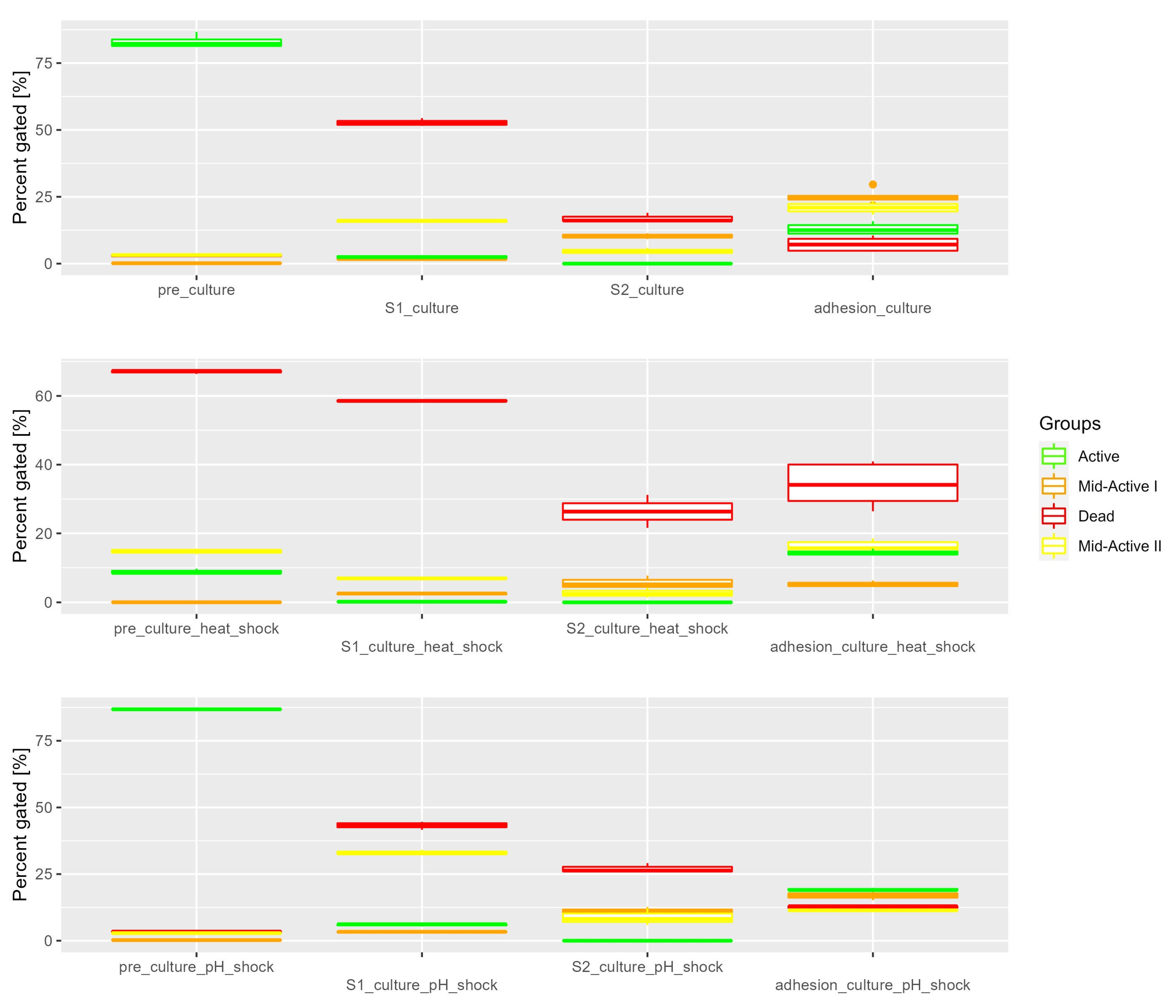
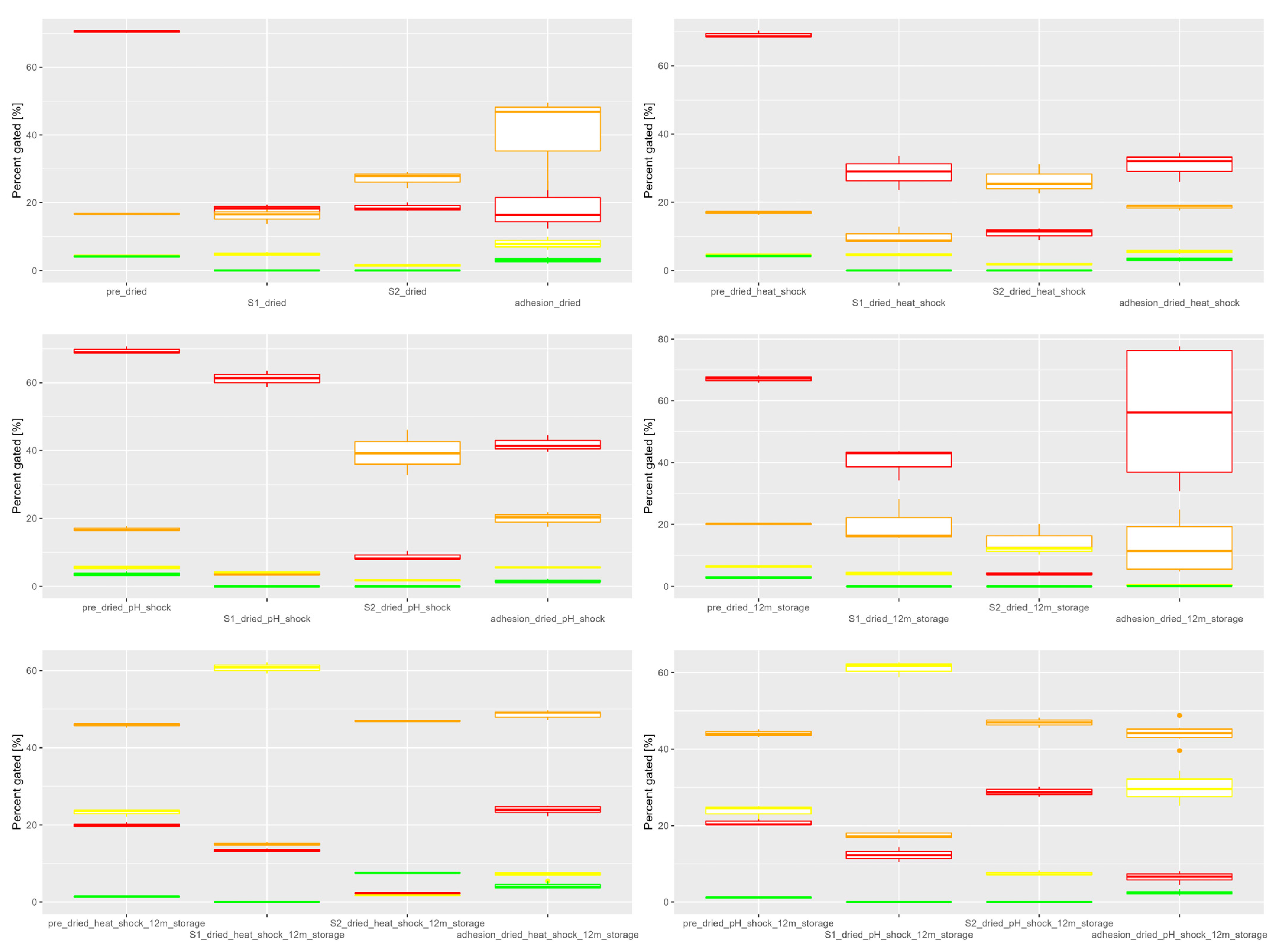
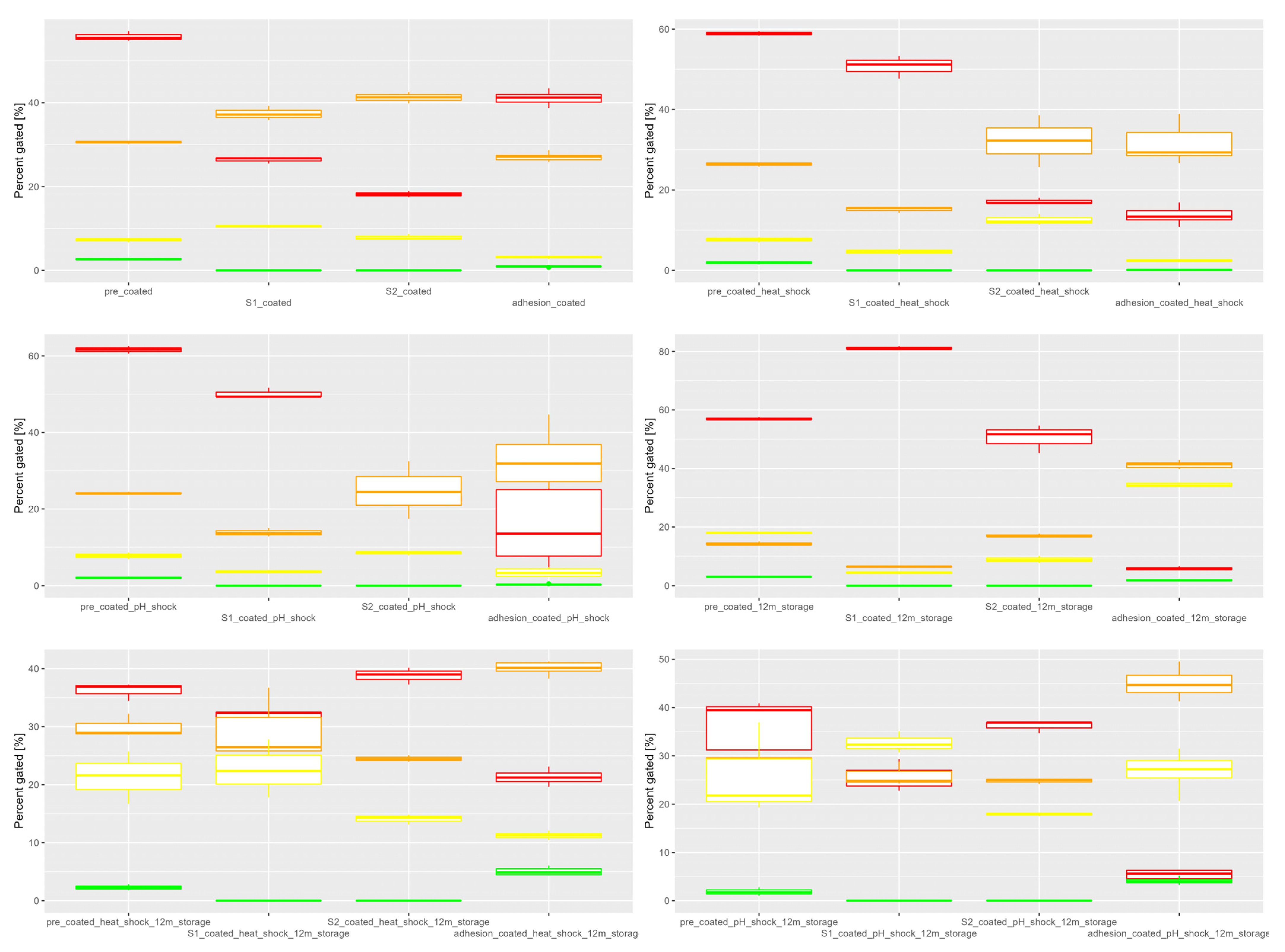
3.1. In Vitro Simulated Digestion

In this part of the research, different samples were treated with two-step gastrointestinal digestion and subjected to adhesion assay afterward. The following samples were used: free bacteria cells, cultured in optimal conditions, and treated with heat shock and pH stress (Figure 1); samples after fluid bed drying and coating were analyzed both fresh and after 12 months of storage after no treatment, heat shock, and pH shock (Figure 2 and Figure 3). Detailed results are available as supplementary materials (Table S1). Heat shock and pH shock were chosen as two types of the most common stress factors for lactic acid bacteria. These stresses are also present during drying (heat shock) and digestion (pH shock). Additionally, cross-protection systems can help the cells adapt to different types of stresses sharing similar resistance mechanisms [29]. The obtained results were used in the project of the technological process to represent the subpopulation spread for different variants. Four subpopulations of cells were determined using IFC, based on their metabolic activity (measured by RedoxSensor green) and cellular membrane damage (measured by propidium iodide). The active subpopulation includes the cells with high levels of metabolic activity and no cellular membrane damage, the mid-active I cells show low levels of both metabolic activity and cellular membrane damage, while the mid-active II cells show high levels of both; the dead cell subpopulation contains cells with no metabolic activity and a high degree of cellular membrane damage.
Free cells presented in Figure 1 show high levels of activity before treatment and after pH shock. The cells after heat shock already show much lower levels of active cells than two other variants. After in vitro digestion, all free cell samples show a significant decrease in active cell subpopulation, especially after the second stage, representing the small intestine. For both dried and coated samples (also after storage) the results show that the first stage of digestion (S1), representing the gastric conditions, led to a decrease in active cells in all samples. For free cells cultured in optimal conditions and after pH shock, the mid-active II subpopulation has increased after S1, which shows that the damage may not be permanent, as these cells have shown the ability to regenerate in optimal conditions [24]. A similar increase in mid-active II cells was also observed in dried cells treated with pH and heat shock after 12 months of storage. The second stage of digestion (S2), representing the small intestine, was more lethal for cells than S1, as also observed by Rodrigues et al. [30]. In all samples reduction of active cells was observed with a simultaneous decrease in overall observed cells, caused by cell breakdown. An increase in cellular debris that could not be assigned to any of the four subpopulations was observed in IFC as a result of the cell decomposition. Cells after coating were overall less affected by the GI conditions than uncoated cells. Similar results were observed for Lactobacillus salivarius NRRL B-30514 coated with rice protein–shellac composite [31]. Cells after S2 were subjected to adhesion assay, which showed an increase in active subpopulation, further proving that the mid-active cells were able to resume their activity after being placed in optimal conditions during adhesion assay. Mid-active cells would not be enumerated using plate cell counts, and with such methods, a reduction in the number of micro-organisms would be noticed. However, the preparations evaluated using our method show activity even after a year, thanks to the detection of VBNC cells. Samples after 12 months of storage show an increase in the proportion of cells with intermediate metabolic activity and small amounts of cellular damage. After such selection, the remaining cells are more resistant and tolerate the digestion conditions better than cells directly after drying.

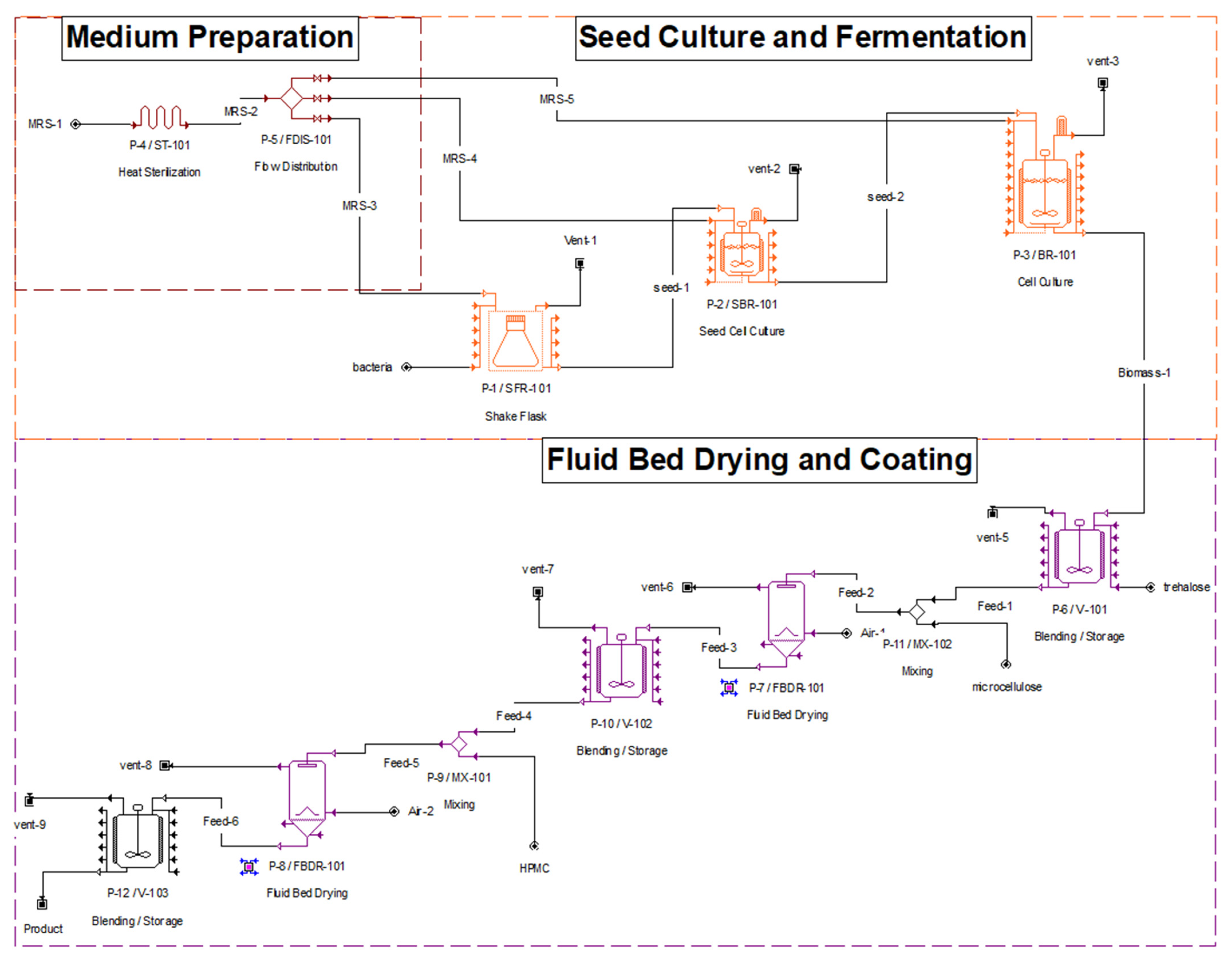
3.2. Process Design and Economic Analysis

The preliminary project of the technological process for dried probiotic production was prepared based on laboratory-scale experiments. Its process flow diagram shows the necessary steps and procedures in the variant, including fluid bed drying and coating (Figure 4). Overall, 6 project variants were prepared—for drying with no stress, with pH shock, and with heat shock, and similarly for coating with no stress, with pH shock, and with heat shock. Triggering bacterial stress adaptation was introduced as a way of improving the viability of probiotics during drying and storage [32]. All projects were divided into 3 sections: medium preparation, seed culture and fermentation, and fluid bed drying and coating. Medium preparation is a step where the MRS medium for culturing is mixed from base ingredients, heat sterilized, and distributed to corresponding culturing vessels. The second section contains culturing containers with increasing volumes—from 2 L shake flasks to 250 L bioreactor. In the final stage, the biomass is mixed with a protective substance (5% trehalose) and dried in a fluid bed dryer with crystalline microcellulose as a matrix. The protective effects of using trehalose come from its ability to replace the water in the intracellular macromolecules during drying [33]. In an additional step, the powder can then be coated with 2% HPMC. The final number of cells in the product was set at 108 cfu/g, as recommended by industry standards [34]. Economic calculations have been performed for the process, taking into consideration the cost of raw materials, waste treatment, energy consumption and heat transfer agents, equipment cost, and the direct fixed capital (DFC). DFC consists of total plant direct cost (TPDC), total plant indirect cost (TPIC), and contractors fee and contingency (CFC). The economic evaluation containing all of the mentioned costs has been summarized in Table 2. The cost of purchasing equipment needed for the process is summarized in Table 3. Additionally, the cost of installation of the equipment was calculated as 1.5× the purchase cost. To evaluate the economic profitability of certain technological treatments, such as coating and stresses during culturing, the production costs of obtaining preparations for 6 variants (dried, dried with heat shock, dried with pH shock, coated, coated with heat shock, and coated with pH shock) were compared. As proven by our previous research, the cells that are beneficial for the patient, namely, metabolically active and adherent to the Caco-2 cells, are present in two of the described subpopulations—active and mid-active II. Only those two groups were taken into consideration when assessing the cost of production of probiotic powder, based on the definition of probiotics, which describes them as live cells [35]. Using imaging flow cytometry the mid-active II cell subpopulation was determined to suit the description of VBNC [36] and, therefore, was also included in the project. The results of simulated in vitro digestion experiments were used, where the subpopulation composition of preparation after ingestion was assessed. Using those values, we determined the viable and beneficial (VB) cells, which are the cells that remain useful and confer health benefits to the host after technological processes and digestion. The individual cost of production of 1 kg of VB cells using different technological variants was calculated in Table 4. For the samples measured directly after drying the coated variant treated with heat shock provided the best value in terms of price per 1 kg of VB cells after digestion. As for the samples after 12 months of storage, the coated samples after pH shock showed the best value. The results for samples after adhesion were not included in the economic assessment of the project, since only the non-adherent cells present in the suspension after incubation could be measured using flow cytometry. Based on the results it can be concluded that the number of viable cells after drying is not a good indicator of the product quality. For better assessment, the number of cells surviving the technological process, storage, and digestion should be most important for the consumer. Economic evaluation based on the price of achieving preparation containing 1 kg of cells that meet those requirements should also be considered by the producer while planning the process.
Sensitivity analysis was provided for key assumptions in reference to the unit production cost and is presented in Figure 5. The baseline unit production price was 2.75 $/kg and the baseline number of batches was 107. Those values were then tested in the range of −20% to 20%. Results show that the main impact on product cost has the medium price, which confirms the need for seeking alternative carbon sources and media based on recycled waste materials. Additionally, the drying process has some impact, mainly because of the dryer purchase price, as well as the amount of trehalose and energy used. Trehalose cost, HPMC cost, and fermentation time both show the expected impact on the price. Little to no change was observed for coating, mainly since this process utilizes the same equipment as the drying step and, therefore, only the resource and energy demand are considered. Figure 6 and Figure 7 both show the probability distribution based on 10,000 trials for the unit production cost and annual number of batches, accordingly. The probability of achieving unit production prices under 3.01 was calculated at 91.88%. The probability of achieving the desired annual number of batches in the range of 102–112 was certain in 86.79%. The variability of the main product cost and number of batches per year has been demonstrated in Figure 8.

4. Conclusions

This paper explores the in vitro digestion and production of probiotic LAB, with a focus on the physiological and economic aspects of this process. Widely adopted enumeration methods (such as pour plate counts) should be supported by modern tools (IFC) for a better assessment of the quality of probiotics. The importance of cell subpopulations with intermediate metabolic activity (VBNC) cannot be understated, as they vastly contribute to the number of beneficial cells in the preparation after technological processes, storage, and digestion. Economic analysis was conducted to identify the key factors that influence the cost of production. These factors include the cost of the medium, drying time, and costs of trehalose (protective substance) and HPMC (coating substance). This study contributes to the holistic understanding of probiotics as a bridge between scientific innovation and consumer well-being. By synthesizing scientific insights with practical applications and financial considerations, this study provides a multidimensional perspective on the probiotics field. A comprehensive approach that encompasses scientific, technological, and economic dimensions is essential for advancing the probiotics field and obtaining more beneficial products.

Supplementary Materials

The following supporting information can be downloaded at: https://www.mdpi.com/article/10.3390/app132011541/s1, Table S1: Mean values with standard deviation for all subpopulations after in vitro digestion and adhesion tests. Means and SD were calculated based on 3 repetitions.

Author Contributions

Conceptualization, J.K.; methodology, J.K., A.O., W.J. and R.D.; software, J.K.; validation, J.K.; formal analysis, J.K.; investigation, J.K.; resources, J.K., A.O. and W.J.; data curation, J.K.; writing—original draft preparation, J.K.; writing—review and editing, J.K. and R.D.; visualization, J.K.; supervision, R.D. All authors have read and agreed to the published version of the manuscript.

Funding

This research received no external funding.

Institutional Review Board Statement

Not applicable.

Informed Consent Statement

Not applicable.

Data Availability Statement

Data are contained within the article.

Conflicts of Interest

The authors declare no conflict of interest.

References

  1. Probiotics in Food: Health and Nutritional Properties and Guidelines for Evaluation; Report of a Joint FAO/WHO Expert Consultation on Evaluation of Health and Nutritional Properties of Probiotics in Food Including Powder Milk with Live Lactic Acid Bacteria, Córdoba, Argentina, 1–4 October 2001; Report of a Joint FAO/WHO Working Group on Drafting Guidelines for the Evaluation of Probiotics in Food, London, Ontario, Canada, 30 April–1 May 2002; Joint FAO WHO Working Group on Drafting Guidelines for the Evaluation of Probiotics in Food (Ed.) FAO Food and Nutrition Paper; Food and Agriculture Organization of the United Nations [u.a]: Rome, Italy, 2006; ISBN 978-92-5-105513-7. [Google Scholar]
  2. Pereira, J.V.; Gamage, H.K.A.H.; Cain, A.K.; Hayes, E.; Paulsen, I.T.; Tetu, S.G. High-Throughput Viability Testing of Microbial Communities in a Probiotic Product Using Flow Cytometry. Appl. Microbiol. 2023, 3, 1068–1082. [Google Scholar] [CrossRef]
  3. George Kerry, R.; Patra, J.K.; Gouda, S.; Park, Y.; Shin, H.-S.; Das, G. Benefaction of Probiotics for Human Health: A Review. J. Food Drug Anal. 2018, 26, 927–939. [Google Scholar] [CrossRef] [PubMed]
  4. Broeckx, G.; Vandenheuvel, D.; Claes, I.J.J.; Lebeer, S.; Kiekens, F. Drying Techniques of Probiotic Bacteria as an Important Step towards the Development of Novel Pharmabiotics. Int. J. Pharm. 2016, 505, 303–318. [Google Scholar] [CrossRef]
  5. Fu, N.; Chen, X.D. Towards a Maximal Cell Survival in Convective Thermal Drying Processes. Food Res. Int. 2011, 44, 1127–1149. [Google Scholar] [CrossRef]
  6. Vass, P.; Domokos, A.; Pantea, E.; Szilágyi, B.; Molnár, M.; Záhonyi, P.; Nagy, B.; Nagy, Z.K. Processing of Thermosensitive Biological API from Suspension Using an Integrated Continuous Granulation—Drying—Milling Line into Powder Ready for Tableting. Dry. Technol. 2023, 41, 492–502. [Google Scholar] [CrossRef]
  7. Assari, M.R.; Basirat Tabrizi, H.; Najafpour, E. Energy and Exergy Analysis of Fluidized Bed Dryer Based on Two-Fluid Modeling. Int. J. Therm. Sci. 2013, 64, 213–219. [Google Scholar] [CrossRef]
  8. Mohideen Batcha, M.F.; Amirnordin, S.H.; Md Yudin, A.S. 4—Fluidized Bed Dryers. In Drying Technology in Food Processing; Jafari, S.M., Malekjani, N., Eds.; Woodhead Publishing: Sawston, UK, 2023; pp. 67–122. ISBN 978-0-12-819895-7. [Google Scholar]
  9. Hathi, Z.; Mettu, S.; Priya, A.; Athukoralalage, S.; Lam, T.N.; Choudhury, N.R.; Dutta, N.K.; El-Omar, E.M.; Gong, L.; Mohan, G.; et al. Methodological Advances and Challenges in Probiotic Bacteria Production: Ongoing Strategies and Future Perspectives. Biochem. Eng. J. 2021, 176, 108199. [Google Scholar] [CrossRef]
  10. Han, S.; Lu, Y.; Xie, J.; Fei, Y.; Zheng, G.; Wang, Z.; Liu, J.; Lv, L.; Ling, Z.; Berglund, B.; et al. Probiotic Gastrointestinal Transit and Colonization After Oral Administration: A Long Journey. Front. Cell. Infect. Microbiol. 2021, 11, 609722. [Google Scholar] [CrossRef]
  11. Qi, Y.; Huang, L.; Zeng, Y.; Li, W.; Zhou, D.; Xie, J.; Xie, J.; Tu, Q.; Deng, D.; Yin, J. Pediococcus Pentosaceus: Screening and Application as Probiotics in Food Processing. Front. Microbiol. 2021, 12, 762467. [Google Scholar] [CrossRef]
  12. Song, A.-X.; Mao, Y.-H.; Siu, K.-C.; Tai, W.C.S.; Wu, J.-Y. Protective Effects of Exopolysaccharide of a Medicinal Fungus on Probiotic Bacteria during Cold Storage and Simulated Gastrointestinal Conditions. Int. J. Biol. Macromol. 2019, 133, 957–963. [Google Scholar] [CrossRef] [PubMed]
  13. Wang, X.; Cao, Z.; Zhang, M.; Meng, L.; Ming, Z.; Liu, J. Bioinspired Oral Delivery of Gut Microbiota by Self-Coating with Biofilms. Sci. Adv. 2020, 6, eabb1952. [Google Scholar] [CrossRef] [PubMed]
  14. Lu, Y.; Han, S.; Zhang, S.; Wang, K.; Lv, L.; McClements, D.J.; Xiao, H.; Berglund, B.; Yao, M.; Li, L. The Role of Probiotic Exopolysaccharides in Adhesion to Mucin in Different Gastrointestinal Conditions. Curr. Res. Food Sci. 2022, 5, 581–589. [Google Scholar] [CrossRef] [PubMed]
  15. Chai, L.-N.; Wu, H.; Wang, X.-J.; He, L.-J.; Guo, C.-F. The Mechanism of Antimicrobial Activity of Conjugated Bile Acids against Lactic Acid Bacilli. Microorganisms 2023, 11, 1823. [Google Scholar] [CrossRef]
  16. Mukherjee, S.; Hooper, L.V. Antimicrobial Defense of the Intestine. Immunity 2015, 42, 28–39. [Google Scholar] [CrossRef]
  17. Long, S.L.; Gahan, C.G.M.; Joyce, S.A. Interactions between Gut Bacteria and Bile in Health and Disease. Mol. Asp. Med. 2017, 56, 54–65. [Google Scholar] [CrossRef]
  18. Zielińska, D.; Rzepkowska, A.; Radawska, A.; Zieliński, K. In Vitro Screening of Selected Probiotic Properties of Lactobacillus Strains Isolated from Traditional Fermented Cabbage and Cucumber. Curr. Microbiol. 2015, 70, 183–194. [Google Scholar] [CrossRef]
  19. Safeer Abbas, M.; Afzaal, M.; Saeed, F.; Asghar, A.; Jianfeng, L.; Ahmad, A.; Ullah, Q.; Elahi, S.; Ateeq, H.; Shah, Y.A.; et al. Probiotic Viability as Affected by Encapsulation Materials: Recent Updates and Perspectives. Int. J. Food Prop. 2023, 26, 1324–1350. [Google Scholar] [CrossRef]
  20. Yang, X.; Wang, C.; Wang, Q.; Zhang, Z.; Nie, W.; Shang, L. Armored Probiotics for Oral Delivery. Smart Med. 2023. [Google Scholar] [CrossRef]
  21. Buss, M.T.; Ramesh, P.; English, M.A.; Lee-Gosselin, A.; Shapiro, M.G. Spatial Control of Probiotic Bacteria in the Gastrointestinal Tract Assisted by Magnetic Particles. Adv. Mater. 2021, 33, 2007473. [Google Scholar] [CrossRef] [PubMed]
  22. Son, Y.S.; Kwon, M.; Son, N.; Kim, S.-K.; Son, M.-Y. Multilayer Coating with Red Ginseng Dietary Fiber Improves Intestinal Adhesion and Proliferation of Probiotics in Human Intestinal Epithelial Models. J. Microbiol. Biotechnol. 2023, 33, 1–10. [Google Scholar] [CrossRef] [PubMed]
  23. Rajam, R.; Subramanian, P. Encapsulation of Probiotics: Past, Present and Future. Beni-Suef Univ. J. Basic Appl. Sci. 2022, 11, 46. [Google Scholar] [CrossRef]
  24. Kiepś, J.; Juzwa, W.; Dembczyński, R. Imaging Flow Cytometry Demonstrates Physiological and Morphological Diversity within Treated Probiotic Bacteria Groups. Int. J. Mol. Sci. 2023, 24, 6841. [Google Scholar] [CrossRef]
  25. Anandharaj, M.; Rani, R.P.; Swain, M.R. Production of High-Quality Probiotics by Fermentation. In Microbial Functional Foods and Nutraceuticals; Gupta, V.K., Treichel, H., Shapaval, V., Antonio De Oliveira, L., Tuohy, M.G., Eds.; Wiley: Hoboken, NJ, USA, 2017; pp. 235–266. ISBN 978-1-119-04901-2. [Google Scholar]
  26. Fiore, W.; Arioli, S.; Guglielmetti, S. The Neglected Microbial Components of Commercial Probiotic Formulations. Microorganisms 2020, 8, 1177. [Google Scholar] [CrossRef]
  27. Minekus, M.; Alminger, M.; Alvito, P.; Ballance, S.; Bohn, T.; Bourlieu, C.; Carrière, F.; Boutrou, R.; Corredig, M.; Dupont, D.; et al. A Standardised Static in Vitro Digestion Method Suitable for Food—An International Consensus. Food Funct. 2014, 5, 1113–1124. [Google Scholar] [CrossRef] [PubMed]
  28. Kiepś, J.; Juzwa, W.; Olejnik, A.; Sip, A.; Tomaszewska-Gras, J.; Dembczyński, R. The Effects of Cellular Membrane Damage on the Long-Term Storage and Adhesion of Probiotic Bacteria in Caco-2 Cell Line. Nutrients 2023, 15, 3484. [Google Scholar] [CrossRef] [PubMed]
  29. Bucka-Kolendo, J.; Sokołowska, B. Lactic Acid Bacteria Stress Response to Preservation Processes in the Beverage and Juice Industry. Acta Biochim. Pol. 2017, 64, 459–464. [Google Scholar] [CrossRef]
  30. da Cruz Rodrigues, V.C.; da Silva, L.G.S.; Simabuco, F.M.; Venema, K.; Antunes, A.E.C. Survival, Metabolic Status and Cellular Morphology of Probiotics in Dairy Products and Dietary Supplement after Simulated Digestion. J. Funct. Foods 2019, 55, 126–134. [Google Scholar] [CrossRef]
  31. Wang, A.; Lin, J.; Zhong, Q. Enteric Rice Protein-Shellac Composite Coating to Enhance the Viability of Probiotic Lactobacillus Salivarius NRRL B-30514. Food Hydrocoll. 2021, 113, 106469. [Google Scholar] [CrossRef]
  32. Huang, S.; Vignolles, M.-L.; Chen, X.D.; Le Loir, Y.; Jan, G.; Schuck, P.; Jeantet, R. Spray Drying of Probiotics and Other Food-Grade Bacteria: A Review. Trends Food Sci. Technol. 2017, 63, 1–17. [Google Scholar] [CrossRef]
  33. Fu, N.; Huang, S.; Xiao, J.; Chen, X.D. Producing Powders Containing Active Dry Probiotics With the Aid of Spray Drying. Adv. Food Nutr. Res. 2018, 85, 211–262. [Google Scholar] [CrossRef] [PubMed]
  34. Haffner, F.B.; Diab, R.; Pasc, A.; Haffner, F.B.; Diab, R.; Pasc, A. Encapsulation of Probiotics: Insights into Academic and Industrial Approaches. Aimsmates 2016, 3, 114–136. [Google Scholar] [CrossRef]
  35. Hill, C.; Guarner, F.; Reid, G.; Gibson, G.R.; Merenstein, D.J.; Pot, B.; Morelli, L.; Canani, R.B.; Flint, H.J.; Salminen, S.; et al. The International Scientific Association for Probiotics and Prebiotics Consensus Statement on the Scope and Appropriate Use of the Term Probiotic. Nat. Rev. Gastroenterol. Hepatol. 2014, 11, 506–514. [Google Scholar] [CrossRef] [PubMed]
  36. Ayrapetyan, M.; Williams, T.; Oliver, J.D. Relationship between the Viable but Nonculturable State and Antibiotic Persister Cells. J. Bacteriol. 2018, 200, e00249-18. [Google Scholar] [CrossRef] [PubMed]
Disclaimer/Publisher’s Note: The statements, opinions, and data contained in all publications are solely those of the individual author(s) and contributor(s), and not of MDPI and/or the editor(s). MDPI and/or the editor(s) disclaim responsibility for any injury to people or property resulting from any ideas, methods, instructions, or products referred to in the content.
Figure 1. Subpopulation distribution in samples from bacterial cultures grown in optimal conditions, with heat shock and pH shock. Shown in four stages—pre-adhesion, after the first stage of in vitro digestion, after the second stage of in vitro digestion, and after adhesion.
Applsci 13 11541 g001
Figure 2. Subpopulation distribution in fluid bed-dried samples treated with heat shock, pH shock, and untreated. Shown in four stages—pre-adhesion, after the first stage of in vitro digestion, after the second stage of in vitro digestion, and after adhesion.
Applsci 13 11541 g002
Figure 3. Subpopulation distribution in fluid bed-dried and coated samples treated with heat shock, pH shock, and untreated. Shown in four stages—pre-adhesion, after the first stage of in vitro digestion, after the second stage of in vitro digestion, and after adhesion.
Applsci 13 11541 g003
Figure 4. Process flow diagram for fluid bed drying and coating of probiotics.
Applsci 13 11541 g004
Figure 5. Tornado plot depicting the impact of variables on unit production cost.
Applsci 13 11541 g005
Figure 6. Probability distribution of the unit production cost, based on 10,000 trials.
Applsci 13 11541 g006
Figure 7. Probability distribution of the annual number of batches, based on 10,000 trials.
Applsci 13 11541 g007
Figure 8. Sensitivity of the main product cost (A) and the number of batches per year (B) to deviation in variables.
Applsci 13 11541 g008
Table 1. Simulated digestion fluids composition.
ConstituentSGFSIF
pepsin2000 U/mL-
pancreatin-100 U/mL
(based on trypsin)
bile-10 mM
CaCl20.075 mM0.3 mM
pH37
Table 2. Economic evaluation summary.
ParameterUnit
DFC$1,584,000
TPDC$861,000
TPIC$516,000
CFC$207,000
Operating cost$100,000
Batch sizekg326.91
Cost basis annualkg/year34,979
Gross margin%71.98
Return on investment (ROI)%11.51
Payback time (PBT)year4.33
Net present value (NPV at 7%)$909,000
Revenues (per year)$350,000
Table 3. Equipment and its purchase cost summary.
EquipmentSizePurchase cost (PC) ($)
Fluid bed dryer415.97 L107,000
Bioreactor296.11 L150,000
Seed bioreactor18.34 L29,950
Heat sterilizer66.17 L/h rated
by throughput
30,000
Blending tank308.29 L11,400
Shake flask rack2 L1000
Table 4. Economic evaluation of different variants.
VariantUnit
Production
Cost ($/kg)
Production Cost
($/1 kg VB Cells Pre-Digestion)
Production Cost
($/1 kg VB Cells after Digestion)
Dried A2.5930.58 cde ± 1.05173.83 a ± 32.59
Dried, 12 months B2.5928.09 cde ± 2.4522.19 cde ± 2.11
Dried heat shock A2.5929.40 cde ± 0.13134.20 b ± 12.05
Dried heat shock 12 months B2.5910.52 e ± 0.3627.91 cde ± 0.98
Dried pH shock A2.6329.35 cde ± 1.99144.51 b ± 10.82
Dried pH shock 12 months B2.6310.58 e ± 0.3735.11 cd ± 2.87
Coated A3.0130.31 cde ± 1.7338.18 c ± 3.36
Coated 12 months B3.0114.36 cde ± 0.2734.13 cde ± 4.06
Coated heat shock A3.0131.45 cde ± 2.7724.03 cde ± 2.48
Coated heat shock 12 months B3.0112.73 de ± 1.8721.38 cde ± 1.41
Coated pH shock A3.0531.00 cde ± 1.7736.22 cd ± 2.44
Coated pH shock 12 months B3.0510.95 e ± 1.2417.02 de ± 0.35
A measured directly after drying/coating. B measured after 12 months of storage. Results are mean values of three repetitions ± standard deviation. Values sharing the same lowercase letter are not significantly different (p ≤ 0.05).
Disclaimer/Publisher’s Note: The statements, opinions and data contained in all publications are solely those of the individual author(s) and contributor(s) and not of MDPI and/or the editor(s). MDPI and/or the editor(s) disclaim responsibility for any injury to people or property resulting from any ideas, methods, instructions or products referred to in the content.

