Article

Phytochemical Profile, Antilipase, Hemoglobin Antiglycation, Antihyperglycemic, and Anti-Inflammatory Activities of Solanum elaeagnifolium Cav.

1
Laboratories of Natural Substances, Pharmacology, Environment, Modeling, Health and Quality of Life (SNAMOPEQ), Faculty of Sciences, Sidi Mohamed Ben Abdellah University, Fez 30000, Morocco
2
Laboratory of Bioresources, Biotechnology, Ethnopharmacology, and Health, Faculty of Sciences, Mohammed First University, Oujda 60000, Morocco
3
Laboratory of Applied Analytical Chemistry, Materials and Environment, Department of Chemistry, Faculty of Sciences, Mohammed First University, Oujda 60000, Morocco
4
Department of Pharmacognosy, College of Pharmacy, King Saud University, Riyadh 11451, Saudi Arabia
5
Department of Agricultural Engineering, College of Food and Agriculture Sciences, King Saud University, Riyadh 11451, Saudi Arabia
6
Laboratory of Biotechnology, Environment, Agri-Food and Health, Faculty of Sciences Dhar El Mahraz, Sidi Mohamed Ben Abdellah University, Fez 30000, Morocco
7
Team of Functional and Pathological Biology, Laboratory of Biological Engineering, Faculty of Sciences and Technology Beni Mellal, University Sultan Moulay Slimane, Beni Mellal 23000, Morocco
8
Laboratoire TBC, Laboratory of Pharmacology, Pharmacokinetics and Clinical Pharmacy, Faculty of Pharmacy, University of Lille, 3. Rue du Professeur Laguesse, 59000 Lille, France
*
Authors to whom correspondence should be addressed.
Appl. Sci. 2023, 13(20), 11519; https://doi.org/10.3390/app132011519
Submission received: 27 August 2023 / Revised: 21 September 2023 / Accepted: 28 September 2023 / Published: 20 October 2023
(This article belongs to the Special Issue Natural Products: Sources and Applications)

Abstract

:
In the present investigation, the phenolic compounds of Solanum elaeagnifolium were identified, and the plant’s anti-lipase and anti-glycation effects on hemoglobin were discovered through in vitro experiments, as well as its short-term antihyperglycemic and anti-inflammatory effects. The chemical compound composition was detected using HPLC-DAD, the anti-lipase activity was tested in vitro using 4-nitrophenyl butyrate as a substrate, and the antiglycation activity of the plant extracts was also tested in vitro using a haemoglobin model. The antihyperglycemic effect was determined by inhibiting pancreatic α-amylase and α-glycosidase activity and performing an in vivo glucose tolerance test on normal rats, and the anti-inflammatory activity was determined by inducing paw inflammation with carrageenan. In both the SEFR (fruit) and SEFE (leaf) extracts, chromatographic analysis revealed the presence of quercetin 3-O-β-D-glucoside, rutin, and quercetin. SEFR inhibited the pancreatic lipase enzyme more effectively, with an IC50 of 0.106 ± 0.00 mg/mL. S. elaeagnifolium extracts demonstrated significant antiglycation activity, with 3.990 ± 0.23 mg/mL of SEFE and 3.997 ± 0.14 mg/mL of SEFR. When compared to positive and negative controls, plant extracts had very significant anti-diabetic and anti-inflammatory effects. The findings in this study and previous research on this plant encourage us to investigate other pharmacological activities of this plant besides its duiretic, cictrisant, and anti-ulcer activity.

1. Introduction

Diabetes is a persistent illness that causes a disruption in glucose metabolism, resulting in persistently high blood sugar levels, known as hyperglycemia. This disease is classified into two types: Type 1 diabetes arising from the deterioration of pancreatic beta cells, which are responsible for producing insulin, and type 2 diabetes, in which cells become insulin-resistant [1]. Glycemic regulation is a complex process involving multiple organs and hormones. Insulin, which is secreted by the pancreas, is critical in facilitating glucose absorption by cells for energy use or glycogen storage. Concurrently, glucagon, which is also produced by the pancreas, raises blood sugar levels by encouraging the liver to release glucose into the bloodstream [2,3].
Inflammation is a critical and complex biological response elicited by the body’s immune system in order to protect itself from potentially harmful stimuli such as pathogens, injury, or damaged cells [4]. The procedure entails a series of co-ordinated events aimed at removing the source of the injury or infection and initiating tissue repair. The secretion of inflammatory signaling molecules such as cytokines, chemokines, and prostaglandins, which promote vasodilation and increase vascular permeability, is typical of the mechanisms of inflammation [5].
The Solanum genus, which includes various plant species, has attracted considerable interest due to its potential therapeutic effects against diabetes, obesity, and inflammation. Numerous studies have highlighted the bioactive compounds present in Solanum plants, which demonstrate anti-diabetic properties by promoting insulin sensitivity and reducing blood glucose levels [6,7]. Research has indicated that certain compounds in Solanum plants can inhibit fat accumulation, promote lipid metabolism, and suppress one’s appetite, making them valuable candidates in the fight against obesity [8,9,10]. Studies have revealed that extracts from certain Solanum species, such as Solanum nigrum, exhibit significant antioxidant properties due to their content of flavonoids and polyphenols [11]. These compounds help neutralize free radicals and protect cells against oxidative stress, which is implicated in aging and the development of certain chronic diseases [12]. Furthermore, some Solanum species have demonstrated potential anti-inflammatory activities. For instance, studies on extracts from Solanum lycopersicum have shown the inhibition of inflammation by suppressing pro-inflammatory cytokines in cellular models [13].
Solanum elaeagnifolium, also known as silverleaf nightshade, is a perennial shrub in the Solanaceae family and solanum genre. This plant species is native to North America, with its geographic range primarily encompassing the southwestern United States and northern Mexico [14]. It thrives in a wide range of habitats, including arid and semi-arid regions, disturbed areas, roadsides, and agricultural lands. Its ability to adapt to a variety of climates and soil conditions has contributed to its widespread distribution across these regions [15,16].
In terms of climate, S. elaeagnifolium exhibits remarkable tolerance to harsh environmental conditions. It can survive in areas with high temperatures, low precipitation, and high salinity levels, making it ideal for arid and drought-prone environments [17]. S. elaeagnifolium has a variety of beneficial biological activities, including analgesic, anti-inflammatory, antioxidant, insecticidal, molluscicidal, larvicidal, antimicrobial, chemopreventive, and antitumor properties [15,16,18,19,20,21,22,23,24]. These various activities can be attributed to the presence of key compounds such as quercetin, gallic acid, kaempferol, and naringenin, which play an important role in mediating these effects [15,16]. The chemical composition of the plant is rich and diverse, which contributes to its therapeutic potential and makes it a valuable subject of interest for further research in a variety of fields, including medicine and agriculture. In this study, we begin by identifying the phenolic compounds present in Solanum elaeagnifolium. Subsequently, we evaluate the anti-lipase and anti-glycation effects on hemoglobin. Finally, we examine the short-term hypoglycemic and anti-inflammatory effects of this plant.

2. Materials and Methods

2.1. Plant Materiel

S. elaeagnifolium is an invasive species that falls under the Solanaceae family and the Solanum genus. The plant specimens were collected from the city of Fes, with leaf harvesting taking place in December 2021 and mature fruit harvesting occurring in April 2022 (Table 1). Both the leaves and fruits underwent a drying process, with a portion dried outdoors and the rest in shaded conditions.

2.2. Preparation of Plant Extracts

Hydro-ethanolic extracts of S. elaeagnifolium leaves and fruits were prepared through a maceration process. In brief, 50 g of powdered plant material was combined with 500 mL of 70% ethanol. The resulting mixture underwent maceration for 72 h at ambient temperature, followed by filtration using Whatman paper.

2.3. HPLC-DAD Analysis

Extracts from leaves and fruits were prepared at a concentration of 50 mg/mL and passed through microfilters with a pore size of 0.45 μm. The phenolic components were analyzed using high-performance liquid chromatography (HPLC) coupled to a UV detector operating within the range of 210–400 nm. The separation was performed by an Alliance ew2695 C18 column (250 × 4 mm, 5 µm) in the normal phase, using a gradient solvent system of A (water/0.5% acetic acid) and B (methanol), with a flow rate of 1 mL/min. The detector used is a UV-visible diode detector (PDA Waters 2996). The chosen wavelength depends on the extract’s nature and the targeted substances. The reading was at 254, 280, 320, and 340 nm, and the identification of compounds was performed at 280 nm. The contents of phenolic compounds are determined from the calibration curves for each compound (1 mg/mL). All calibration curves showed high linearity (r2 > 0.99). The identification and quantification of the compounds are performed by comparison of their relative retention times with those of the standards. The results were presented in mg/100 g per extract [15,25,26].

2.4. In Vitro Activities

2.4.1. Lipase Inhibitory Activity

Lipase inhibitory activity was measured using a modified Hu et al. method. In a brief, 150 μL of orlistat or extract were combined with 500 μL of lipase enzyme (2 U) dissolved in Tris-HCl buffer (1 mM, pH 8). After 30 min at 37 °C, 450 μL of 1 mM 4-nitrophenyl butyrate substrate was added, followed by another 30 min at 37 °C [27]. The value of absorbance had been determined at 405 nm. The percentage lipase inhibition was calculated using the following Formula (1):
Inhnibition   ( % ) = [ ( ac acb ) ( as asb ) ] ac acb × 100

2.4.2. Antiglycation Activity

The antiglycation activity of S. elaeagnifolium has been assessed using the method developed by Elrherabi et al. In test tubes, different doses of the extract (100, 200, 400, 800, and then 1000 μg/mL) have been mixed with 5 μL of gentamycin and 1 ml of hemoglobin solution. After 20 min, we added 1 mL of 20 mM glucose in 10 mM phosphate buffer with pH 4.7 to start the reaction. Subsequently, the tubes were left to incubate at ambient temperature for a period of 72 h. Finally, the percentage of glycosylation of haemoglobin was calculated using a spectrophotometer set to 443 nm. Using the same protocol described above, gallic acid was used as a standard at various concentrations (0.1, 0.2, 0.4, 0.8, and 1 mg/mL) [28].

2.5. Pharmacological Activities

2.5.1. Animals

Male Wistar rats measuring between 125 and 150 g were obtained from the Biology Department of the Dhar El-Mahraz Faculty of Science at Sidi Mohamed Ben Abdellah University in Fez, Morocco. The rats were placed in temperature, humidity, and treatment procedures that strictly adhered to the European Community standard for the protection of animals used for experimental purposes (Council Directive of 24 November 1986) [29]. The doses were selected depending on previous research; indeed, extract from the aerial parts of S. elaeagnifolium was found to be virtually non-toxic when administered orally to mice, with an LD50 value greater than 500 mg/kg body weight (BW). This allowed us to choose safe doses to test for anti-diabetic and anti-inflammatory effects in rats [19,23]. The anti-diabetic activity tests used doses ranging from 150 mg/kg to 300 mg/kg body weight. The anti-inflammatory activity test used doses ranging from 200 mg/kg to 400 mg/kg body weight.

2.5.2. Oral Glucose Tolerance Test (OGTT)

The researchers conducted an oral glucose tolerance test to evaluate the in vivo antihyperglycemic (postprandial glucose) effects [30,31]. Normal rats were divided into six groups (n = 5): a control group receiving distilled water (10 mL/kg), and test groups comprising normal rats administered two doses of 150 mg/kg and 300 mg/kg of SEFR extract, two doses of SEFE extract, and glibenclamide (2 mg/kg). Initially, blood glucose levels were measured at t0, just before administering the test products (distilled water, extract, or glibenclamide). After 30 min, another blood glucose measurement was taken, following which the rats were given an overload of D-glucose (2 g/kg). Subsequently, blood glucose levels were measured every half-hour until 120 min.

2.5.3. Inhibition of Carbohydrate Hydrolase Enzymes, In Vivo

Pancreatic α-Amylase

This study utilized healthy Wistar rats that underwent a 16 h fasting period. The rats were divided into six groups (n = 5 each). The control group received only distilled water (10 mL/kg orally). The positive group received Acarbose (10 mg/kg orally). Four treated groups received either SEFR or SEFE at two different doses—150 mg/kg orally and 300 mg/kg orally. After administering the solutions, the rats from different groups were orally loaded with starch (2 g/kg) and their blood glucose levels were measured at 0, 30, 60, and 120 min using the glucose oxidase method [30,32].

Pancreatic α-Glucosidase

In this study, healthy Wistar rats were used, and they underwent a 16 h fasting period. The rats were divided into six groups (with five rats in each group). The control group received only distilled water (10 mL/kg orally), while the positive group received Acarbose (10 mg/kg orally). Additionally, there were four treated groups, which received either SEFR or SEFE at two different doses—150 mg/kg orally and 300 mg/kg orally. After administering the solutions, the rats in all groups were orally given sucrose (2 g/kg), and their blood glucose levels were measured at 0, 30, 60, and 120 min using the glucose oxidase method [3].

2.5.4. Anti-Inflammatory Potential

The anti-inflammatory potential of each extract was investigated using 1% carrageenan as an edema-inducing drug, as modified by Mssilou et al. [33]. Six groups of five rats each were formed at random. The first group was given physiological water (NaCl 0.9%) as a negative control. SEFE was given to groups 2 and 3 at doses of 200 mg/kg and 400 mg/kg, respectively. SEFR was given to groups 4 and 5 at doses of 200 mg/kg and 400 mg/kg, respectively. Group 6 was given indomethacin (10 mg/kg) orally as a control. After one hour of treatment, inflammation was induced by carrageenan. Footpad circumferences were measured in millimeters (mm) using calipers before and after 3 h, 4 h, 5 h, and 6 h of carrageenan injection (Sigma Aldrich, Ref. 3B-C1804, Barcelona, Spain). Each treatment’s inhibitory activity was determined as follows (2):
Inhibition   ( % ) = [ 1 ( Dc D 0 ) sample ( Dc D 0 ) control ] × 100
where Dc = the mean of paw volume following carrageenan administration; and D0 = the mean of paw volume before the injection.

2.6. Statistical Investigation

Tukey’s test was used to identify significant differences between treatments and controls, and the results were subjected to analysis of variance (ANOVA). The results were presented as mean ± s.e.m.

3. Results and Discussion

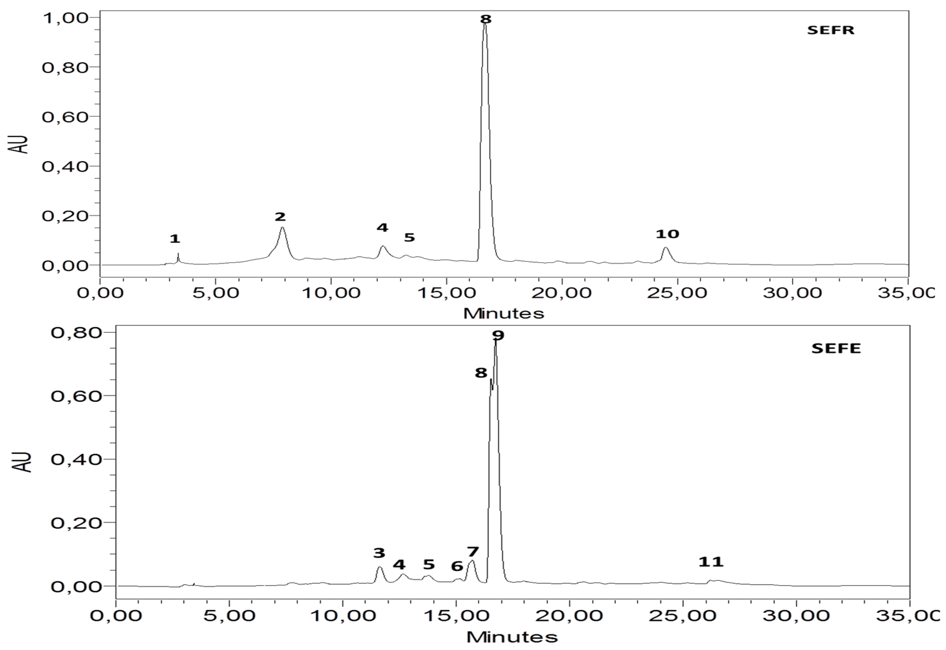
3.1. HPLC-DAD Analysis

The analysis of the S. elaeagnifolium extracts revealed that they are high in bioactive molecules such as flavonoids, polyphenols, and phenolic acids; Table 2 and Figure 1 show the results of the compound identification in the two extracts studied. Fruit extract (SEFR) revealed the presence of catechin, vanillin, quercetin 3-O-β-D-glucoside, rutin, quercetin, and kaempferol. Leaf extract (SEFE) showed an abundance of naringin, quercetin 3-O-β-D-glucoside, rutin, cinnamic acid, p-coumaric acid, quercetin, trans-3-hydroxycinnamic acid, and flavones. The chromatogram analysis provides a difference in the chemical composition of each of the extracts, with the same molecules present in different concentrations depending on the part of the plant. Research has revealed the presence of compounds similar to those discovered in our study [15,16,24]. An analysis of the results revealed the presence of quercetin in the two plant extracts at a very high concentration of 429.02 ± 0.97 mg/100 g for SEFR and 114.15 ± 0.34 mg/100 g for SEFE. Quercitin is a biactive molecule with a variety of pharmacological activities. Because of the inclusion of a phenolic hydroxyl group, it is considered to be a very strong antioxidant, and this compound has anti-inflammatory, antimicrobial, antitumor, and cardioprotective properties [34,35].

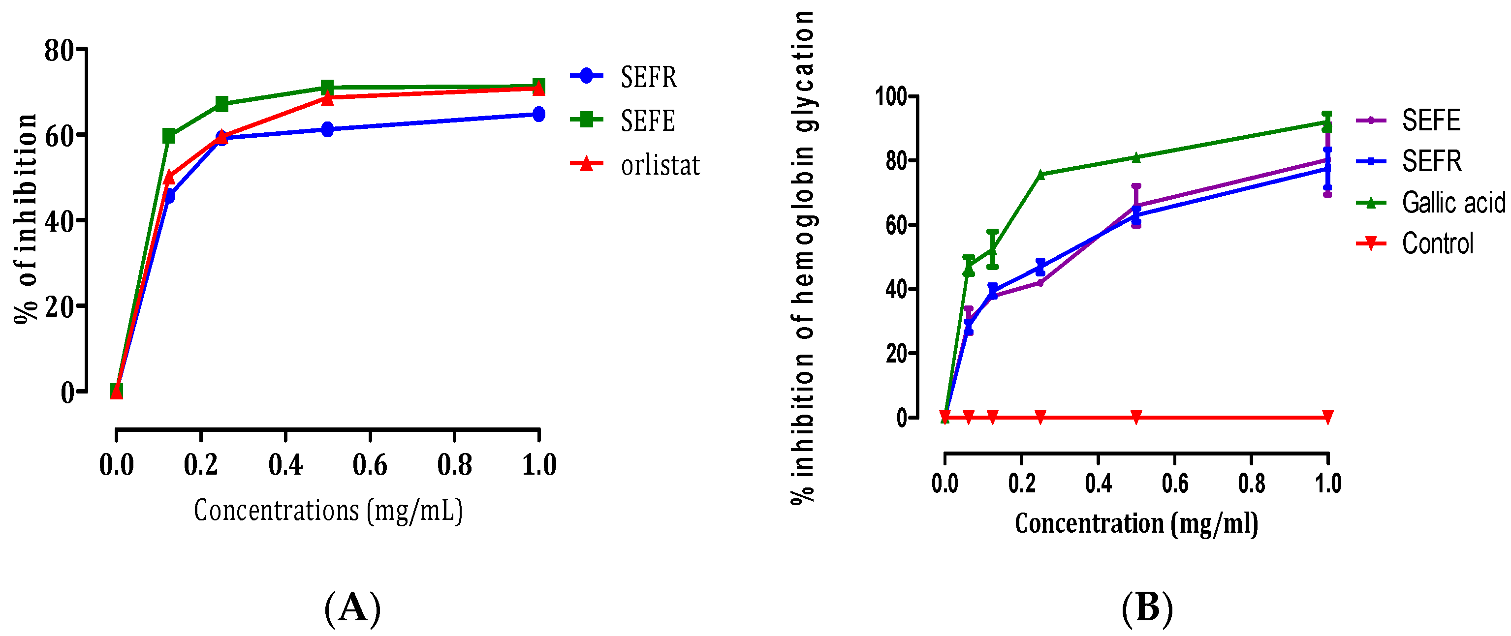
3.2. Lipase Inhibitory and Hemoglobin Antiglycation Activities

The anti-lipase and hemoglobin antiglycation activities of extracts from S. elaeagnifolium are presented in Table 3. The inhibitory lipase enzyme of the extracts was evaluated at various concentrations (Figure 2A). The reference compound, Orlistat (with an IC50 of 0.128 ± 0.003 mg/mL), demonstrated stronger inhibitory activity compared to SEFE (with an IC50 of 0.167 ± 0.0006 mg/mL). Conversely, SEFR exhibited a higher inhibitory impact against the pancreatic lipase enzyme in comparison to both SEFE and Orlistat, boasting an IC50 of 0.106 ± 0.0008 mg/mL (p < 0.01). These results are consistent with the results of Al-Hamaideh et al. who tested the anti-lipase activity of S. elaeagnifolium berries at two stages of ripening; they found that the anti-lipase activity of ripe berries is 0.164 ± 0.023 mg/mL [9].
The in vitro haemoglobin glycosylation inhibition assessment indicated a significant decrease in glycosylation as a function of concentration (Figure 2B). S. elaeagnifolium extracts demonstrated significant antiglycation activity, with 3.990 ± 0.236 mg/mL of SEFE and 3.997 ± 0.140 mg/mL of SEFR. Gallic acid also demonstrated significant antiglycation activity, with a value of 3.008 ± 0.030 mg/mL. Solanum nigrum, which is characterized by the presence of molecules with anti-glycation properties such as solamargine and solasonine [22], which are also found in our plant, is another species with an anti-glycation effect. The extract of S. nigrum inhibited the formation of advanced glycation end products (AGEs) [36].

3.3. Pharmacological Activities

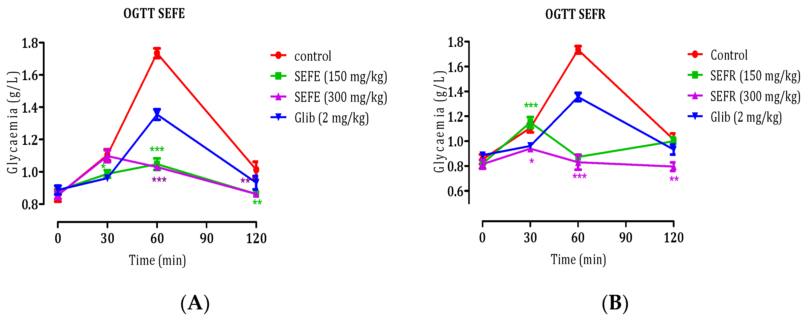
3.3.1. Oral Glucose Tolerance Test (OGTT)

The impact of the two extracts from S. elaeagnifolium on normal rats subjected to oral glucose loading is depicted in Figure 3. Prior to administering their respective treatments, there were no notable variations in fasting blood glucose levels across the groups. The outcomes of an oral glucose tolerance test conducted on healthy rats demonstrate that the administration of 2 g/kg of glucose resulted in a baseline blood sugar level of 0.92 g/L for rats in the control group, which received only distilled water. The levels of postprandial hyperglycemia reached a peak at 1.76 ± 0.03 g/L after 60 min, and subsequently decreased to an average of 1.02 ± 0.08 g/L after 120 min. While a substance known as SEFE was administered at a dose of 150 mg/kg, it significantly suppressed postprandial hyperglycemia in comparison to the control group’s rats. This suppression was observed at multiple time points: after 30 min (p < 0.01) with a reading of 0.98 ± 0.02 g/L, after 60 min (p < 0.001) with a reading of 1.04 ± 0.03 g/L, and at 120 min (p < 0.01) with a reading of 0.85 ± 0.01 g/L. In contrast, when SEFE was administered at a higher dose of 300 mg/kg, it also significantly suppressed postprandial hyperglycemia in comparison to the control group’s rats. This suppression was observed at multiple time points: after 60 min (p < 0.001) with a reading of 1.03 ± 0.009 g/L, and at 120 min (p < 0.01) with a reading of 0.86 ± 0.01 g/L. The outcomes of an oral glucose tolerance test conducted on healthy rats demonstrate that the administration of 2 g/kg of glucose resulted in a baseline blood sugar level of 0.92 g/L for rats in the control group, which received only distilled water. The levels of postprandial hyperglycemia reached a peak at 1.76 ± 0.03 g/L after 60 min, and subsequently decreased to an average of 1.02 ± 0.08 g/L after 120 min.
While a substance known as SEFR was administered at a dose of 150 mg/kg, it significantly suppressed postprandial hyperglycemia in comparison to the control group’s rats. This suppression was observed after 30 min (p < 0.001) with a reading of 1.15 ± 0.04 g/L. In contrast, when SEFR was administered at a higher dose of 300 mg/kg, it also significantly suppressed postprandial hyperglycemia in comparison to the control group’s rats. This suppression was observed at multiple time points: after 30 min (p < 0.05) with a reading of 0.94 ± 0.02 g/L, at 60 min (p < 0.001) with a reading of 0.83 ± 0.06 g/L, and at 120 min (p < 0.01) with 0.79 ± 0.03 g/L. The SEFR extract at a dose of 300 mg/kg appears to exhibit the most significant antihyperglycemic potential among the tested extracts and glibenclamide. This is evident from its more pronounced suppression of postprandial hyperglycemia at multiple time points and lower blood glucose levels compared to the other tested extracts and doses. Oral glucose loading causes diabetes mellitus by gradually raising blood glucose levels without harming pancreatic cells. As a result, whether an extract reduced blood glucose levels in rats after an oral glucose load indicates its therapeutic potential in diabetes management [37].

3.3.2. Inhibition of Carbohydrate Hydrolase Enzymes, In Vivo

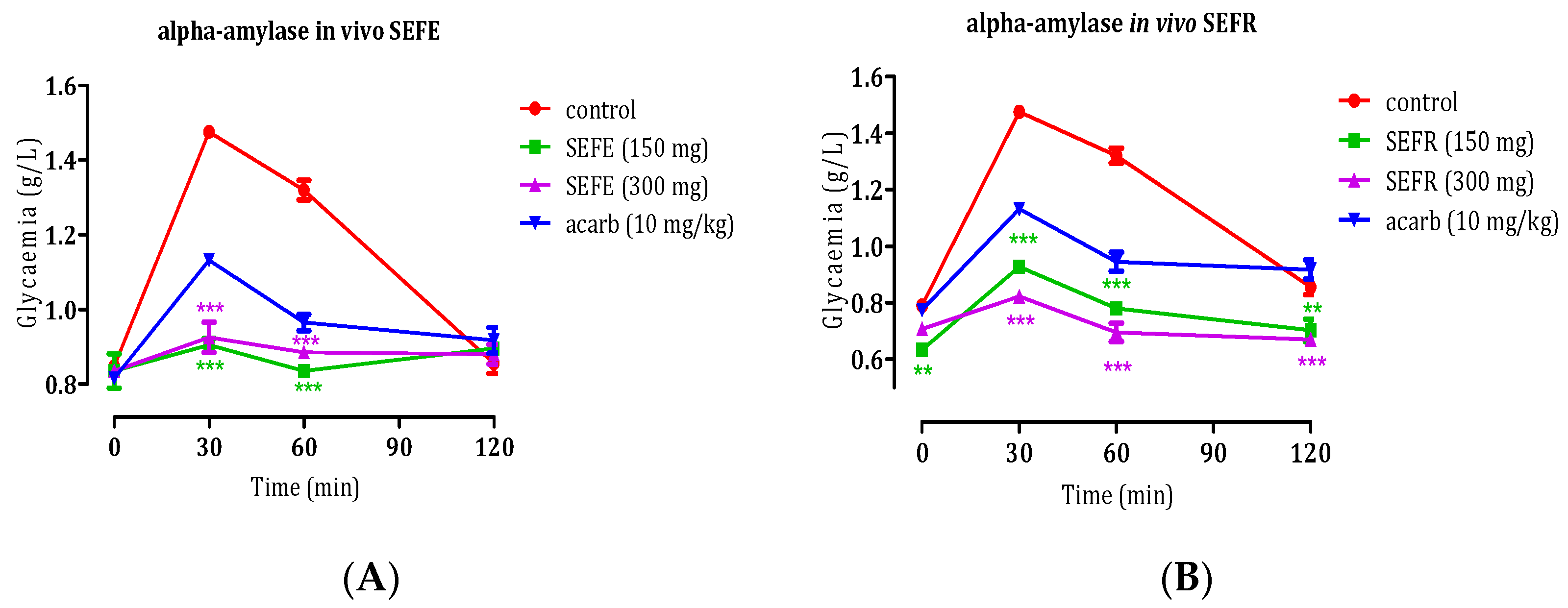
In Vivo Anti-α-Amylase Activity of S. elaeagnifolium Leaves and Fruits

In the pursuit of treating diabetes mellitus, one of the targeted pathways involves inhibiting sugar-digesting enzymes, specifically pancreatic α-amylase. This enzyme plays a crucial role in the initial digestion of disaccharides, which undergo a transformation process comprising both intraluminal and straightforward mechanisms, ultimately resulting in the formation of naturally occurring oligosaccharides [38].
After studying the anti-α-amylase activity of S. elaeagnifolium extracts in vitro, and to confirm the inhibitory activity of S. elaeagnifolium SEFE and SEFR extracts on the pancreatic α-amylase enzyme, the test was performed in vivo using the protocol defined by Elrherabi et al. [39], and the results are shown in Figure 4. After administering 2 g/kg of starch, there was a notable increase in blood glucose levels, peaking at 1.49 ± 0.02 g/L. This peak gradually subsided to 1.32 ± 0.07 g/L at the 60 min mark. However, upon the administration of SEFE at a dose of 150 mg/kg, a substantial reduction in postprandial blood glucose levels was observed. The reduction was statistically significant at both 30 min (p < 0.001) and 60 min (p < 0.001), with recorded values of 0.9 ± 0.01 g/L and 0.83 ± 0.002 g/L, respectively, in comparison to the control group. Interestingly, there was no significant difference in blood glucose levels at the 120 min mark. In contrast, when SEFE was administered at a higher dose of 300 mg/kg, it also significantly suppressed postprandial hyperglycemia in comparison to the control group’s rats. This suppression was observed at multiple time points: after 30 min (p < 0.001) with a reading of 0.9 ± 0.04 g/L, and at 60 min (p < 0.011) with a reading of 0.88 ± 0.007 g/L.
After administering 2 g/kg of starch, there was a notable increase in blood glucose levels, peaking at 1.49 ± 0.02 g/L. This peak gradually subsided to 1.32 ± 0.07 g/L at the 60 min mark. However, upon the administration of SEFR at a dose of 150 mg/kg, a substantial reduction in postprandial blood glucose levels was observed. The reduction was statistically significant at 30 min (p < 0.001), 60 min (p < 0.001), and 120 min (p < 0.01), with recorded values of 0.92 ± 0.02 g/L, 0.78 ± 0.02 g/L, and 0.7 ± 0.03 g/L respectively, in comparison to the control group. Interestingly, there was no significant difference in blood glucose levels at the 120 min mark. In contrast, when SEFE was administered at a higher dose of 300 mg/kg, it also significantly suppressed postprandial hyperglycemia in comparison to the control group’s rats. This suppression was observed at multiple time points: after 30 min (p < 0.001) with a reading of 0.82 ± 0.009 g/L, at 60 min (p < 0.001) with a reading of 0.69 ± 0.03 g/L, and at 120 min (p < 0.001) with 0.76 ± 0.01 g/L. Furthermore, the introduction of 10 mg/kg of acarbose significantly hindered starch-induced hyperglycemia. The inhibition was significant at both 30 min (p < 0.001) and 60 min (p < 0.001), and also showed significance at 120 min (p < 0.05). The measured blood glucose values were 1.11 ± 0.06 g/L, 0.95 ± 0.004 g/L, and 0.91 ± 0.03 g/L for the respective time points, compared to the control group. The SEFR extract, at a dose of 300 mg/kg, consistently demonstrated the most significant and consistent antihyperglycemic activity across multiple time points, demonstrating that it has the highest antihyperglycemic potency of the tested extracts. Limiting the activity of carbohydrate digestive enzymes in the intestine is one strategy used to reduce postprandial hyperglycemia [40]. α-amylase inhibitors prevent carbohydrate digestion and absorption in the intestine, resulting in hypoglycemia [41].

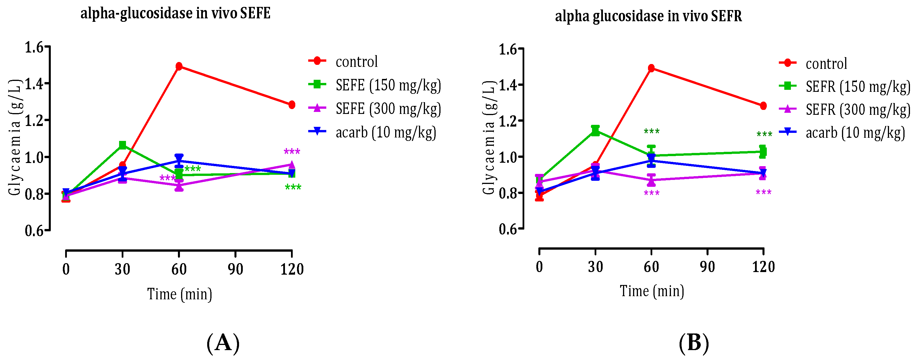
In Vivo Anti-α-Glycosidase Activity of S. elaeagnifolium Leaves and Fruits

Under typical circumstances, postprandial blood D-glucose levels are influenced by two primary processes: the conversion of complex, long-chain sugars into readily absorbable units, and the subsequent absorption of these carbohydrate units within the intestine. Following a meal, polysaccharides undergo enzymatic breakdown initiated by α-amylase, leading to their transformation into oligosaccharides. Subsequently, the final phase of this process involves the action of intestinal α-glucosidase, resulting in the production of absorbable monosaccharides. For these monosaccharides to enter the bloodstream, they must pass through the SGLT1 transporters situated on the brush border membrane (BBM) of the absorbing epithelial cells. Then, with the assistance of facilitated diffusion facilitated by GLUT2 transporters located on the basolateral membrane (BLM) of the epithelial cells, they are able to traverse into the bloodstream [42].
S. elaeagnifolium leaves and fruits have been examined for their potential to inhibit the activity of intestinal α-glucosidase, following the protocol outlined by Elrherabi et al. [39]. The ingestion of sucrose (2 g/kg) led to a marked elevation in postprandial blood glucose levels, reaching 1.45 ± 0.03 g/L at the 60 min mark and 1.36 ± 0.01 g/L at 90 min. However, upon the administration of SEFE at a dosage of 150 mg/kg, a significant decrease in postprandial blood glucose levels was observed. This reduction reached statistical significance at both 60 min (p < 0.001) and 120 min (p < 0.001), with recorded values of 0.9 ± 0.02 g/L and 0.91 ± 0.01 g/L, respectively, in comparison to the control group. In contrast, when a higher dose of SEFE (300 mg/kg) was administered, it also exerted a noteworthy suppression on postprandial hyperglycemia in comparison to the control group’s rats. This effect was evident at two key time points: at 60 min (p < 0.001) with a measured level of 0.84 ± 0.02 g/L, and at 120 min (p < 0.001) with a level of 0.95 ± 0.01 g/L. Similarly, the administration of 10 mg/kg of acarbose also significantly inhibited starch-induced hyperglycemia (p < 0.001 and p < 0.001) at 60 and 120 min, yielding values of 0.98 g/L and 1 g/L, respectively (Figure 5). The ingestion of sucrose (2 g/kg) led to a marked elevation in postprandial blood glucose levels, reaching 1.45 ± 0.03 g/L at the 60 min mark and 1.36 ± 0.01 g/L at 90 min. However, upon the administration of SEFR at a dosage of 150 mg/kg, a significant decrease in postprandial blood glucose levels was observed. This reduction reached statistical significance at both 60 min (p < 0.001) and 120 min (p < 0.001), with recorded values of 1 ± 0.05 g/L and 1.02 ± 0.02 g/L, respectively, in comparison to the control group.
In contrast, when a higher dose of SEFR (300 mg/kg) was administered, it also exerted a noteworthy suppression on postprandial hyperglycemia in comparison to the control group’s rats. This effect was evident at two key time points: at 60 min (p < 0.001) with a measured level of 0.87 ± 0.02 g/L, and at 120 min (p < 0.001) with a level of 0.9 ± 0.02 g/L. Similarly, the administration of 10 mg/kg of acarbose also significantly inhibited starch-induced hyperglycemia (p < 0.001 and p < 0.001) at 60 and 120 min, yielding values of 0.98 g/L and 1 g/L, respectively. SEFE and SEFR extracts have anti-alpha glycosidase activity, as evidenced by their ability to lower postprandial blood glucose levels, with their effects being comparable to acarbose, a known alpha glycosidase inhibitor. The inhibition of sugar-digesting enzymes, such as intestinal glucosidase and pancreatic amylase, as well as slowing intestinal D-glucose absorption and lowering postprandial D-glucose, are among the areas of focus aimed at glycemic regulation [43].

3.3.3. Anti-Inflammatory Potential

Table 4 illustrates the results of the anti-inflammatory activity evaluation using the method of inducing inflammation in the rat paw using carrageenan. These include plant extracts that have been shown to have anti-inflammatory properties, though the effect varies depending on the part of the plant used and the dose used. SEFR at 400 mg/kg showed an anti-inflammatory effect (86%) comparable to the inflammation-inhibiting effect of indomethacin (92%); SEFE at the two doses also showed an anti-inflammatory effect, but to a lesser extent than SEFR. Badawy and colleagues studied the anti-inflammatory properties of S. elaeagnifolium [19]. A few species of the Solanum genus have been shown in scientific studies to have anti-inflammatory properties [44,45,46]. Furthermore, studies have shown that a hydro-ethanolic extract of the aerial parts of Solanum lycocarpum has anti-inflammatory activity against carrageenan-induced paw inflammation [47]. The anti-inflammatory capacity of plant extracts depends on their phytochemical composition. The chytochemical study revealed the presence of quercetin in large quantities, and this molecule demonstrated anti-inflammatory activity via the mechanisms of regulation of COX-2 protein expression, inhibition of NF activation, and inhibition of NO production [48].

4. Conclusions

S. elaeagnifolium has been found to have a variety of pharmacological activities and bioactive molecules. Our findings show that extracts derived from this plant’s leaves and fruits contain bioactive molecules such as naringin, quercetin 3-O--D-glucoside, rutin, and cinnamic acid, and that extracts from this plant have anti-lipase activity and anti-glycation effects on haemoglobin, as well as anti-hyperglycemic and anti-inflammatory properties. These and other findings help us gain a better understanding of its potential benefits. These findings compel us to investigate other pharmacological activities of this plant.

Author Contributions

Conceptualisation, E.H.L., M.B. (Mohammed Bouslamti) and A.E.; methodology, E.H.L., M.B. (Mohammed Bouslamti) and A.E.; software, M.B. (Mohamed Bouhrim) and M.S.; validation, M.B. (Mohamed Bouhrim) and M.S.; formal analysis, R.A. and M.B. (Mohammed Bouslamti); investigation, E.H.L., M.S., A.E. and M.B. (Mohammed Bouslamti); resources, E.H.L., M.S., A.E. and R.A.; data retention, E.H.L., R.A., M.B. (Mohamed Bouhrim) and A.E.; writing the original draft, M.B. (Mohammed Bouslamti), A.E. and E.H.L.; revising and editing, A.S.B., B.L. and M.B. (Mohamed Bnouham); visualization, E.H.L., supervision, O.M.N., R.A.M. and M.N.I., project administration, M.B. (Mohammed Bouslamti); fund acquisition, O.M.N., R.A.M., and M.N.I. All authors have read and agreed to the published version of the manuscript.

Funding

Researchers Supporting Project number (RSP2023R119), King Saud University, Riyadh, Saudi Arabia.

Institutional Review Board Statement

Not applicable.

Informed Consent Statement

Not applicable.

Data Availability Statement

The data are contained within the article.

Acknowledgments

The authors extend their appreciation to Researchers Supporting Project number (RSP2023R119), King Saud University, Riyadh, Saudi Arabia, for their financial support. The authors also expressed their gratitude to Laboratoire TBC (France) for their technical support.

Conflicts of Interest

The authors declare no conflict of interest.

References

  1. Xiong, W.-T.; Gu, L.; Wang, C.; Sun, H.-X.; Liu, X. Anti-Hyperglycemic and Hypolipidemic Effects of Cistanche Tubulosa in Type 2 Diabetic Db/Db Mice. J. Ethnopharmacol. 2013, 150, 935–945. [Google Scholar] [CrossRef] [PubMed]
  2. El Ghouizi, A.; Ousaaid, D.; Laaroussi, H.; Bakour, M.; Aboulghazi, A.; Soutien, R.; Hano, C.; Badiaa, L. Ficus carica (Linn.) Leaf and Bud Extracts and Their Combination Attenuates Type-1 Diabetes and Its Complications via the Inhibition of Oxidative Stress. Foods 2023, 12, 759. [Google Scholar] [CrossRef] [PubMed]
  3. Hbika, A.; Daoudi, N.E.; Bouyanzer, A.; Bouhrim, M.; Mohti, H.; Loukili, E.H.; Mechchate, H.; Al-salahi, R.; Nasr, F.; Bnouham, M.; et al. Artemisia absinthium L. Aqueous and Ethyl Acetate Extracts: Antioxidant Effect and Potential Activity In Vitro and In Vivo against Pancreatic α-Amylase and Intestinal α-Glucosidase. Pharmaceutics 2022, 14, 481. [Google Scholar] [CrossRef] [PubMed]
  4. Medzhitov, R.; Medzhitov, R. Origin and Physiological Roles of Inflammation. Nature 2008, 454, 428–435. [Google Scholar] [CrossRef] [PubMed]
  5. Medzhitov, R. Inflammation 2010: New Adventures of an Old Flame. Cell 2010, 140, 771–776. [Google Scholar] [CrossRef]
  6. Umamageswari, M.; Karthikeyan, T.; Maniyar, Y.A. Antidiabetic Activity of Aqueous Extract of Solanum nigrum linn berries in Alloxan Induced Diabetic Wistar Albino Rats. J. Clin. Diagn. Res. 2017, 11, FC16–FC19. [Google Scholar] [CrossRef]
  7. Dasgupta, N.; Muthukumar, S.P.; Murthy, P.S. Solanum nigrum Leaf: Natural Food Against Diabetes and Its Bioactive Compounds. Res. J. Med. Plants 2016, 10, 181–193. [Google Scholar] [CrossRef]
  8. Nderitu, K.W.; Mecha, E.; Nyachieo, A. Solanum nigrum Show Anti-Obesity Effects on High-Fat Diet-Fed Sprague Dawley Rats in a Randomized Study. Sci. Afr. 2023, 20, e01713. [Google Scholar] [CrossRef]
  9. Aabideen, Z.U.; Mumtaz, M.W.; Akhtar, M.T.; Raza, M.A.; Mukhtar, H.; Irfan, A.; Raza, S.A.; Nadeem, M.; Ling, Y.S. Anti-Obesity Effect and UHPLC-QTOF-MS/MS Based Metabolite Profiling of Solanum nigrum Leaf Extract. Asian Pac. J. Trop. Biomed. 2022, 12, 164–174. [Google Scholar] [CrossRef]
  10. Peng, C.-H.; Cheng, J.-J.; Yu, M.-H.; Dai-Jung, C.; Huang, C.-N.; Wang, C.-J. Solanum nigrum Polyphenols Reduce Body Weight and Body Fat by Affecting Adipocyte and Lipid Metabolism. Food Funct. 2019, 11, 483–492. [Google Scholar] [CrossRef]
  11. Yeom, Y.-E.; Kim, M.A.; Kim, J.; Lee, C.-M. Anti-Inflammatory Effects of the Extract of Solanum nigrum L. on an Acute Ear Edema Mouse Model. Mater. Technol. 2019, 34, 851–857. [Google Scholar] [CrossRef]
  12. Campisi, A.; Acquaviva, R.; Raciti, G.; Duro, A.; Rizzo, M.; Santagati, N.A. Antioxidant Activities of Solanum nigrum L. Leaf Extracts Determined in In Vitro Cellular Models. Foods 2019, 8, 63. [Google Scholar] [CrossRef] [PubMed]
  13. Navarrete, S.; Alarcón, M.; Palomo, I. Aqueous Extract of Tomato (Solanum lycopersicum L.) and Ferulic Acid Reduce the Expression of TNF-α and IL-1β in LPS-Activated Macrophages. Molecules 2015, 20, 15319–15329. [Google Scholar] [CrossRef] [PubMed]
  14. Houda, M.; Derbré, S.; Jedy, A.; Tlili, N.; Legault, J.; Richomme, P.; Limam, F.; Saidani-Tounsi, M. Combined Anti-AGEs and Antioxidant Activities of Different Solvent Extracts of Solanum elaeagnifolium Cav. (Solanaceae) Fruits during Ripening and Related to Their Phytochemical Compositions. EXCLI J. 2014, 13, 1029–1042. [Google Scholar] [CrossRef]
  15. Bouslamti, M.; Loukili, E.H.; Elrherabi, A.; Moussaoui, A.; Chebaibi, M.; Bencheikh, N.; Nafidi, H.-A.; Bin Jardan, Y.; Bourhia, M.; Bnouham, M.; et al. Phenolic Profile, Inhibition of α-Amylase and α-Glucosidase Enzymes, and Antioxidant Properties of Solanum elaeagnifolium Cav. (Solanaceae): In Vitro and In Silico Investigations. Processes 2023, 11, 1384. [Google Scholar] [CrossRef]
  16. Bouslamti, M.; Chelouati, T.; El Moussaoui, A.; El Barnossi, A.; Badiaa, L.; Benjelloun, A. Solanum elaeagnifolium Var. Obtusifolium (Dunal) Dunal: Antioxidant, Antibacterial, and Antifungal Activities of Polyphenol-Rich Extracts Chemically Characterized by Use of In Vitro and In Silico Approaches. Molecules 2022, 27, 8688. [Google Scholar] [CrossRef]
  17. Balah, M.A.; Hassany, W.M.; Kopici, A.A. Allelopathy of Invasive Weed Solanum elaeagnifolium Cav.: An Investigation in Germination, Growth and Soil Properties. J. Plant Prot. Res. 2022, 62, 58–70. [Google Scholar] [CrossRef]
  18. Bouslamti, M.; El Barnossi, A.; Kara, M.; Alotaibi, B.; Kamaly, O.; Assouguem, A.; Badiaa, L.; Benjelloun, A. Total Polyphenols Content, Antioxidant and Antimicrobial Activities of Leaves of Solanum elaeagnifolium Cav. from Morocco. Molecules 2022, 27, 4322. [Google Scholar] [CrossRef]
  19. Badawy, A.; Zayed, R.; Ahmed, S.; Hassanean, H. Phytochemical and Pharmacological Studies of Solanum elaeagnifolium Growing in Egypt. J. Nat. Prod. 2013, 6, 156–167. [Google Scholar]
  20. Radwan, M.; Badawy, A.; Zayed, R.; Hassanin, H.; Elsohly, M.; Ahmed, S. Cytotoxic Flavone Glycosides from Solanum elaeagnifolium. Med. Chem. Res. 2014, 24, 1326–1330. [Google Scholar] [CrossRef]
  21. Feki, H.; Koubaa, I.; Damak, M. Secondary Metabolites and Antioxidant Activity of Seed Extracts from Solanum elaeagnifolium Cav. Mediterr. J. Chem. 2014, 2, 639–647. [Google Scholar] [CrossRef]
  22. Njeh, F.; Feki, H.; Koubaa, I.; Hamed, N.; Mohamed, D.; Ayadi, A.; Hammami, H.; Jarraya, R. Molluscicidal Activity of Solanum elaeagnifolium Seeds against Galba Truncatula Intermediate Host of Fasciola Hepatica: Identification of β-Solamarine. Pharm. Biol. 2015, 54, 726–731. [Google Scholar] [CrossRef] [PubMed]
  23. Hawas, U.W.; Soliman, G.M.; Abou El-Kassem, L.T.; Farrag, A.R.H.; Mahmoud, K.; León, F. A New Flavonoid C-Glycoside from Solanum elaeagnifolium with Hepatoprotective and Curative Activities against Paracetamol-Induced Liver Injury in Mice. Z. Naturforschung C J. Biosci. 2013, 68, 19–28. [Google Scholar]
  24. Al-hamaideh, K.; Dmour, I.; El-Elimat, T.; Afifi, F. UPLC-MS Profile and Anti-Proliferative Activity of the Berries of an Aggressive Wild-Growing Weed: Solanum elaeagnifolium Cav. (Solanaceae). Trop. J. Nat. Prod. Res. 2020, 4, 1131–1138. [Google Scholar] [CrossRef]
  25. Loukili, E.H.; Bouchal, B.; Bouhrim, M.; Abrigach, F.; Genva, M.; Kahina, Z.; Bnouham, M.; Bellaoui, M.; Hammouti, B.; Addi, M.; et al. Chemical Composition, Antibacterial, Antifungal and Antidiabetic Activities of Ethanolic Extracts of Opuntia Dillenii Fruits Collected from Morocco. J. Food Qual. 2022, 2022, 9471239. [Google Scholar] [CrossRef]
  26. Ouahabi, S.; Loukili, E.H.; Daoudi, N.E.; Chebaibi, M.; Ramdani, M.; Rahhou, I.; Bnouham, M.; Fauconnier, M.-L.; Hammouti, B.; Rhazi, L.; et al. Study of the Phytochemical Composition, Antioxidant Properties, and In Vitro Anti-Diabetic Efficacy of Gracilaria Bursa-Pastoris Extracts. Mar. Drugs 2023, 21, 372. [Google Scholar] [CrossRef]
  27. Benrahou, K.; Naceiri Mrabti, H.; Bouyahya, A.; Daoudi, N.E.; Bnouham, M.; Mezzour, H.; Mahmud, S.; Alshahrani, M.; Obaidullah, A.; Cherrah, Y.; et al. Inhibition of α-Amylase, α-Glucosidase, Lipase, Intestinal Glucose Absorption and Antidiabetic Properties by Extracts of Erodium Guttatum. Evid. Based Complement. Alternat. Med. 2022, 2022, 5868682. [Google Scholar] [CrossRef]
  28. Elrherabi, A.; Bouhrim, M.; Rhizlan, A.; Berraaouan, A.; Ziyyat, A.; Mekhfi, H.; Legssyer, A.; Bnouham, M. Phenolic Content, Antioxidant, Hemidiaphragm Glucose Consumption, and Hemoglobin Glycosylation Inhibitory Activities of Lavandula stoechas L. Aqueous Extract. J. Nat. Remedies 2023, 23, 653–660. [Google Scholar] [CrossRef]
  29. Council of the European Union. Council Directive 86/609/EEC of 24 November 1986 on the Approximation of Laws, Regulations and Administrative Provisions of the Member States Regarding the Protection of Animals Used for Experimental and Other Scientific Purposes, CELEX1. Available online: https://op.europa.eu/en/publication-detail/-/publication/cc3a8ccb-5a30-4b6e-8da8-b13348caeb0c/language-en (accessed on 25 July 2023).
  30. Daoudi, N.E.; Bouhrim, M.; Ouassou, H.; Abdelkhaleq, L.; Mekhfi, H.; Ziyyat, A.; Aziz, M.; Bnouham, M. Inhibitory Effect of Roasted/Unroasted Argania Spinosa Seeds Oil on α-Glucosidase, α-Amylase and Intestinal Glucose Absorption Activities. S. Afr. J. Bot. 2020, 135, 413–420. [Google Scholar] [CrossRef]
  31. El Moussaoui, A.; Mechchate, H.; Bourhia, M.; Es-safi, I.; Salamatullah, A.; Alkaltham, M.; Alyahya, H.; Bousta, D.; Bari, A. Glycemic Control Potential of Chemically Characterized Extract from Withania frutescens L. Roots in Severe Diabetes-Induced Mice. Appl. Sci. 2021, 11, 3998. [Google Scholar] [CrossRef]
  32. Remok, F.; Saidi, S.; Gourich, A.; Zibouh, K.; Maouloua, M.; Makhoukhi, F.; el Menyiy, N.; Touijer, H.; Bouhrim, M.; Sahpaz, S.; et al. Phenolic Content, Antioxidant, Antibacterial, Antihyperglycemic, and α-Amylase Inhibitory Activities of Aqueous Extract of Salvia Lavandulifolia Vahl. Pharmaceuticals 2023, 16, 395. [Google Scholar] [CrossRef] [PubMed]
  33. Mssillou, I.; Agour, A.; Slighoua, M.; Chebaibi, M.; Amrati, F.E.-Z.; Alshawwa, S.; Kamaly, O.; El Moussaoui, A.; Badiaa, L.; Derwich, E. Ointment-Based Combination of Dittrichia viscosa L. and Marrubium vulgare L. Accelerate Burn Wound Healing. Pharmaceuticals 2022, 15, 289. [Google Scholar] [CrossRef] [PubMed]
  34. Hanasaki, Y.; Ogawa, S.; Fukui, S. The Correlation between Active Oxygens Scavenging and Antioxidative Effects of Flavonoids. Free Radic. Biol. Med. 1994, 16, 845–850. [Google Scholar] [CrossRef]
  35. Wang, G.; Wang, Y.; Yao, L.; Gu, W.; Zhao, S.; Shen, Z.; Lin, Z.; Liu, W.; Yan, T. Pharmacological Activity of Quercetin: An Updated Review. Evid. Based Complement. Alternat. Med. 2022, 2022, e3997190. [Google Scholar] [CrossRef]
  36. Hou, T.-H.; Chung, J.-P.; Chen, S.-S.; Chang, T. Antioxidation and Antiglycation of 95% Ethanolic Extracts Prepared from the Leaves of Black Nightshade (Solanum nigrum). Food Sci. Biotechnol. 2013, 22, 839–844. [Google Scholar] [CrossRef]
  37. Singh, M.P.; Pathak, K. Animal Models for Biological Screening of Anti-Diabetic Drugs: An Overview. Euro. J. Exp. Biol. 2015, 5, 37–48. [Google Scholar]
  38. Shai, L.J.; Masoko, P.; Mokgotho, M.P.; Magano, S.R.; Mogale, A.M.; Boaduo, N.; Eloff, J.N. Yeast Alpha Glucosidase Inhibitory and Antioxidant Activities of Six Medicinal Plants Collected in Phalaborwa, South Africa. S. Afr. J. Bot. 2010, 76, 465–470. [Google Scholar] [CrossRef]
  39. Elrherabi, A.; Bouhrim, M.; Rhizlan, A.; Berraaouan, A.; Ziyyat, A.; Mekhfi, H.; Legssyer, A.; Bnouham, M. Antihyperglycemic Potential of the Lavandula stoechas Aqueous Extract via Inhibition of Digestive Enzymes and Reduction of Intestinal Glucose Absorption. J. Ayurveda Integr. Med. 2023, 14, 100795. [Google Scholar] [CrossRef]
  40. Gong, L.; Feng, D.; Wang, T.; Ren, Y.; Liu, Y.; Wang, J. Inhibitors of α-Amylase and α-Glucosidase: Potential Linkage for Whole Cereal Foods on Prevention of Hyperglycemia. Food Sci. Nutr. 2020, 8, 6320–6337. [Google Scholar] [CrossRef]
  41. Dona, A.; Pagès, G.; Kuchel, P. Digestion of Starch: In Vivo and in Vitro Kinetic Models Used to Characterise Oligosaccharide or Glucose Release. Carbohydr. Polym. 2010, 80, 599–617. [Google Scholar] [CrossRef]
  42. Tomimatsu, T.; Horie, T. Enhanced Glucose Absorption in the Rat Small Intestine Following Repeated Doses of 5-Fluorouracil. Chem. Biol. Interact. 2005, 155, 129–139. [Google Scholar] [CrossRef] [PubMed]
  43. Benayad, O.; Bouhrim, M.; Tiji, S.; Kharchoufa, L.; Addi, M.; Drouet, S.; Hano, C.; Lorenzo, J.M.; Bendaha, H.; Bnouham, M.; et al. Phytochemical Profile, α-Glucosidase, and α-Amylase Inhibition Potential and Toxicity Evaluation of Extracts from Citrus aurantium (L) Peel, a Valuable By-Product from Northeastern Morocco. Biomolecules 2021, 11, 1555. [Google Scholar] [CrossRef] [PubMed]
  44. Ndebia, E.; Kamgang, R.; Nkeh-ChungagAnye, B. Analgesic and Anti-Inflammatory Properties of Aqueous Extract from Leaves of Solanum torvum (Solanaceae). Afr. J. Tradit. Complement. Altern. Med. AJTCAM Afr. Netw. Ethnomedicines 2006, 4, 240–244. [Google Scholar] [CrossRef] [PubMed]
  45. Piana, M.; Camponogara, C.; Boligon, A.; Oliveira, S. Solanum Paranense Extracts and Solanine Present Anti-Inflammatory Activity in an Acute Skin Inflammation Model in Mice. Evid. Based Complement. Alternat. Med. 2017, 2017, 4295680. [Google Scholar] [CrossRef]
  46. Rahman, N.; Haris, M.; Mahmood, R.; ur Rahman, H. Antioxidant and Anti-Inflammatory Potentials of Solanum pubescens Willd an Ethnomedicinal Plant of South Western Andhra Pradesh, India. J. Res. Pharm. 2019, 23, 187–197. [Google Scholar] [CrossRef]
  47. da Costa, G.A.F.; Morais, M.G.; Saldanha, A.A.; Assis Silva, I.C.; Aleixo, Á.A.; Ferreira, J.M.S.; Soares, A.C.; Duarte-Almeida, J.M.; Lima, L.A.R.d.S. Antioxidant, Antibacterial, Cytotoxic, and Anti-Inflammatory Potential of the Leaves of Solanum Lycocarpum A. St. Hil. (Solanaceae). Evid. Based Complement. Alternat. Med. 2015, 2015, e315987. [Google Scholar] [CrossRef]
  48. Güran, M.; Şanlıtürk, G.; Kerküklü, N.; Altundag, E.; Yalçin, A.S. Combined Effects of Quercetin and Curcumin on Anti-Inflammatory and Antimicrobial Parameters in Vitro. Eur. J. Pharmacol. 2019, 859, 172486. [Google Scholar] [CrossRef]
Figure 1. HPLC-DAD chromatogram of S. elaeagnifolium extracts at 254 nm (fruits (SEFR) and leaves (SEFE)) and identified with the standards (catechin (1), vanillin (2), naringin (3), quercetin 3-O-β-D-glucoside (4), rutin (5), cinnamic acid (6), p-coumaric acid (7), quercetin (8), trans-3-hydroxycinnamic acid (9), kaempferol (10), and flavone (11)).
Applsci 13 11519 g001
Figure 2. (A) Anti-lipase effect of SEFR, SEFE, and Orlistat; and (B) inhibitory effect of SEFR, SEFE, and gallic acid (positive control) on glycation activity of hemoglobin. Results are shown as mean ± SEM (n = 3).
Applsci 13 11519 g002
Figure 3. (A) Effect of SEFE on rats orally loaded with glucose; and (B) effect of SEFR on rats orally loaded with glucose. * p < 0.05;** p < 0.01; *** p < 0.001 as compared to the control.
Applsci 13 11519 g003
Figure 4. Glycemic postprandial results in normal rats after administration of S. elaeagnifolium extract and acarbose): (A) effect of leaf extract, and (B) effect of fruit extract. ** p < 0.01; *** p < 0.001 as compared to the control.
Applsci 13 11519 g004
Figure 5. Glycemic postprandial response in normal rats to S. elaeagnifolium extract and acarbose): (A) effect of leaf extract, and (B) effect of fruit extract. *** p < 0.001 as compared to the control.
Applsci 13 11519 g005
Table 1. Information about the study plant.
Plant SpeciesFamilyVoucher N°Used Organ RegionCo-OrdinatesDate
Solanum elaeagnifoliumSolanaceaeE17/1405Leaves (SEFE)Fez34°04′04.2 N, 5°01′26.4 WDecember 2021
Fruits (SEFR)April 2022
Table 2. HPLC chromatographic examination of the chemicals found in S. elaeagnifolium fruit and leaf extracts.
PeakCompoundsSEFRSEFE
% Areamg/100 g% Areamg/100 g
1Catechin0.490.49 ± 0.21NdNd
2Vanillin12.1346.72 ± 0.69NdNd
3NaringinNdNd4.1612.00 ± 0.01
4Quercetin 3-O-β-D-glucoside4.3616.72 ± 0.551.591.84 ± 0.05
5Rutin3.2298.17 ± 0.6511.343.83 ± 0.23
6Cinnamic acidNdNd0.812.17 ± 0.44
7p-coumaric acidNdNd6.5675.85 ± 0.13
8Quercetin75.84429.02 ± 0.9726.29114.15 ± 0.34
9Trans-3-hydroxycinnamic acidNdNd55.95243.51 ± 0.46
10Kaempferol3.9612.09 ± 0.08NdNd
11FlavoneNdNd3.309.28 ± 0.25
Table 3. Determination of anti-lipase and hemoglobin antiglycation activities of the leaf extracts (SEFE) and fruit extracts (SEFR) of S. elaeagnifolium. Results are shown as mean ± SEM (n = 3).
SEFESEFROrlistatGallic Acid
Lipase inhibitory
IC50 (mg/mL)
0.167 ± 0.00060.106 ± 0.00080.128 ± 0.003-
Antiglycation IC50 (mg/mL)3.990 ± 0.2363.997 ± 0.140-3.008 ± 0.03
Table 4. Edema-inhibiting effect of carrageenan from S. elaeagnifolium extracts and indomethacin. The values are the means ± SEM (n = 6). * p < 0.05; ** p < 0.01; *** p < 0.001.
Diameter (cm) and % of Inhibition
TreatmentDose (mg/kg)0 h3 h4 h5 h6 h
Control (NaCl 0.9%)-2.43 ± 0.123.03 ± 0.03 ***3.00 ± 0.00 ***2.96 ± 0.03 ***2.90 ± 0.05 **
Indomethacine102.30 ± 0.032.54 ± 0.03 ** (60%)2.45 ± 0.03 *
(74%)
2.41 ± 0.02
(80%)
2.34 ± 0.03
(92%)
SEFR
Extract
2002.36 ± 0.062.86 ± 0.06 ***
(17%)
2.76 ± 0.06 **
(30%)
2.63 ± 0.03 *
(50%)
2.53 ± 0.03
(64%)
4002.56 ± 0.062.96 ± 0.03 ***
(34%)
2.83 ± 0.03 *
(53%)
2.70 ± 0.00
(74%)
2.63 ± 0.03
(86%)
SEFE Extract2002.60 ± 0.153.20 ± 0.00 ***
(0%)
3.06 ± 0.03 **
(20%)
3.00 ± 0.05 *
(25%)
2.93 ± 0.03 *
(30%)
4002.36 ± 0.082.83 ± 0.06 **
(22%)
2.73 ± 0.08 *
(36%)
2.60 ± 0.05
(55%)
2.56 ± 0.03
(58%)
Disclaimer/Publisher’s Note: The statements, opinions and data contained in all publications are solely those of the individual author(s) and contributor(s) and not of MDPI and/or the editor(s). MDPI and/or the editor(s) disclaim responsibility for any injury to people or property resulting from any ideas, methods, instructions or products referred to in the content.

