Article

Roasting Extract of Handroanthus impetiginosus Enhances Its Anticancer Activity in A549 Lung Cancer Cells and Improves Its Antioxidant and Anti-Inflammatory Effects in Normal Cells

1
Graduate School of Biotechnology, College of Life Sciences, Kyung Hee University, Yongin-si 17104, Republic of Korea
2
Department of Oriental Medicinal Biotechnology, College of Life Sciences, Kyung Hee University, Yongin-si 17104, Republic of Korea
3
Department of Biopharmaceutical Biotechnology, College of Life Science, Kyung Hee University, Yongin-si 17104, Republic of Korea
4
NK TAHEEBO Company, 27, Dongtancheomdansaneop 1-ro, Hwaseong-si 18445, Republic of Korea
5
Department of Chemistry Education, Graduate Department of Chemical Materials, Pusan National University, Busan 46241, Republic of Korea
6
Hanbangbio Inc., 13, Heungdeok 1-ro, Giheung-gu, Yongin-si 16954, Republic of Korea
7
State Local Joint Engineering Research Center of Ginseng Breeding and Application, Jilin Agriculture University, Changchun 130118, China
8
Department of Horticulture, Kongju National University, Yesan 32439, Republic of Korea
*
Authors to whom correspondence should be addressed.
These authors contributed equally to this work.
Appl. Sci. 2023, 13(24), 13171; https://doi.org/10.3390/app132413171
Submission received: 18 October 2023 / Revised: 29 November 2023 / Accepted: 8 December 2023 / Published: 12 December 2023
(This article belongs to the Special Issue Natural Products: Sources and Applications)

Abstract

:
The family Bignoniaceae includes Handroanthus impetiginosus trees, which are sparsely distributed in the northeast of Brazil. Natural products play a vital role in the discovery of drugs for various diseases. Many plants have been used as sources of medicines because of their chemical diversity and potent bioactivity. Handroanthus impetiginosus has been used traditionally to cure a wide range of illnesses, such as cancer, oxidative stress, and inflammation. This work highlights the cytotoxicity, cell death, and routes of apoptosis in lung cancer cells (A549) and the anti-inflammatory and antioxidant effects of roasted Handroanthus impetiginosus (lapacho/taheebo) in normal cells. The cell viability assay indicated that puffing roasted taheebo is nontoxic to a normal cell line up to 500 µg/mL but significantly toxic to A549 cells. The roasted lapacho/taheebo also increases reactive oxygen species (ROS) generation in A549 lung cancer cells, and cellular apoptosis via a mitochondrial intrinsic pathway was confirmed. The roasted lapacho/taheebo significantly inhibited both colony formation and cell migration ability, highlighting its potential as an anticancer agent. Additionally, this study demonstrates that roasted taheebo enhanced the expression of genes for BAX accumulation and decreased Bcl-2 gene expression through the p53 signaling pathway. Furthermore, research on the anti-inflammatory properties of roasted taheebo revealed a strong NO inhibition as well as the inhibition of inflammatory mediators (TNF-α, iNOS, COX-2, IL-6, and IL-8) through the NF-κB signaling pathway. However, in H2O2-induced HaCaT cells, roasted taheebo extract significantly reduced oxidative stress by upregulating the level of expression of antioxidative markers (SOD, CAT, GPx, and GST) at 50 μg/mL. As a result, roasted taheebo justifies investigation in animal and clinical trials as a possible source of antioxidants, anti-inflammatory substances, and anti-cancer compounds.

1. Introduction

People have long relied on biological resources such as plants, animals, and microbes to treat and prevent illnesses [1]. In recent years, the Western medical system has become more accepting of herbal remedies and has begun testing their chemical constituents as treatments for a variety of disorders as it has become educated about their nutritional qualities, biological activity, and health advantages [2]. Several commercial pharmaceuticals currently on the market, including digoxin, atropine, silymarin, teniposide, and aescin, were created from plants and other biological resources. Consequently, understanding how natural products have traditionally been used is crucial for both the discovery and development of new drugs [3]. Handroanthus impetiginosus, a member of the Bignoniaceae family, is native to and distributed throughout North, Central, and South America, from northern Mexico south to northern Argentina, and features purple flowers [4,5]. Handroanthus impetiginosum (Mart. ex DC.) Mattos, synonym tabebuia impetiginosa (Mart. ex DC.) Standl., or tabebuia avellanedae Lor. ex Griseb [6]. Herbal items derived from Tabebuia bark are referred to as taheebo, ipe roxo, pau d’arco, and lapacho [7]. Taheebo/lapacho has historically been used to treat digestion-related problems, diabetes, prostatitis, syphilis, cancer, fungal infections, and allergies [8]. For centuries, Brazilians have used this herb as an analgesic, anti-inflammatory, and antiophidic against the venom of snakes [9,10]. Additionally, the inner part of the bark is used to cure boils, ulcers, fever, diarrhea, arthritis, and prostate inflammation [11]. This inner bark can be used to make a poultice or concentrated tea to relieve a variety of skin-inflammatory conditions [12]. The FDA (Food and Drug Administration) has designated taheebo/lapacho tea as a nutritional supplement “herb recommended to treat diseases and cancer indications” [13]. The FDA has classified taheebo/lapacho as “generally recognized as safe" (GRAS). Although single chemicals, such as lapachol, have demonstrated Vitamin K toxicity, the combination of ingredients in Red Lapacho tea appears to negate that toxicity because it contains some pro-vitamin K compounds [14].
Various components, including iridoids, benzoic acids, coumarin derivatives, flavonoids, quinones, furanonaphthoquinones, anthraquinones, cyclopentene, dialdehydes, benzaldehyde derivatives, and naphthoquinones, have been identified in taheebo [13,15]. Some members of this genus contain lapacol and β-lapachone, which act by preventing the growth of tumor cells during the glycolysis process [16,17]. Both have shown significant anticancer effects in vitro [13]. Recent studies have shown that both crude extracts of taheebo and compounds isolated from those extracts have demonstrated a variety of pharmacological functions, such as anti-obesity, anti-psoriatic, antifungal, anti-inflammatory, and anti-cancer effects [3]. Previously, it was reported that water and ethanol extracts of taheebo showed anti-inflammatory activity [12,18]. Also, methanol, butanol, and water extracts of taheebo displayed dose-dependent antioxidant activity [19]. Furthermore, an n-butanol extract of taheebo prevented adipocyte accumulation, promoted weight loss, and decreased fat mass in mice [20]. Moreover, a water extract of taheebo suppressed the growth of the breast cancer cell line MCF-7 [21]. Ethanol extracts of taheebo exhibited antinociceptive activity [18], immunomodulatory efficacy by inhibiting proliferation and activation of IL-2-independent T-lymphocytes [22], anti-osteoarthritis effects by minimizing cartilage degeneration [23], and anti-depression activity in forced swimming and tail suspension experiments [24].
Puffing is a food processing method that treats high temperatures in a short period of time to gelatinize starch present in food, inactivate enzymes that cause rancidity of lipids, destroy naturally occurring harmful substances, and deodorize and transform raw ingredients. Consequently, puffing is the act of abruptly lowering pressure while maintaining a high temperature and pressure, which lowers moisture content and raises the particular quantity of a food matrix [25,26]. The advantages of the roasting puffing method are that it increases the amount of nutrients in raw samples with a high absorption rate in the body [27,28].
However, the antioxidant, anti-inflammatory, and anti-lung cancer activity of puffing whole roasted taheebo extract in A549 cells has not yet been documented. In the current investigation, in order to enhance the nutritional content of the roasted sample, we have clarified the preparation of novel techniques using the puffing process. Therefore, an in vitro study reports the antioxidant, anti-inflammatory, and anti-lung cancer activity of puffing whole roasted taheebo extract in normal cells and cancerous cells.

2. Materials and Methods

2.1. Chemicals and Reagents

The NK TAHEEBO Company (Hwaseong-si, Gyeonggi-do, Republic of Korea) graciously supplied samples of the Taheebo lung cancer cell line (A549), the HaCaT keratinocyte cells, and the Raw 264.7 murine macrophage cells employed in this work were all obtained from the Korean Cell Line Bank. FBS (Fetal bovine serum) and penicillin-streptomycin solution were obtained from Gen DEPOT (Barker, TX, USA). Gibco (Waltham, MA, USA) provided Dulbecco’s Modified Eagle’s Medium with high glucose and pyruvate (DMEM). The MTT (3-(4, 5-dimethylthiazol-2-yl)-2, 5-diphenyltetrazolium bromide) is from Life Technologies in Suwon, Republic of Korea. Both the 2,2-diphenyl-1-picrylhydrazyl (DPPH) and Folin–Ciocalteu (FC) reagents were purchased from Sigma-Aldrich in the Republic of Korea.

2.2. Preparation of Roasted Taheebo Extract

Collection of Samples and Processing Procedure
The obtained samples were cleaned in distilled water and then dried for 20 h at 35 °C in an L’EQUIP dehydrator. After the materials were dried, they were crushed into a fine powder using a home grinder. To extract the bioactive components, one gram of the powder was diluted in 60% ethanol and processed ultrasonically for 140 min. After sonication, the crude extract was obtained using a rotary evaporator, and then it was converted to a 20 mg/mL solution by dissolving it in EtOH. Puffing techniques are used to make roasted taheebo. The first step was drying regular taheebo for 1–2 h at 60–70 °C and 50–60% humidity. In the second process, roasted tahebbo was dried at 50–60 °C with humidity (20–30%) for 3–4 h. Thirdly, dry the samples once more for 10–15 h at 50 °C and 10% humidity. The roasted puffing method was then applied to the roasted samples for 40–70 s when the speed of wind was 25–60 BLOW and the temperature range was 210–250 °C. Finally, the roasted puffing samples were prepared using the high-pressure extraction technique for two to four hours at a temperature of 120 to 140 °C.

2.3. Antioxidant Screening

2.3.1. Total Phenolic Content (TPC) Determination

As a modest adjustment to the previously published FC technique [29], ascorbic acid (5–80 µg/mL) was used as the benchmark for determining the phenolic contents. Briefly, 10% 2 N FC reagent (150 µL) was mixed with the extract solution (30 µL). After shaking it thoroughly, a 7.5% solution of sodium carbonate (160 µL) was incorporated into the mixture. Following incubation, the absorbance was assessed at 715 nm by BioTek Instruments, Inc. (Winooski, VT, USA).

2.3.2. Total Flavonoid Content (TFC) Determination

TFC was identified by significantly modifying the aluminum chloride colorimetric technique [29]. Briefly, taheebo and roasted taheebo extracts (50 µL) were incorporated with 1 M potassium acetate (10 µL) and 10% aluminum chloride (10 µL) mixed with 430 µL of distilled water. After 30 min of incubation, the absorbance was measured at 415 nm by BioTek Instruments, Inc. in Winooski, VT, USA. The ascorbic acid calibration curve was used to compute the TFC.

2.3.3. DPPH-Free Radical Scavenging Activity Assessment

In accordance with a previously published study [30] and the minor modification of using ascorbic acid as the standard, a mixture of plant extract (20 µL) and 0.2 mM DPPH solution (180 µL) was made and maintained in the dark at room temperature for about thirty minutes to promote a better reaction. Approximately 100% ethanol was used as a blank, and as the control, sample solvent and DPPH solution were used. After the incubation, the absorbance was measured at 517 nm. The DPPH scavenging activity was calculated using the equation below:
DPPH radical scavenging activity (% inhibition) = {(abs control − abs sample)/Abs control} × 100
where
Abs control = the absorbance of the control solution (without sample)
Abs sample = the absorbance of the sample solution.

2.3.4. Procedure for Reducing Power Activity (RPA)

To test the RPA, the conversion of Fe3+ to Fe2+ in the presence of taheebo and roasted taheebo extracts was examined using the conventional procedure [31]. Briefly, the aliquot portions of the extracts and standard (ascorbic acid) were combined with 1% potassium ferricyanide (250 µL) and phosphate buffer (250 µL, pH 6.6). After 30 min of incubation, 250 L of 10% trichloroacetic acid was added, and then the mixture was subjected to 10 min of centrifugation at 3000 rpm. After that, 10 µL of FeCl3 (0.1%) and 50 µL of distilled water were added to the supernatant. The absorbance was measured at 700 nm with an ELISA reader. The findings are presented as μg of ascorbic acid equivalent per mg of sample (μg AAE/mg extract).

2.4. Cell Culture

Human lung carcinoma (A549) cells were grown in culture medium containing 89% Dulbecco’s Modified Eagle’s Medium (DMEM), 10% fetal bovine serum (FBS), and 1% penicillin-streptomycin. Similarly, human keratinocyte HaCaT and Raw 264.7 cells were cultured with RPMI-containing culture medium. All of the cells were cultured for 24 h in an incubator equipped with 95% air and 5% CO2.

2.5. In Vitro Cytotoxicity of Taheebo

MTT solution was used to test the cytotoxicity of taheebo (normal and roasted) extracts to A549, RAW 264.7, and HaCaT cells. A 96-well plate was seeded with cancer cells and healthy cells at a selective density of 1 × 10⁴ cells/well. Then, A549 and Raw 264.7 cells were treated with eight distinct concentrations (0, 15.625, 31.25, 62.5, 125, 250, 500, and 1000 μg/mL). HaCaT cells were exposed to samples at (25, 50, 100, 200, and 400 µg/mL). All cells were then incubated for 24 h. After that, 20 µL of the MTT solution (5 µg/L) was applied to each well. The culture medium was discarded after 4 h of incubation, and 100 µL of dimethyl sulfoxide (DMSO) was then added to solubilize the formazan. Finally, the samples were analyzed by an ELISA reader at 570 nm. Calculations of cell viability were conducted in comparison to the control (100%).

2.6. Measurement of Antioxidant Enzyme Activity

HaCaT cells in six-well cell culture dishes were inoculated with 250 µg/mL of taheebo and roasted taheebo extracts. After 24 h, 500 µM H2O2 was added to each well, and the cells were allowed to incubate for an additional 12 h. The cells were twice washed in PBS. After that, Triton X-100 (1%) was applied to the cells, which were kept on ice for 10 min. After defrosting the cells for 1 h, the lysates were spun in a centrifuge at 12,000 rpm for 30 min at 4 °C to eliminate the debris from the cells. The Bradford protein test, developed by Bio-Rad (Hercules, CA, USA), was used to calculate the total amount of protein.

2.6.1. Determination of GPx Activity

A previously published method [32] was used with some changes to measure GPx activity. Briefly, 50 µg of protein were incorporated with a reactant containing 1.5 mM NADPH, 1 mM NaN3, 1 mM EDTA, a single unit of glutathione reductase, 10 mM GSH reduced, and 100 mM phosphate buffer (pH = 7). Absorbance was observed at 340 nm for 5 min. The activity of GPx was identified by monitoring the change in the rate of NADPH oxidation.

2.6.2. Determination of SOD Activity

The measurement of the activity of SOD was carried out using a known method [33], with some modifications. Briefly, in a 96-well plate, 50 µg of protein, phosphate buffer (50 mM) (pH = 10.2), and epinephrine (10 mM) were added in each well with 3 replications. The absorbance of a pink product formed was measured using a micro-plate reader at 490 nm for 10 min. Units per milligram of protein were used to express the enzyme concentration necessary to induce a 50% inhibition of the autoxidation of epinephrine.

2.6.3. Determination of CAT Activity

Catalase activity was assessed using the previously mentioned methods [33], with minor optimization. In a 96-well plate, 50 µg of protein, phosphate buffer (100 mM) (pH = 7), and H2O2 (100 mM) were added. The reaction mixture was then incubated for 2 min at 37 °C. Using a micro-plate reader, the absorbance at 240 nm was recorded for 5 min. The rate of H2O2 oxidation is directly related to variations in absorbance over time. Units/mg of protein are used to convey how much enzyme is needed to break down 1 mM H2O2.

2.6.4. Determination of GST Activity

The method of Sikdar et al. [34] was used with some changes to measure GST activity. The experiment was performed in 100 mM phosphate buffer (pH 7.5) with 1 mM reduced GSH and 1 mM CDNB. Glutathione and CDNB were conjugated, and the absorbance change at 340 nm was observed for 10 min in a microplate reader.

2.6.5. Reactive Oxygen Species (ROS) Generation

A total of 2′,7′Dichloro-dihydro-fluorescein diacetate (DCFH-DA) probes were utilized to measure ROS generation in A549 adenocarcinomic cells and normal Raw 264.7 cells. The cells were plated in 96 wells (1 × 10⁴ cells/well) for 24 h at 37 °C and 5% CO2. After seeding, lipopolysaccharide (LPS) and/or samples were applied to Raw 264.7 cells for 24 h at various concentrations (0 to 1000 g/mL). To stain the cells, 10 µM DCFH-DA (100 µL) solution was added to each well. The cells were then left in the dark for 30 min. Before discarding the old media, these cells had been washed with PBS (100 L/well). The fluorescence intensity of ROS generation was measured using a spectrofluorometer at 485 nm and 528 nm for excitation and emission, respectively.

2.7. Nitrite Levels (NO) Determination

To measure nitrile, previously published techniques were used [35]. Shortly, a 96-well plate was seeded with raw cells and incubated for one day. Then, samples were administered in different dosages for 1 h. After treatment, E. coli LPS was used for 1 day to stimulate the cells (to stimulate and produce inflammation). Following the manufacturer’s recommended protocol, 100 µL of stimulated supernatant was mixed with 100 µL of Griess reagent. The resulting absorbance at 540 nm was measured in a microplate reader (Bio-Tek Instruments, Inc., Winooski, VT, USA) and compared with a standard curve created using sodium nitrite. The standard inhibitor, or positive control, of this experiment was 50 µM L-NMMA. From three executions of each experiment, the data are shown as NO production (%).

2.8. Wound-Healing Assay

Using a wound-healing experiment, the effectiveness of A549 lung cancer cell migration was examined. A549 cells were seeded at an amount of 2 × 10⁴ cells per well in a 6-well plate and incubated for 24 h at 37 °C. Using a 200 µL sterile pipette tip, the cell monolayer was scuffed vertically, and PBS was used to extract the fragmented cells. After that, the cells were exposed to roasted and unroasted taheebo extracts at doses of 500 and 1000 µg/mL. Images were captured with an implanted 5.0-megapixel MC 170 HD camera (Wetzlar, Germany) three days after the therapy period started.

2.9. Reverse Transcription-Polymerase Chain Reaction (RT-PCR and qRT-PCR)

A549 cells were seeded in a 6-well plate (1 × 10⁴) and incubated for 24 h. The cell culture media were changed, and a new medium containing samples at various concentrations was added. After that, QIAzol lysis reagent (QIAGEN, Germantown, MD, USA) was used to extract RNA from the cells in accordance with the manufacturer’s instructions. Reverse transcription was carried out using 1 µg of RNA and 20 µL of amfiRivert reverse transcription reagents (GenDepot, Barker, TX, USA). The procedure took place at the following temperatures: 25 °C for 5 min, 42 °C for 60 min, and 70 °C for 15 min. The RT-PCR procedure was run 35 times at 95 °C, 60 °C, and 72 °C for 30 s each. The 1% agarose gels with Safe Pinky DNA gel staining (GenDepot, Barker, TX, USA) were used to examine the amplified RT-PCR data and photographed under ultraviolet light. For the qRT-PCR, Enzynomics (Daejeon, Republic of Korea) SYBR TOPreal qPCR2X premix was used. In summary, the reactions were performed three times, and the ultimate 10 µL reaction volume included 1 µL of each of the forward and reverse primers, a 2× Master Mix, and 1 µL of template cDNA. All real-time measurements were performed using a CFX Connect real-time PCR system (Bio Rad, Hercules, CA, USA). Amplification reactions were performed under the following conditions: 95 °C for 10 min, a total of forty cycles of 95 °C for 20 s, then 55 °C for 30 s, and 72 °C for 15 s. The comparative 2−∆∆Ct method was used to measure the relative amounts of mRNA, and the GAPDH gene was used to standardize the results. The lists of primer sequences (GenoTech, Daejeon, Republic of Korea) are provided in Table 1.

3. Results

3.1. Antioxidant Activities of Taheebo and Roasted Taheebo

TPC, TFC, and Antioxidant Effects

It is commonly known that antioxidants can scavenge reactive species such as ROS and RNS, etc., or prevent cell oxidation to protect against disease. Antioxidants can block or delay oxidative reactions and thereby manage the excessive generation of oxidants [36]. Phenolic compounds are widely used against oxidative stress, skin infections, wounds, immune dysfunction, inflammation, hypoglycemia, cancer, etc. [37]. Similarly, numerous health benefits, including antioxidant, anticancer, antiviral, and anti-inflammatory effects, are provided by flavonoids. They also possess cardioprotective and neuroprotective properties [38]. DPPH and reducing power assays are often used to determine the antioxidant potential and electron donation capability, which are essential to phenolic antioxidant activity, of an extract, compound, or other biological source [39]. TPC and TFC were estimated in each sample using the FC and aluminum chloride colorimetric techniques. Table 2 indicates that the phenolic content of roasted and unroasted taheebo extracts varied widely. The TPC of roasted taheebo was 65,263.64 ± 0.029, and that of unroasted taheebo was 54,877.27 ± 0.069 µg AAE/mg extract. The total flavonoid contents also varied: 1526.82 ± 0.001 and 1185.36 ± 0.001 µg AAE/mg extract for roasted and unroasted taheebo extracts, respectively. Clearly, roasted taheebo contains more TPC and TFC than unroasted taheebo.
The results of the DPPH test indicate that the antioxidant efficacy of roasted and unroasted taheebo was 989.62 ± 0.015 and 767.73 ± 0.025 µg AAE/mg extract, respectively. The radical scavenging activity (% inhibition) vs. concentration curve shows that roasted taheebo has greater antioxidant capacity than unroasted taheebo in comparison to ascorbic acid (Figure 1A). Similarly, the results of the RPA reveal that the antioxidant reduction capability of the roasted and unroasted samples was 15.59 ± 0.006 and 11.66 ± 0.029 µg AAE/mg extract, respectively. The absorbance vs. concentration curve shows that roasted taheebo has a higher reducing capability than unroasted taheebo (Figure 1B).

3.2. Evaluation of Cytotoxicity

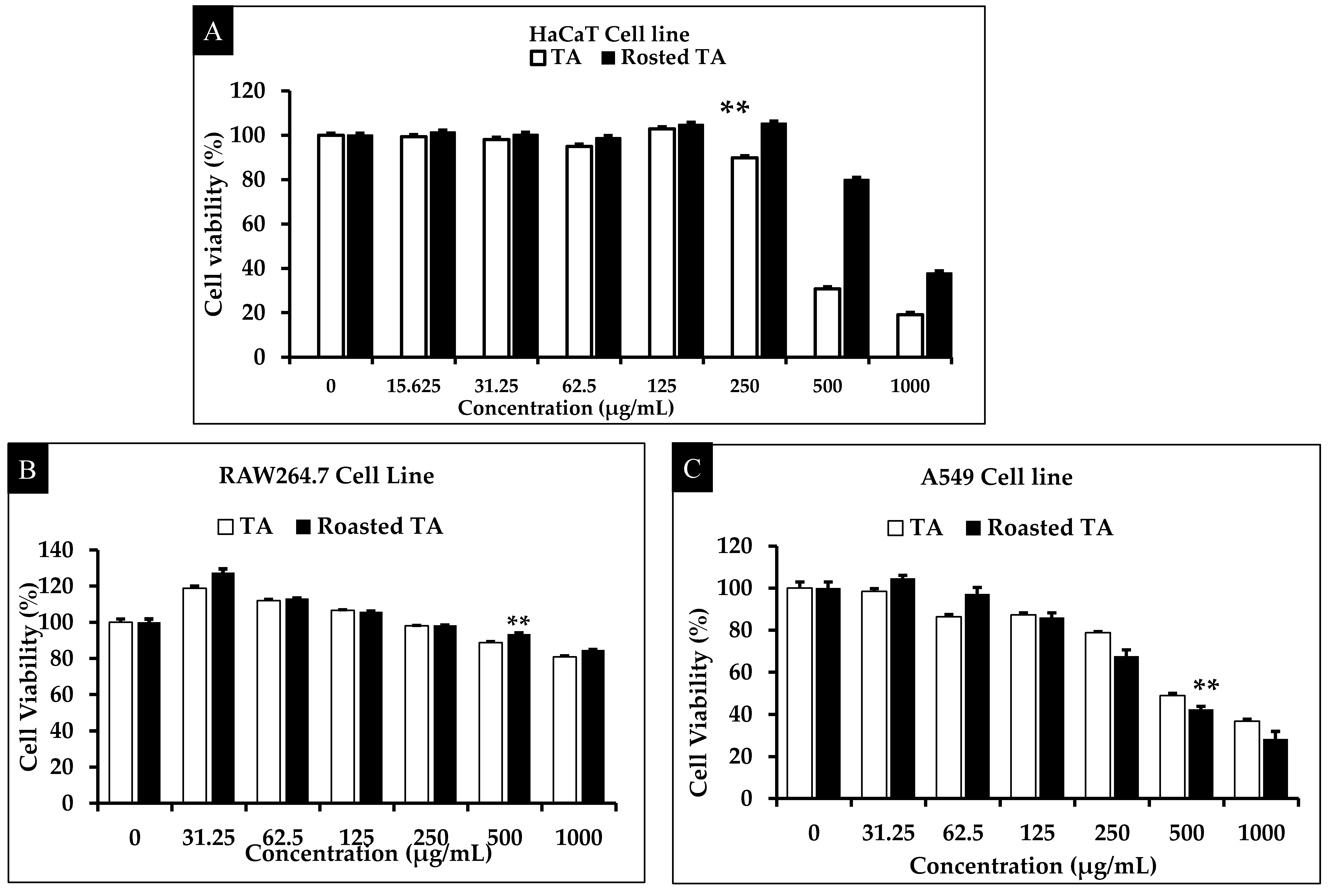
Intense cellular heterogeneity is a hallmark of cancer, and it makes therapy challenging and reduces the efficacy of current medications. To create new therapeutics, plants are major sources of natural, biologically active compounds [40,41]. Several indigenous plant species have pharmacological advantages, including anti-inflammatory, anti-cancer, and antioxidant properties [42,43]. For a more effective and secure course of therapy, it is fundamental to assess the anticancer activity of plant extracts. For the antioxidant enzyme assays, HaCaT cells were treated with extract concentrations varying from (0 to 1000) μg/mL for 24 h. At 250 μg/mL, the roasted TA sample showed less toxicity to normal cells than the unroasted sample (Figure 2A). The cytotoxicity of TA and roasted TA against RAW 264.7 macrophage cells and A549 cancer cells was evaluated using the MTT test. At concentrations of 0–1000 μg/mL, the roasted TA extract inhibited the growth of A549 cells significantly more than unroasted TA. Compared with the control, 500 μg/mL of roasted TA decreased the growth of the A549 cells by 55–60%. Although roasted TA at various doses reduced the viability of A549 cells, treatment of normal cells with TA or roasted TA at different concentrations caused no significant cell death, as depicted in Figure 2B,C. Therefore, the roasted TA sample can be considered safe for normal cells and incredibly toxic to cancer cells.

3.3. Enzymatic Activity of Antioxidants

ROS can induce skin cell oxidative damage. An increase in ROS levels can lead to DNA damage, oxidative stress, tumor progression, and disruption of redox homeostasis, which are linked to the emergence of numerous diseases. The function of antioxidants is essential to preventing oxidative cell damage [36]. Antioxidant mechanisms rely on enzymes (SOD, CAT, GPx, GST, etc.) that inhibit excessive ROS production [44]. SOD slows the aging process by reducing the generation of anion superoxide, which is produced during the early stages of oxidative damage [45]. CAT transforms superoxide into oxygen and hydrogen peroxide. Cells that use CAT and GPx often catalyze the quick breakdown of hydrogen peroxide. GSTs primarily function as cytoprotectors by inducing the association of reduced glutathione (GSH), which results in reactive electrophiles.
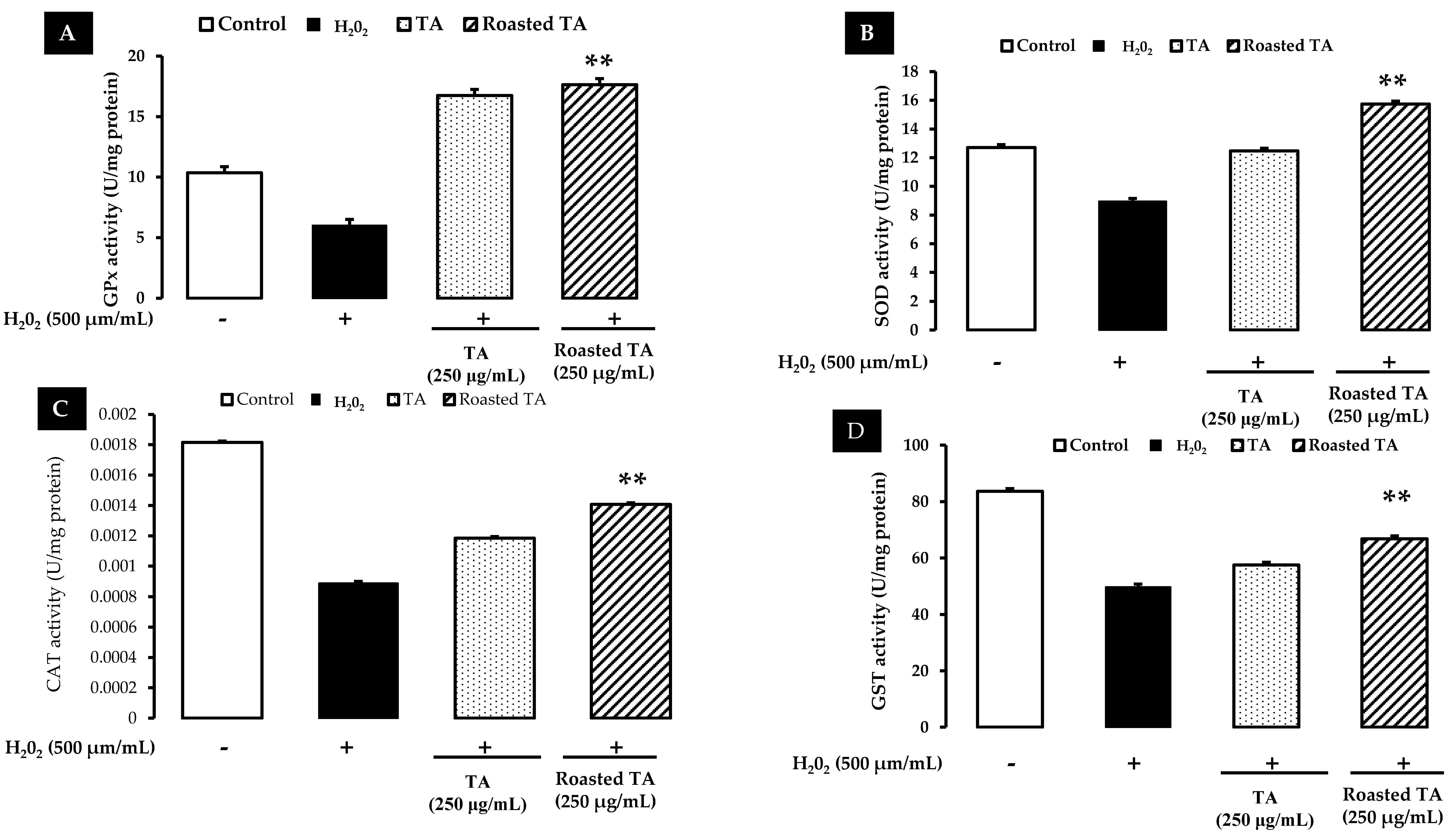
To investigate the antioxidant enzymatic activity caused by taheebo and roasted taheebo, HaCaT cells were treated with both extracts, and GPx, SOD, CAT, and GST levels were examined. Roasted taheebo increased the protein expression of GPx, SOD, CAT, and GST more than unroasted taheebo extract. H2O2 reduced the expression of these proteins, and treatment with roasted taheebo reversed that effect of H2O2. Figure 3 shows that after treatment with H2O2 (100 µM), the activities of the four enzymes were downregulated. Treatment with taheebo or roasted taheebo recovered the enzyme activities compared with the control. Moreover, roasted taheebo appeared to have more antioxidant properties because it increased the activity of the antioxidant enzymes more than unroasted taheebo did (Figure 3).

3.4. In Vitro ROS Induced by Roasted Taheebo on Cancer Cells

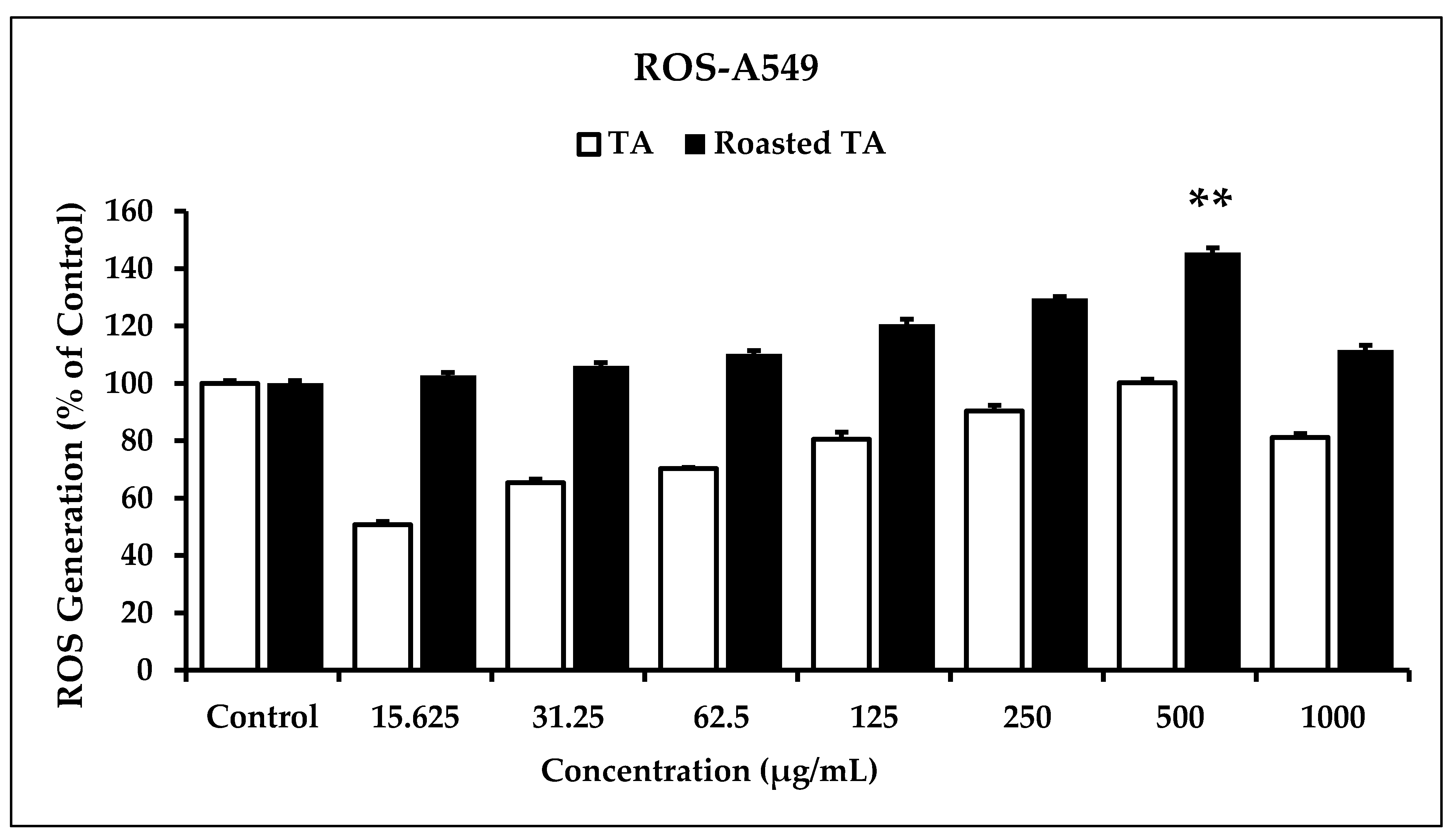
Free radicals negatively affect biological systems. The rapid combination and oxidation of cellular macromolecules by ROS renders them hazardous to cells, tissues, and organs and accelerates the growth of cancer [46,47]. As naturally occurring byproducts of routine cell activity, ROS plays a role in cellular signaling [48]. Cancer cells frequently have higher basal ROS levels than healthy cells due to an imbalance between oxidants and antioxidants. ROS in moderate concentrations are good for cancer cells because they boost cancer cell metabolism and development signals while inhibiting antioxidants that aid in oncogenesis. However, high ROS concentrations can cause DNA damage-related cell death [49,50]. In the A549 cell line, the DCFH-DA fluorescent probe was applied to evaluate the changes in cellular ROS generation following exposure to various doses of TA and roasted TA. As shown in Figure 4, oxidative stress in the A549 cell line was higher after the 500 µg/mL roasted-TA treatment than in the control condition. Previous research has found that a rise in ROS production and apoptosis is associated with a decrease in the membrane potential of mitochondria. Mitochondria produce ROS during apoptosis [51,52]. Furthermore, an increase in ROS levels may result in the production of p53, which regulates the beginning of apoptosis by transforming proteins that promote apoptosis (Bax) or interacting with anti-apoptotic proteins in the mitochondria (Bcl-2) [53,54]. Because they both inhibit cell proliferation and generate ROS, roasted TA extracts might cause apoptosis in A549 lung cancer cells by activating the mitochondrial pathway.

3.5. Cancer Cells Migration Is Inhibited by Taheebo

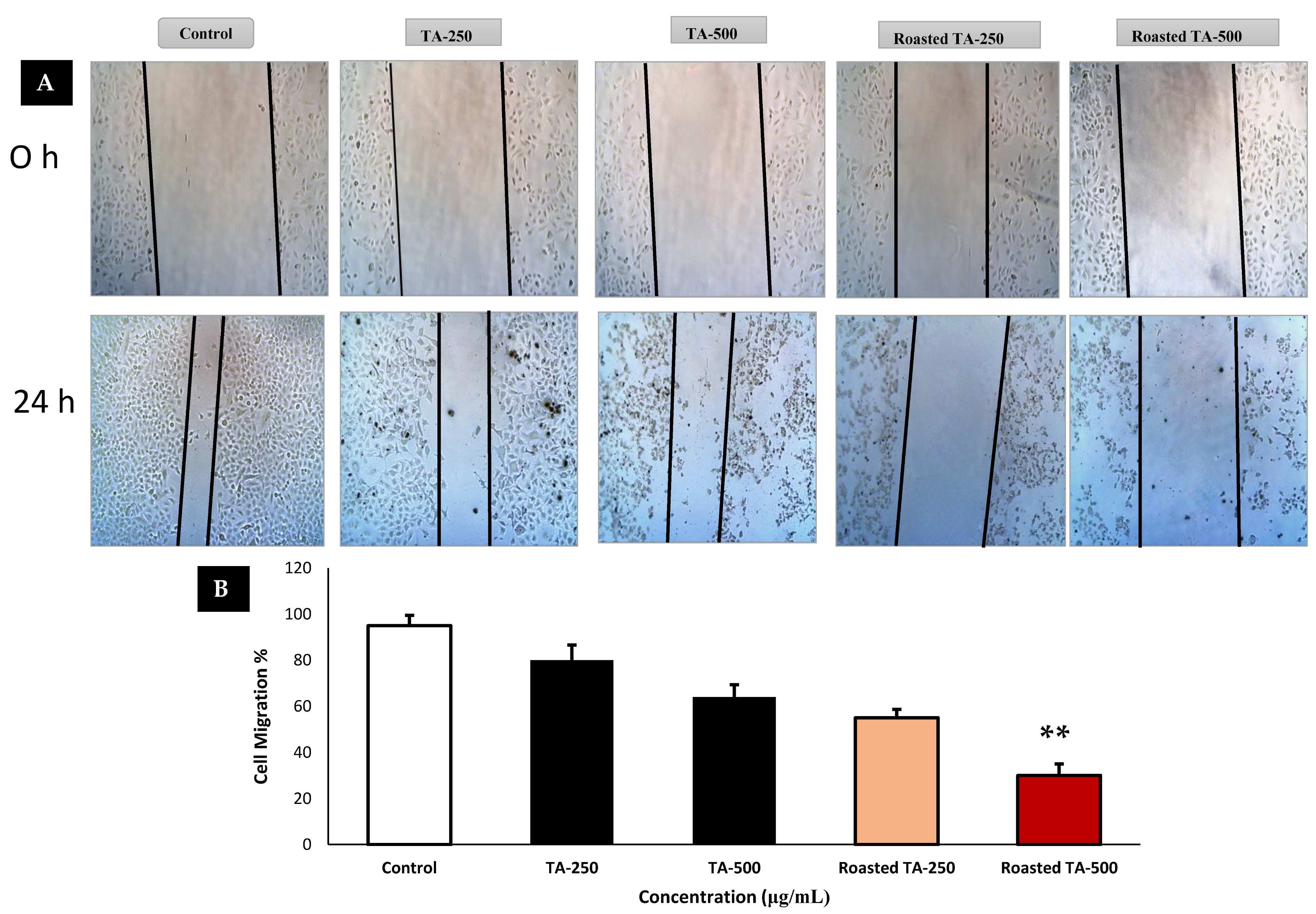
Most cancers have some genetic predisposition through which mutations turn healthy cells into cancerous cells. One fundamental feature of malignancy is the uncontrolled multiplication of cells. The ability of tumor cells to infiltrate and propagate is a cancer hallmark that is governed by genomic instability and mutations [55]. Metastasis is the main cause of cancer-related mortality. As tumor cells spread throughout the body through metastasis, they produce secondary tumor sites and seriously compromise organ function [56]. Because cancer spread accounts for 90% of all cancer-related fatalities, preventing cancer from spreading is a fundamental objective of cancer treatment [57]. The most widely used and reliable in vitro technique for monitoring and evaluating cell migration after therapy is the wound-healing scratch test. To measure cell mobility (%) before and after treatment, A549 lung cancer cells were treated with TA or roasted TA and subjected to a wound closure test (Figure 5). After one day of treatment with roasted TA at a dose of 500 µg/mL, A549 cells had less migration than the control group, which filled nearly all of the spaces between layers with migratory cells after 24 h. The TA treatment was not as successful at stopping migration as the roasted TA treatment. These research findings imply that roasted TA can prevent cancer cells from migrating and metastasizing.

3.6. Inhibition of Colony Formation in Cancer Cells

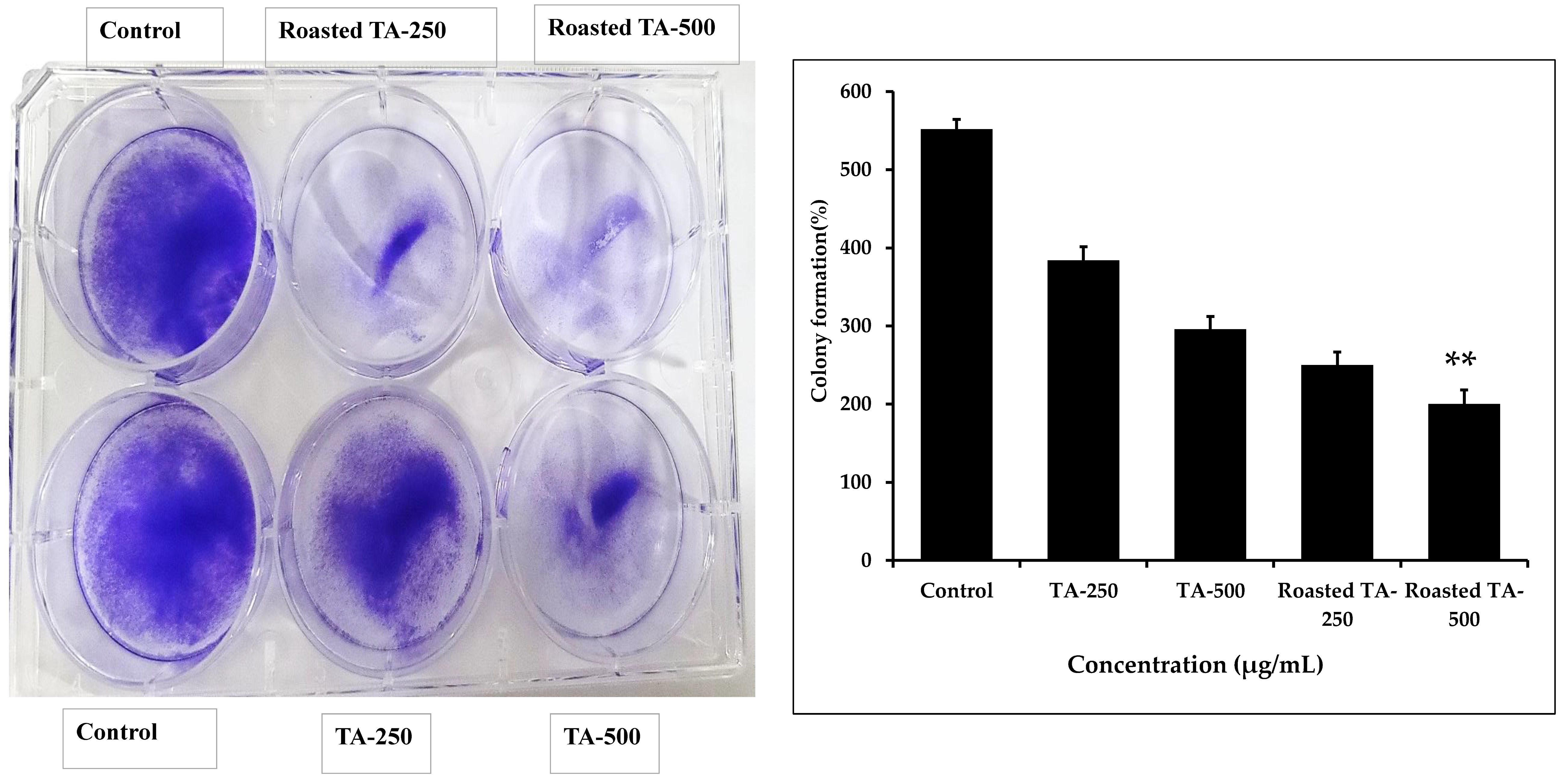
The clonogenic assay, an in vitro test of cell survival, evaluates a cell’s ability to form colonies [58]. This technique tests how medications affect cell growth and proliferation. A microscopic analysis was used to assess the shape and colony formation of A549 cells. As shown in Figure 6, untreated cells produced more colonies than cells treated with roasted TA at 250 or 500 μg/mL. The roasted TA group at 500 μg/mL produced fewer colonies of A549 cells than the TA group. Therefore, the anticancer capabilities of roasted TA include the suppression of colony formation.

3.7. Taheebo Induces Apoptosis by Regulating Apoptotic Gene Expression

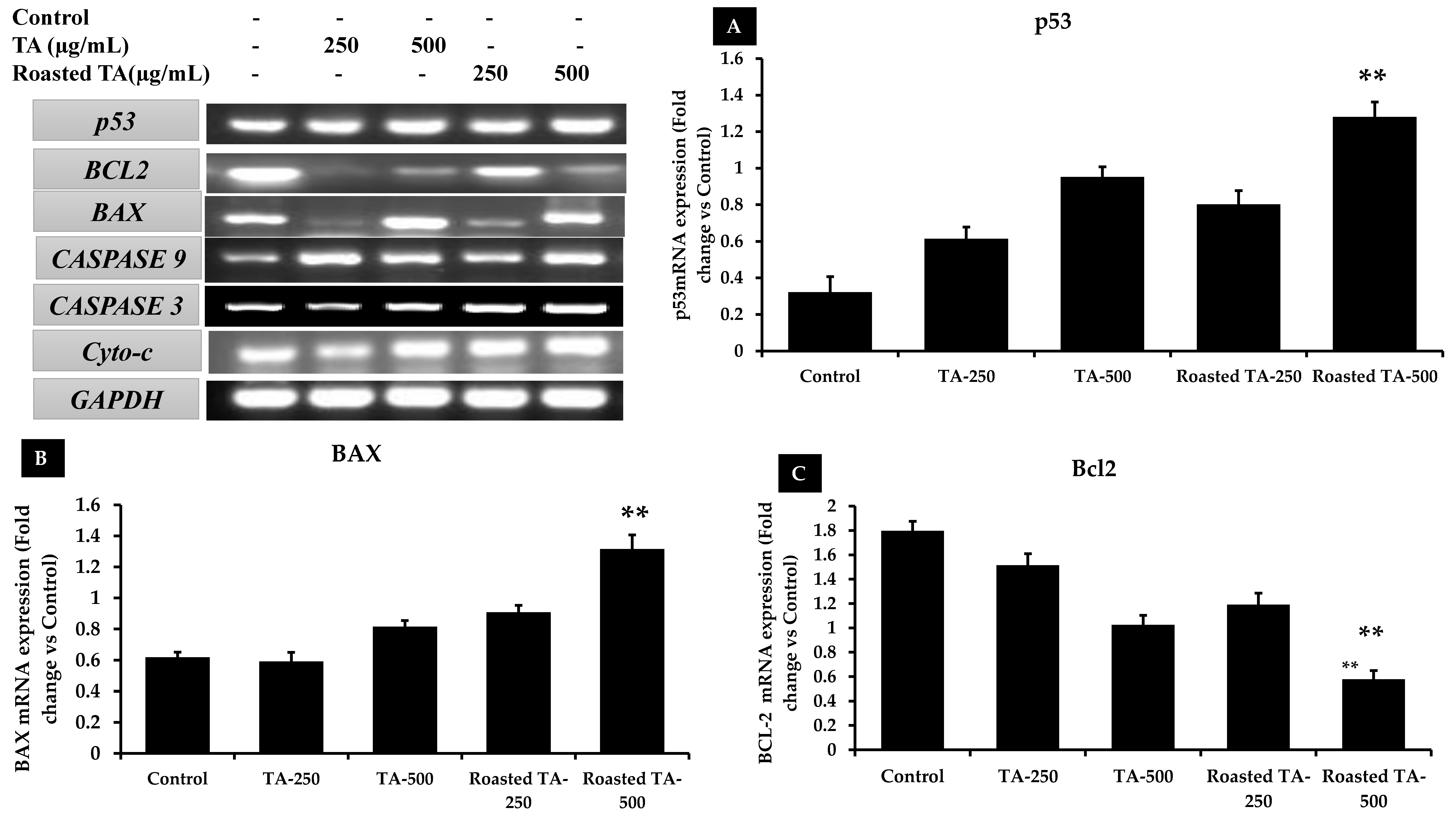
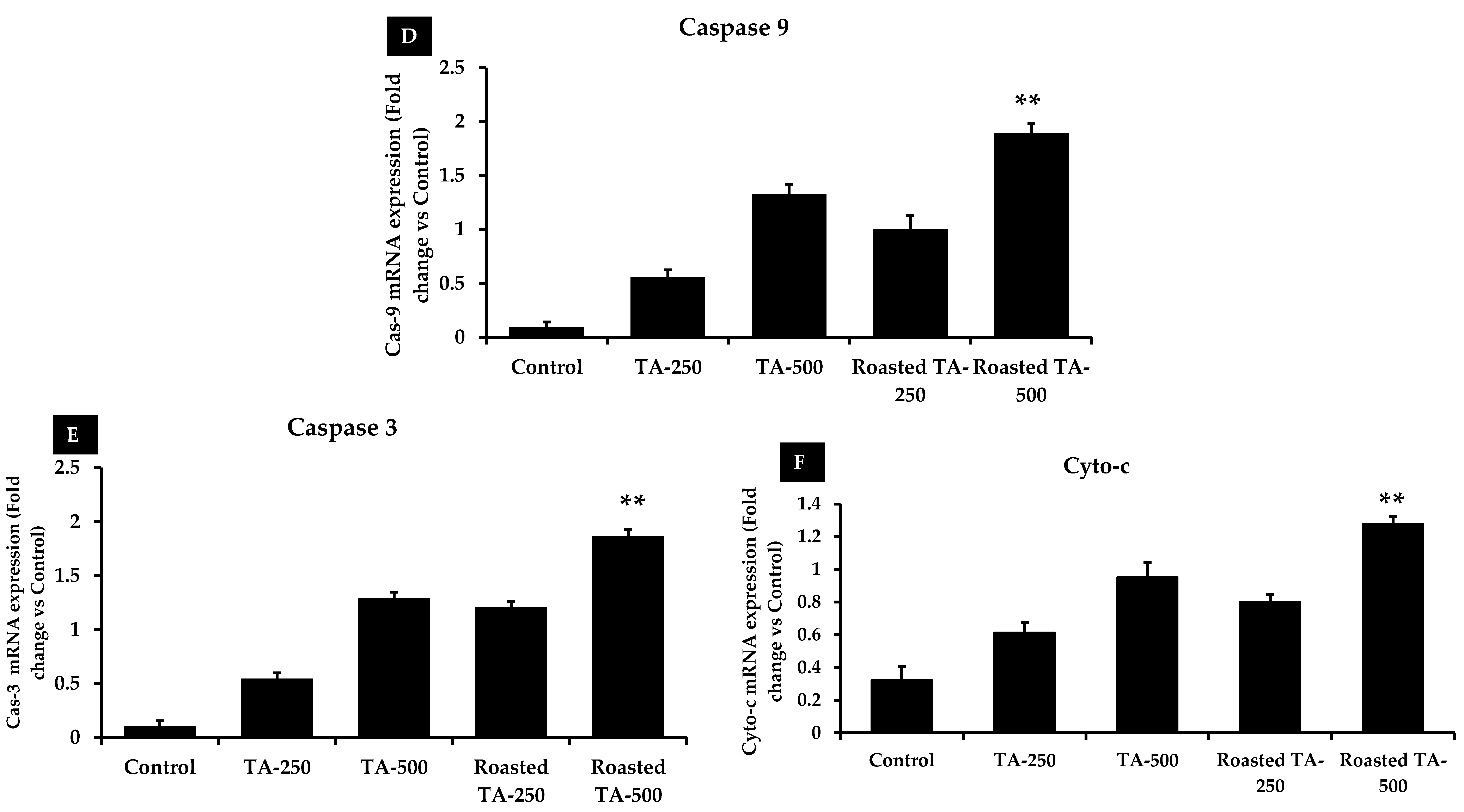
By eliminating damaged, old, or autoimmune cells, apoptosis plays a crucial role in the development and maintenance of multicellular organisms [59]. Anticancer mediators are vital for inducing apoptosis, which is necessary for destroying tumor cells. A substance that kills or prevents the multiplication of cancer cells by inducing apoptosis is an efficient anticancer agent [60]. Cancer cells can undergo apoptosis through intrinsic and extrinsic routes that are regulated by the caspase-dependent proteolysis of hundreds of cellular proteins, membrane blebbing, and endonucleolytic breakage of chromosomal DNA [61]. Phytochemicals that promote apoptosis via intrinsic apoptotic pathways trigger a range of intracellular stimuli. ROS produced by mitochondria are essential for redox signaling, and p53 inhibits cancer by becoming redox active. ROS cause p53 to become active, which in turn causes cancer cells to go through apoptosis [62,63]. p53 controls the expression of the pro- and anti-apoptotic genes bax and bcl2, respectively [64]. When Bax expression is increased, it can lead to apoptosis, but when Bax expression is reduced, Bcl-2 can stop apoptosis [65]. Variation in the range of those proteins damages the mitochondrial membrane, which triggers the release of cytochrome-c and regulates the activation of caspase-9 [66]. Caspases, one of the most important types of proteases, are essential for cell death. The bax protein is an essential mitochondrial membrane component that facilitates the transport of cytochrome-c between membranes, leading to the formation of apoptotic remnants, and it activates caspase-9 and caspase-3 to produce apoptosis [67,68,69]. As shown by the RT-PCR results, roasted TA substantially increased the expression of the genes for p53, Bax, cyto-c, caspase 3, and caspase 9 and decreased the expression of the gene for Bcl-2, compared with both the untreated control and TA samples (Figure 7). Similarly, roasted TA downregulated the expression of bcl-2 (0.57 fold) and upregulated the expression of p53 (1.18 fold), bax (1.31 fold), cyto-c (1.28 fold), caspase 3 (1.86 fold), and caspase 9 (1.89 fold). The gene expression changes in the TA sample groups were smaller than those in the roasted TA sample groups. Thus, roasted TA can reduce the expression of anti-apoptosis genes in lung cancer cells. To completely comprehend those biological processes, Western blot analyses and more study of the molecular mechanisms are needed.

3.8. Taheebo Extract Increased NO Production and Inhibited ROS Generation Induced by LPS

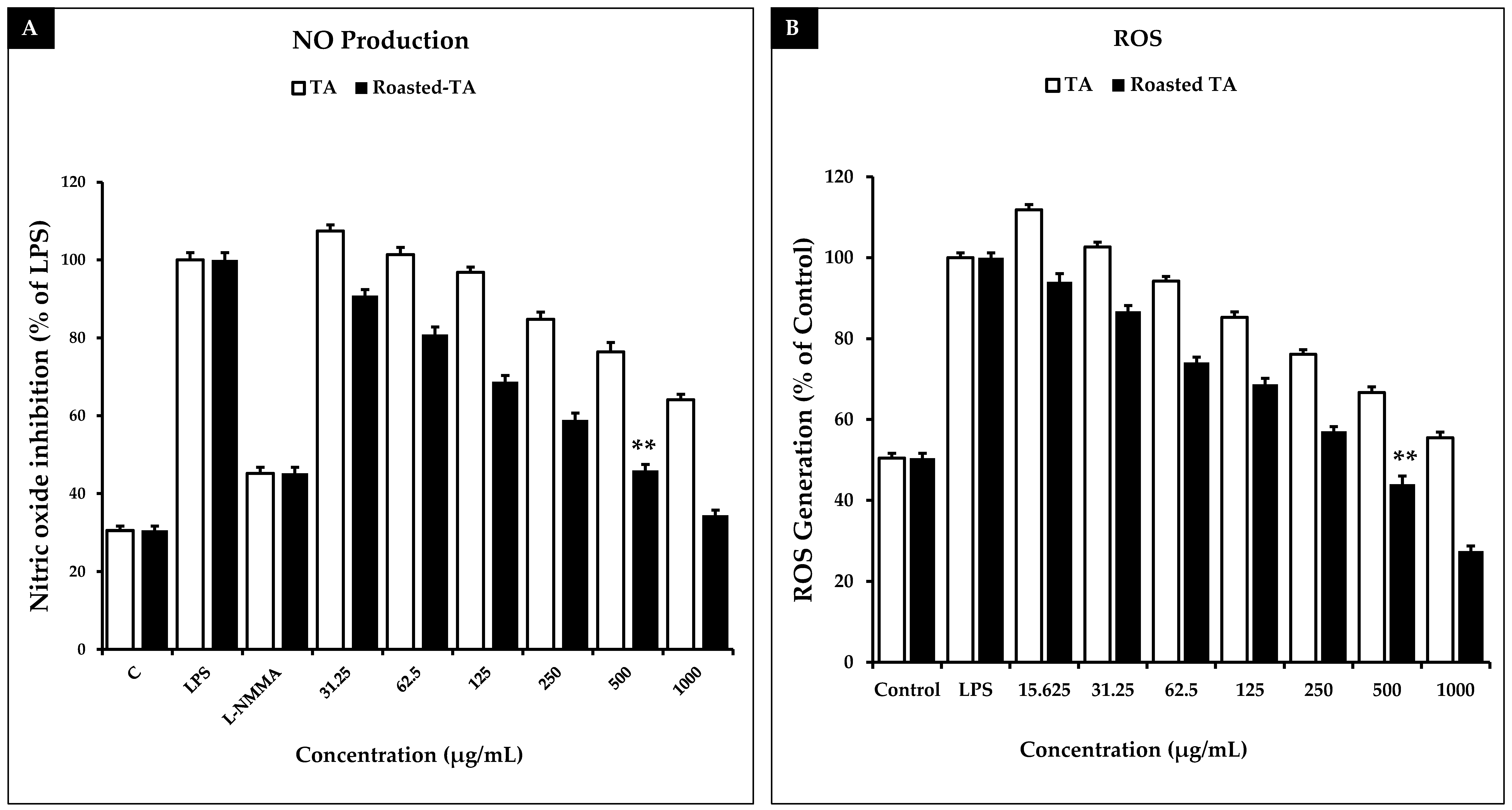
Macrophages are beneficial for both host defensive mechanisms and inflammation. Many inflammatory disorders, including cancer, create too many macrophages [70]. NO, TNF-α, IL-1, and IL-6 are among the inflammatory mediators secreted by activated macrophages. Therefore, preventing macrophage activation is a key goal for treating inflammatory illnesses. NO is a common cellular mediator of physiological and pathological processes that is mostly produced in inflamed areas [71]. When macrophages are stimulated by LPS, the inducible isoform of nitric oxide synthase (iNOS) produces a considerable amount of NO [72]. Numerous fundamental physiological processes in mammalian cells depend on appropriate NO production [73]. The ROS created by activated neutrophils and macrophages are another significant pathogenic agent that can harm many tissues and organs [74]. Excessive ROS generation can result in protein and nucleic acid oxidation and have harmful effects on cell structures in a variety of pathogenic and infectious situations [75]. A redox imbalance caused by ROS buildup from the endogenous antioxidant response causes oxidative stress [76]. ROS generation may potentially be responsible for the induction of iNOS, as evidenced by previous findings that oxygen radical scavengers could suppress TNF-α induced iNOS expression [77]. In this study, LPS-stimulated RAW 264.7 cells were exposed to TA and roasted TA samples at concentrations ranging from 15.625 to 1000 μg/mL, and the amount of NO production was measured. At 500 μg/mL, the roasted-TA sample produced significantly greater decreases in LPS-induced NO generation (54.07%) than the TA sample (23.56%). In this study, L-NMMA was used as the positive control for preventing NO generation (Figure 8A). RAW 264.7 macrophage cells were exposed to varying doses of the samples in the presence and absence of LPS for 24 h to test how well the samples reduced the ROS production caused by LPS. Figure 8B demonstrates that the LPS-treated groups produced more ROS than the control group. The results show that roasted TA inhibited the LPS-induced inflammatory response better than the TA sample at 500 μg/mL.

3.9. Taheebo Extract Inhibited the Increased Levels of Inflammation-Related Cytokines

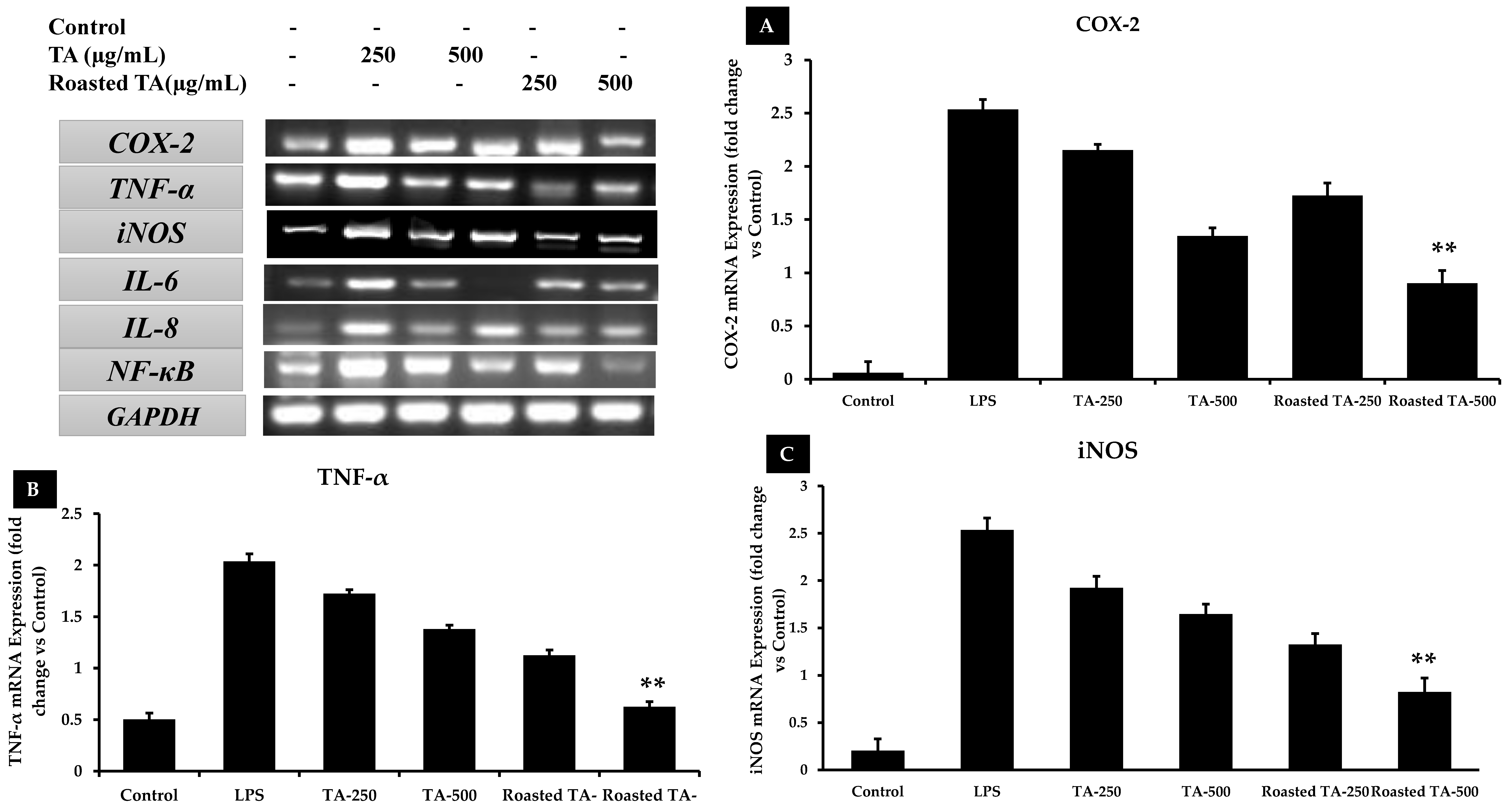
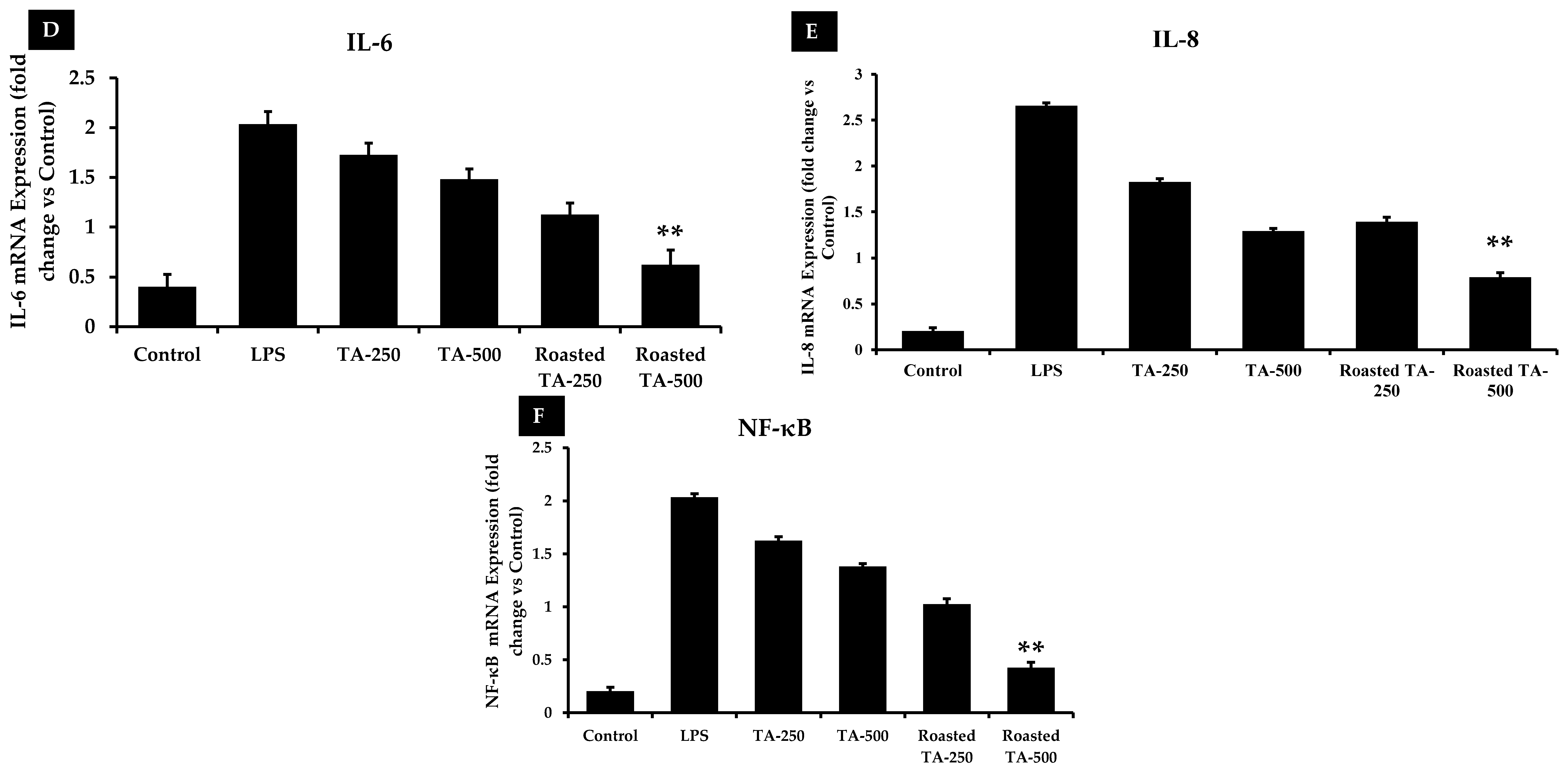
An organism’s universal response to modifications in its local tissues is inflammation. Pro-inflammatory mediators play a significant role in the development of inflammatory disorders [78]. Important inflammatory response signaling pathways such as nuclear factor kappa B (NF-κB) and mitogen-activated protein kinase (MAPK) regulate a range of innate and adaptive immune responses. During the inflammatory response, both NF-κB and MAPK signaling significantly regulate the generation of pro-inflammatory mediators. LPS can activate nuclear transcription factors such as NF-κB and MAPK, which in turn stimulate signaling pathways in macrophage cells. When LPS is expressed, macrophages start producing iNOS, TNF-α, IL-6, COX-2, and other inflammatory mediators [79]. Important inflammatory response signaling pathways such as nuclear factor kappa B (NF-κB) and mitogen-activated protein kinase (MAPK) regulate a range of innate and adaptive immune responses. During the inflammatory response, both NF-κB and MAPK signaling significantly regulate the generation of pro-inflammatory mediators. LPS can activate nuclear transcription factors such as NF-κB and MAPK, which in turn stimulate signaling pathways in macrophage cells. When LPS is expressed, macrophages start producing iNOS, TNF-α, IL-6, COX-2, and other inflammatory mediators [80]. This study used RT-PCR and qRT-PCR to examine how TA and roasted TA influenced the gene expression for those factors. Significantly higher mRNA expression of COX-2, TNF-α, iNOS, IL-6, IL-8, and NF-κB was seen in the LPS-treated groups than in the control group, and roasted TA extract suppressed that gene expression better than TA in a dose-dependent manner. Compared with TA, roasted TA dramatically reduced the expression of the COX-2, TNF-α, iNOS, IL-6, IL-8, and NF-κB genes by 0.90, 0.62, 0.82, 0.62, 0.79, and 0.42-fold, respectively (Figure 9). Therefore, the anti-inflammatory activity of roasted TA is greater than that of TA alone.

4. Conclusions

This study demonstrates that in vitro roasted taheebo samples from Handroanthus impetiginosus species showed more antioxidant, anti-inflammatory, and anti-lung cancer activity than only the extract of taheebo in RAW 264.7, Hacat, and A549 cell lines through the NF-κB and p53 signaling pathways. The genus Handroanthus impetiginosus (Bignoniaceae) is widely used for the treatment of cancer, inflammation, and anti-oxidants in traditional medicine in Brazil and other South American countries. In the present study, roasted taheebo exhibited more phenolic and flavonoid content and stronger antioxidant activity than unroasted taheebo. Moreover, roasted taheebo extract significantly reduced oxidative stress by increasing the expression of antioxidant enzymes in H2O2-induced HaCaT cells. We also found that roasted taheebo is not toxic to RAW 246.7 cells, in which it inhibited pro-inflammatory mediators such as COX-2, TNF-α, iNOS, IL-6, IL-8, and NF-κB and inhibited NO synthesis and intercellular ROS production. Intriguingly, our anticancer studies show that roasted taheebo has considerable toxicity to A549 lung cancer cells, in which its upregulation of ROS levels impedes cell migration and induces apoptosis. The expression of the p53, Bax, cyto-c, caspase 3, and caspase 9 genes was upregulated by roasted taheebo extracts, and Bcl-2 gene expression was downregulated via the intrinsic mitochondrial pathway. Our findings encourage future research to identify potential therapeutic medicinal compounds from the Bignoniaceae family for the development of herb-based anti-inflammatory, antioxidant, and anticancer medicines.

Author Contributions

Conceptualization, D.C.Y., K.J.H., L.J.S., E.J.R. and S.-K.J.; methodology, J.N., E.J.R., M.N.M., J.H.L., A.K.V. and M.A., software, J.N., E.J.R., A.K.V. and M.A., validation, E.J.R., D.C.Y. and S.-K.J., formal analysis, J.N., E.J.R., M.A.-A. and J.C.A.; resources, D.C.Y., K.J.H., L.J.S. and S.-K.J.; data curation, E.J.R., J.N., M.N.M., A.K.V. and M.A.; investigation, D.C.Y., J.C.A., E.J.R. and S.-K.J.; writing—original draft preparation, J.N., M.N.M. and E.J.R.; writing—review and editing, J.N., M.N.M., E.J.R., M.A.-A. and S.-K.J.; supervision, D.C.Y., E.J.R. and S.-K.J.; project administration, D.C.Y., K.J.H., L.J.S., E.J.R. and S.-K.J. All authors have read and agreed to the published version of the manuscript.

Funding

The work is supported by the NK TAHEEBO company, Hwaseong-si, Gyeonggi-do, Republic of Korea.

Institutional Review Board Statement

The study was conducted in accordance with the Declaration of Helsinki, and approved by the Kyung Hee University, South Korea. Ethical review and approval were waived for this study due to we didn’t do any in vivo or human experiments.

Informed Consent Statement

Patient consent was waived due to the following reasons: The human lung cancer cell was collected from Korean Cell Line Bank (https://cellbank.snu.ac.kr/main/index.html, accessed on 1 October 2023) and was previously used for various research related to lung cancer at Kyung Hee University at Han-bang Bio Lab.

Data Availability Statement

The authors confirm that the data supporting the findings of this study are available within the article.

Conflicts of Interest

Authors Ko Jeong Hun and Lee Ji Sook were employed by the NK TAHEEBO Company. Author Deok Chun Yang was employed by the Hanbangbio Inc. The remaining authors declare that the research was conducted in the absence of any commercial or financial relationships that could be construed as a potential conflict of interest. The authors declare that they have no known competing financial interests or personal relationships that could have appeared to influence the work reported in this paper. Ko Jeong Hun and Lee Ji Sook are employee of NK TAHEEBO Company, who provided funding and technical support for the work. The funder had no role in the design of the study; in the collection, analysis, or interpretation of data, in the writing of the manuscript, or in the decision to publish the results.

References

  1. Ngo, L.T.; Okogun, J.I.; Folk, W.R. 21st Century natural product research and drug development and traditional medicines. Nat. Prod. Rep. 2013, 30, 584–592. [Google Scholar] [CrossRef]
  2. Akter, R.; Yang, D.U.; Ahn, J.C.; Awais, M.; Nahar, J.; Ramadhania, Z.M.; Kim, J.Y.; Lee, G.J.; Kwak, G.-Y.; Lee, D.W.; et al. Comparison of In Vitro Estrogenic Activity of Polygoni multiflori Radix and Cynanchi wilfordii Radix via the Enhancement of ERα/β Expression in MCF7 Cells. Molecules 2023, 28, 2199. [Google Scholar] [CrossRef] [PubMed]
  3. Zhang, J.; Hunto, S.T.; Yang, Y.; Lee, J.; Cho, J.Y. Tabebuia impetiginosa: A comprehensive review on traditional uses, phytochemistry, and immunopharmacological properties. Molecules 2020, 25, 4294. [Google Scholar] [CrossRef]
  4. Sobreiro, M.B.; Vieira, L.D.; Nunes, R.; Novaes, E.; Coissac, E.; Silva-Junior, O.B.; Grattapaglia, D.; Collevatti, R.G. Chloroplast genome assembly of Handroanthus impetiginosus: Comparative analysis and molecular evolution in Bignoniaceae. Planta 2020, 252, 1–16. [Google Scholar] [CrossRef] [PubMed]
  5. Jausoro, V.; Llorente, B.E.; Apóstolo, N.M. Structural differences between hyperhydric and normal in vitro shoots of Handroanthus impetiginosus (Mart. ex DC) Mattos (Bignoniaceae). Plant Cell Tissue Organ Cult. (PCTOC) 2010, 101, 183–191. [Google Scholar] [CrossRef]
  6. Edwards, S.E.; da Costa Rocha, I.; Williamson, E.M.; Heinrich, M. Lapacho Handroanthus impetiginosus (Mart. ex DC.) Mattos. In Phytopharmacy: An Evidence-Based Guide to Herbal Medicinal Products; Wiley: Hoboken, NJ, USA, 2015; p. 234. [Google Scholar]
  7. Jones, K. Pau d’Arco: Immune Power from the Rain Forest; Inner Traditions/Bear & Co: Rochester, VT, USA, 1995. [Google Scholar]
  8. Park, B.-S.; Lee, K.-G.; Shibamoto, T.; Lee, S.-E.; Takeoka, G.R. Antioxidant Activity and Characterization of Volatile Constituents of Taheebo (Tabebuia impetiginosa Martius ex DC). J. Agric. Food Chem. 2002, 51, 295–300. [Google Scholar] [CrossRef] [PubMed]
  9. Malange, K.F.; dos Santos, G.G.; Kato, N.N.; Toffoli-Kadri, M.C.; Carollo, C.A.; Silva, D.B.; Portugal, L.C.; Alves, F.M.; Rita, P.H.S.; Parada, C.A.; et al. Tabebuia aurea decreases hyperalgesia and neuronal injury induced by snake venom. J. Ethnopharmacol. 2018, 233, 131–140. [Google Scholar] [CrossRef] [PubMed]
  10. Suo, M.; Isao, H.; Kato, H.; Takano, F.; Ohta, T. Anti-inflammatory constituents from Tabebuia avellanedae. Fitoterapia 2012, 83, 1484–1488. [Google Scholar] [CrossRef]
  11. Pires, T.C.S.P.; Dias, M.I.; Calhelha, R.C.; Carvalho, A.M.; Queiroz, M.-J.R.P.; Barros, L.; Ferreira, I.C.F.R. Bioactive Properties of Tabebuia impetiginosa-Based Phytopreparations and Phytoformulations: A Comparison between Extracts and Dietary Supplements. Molecules 2015, 20, 22863–22871. [Google Scholar] [CrossRef]
  12. Byeon, S.E.; Chung, J.Y.; Lee, Y.G.; Kim, B.H.; Kim, K.H.; Cho, J.Y. In vitro and in vivo anti-inflammatory effects of taheebo, a water extract from the inner bark of Tabebuia avellanedae. J. Ethnopharmacol. 2008, 119, 145–152. [Google Scholar] [CrossRef]
  13. Castellanos, J.R.G.; Prieto, J.M.; Heinrich, M. Red Lapacho (Tabebuia impetiginosa)—A global ethnopharmacological commodity? J. Ethnopharmacol. 2009, 121, 1–13. [Google Scholar] [CrossRef] [PubMed]
  14. Mowrey, D. Ancient Herb. Modern Medicine; Mountainwest Institute of Herbal Sciences: Salt Lake City, UT, USA, 2001. [Google Scholar]
  15. Jin, Y.; Jeong, K.M.; Lee, J.; Zhao, J.; Choi, S.-Y.; Baek, K.-S. Development and Validation of an Analytical Method Readily Applicable for Quality Control of Tabebuia impetiginosa (Taheebo) Ethanolic Extract. J. AOAC Int. 2018, 101, 695–700. [Google Scholar] [CrossRef] [PubMed]
  16. Bezerra, J.J.L.; Johanes, I.; Pinheiro, A.A.V. Anticancer potential and toxicity of the genus Handroanthus Mattos (Bignoniaceae): A systematic review. Toxicon 2022, 217, 131–142. [Google Scholar] [CrossRef] [PubMed]
  17. Valle, A.C.V.; Carvalho, A.; Rahme, S.W.; Araújo, A.d.R.B.; Malard, P.F.; Brunel, H.S.S. Evaluation of cell viability and nitric oxide release after treatment of human hepatocellular carcinoma cells with a homeopathic compound of Graviola (Anonna muricata) and Purple Ipe (Handroanthus impetiginosus). Med. Res. Arch. 2023, 11, 4091. [Google Scholar] [CrossRef]
  18. Lee, M.H.; Choi, H.M.; Hahm, D.-H.; Her, E.; Yang, H.-I.; Yoo, M.C.; Kim, K.S. Analgesic and anti-inflammatory effects in animal models of an ethanolic extract of Taheebo, the inner bark of Tabebuia avellanedae. Mol. Med. Rep. 2012, 6, 791–796. [Google Scholar] [CrossRef] [PubMed]
  19. Lee, S.-E.; Park, B.-S.; Yum, J.H. Antioxidative activity of taheebo (Tabebuia impetiginosa Martius ex DC.) extracts on the H2O2-induced NIH3T3 cells. J. Med. Plants Res. 2012, 6, 5258–5265. [Google Scholar]
  20. Iwamoto, K.; Fukuda, Y.; Tokikura, C.; Noda, M.; Yamamoto, A.; Yamamoto, M.; Yamashita, M.; Zaima, N.; Iida, A.; Moriyama, T. The anti-obesity effect of Taheebo (Tabebuia avellanedae Lorentz ex Griseb) extract in ovariectomized mice and the identification of a potential anti-obesity compound. Biochem. Biophys. Res. Commun. 2016, 478, 1136–1140. [Google Scholar] [CrossRef]
  21. Mukherjee, B.; Telang, N.; Wong, G.Y.C. Growth inhibition of estrogen receptor positive human breast cancer cells by Taheebo from the inner bark of Tabebuia avellandae tree. Int. J. Mol. Med. 1998, 24, 253–260. [Google Scholar] [CrossRef]
  22. Böhler, T.; Nolting, J.; Gurragchaa, P.; Lupescu, A.; Neumayer, H.-H.; Budde, K.; Kamar, N.; Klupp, J. Tabebuia avellanedae extracts inhibit IL-2-independent T-lymphocyte activation and proliferation. Transpl. Immunol. 2008, 18, 319–323. [Google Scholar] [CrossRef]
  23. Tahara, T.; Watanabe, A.; Yutani, M.; Yamano, Y.; Sagara, M.; Nagai, S.; Saito, K.; Yamashita, M.; Ihara, M.; Iida, A. STAT3 inhibitory activity of naphthoquinones isolated from Tabebuia avellanedae. Bioorganic Med. Chem. 2020, 28, 115347. [Google Scholar] [CrossRef]
  24. Freitas, A.E.; Budni, J.; Lobato, K.R.; Binfaré, R.W.; Machado, D.G.; Jacinto, J.; Veronezi, P.O.; Pizzolatti, M.G.; Rodrigues, A.L.S. Antidepressant-like action of the ethanolic extract from Tabebuia avellanedae in mice: Evidence for the involvement of the monoaminergic system. Prog. Neuro-Psychopharmacol. Biol. Psychiatry 2010, 34, 335–343. [Google Scholar] [CrossRef] [PubMed]
  25. Lee, S.W.; Lee, J.H. Effects of oven-drying, roasting, and explosive puffing process on isoflavone distributions in soybeans. Food Chem. 2009, 112, 316–320. [Google Scholar] [CrossRef]
  26. Kim, W.; Kim, S.-Y.; Kim, D.-O.; Kim, B.-Y.; Baik, M.-Y. Puffing, a novel coffee bean processing technique for the enhancement of extract yield and antioxidant capacity. Food Chem. 2018, 240, 594–600. [Google Scholar] [CrossRef] [PubMed]
  27. Choi, G.-S.; Shin, J.-S.; Kim, W.; Baik, M.-Y.J.F. Increases in Ginsenoside Rg3, Compound K, and Antioxidant Activity of Cultivated Wild Panax Ginseng (CWPG) by Puffing. Foods 2022, 11, 2936. [Google Scholar] [CrossRef] [PubMed]
  28. Kaur, R.; Kumar, A.; Kumar, V.; Kumar, S.; Saini, R.K.; Nayi, P.; Gehlot, R. Recent advancements and applications of explosion puffing. Food Chem. 2023, 403, 134452. [Google Scholar] [CrossRef] [PubMed]
  29. Ramadhania, Z.M.; Yang, D.U.; Moektiwardojo, M.; Han, Y.; Park, J.K.; Rupa, E.J.; Yang, D.C.; Lee, S.J.; Kang, S.C. Enhanced Anti-Skin Aging Effects of Fermented Black Ginseng (Panax ginseng C.A. Meyer) by Aspergillus niger KHNT-1. Appl. Sci. 2022, 13, 550. [Google Scholar] [CrossRef]
  30. Nahar, J.; Boopathi, V.; Rupa, E.J.; Awais, M.; Valappil, A.K.; Morshed, M.N.; Murugesan, M.; Akter, R.; Yang, D.U.; Mathiyalagan, R. Protective Effects of Aquilaria agallocha and Aquilaria malaccensis Edible Plant Extracts against Lung Cancer, Inflammation, and Oxidative Stress—In Silico and In Vitro Study. Appl. Sci. 2023, 13, 6321. [Google Scholar] [CrossRef]
  31. Akter, R.; Kwak, G.-Y.; Ahn, J.C.; Mathiyalagan, R.; Ramadhania, Z.M.; Yang, D.C.; Kang, S.C. Protective Effect and Potential Antioxidant Role of Kakadu Plum Extracts on Alcohol-Induced Oxidative Damage in HepG2 Cells. Appl. Sci. 2021, 12, 236. [Google Scholar] [CrossRef]
  32. Bak, M.-J.; Jun, M.; Jeong, W.-S. Antioxidant and Hepatoprotective Effects of the Red Ginseng Essential Oil in H2O2-Treated HepG2 Cells and CCl4-Treated Mice. Int. J. Mol. Sci. 2012, 13, 2314–2330. [Google Scholar] [CrossRef]
  33. Piao, M.J.; Yoo, E.S.; Koh, Y.S.; Kang, H.K.; Kim, J.; Kim, Y.J.; Kang, H.H.; Hyun, J.W. Antioxidant Effects of the Ethanol Extract from Flower of Camellia japonica via Scavenging of Reactive Oxygen Species and Induction of Antioxidant Enzymes. Int. J. Mol. Sci. 2011, 12, 2618–2630. [Google Scholar] [CrossRef]
  34. Sikdar, S.; Lallemand, B.; Dubois, J. Induction of Phase II Enzymes Glutathione-S-Transferase and NADPH: Quinone Oxydoreductase 1 with Novel Sulforaphane Derivatives in Human Keratinocytes: Evaluation of the Intracellular GSH Level. Pharmacol. Pharm. 2014, 05, 937–943. [Google Scholar] [CrossRef]
  35. Nagano, T.; Tachihara, M.; Nishimura, Y. Mechanism of Resistance to Epidermal Growth Factor Receptor-Tyrosine Kinase Inhibitors and a Potential Treatment Strategy. Cells 2018, 7, 212. [Google Scholar] [CrossRef] [PubMed]
  36. Morshed, N.; Ahn, J.C.; Mathiyalagan, R.; Rupa, E.J.; Akter, R.; Karim, R.; Jung, D.H.; Yang, D.U.; Yang, D.C.; Jung, S.K. Antioxidant Activity of Panax ginseng to Regulate ROS in Various Chronic Diseases. Appl. Sci. 2023, 13, 2893. [Google Scholar] [CrossRef]
  37. Saeed, A.; Marwat, M.S.; Shah, A.H.; Naz, R.; Abidin, S.Z.-U.; Akbar, S.; Khan, R.; Navid, M.T.; Saeed, A. Assessment of total phenolic and flavonoid contents of selected fruits and vegetables. Indian J. Tradit. Knowl. (IJTK) 2019, 18, 686–693. [Google Scholar]
  38. Ullah, A.; Munir, S.; Badshah, S.L.; Khan, N.; Ghani, L.; Poulson, B.G.; Emwas, A.-H.; Jaremko, M. Important Flavonoids and Their Role as a Therapeutic Agent. Molecules 2020, 25, 5243. [Google Scholar] [CrossRef] [PubMed]
  39. Lue, B.-M.; Nielsen, N.S.; Jacobsen, C.; Hellgren, L.; Guo, Z.; Xu, X. Antioxidant properties of modified rutin esters by DPPH, reducing power, iron chelation and human low density lipoprotein assays. Food Chem. 2010, 123, 221–230. [Google Scholar] [CrossRef]
  40. Mushtaq, S.; Abbasi, B.H.; Uzair, B.; Abbasi, R. Natural products as reservoirs of novel therapeutic agents. EXCLI J. 2018, 17, 420–451. [Google Scholar] [CrossRef]
  41. Cragg, G.M.; Newman, D.J. Plants as a source of anti-cancer agents. J. Ethnopharmacol. 2005, 100, 72–79. [Google Scholar] [CrossRef]
  42. Su, X.-L.; Wang, J.-W.; Che, H.; Wang, C.-F.; Jiang, H.; Lei, X.; Zhao, W.; Kuang, H.-X.; Wang, Q.-H. Clinical application and mechanism of traditional Chinese medicine in treatment of lung cancer. Chin. Med. J. 2020, 133, 2987–2997. [Google Scholar] [CrossRef]
  43. Ghuman, S.; Ncube, B.; Finnie, J.; McGaw, L.; Njoya, E.M.; Coopoosamy, R.; Van Staden, J. Antioxidant, anti-inflammatory and wound healing properties of medicinal plant extracts used to treat wounds and dermatological disorders. S. Afr. J. Bot. 2019, 126, 232–240. [Google Scholar] [CrossRef]
  44. Angusti, A.; Manfredini, S. The Antioxidants and Pro-Antioxidants Network: An Overview. Curr. Pharm. Des. 2004, 10, 1677–1694. [Google Scholar] [CrossRef]
  45. Alscher, R.G.; Erturk, N.; Heath, L.S. Role of superoxide dismutases (SODs) in controlling oxidative stress in plants. J. Exp. Bot. 2002, 53, 1331–1341. [Google Scholar] [CrossRef] [PubMed]
  46. Siddique, N.A.; Mujeeb, M.; Najmi, A.K.; Khan, H.; Farooqi, H. Evaluation of antioxidant activity, quantitative estimation of phenols and flavonoids in different parts of Aegle marmelos. J. Saudi Chem. Soc. 2010, 4, 5. [Google Scholar] [CrossRef]
  47. Koksal, E.; Bursal, E.; Dikici, E.; Tozoglu, F.; Gulcin, I. Antioxidant activity of Melissa officinalis leaves. J. Med. Plants Res. 2011, 5, 217–222. [Google Scholar]
  48. Rao, P.C.; Begum, S.; Sahai, M.; Sriram, D.S. Coptisine-induced cell cycle arrest at G2/M phase and reactive oxygen species–dependent mitochondria-mediated apoptosis in non-small-cell lung cancer A549 cells. Tumor Biol. 2017, 39, 1010428317694565. [Google Scholar] [CrossRef] [PubMed]
  49. Zhang, J.; Wang, X.; Vikash, V.; Ye, Q.; Wu, D.; Liu, Y.; Dong, W. ROS and ROS-mediated cellular signaling. Oxid. Med. Cell. Longev. 2016, 2016, 4350965. [Google Scholar] [CrossRef] [PubMed]
  50. Perillo, B.; Di Donato, M.; Pezone, A.; Di Zazzo, E.; Giovannelli, P.; Galasso, G.; Castoria, G.; Migliaccio, A. ROS in cancer therapy: The bright side of the moon. Exp. Mol. Med. 2020, 52, 192–203. [Google Scholar] [CrossRef]
  51. Zamzami, N.; Marchetti, P.; Castedo, M.; Decaudin, D.; Macho, A.; Hirsch, T.; A Susin, S.; Petit, P.X.; Mignotte, B.; Kroemer, G. Sequential reduction of mitochondrial transmembrane potential and generation of reactive oxygen species in early programmed cell death. J. Exp. Med. 1995, 182, 367–377. [Google Scholar] [CrossRef]
  52. Guo, R.; Chen, X.; Nguyen, T.; Chai, J.; Gao, Y.; Wu, J.; Li, J.; Abdel-Rahman, M.A.; Chen, X.; Xu, X. The Strong Anti-Tumor Effect of Smp24 in Lung Adenocarcinoma A549 Cells Depends on Its Induction of Mitochondrial Dysfunctions and ROS Accumulation. Toxins 2022, 14, 590. [Google Scholar] [CrossRef]
  53. Hu, T.-Y.; Ju, J.-M.; Mo, L.-H.; Ma, L.; Hu, W.-H.; You, R.-R.; Chen, X.-Q.; Chen, Y.-Y.; Liu, Z.-Q.; Qiu, S.-Q. Anti-inflammation action of xanthones from Swertia chirayita by regulating COX-2/NF-κB/MAPKs/Akt signaling pathways in RAW 264.7 macrophage cells. Phytomedicine 2019, 55, 214–221. [Google Scholar] [CrossRef]
  54. Wang, Z.; Liu, X.; Bao, Y.; Wang, X.; Zhai, J.; Zhan, X.; Zhang, H. Characterization and anti-inflammation of a polysaccharide produced by Chaetomium globosum CGMCC 6882 on LPS-induced RAW 264.7 cells. Carbohydr. Polym. 2021, 251, 117129. [Google Scholar] [CrossRef]
  55. Hanahan, D.; Weinberg, R.A. Hallmarks of cancer: The next generation. Cell 2011, 144, 646–674. [Google Scholar] [CrossRef] [PubMed]
  56. Lambert, A.W.; Pattabiraman, D.R.; Weinberg, R.A. Emerging Biological Principles of Metastasis. Cell 2017, 168, 670–691. [Google Scholar] [CrossRef] [PubMed]
  57. Yarrow, J.C.; E Perlman, Z.E.; Westwood, N.J.; Mitchison, T.J. A high-throughput cell migration assay using scratch wound healing, a comparison of image-based readout methods. BMC Biotechnol. 2004, 4, 21. [Google Scholar] [CrossRef] [PubMed]
  58. Franken, N.A.P.; Rodermond, H.M.; Stap, J.; Haveman, J.; Van Bree, C. Clonogenic assay of cells in vitro. Nat. Protoc. 2006, 1, 2315–2319. [Google Scholar] [CrossRef] [PubMed]
  59. Sorenson, C.M. Bcl-2 family members and disease. Biochim. Biophys. Acta (BBA)-Mol. Cell Res. 2004, 1644, 169–177. [Google Scholar] [CrossRef] [PubMed]
  60. George, B.P.; Abrahamse, H. Increased oxidative stress induced by rubus bioactive compounds induce apoptotic cell death in human breast cancer cells. Oxidative Med. Cell. Longev. 2019, 2019, 6797921. [Google Scholar] [CrossRef] [PubMed]
  61. Carneiro, B.A.; El-Deiry, W.S. Targeting apoptosis in cancer therapy. Nat. Rev. Clin. Oncol. 2020, 17, 395–417. [Google Scholar] [CrossRef] [PubMed]
  62. Johnson, T.M.; Yu, Z.X.; Ferrans, V.J.; Lowenstein, R.A.; Finkel, T. Reactive oxygen species are downstream mediators of p53-dependent apoptosis. Proc. Natl. Acad. Sci. USA 1996, 93, 11848–11852. [Google Scholar] [CrossRef]
  63. Liu, B.; Chen, Y.; St. Clair, D.K. ROS and p53: A versatile partnership. Free Radic. Biol. Med. 2008, 44, 1529–1535. [Google Scholar] [CrossRef]
  64. Omoyeni, O.A.; Hussein, A.; Meyer, M.; Green, I.; Iwuoha, E. Pleiocarpa pycnantha leaves and its triterpenes induce apoptotic cell death in Caco-2 cells in vitro. BMC Complement. Altern. Med. 2015, 15, 224. [Google Scholar] [CrossRef]
  65. Kang, M.H.; Reynolds, C.P. Bcl-2 Inhibitors: Targeting Mitochondrial Apoptotic Pathways in Cancer Therapy. Clin. Cancer Res. 2009, 15, 1126–1132. [Google Scholar] [CrossRef] [PubMed]
  66. Zheng, J.H.; Viacava Follis, A.; Kriwacki, R.W.; Moldoveanu, T. Discoveries and controversies in BCL-2 protein-mediated apoptosis. FEBS J. 2016, 283, 2690–2700. [Google Scholar] [CrossRef] [PubMed]
  67. Julien, O.; Wells, J.A. Differentiation. Caspases and their substrates. Cell Death Differ. 2017, 24, 1380–1389. [Google Scholar] [CrossRef] [PubMed]
  68. Li, W.; Zihan, X.; Yizhe, W.; Yanyang, L.; Zhixi, L.; Xi, Y. Trilobatin induces apoptosis and attenuates stemness phenotype of acquired gefitinib resistant lung cancer cells via suppression of NF-κB pathway. Nutr. Cancer 2022, 74, 735–746. [Google Scholar] [CrossRef] [PubMed]
  69. Li, Y.; Wang, Y.; Yu, X.; Yu, T.; Zheng, X.; Chu, Q. Radix tetrastigma inhibits the non-small cell lung cancer via Bax/Bcl-2/Caspase-9/Caspase-3 pathway. Nutr. Cancer 2022, 74, 320–332. [Google Scholar] [PubMed]
  70. Lin, W.-W.; Karin, M. A cytokine-mediated link between innate immunity, inflammation, and cancer. J. Clin. Investig. 2007, 117, 1175–1183. [Google Scholar] [CrossRef] [PubMed]
  71. Cunha, I.W.; Lopes, A.; Falzoni, R.; Soares, F.A. Sarcomas Often Express Constitutive Nitric Oxide Synthases (NOS) But Infrequently Inducible NOS. Appl. Immunohistochem. Mol. Morphol. 2006, 14, 404–410. [Google Scholar] [CrossRef]
  72. Gupta, S.C.; Sundaram, C.; Reuter, S.; Aggarwal, B.B. Inhibiting NF-κB activation by small molecules as a therapeutic strategy. Biochim. Biophys. Acta (BBA)-Gene Regul. Mech. 2010, 1799, 775–787. [Google Scholar] [CrossRef]
  73. Knott, A.B.; Bossy-Wetzel, E. Nitric oxide in health and disease of the nervous system. Antioxid. Redox Signal. 2009, 11, 541–553. [Google Scholar] [CrossRef]
  74. Cuzzocrea, S.; Riley, D.P.; Caputi, A.P.; Salvemini, D. Antioxidant therapy: A new pharmacological approach in shock, inflammation, and ischemia/reperfusion injury. Pharmacol. Rev. 2001, 53, 135–159. [Google Scholar] [PubMed]
  75. Pizzino, G.; Irrera, N.; Cucinotta, M.; Pallio, G.; Mannino, F.; Arcoraci, V.; Squadrito, F.; Altavilla, D.; Bitto, A. Oxidative Stress: Harms and Benefits for Human Health. Oxid. Med. Cell. Longev. 2017, 2017, 8416763. [Google Scholar] [CrossRef] [PubMed]
  76. Schieber, M.; Chandel, N.S. ROS function in redox signaling and oxidative stress. Curr. Biol. 2014, 24, R453–R462. [Google Scholar] [CrossRef] [PubMed]
  77. Han, Y.-J.; Kwon, Y.-G.; Chung, H.-T.; Lee, S.-K.; Simmons, R.L.; Billiar, T.R.; Kim, Y.-M. Antioxidant enzymes suppress nitric oxide production through the inhibition of NF-κB activation: Role of H2O2 and nitric oxide in inducible nitric oxide synthase expression in macrophages. Nitric Oxide 2001, 5, 504–513. [Google Scholar] [CrossRef] [PubMed]
  78. Cao, J.; Naeem, M.; Noh, J.-K.; Lee, E.H.; Yoo, J.-W. Dexamethasone phosphate-loaded folate-conjugated polymeric nanoparticles for selective delivery to activated macrophages and suppression of inflammatory responses. Macromol. Res. 2015, 23, 485–492. [Google Scholar] [CrossRef]
  79. Yang, M.; Wang, Y.; Patel, G.; Xue, Q.; Njateng, G.S.S.; Cai, S.; Cheng, G.; Kai, G. In vitro and in vivo anti-inflammatory effects of different extracts from Epigynum auritum through down-regulation of NF-κB and MAPK signaling pathways. J. Ethnopharmacol. 2020, 261, 113105. [Google Scholar] [CrossRef] [PubMed]
  80. Huang, H.; Hu, G.; Wang, C.; Xu, H.; Chen, X.; Qian, A. Cepharanthine, an Alkaloid from Stephania cepharantha Hayata, Inhibits the Inflammatory Response in the RAW264.7 Cell and Mouse Models. Inflammation 2013, 37, 235–246. [Google Scholar] [CrossRef]
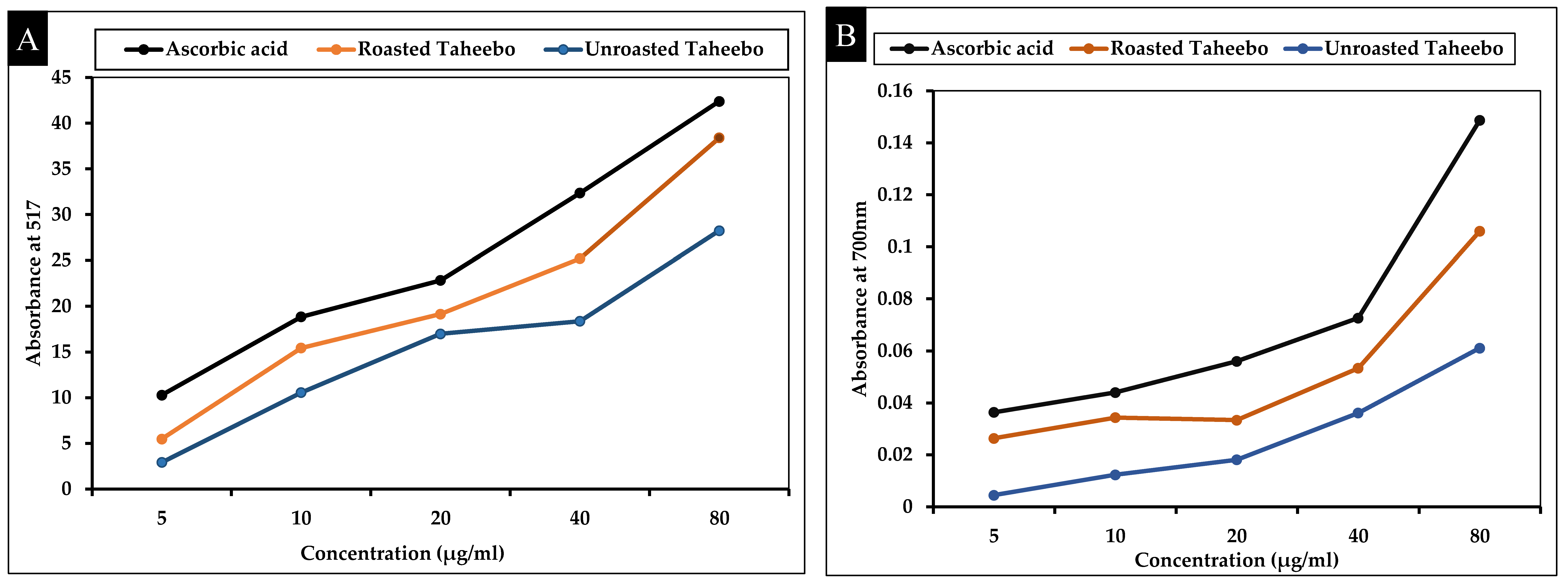
Figure 1. (A) DPPH scavenging activity (% inhibition) vs. concentration; (B) Reducing power activity (Absorbance vs. concentration).
Applsci 13 13171 g001
Figure 2. An assessment of the cytotoxicity of taheebo (TA) and roasted taheebo (roasted TA) to cells (A) on HaCaT cells, (B) on RAW264.7 cells, and (C) on lung cancer A549 cell lines versus non-treated cells. The mean as well as the standard deviation for four different replicates are shown in the graph. ** p < 0.001 denotes significant deviations from control sets.
Applsci 13 13171 g002
Figure 3. In vitro enzymatic activity of antioxidants for taheebo and roasted taheebo (A) GPx, (B) SOD, (C) CAT, and (D) GST on HaCaT cells compared to non-treated control. The graph shows the mean ± SD values of four replicates. ** p < 0.001 denotes significant deviations from control.
Applsci 13 13171 g003
Figure 4. The capability of TA and roasted TA to generate intracellular reactive oxygen species (ROS) in A549 cells was compared to a control. The graph depicts the mean SD values for the three replicates. ** p < 0.001 denotes significant differences across groups.
Applsci 13 13171 g004
Figure 5. (A) ImageJ software (ImageJ bundled with 64-bit Java 8) was used to determine the cell-free region of the scratched region. The proportion of scratching cell migration detected 24 h after administration compared to control values represents the amount of cell migration. (B). Untreated cells are shown as controls. The values are provided as mean standard deviations, and the statistical significance is denoted by ** p < 0.001. The scale bar represents a magnification of ten.
Applsci 13 13171 g005
Figure 6. Colony formation assay in A549 cells at concentrations of 250 and 500 µg/mL of TA and roasted TA The corresponding bar graph of the colony formation assay shows the number of colonies/ dish when A549 cells were treated with TA and roasted with TA. The mean ± SD data of three replicates are shown in the bar graph. ** p < 0.001 shows statistically significant differences from the control groups.
Applsci 13 13171 g006
Figure 7. Efficacy of TA and roasted TA on mRNA expression levels of apoptosis-related genes in A549 cells. For 24 h, TA and roasted TA were administered to A549 cells at concentrations of 250 and 500 g/mL, respectively. Following total RNA extraction, qPCR was performed to assess transcript expression levels with primers targeting (A) p53 (B) BAX (C) Bcl-2 (D) Caspase 9 and (E) Caspase 3 (F) Cyto C. Each bar displays the mean ± SE of duplicate samples from 3 independent experiments (** p < 0.001 using the Student’s t-test compared to the non-treated control).
Applsci 13 13171 g007aApplsci 13 13171 g007b
Figure 8. The effects of TA and roasted TA (A) NO production were assessed by 1 μg/mL LPS-induced RAW 264.7 cells (B) generation of intercellular ROS compared to the control. Data presented as ± SEM, ** p < 0.001 vs. control cell. All treatments were performed three times.
Applsci 13 13171 g008
Figure 9. Effects of TA and roasted TA on pro-inflammatory mediators (A) COX-2, (B) TNF-α, (C) iNOS, (D) IL-6, (E) IL-8, and (F) NF-κB in LPS-induced RAW 264.7 cells. The mRNA expression was determined by qPCR analysis. Data presented as ± SEM, ** p < 0.001 vs. normal. All treatments were performed three times.
Applsci 13 13171 g009aApplsci 13 13171 g009b
Table 1. Sequences of primers used for mRNA gene expression analysis by qRT-PCR.
GenePrimer Sequences (5′-3′)
p53F: TCT TGGGCC TGT GTT ATC TCC
R: CGC CCA TGC AGG AAC TGT TA
Bcl2F: GAA GGG CAG CCG TTA GGAAA
R: GCG CCC AAT ACG ACC AAA TC
BAXF: GGT TGC CCT CTT CTA CTT T
R: AGC CAC CCT GGT CTT G
CASPASE 3F: GAA GGA ACA CGC CAG GAA AC
R: GCA AAG TGA AAT GTA GCA CCA A
CASPASE 9F: GCC CGA GTT TGA GAG GAA AA
R: CAC AGC CAG ACC AGG AC
Cyto-cF: CAGAAGGAAGTTAGGCC
R: CGTCGCAGTGGATGATGTG
COX-2F: CCT GAG CAT CTA CGG TTT GC
R: ACT GCT CAT CAC CCC ATT CA
TNF-αF: GCCAGAATGCTGCAGGACTT
R: GGCCTAAGGTCCACTTGTGTCA
iNOSF: CCT GAG CAT CTA CGG TTT GC
R: ACT GCT CAT CAC CCC ATT CA
IL-6F: AGGGTTGCCAGATGCAATAC
R: AAACCAAGGCACAGTGGAAC
IL-8F: CCGGAGAGGAGACTTCACAG
R: GGAAATTGGGGTAGGAAGGA
NF-κBF: GCAAAGGGAACATTCCGATAT
R: GCGACATCACATGGAAATCTA
GAPDHF: CAA GGT CAT CCA TGA CAA CTT TG
R: GTC CAC CAC CCT GTT GCT GTA G
Table 2. TPC, TFC, and antioxidant efficacy of roasted and unroasted taheebo.
SamplesTotal Phenolic
Contents (TPC)
(µg AAE/mg Extract)
Total Flavonoid
Contents (TFC)
(µg AAE/mg Extract)
DPPH Scavenging
(µg AAE/mg
Extract)
Reducing Power
(µg AAE/mg Extract)
Roasted Taheebo65,263.64 ± 0.0291526.82 ± 0.001989.62 ± 0.01515.59 ± 0.006
Unroasted taheebo54,877.27 ± 0.0691185.36 ± 0.001767.73 ± 0.02511.66 ± 0.029
μg AAE/mg extract: μg ascorbic acid equivalents (μg AAE)/mg extract.
Disclaimer/Publisher’s Note: The statements, opinions and data contained in all publications are solely those of the individual author(s) and contributor(s) and not of MDPI and/or the editor(s). MDPI and/or the editor(s) disclaim responsibility for any injury to people or property resulting from any ideas, methods, instructions or products referred to in the content.