Article

High-Performance Microcomputing Tomography of Chick Embryo in the Early Stages of Embryogenesis

by
Igor Rzhepakovsky
1,
Sergei Piskov
1,
Svetlana Avanesyan
1,
Magomed Shakhbanov
1,
Marina Sizonenko
1,
Lyudmila Timchenko
1,
Mohammad Ali Shariati
2,
Maksim Rebezov
3,4 and
Andrey Nagdalian
5,*
1
Faculty of Medicine and Biology, North-Caucasus Federal University, Pushkina Street 1, 355000 Stavropol, Russia
2
Scientific Department, Semey Branch of Kazakh Research Institute of Processing and Food Industry, Gagarin Avenue 238G, Almaty 050060, Kazakhstan
3
Department of Scientific Research, V. M. Gorbatov Federal Research Center for Food Systems, Talalikhin Street 26, 109316 Moscow, Russia
4
Faculty of Biotechnology and Food Engineering, Ural State Agrarian University, Karl Liebknecht Street 42, 620075 Yekaterinburg, Russia
5
Laboratory of Food and Industrial Biotechnology, North-Caucasus Federal University, Pushkina Street 1, 355000 Stavropol, Russia
*
Author to whom correspondence should be addressed.
Appl. Sci. 2023, 13(19), 10642; https://doi.org/10.3390/app131910642
Submission received: 16 August 2023 / Revised: 20 September 2023 / Accepted: 22 September 2023 / Published: 25 September 2023
(This article belongs to the Special Issue Advances in Molecular Imaging and Its Biomedical Application)

Abstract

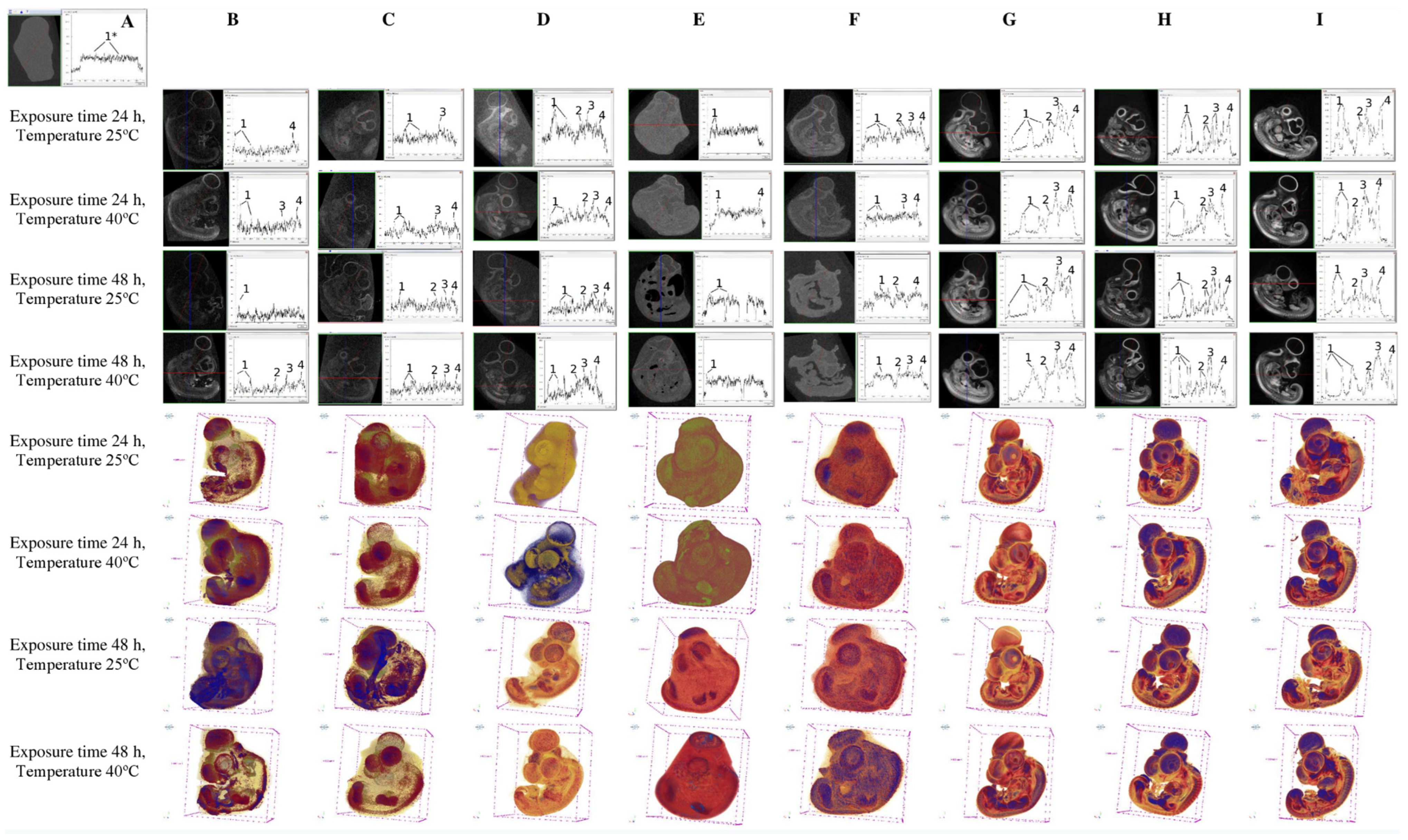
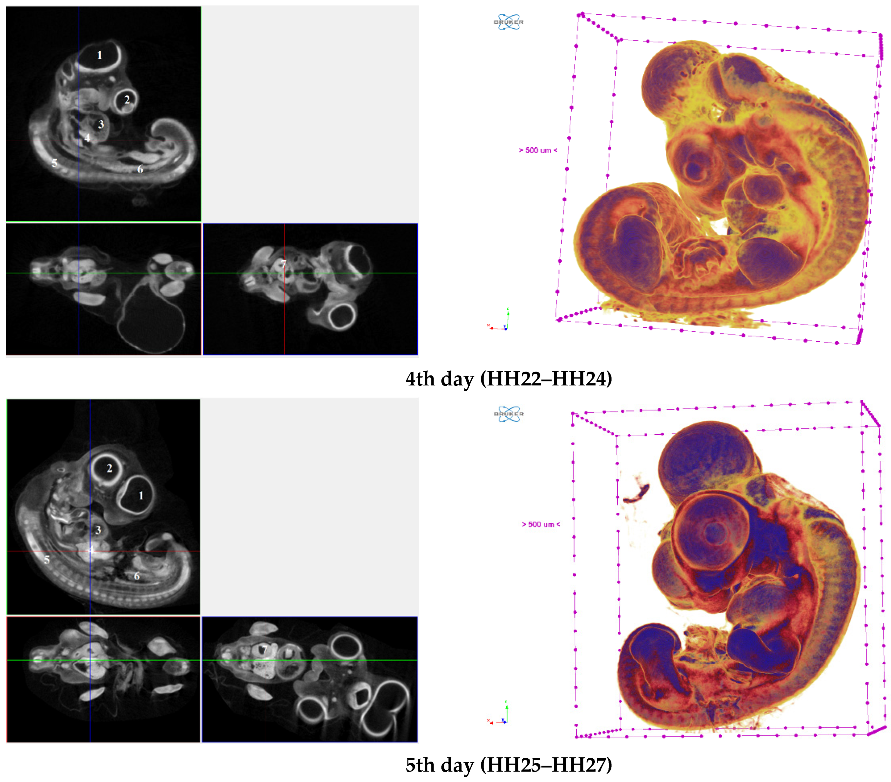
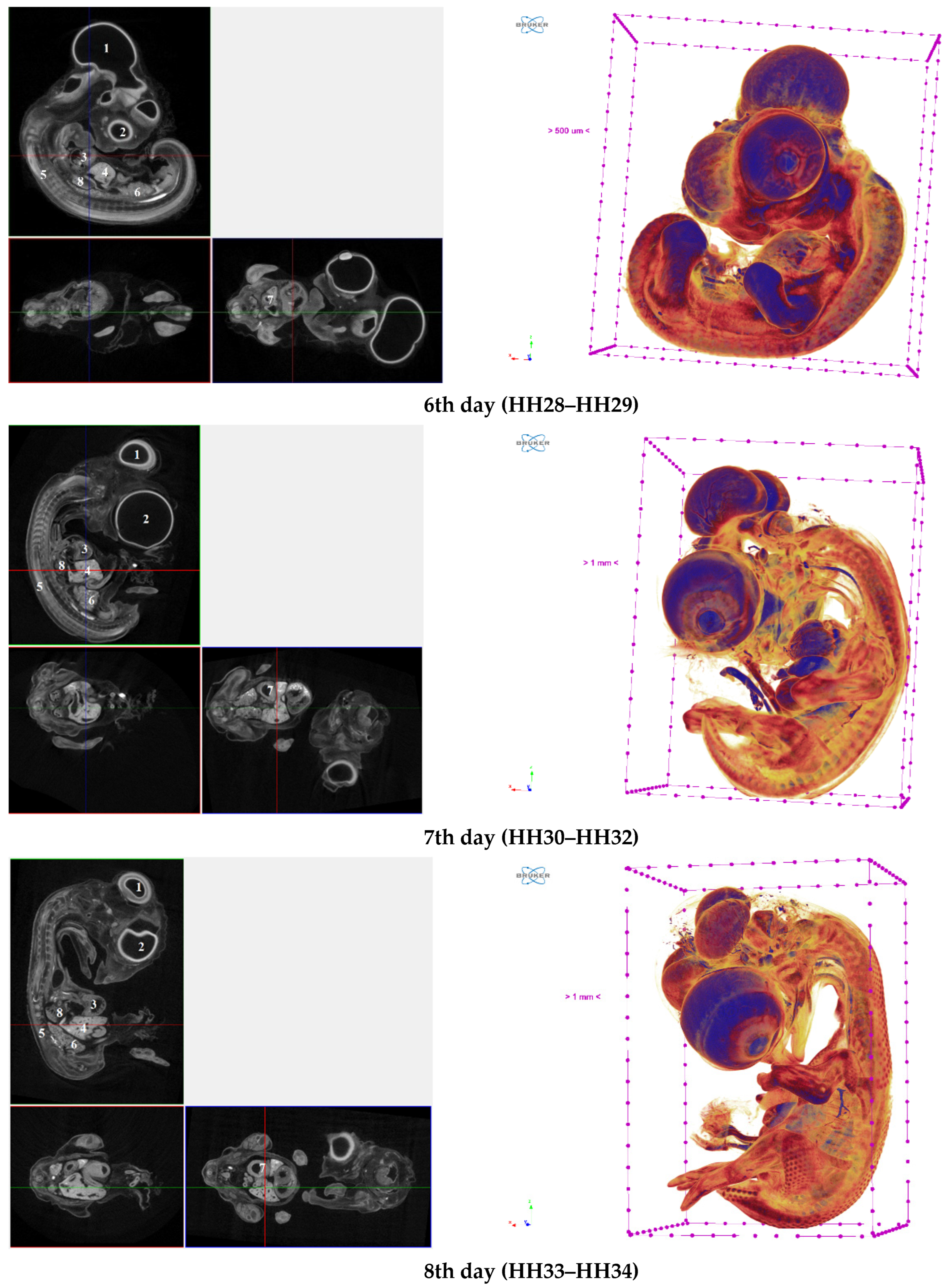
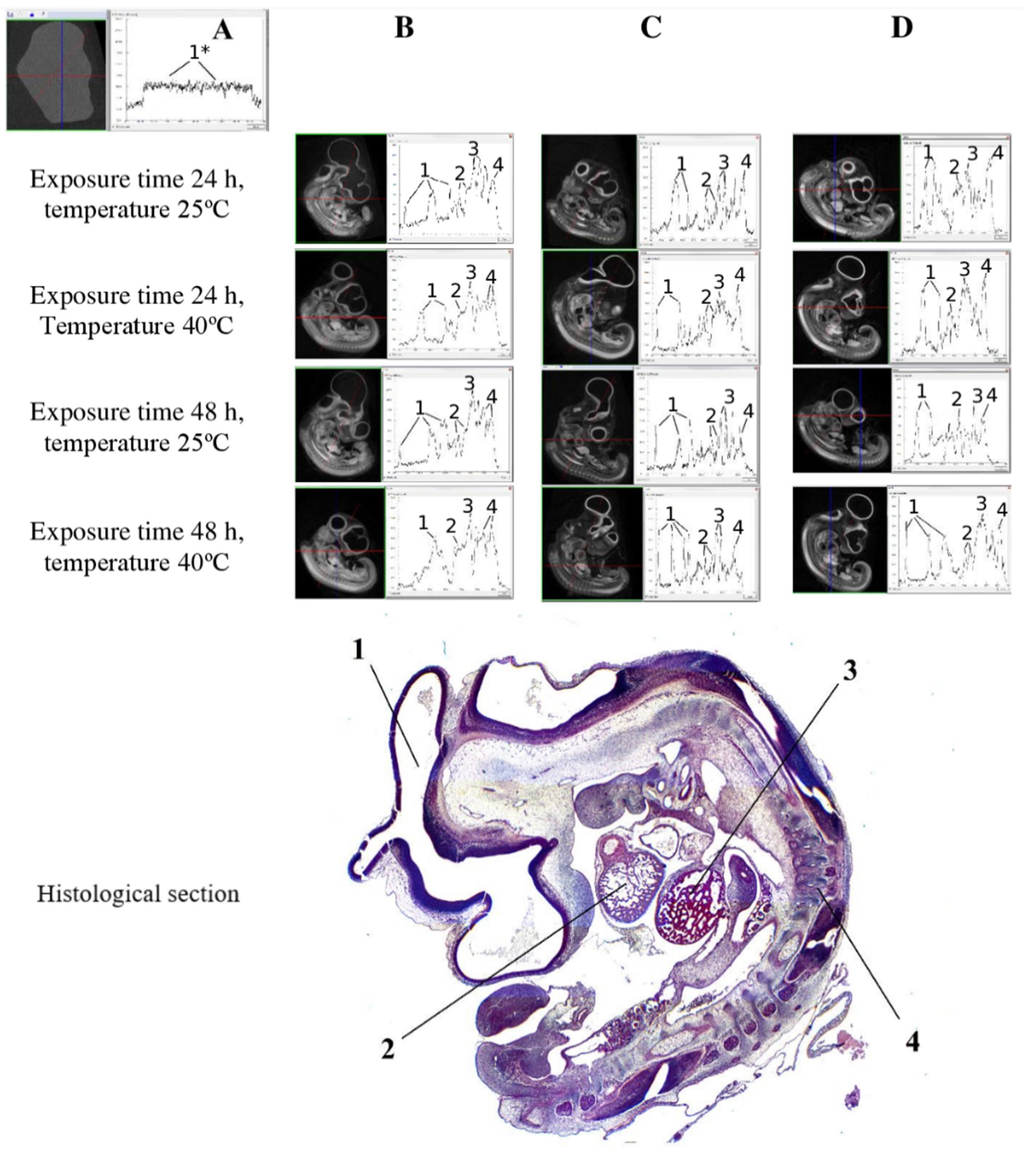
X-ray contrast techniques were tested on the chick embryos in early periods of embryogenesis. For contrast stain, reagents with radiopacity in various concentrations were used: silver proteinate, eosin, Lugol’s solution (I2KI), phosphomolybdic acid and phosphotungstic acid under heating at 25 °C and 40 °C and exposure for 24 and 48 h. The use of silver proteinate, eosin and I2KI in various concentrations in the contrast of the chick embryo in the early period of embryogenesis did not make it possible to obtain microtomographic results that provide reliable microstructural analysis. The most optimal and effective method of embryo staining at the HH22–HH34 embryonic stages reliably determined the staining of 1% phosphotungstic acid at 40 °C heating and exposure for 24 h. Taking into account the size of the chick embryos and their structures at the HH22–HH34 embryonic stages, the features of the development, location of organs, and the minimum permissible parameters of microtomography for obtaining high-quality and reliable results were determined by the isometric spatial resolution of 8.87 μm, X-ray voltage 50 kV, X-ray current 500 μA, and the use of filters started from Al 0.5 mm. Microtomographic results were obtained, characterized by the appearance of the chick embryo at the HH22–HH34 embryonic stages, and they visualized the locations and structures of the chick embryo organs and provided calculation of their volume and X-ray density. The results of the work open up significant prospects for using the chick embryo at the early embryonic period of embryogenesis as an alternative model for screening teratogenicity.
Keywords: microCT; X-ray density; embryo development; embryonic stages; staining; histology; laboratory animals microCT; X-ray density; embryo development; embryonic stages; staining; histology; laboratory animals