Abstract
This article concerns the diagnostic campaign aimed at analyzing the mural painting representing the iconographic theme of the Deesis of the Church of St. Maria Annunziata, Motta San Giovanni, in the province of Reggio Calabria. In 1951, a flood caused the collapse of the building and the consequent breaking of the apse into two parts. The present study focused on the left side of the apse, hosting the figures of Christ and Mary, in order to plan the best conservation intervention strategy. For this purpose, non-invasive investigations and laboratory analytical methods were conducted in order to characterize the constituent materials and to identify the forms of alteration and degradation present on the surface of the painting. In particular, Raman spectroscopy, optical microscopy, scanning electron microscopy coupled to the chemical analysis by an EDS probe, Fourier-transform infrared spectroscopy and ion chromatography were employed. The results highlighted the presence of a single layer of plaster made with a lime-based binder. The chromatic palette of the painting is characterized by ochres and carbon black mixed with lime to obtain the different shades. Finally, the definition of the nature of the deposits and of the overlaid materials was fundamental in order to identify the best products and methods to restore the readability of the work.
1. Introduction
The Medieval pictorial art in southern Italy was heavily influenced by the Byzantine Empire, which controlled the region for much of the Middle Ages. Byzantine art is characterized by its use of vibrant colors and articulate patterns, often depicting religious subjects such as scenes from the Bible or portraits of saints. The site of Santo Niceto, Motta San Giovanni (Reggio Calabria), represents an interesting example of Byzantine art in Calabria (Southern Italy). The church of Santo Niceto dates back, based on the apse painting’s style, to the XI century [1].
The studies conducted on the visible parts of the painting led to its identification as a representation of the Byzantine iconography of the Déesis, which traditionally shows the representations of the Virgin and St. John Baptist in the act of supplication in the presence of Christ, for humanity’s salvation [2].
Even if the eldest example of this iconographic theme arrived to us is the Déesis on the southeastern pillar of St. Maria Antiqua’s church, dated to the VII century and located in Rome, its origins are Constantinopolitan. Due to the iconoclastic crisis, all the oriental Déesis antecedents to the VII century were destroyed, but the experts agree on collocating the birth of this model around the V century, since before this term, the worship of St. John the Baptist was not diffused in the east [3]. In the east, during the post-iconoclastic period, the majority of Déesis was found in the central register of the iconostasis. It was only from the X century onwards that, in some regions of the Byzantine Empire (such as Georgia and Cappadocia), variations to the traditional theme began to emerge, leading to the first examples of Déesis painted in the apse of funerary chapels. The importation of such iconography to Southern Italy can be attributed to the significant migration flows of monks who were escaping from the iconoclast persecution and the Seliuk invasions in Anatolia [4]. These phenomena made possible the establishment, in Calabria, of Basilian monks who heavily influenced the political, religious, cultural and artistic role in the entire region.
Over the years, many variations of Déesis were realized, yet that of St. Maria Annunziata’s church seems to be an example of the original one, which is the simplest, characterized by the presence of the three figures only, on full body or mid-bust. In southern Italy, and in particular, in the Apulian region, several episodes of the Déesis theme can be traced. In this respect, it is worth mentioning the Déesis in the crypt of San Michele delle Grotte at Gravinia (Bari, XIV century), the Déesis in the crypt of San Giovanni at Brindisi (XIII century), the Déesis in the church of San Nicola at Mottola (Taranto, XII-XIII century) and that in the crypt of San Vito Vecchio at Gravina (XIII-XIV century). Another example of such iconography is found in Calabria as well, in the church of San Zaccaria, at Caulonia (Reggio Calabria), dating back to the XIII century. However, even if the compositional structure of the previously mentioned works appears quite similar to that of the Déesis of the church of Santo Niceto, judging from the technique and the style, the other Calabrian and Apulian pictorial evidence achieved a greater artistic maturity and are characterized by a low stylization and better use of proportions. For this reason, according to the most sustained hypothesis, the painting was most likely executed in the XI century [1].
Starting from the late Antiquity, there was a profound change in the mural painting techniques, which led to the reduction of the layers of plaster to two or even just one. Generally speaking, it can be stated that in the Middle Ages, a large variety of materials was used to execute wall paintings [5]. This reflects not only the ingenuity of medieval artists but the circulation between Europe, the Mediterranean, Africa and Asia of knowledge, materials and different techniques as well [6]. The plaster was generally made starting from slaked lime and then adding inert such as sand, ground marble and eventually straw, in different proportions. The range of pigments varies from the inorganic one [7,8,9,10] (preferred for a fresco paints) (which include ochres, bianco San Giovanni, earths, black carbon, Egyptian blue and so on) to the organic ones, usually bound in organic media [11]. Different wall painting techniques are recognized in Middle Ages painting, which differ according to the carbonation level of the plaster at the moment the pigments were applied [12,13]. However, a large number of medieval wall paintings are the result of the combination of a fresco, mezzo fresco and a secco techniques used together at different times during the execution process.
The investigations carried out in recent decades on a large group of medieval wall paintings have contributed to further clarifying the techniques and materials used, allowing the activity of the different workshops to be specified. These are the studies conducted, for example, on Byzantine painting in Cappadocia, that aim to demonstrate that the use and evolution of painting materials and techniques are also rooted in specific localities, defined by unique circumstances of time and place [14]. The mapping of the materials used in medieval painting in Rome [15] and southern Italy [16] provides data that will make it possible to recompose the geography of the painters’ work and the origin of the materials with respect to eastern medieval painting as well [17]. The work carried out in Motta San Giovanni falls within this line of research.
In 1951, the church of Santo Niceto collapsed due to a flood, and because of that, the apse split into two parts, which are still nowadays located between alluvial soil and debris (Figure 1) [18].
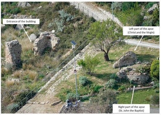
Figure 1.
View of the ruins of St. Maria Annunziata’s church.
On the left portion of the apse, there is the only fully visible painting, in which can be recognized the figures of the Virgin and Christ (Figure 2B,C), while the right part houses the figure of St. John Baptist.
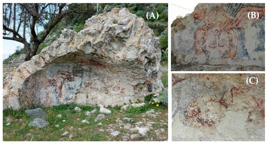
Figure 2.
The left part of the apse before the restoration procedures (A). Details of the figures of Christ (B) and Mary (C) after the restoration work.
In 2021, the left part of the apse was subjected to restoration work aimed to mitigate the degradation processes on the painting in order to stabilize the conditions of the artifact until a future and more complex architectural intervention (Figure 2A).
Due to the abandonment and the exposure to climate factors, before the restoration work, the pictorial evidence of the left part of the apse suffered several degradation and alteration phenomena that appeared to have irremediably affected the integrity of the painting. These included incoherent and coherent deposits, such as dust, soiling and atmospheric particles, and lacunae, abrasions, cracks and deliberate incisions of the plaster. In addition, the painting was strongly affected by the presence of biological colonization caused by many types of microorganisms, such as crustose algae, fungi and lichens, which made the features of Christ’s face almost illegible.
Moreover, a black deposit and a whitish patina, characterized by variable thicknesses and a high degree of compactness, were visible on the painting surface. Another consistent aesthetic damage was caused by the presence of a non-original substance laid upon the pictorial layer, with a brown color and a resinous appearance, widespread in the central area of the painting.
The painting under investigation represents one of the rare examples of Byzantine art in the Calabrian territory still preserved today. In addition, it is the most ancient of the only two still existing Déesis in Calabria (the latest one is that in the church of San Zaccaria, at Caulonia, Reggio Calabria, mentioned above). In this scenario, the diagnostic analyses play a crucial role in expanding knowledge about the techniques and the raw material involved in the realization of the artifacts. This statement acquires greater value if we consider that there are very few mural paintings in the Calabria region attributable to the Medieval period investigated through analytical methods. As a matter of fact, to our best knowledge, in the literature, only a few archaeometric studies about Medieval Calabrian wall paintings are reported [19,20,21]. In the case of the Déesis of Santo Niceto, the diagnostic campaign was aimed at characterizing the pictorial materials used and the executive techniques involved. An additional purpose was to provide information about the degradation processes and the past undocumented restoration works in order to plan the best restoration strategy and to guide the conservative intervention.
The first step of the diagnostic campaign concerned the identification of the main pigments by means of non-destructive investigations (Raman spectroscopy). The results of the spectroscopy analysis also helped gain preliminary information about the alteration phenomena on the painted surface. Then, thanks to the analyses conducted on micro-samples carefully taken from the painting, such as optical microscopy, infrared spectroscopy (FT-IR), scanning electron microscopy coupled with energy-dispersive X-ray spectroscopy (SEM-EDS) and ion chromatography (IC), it was possible to obtain chemical and morphological data on both the constitutive materials, useful to reconstruct the executive processes and the degradation and alteration phenomena that occurred on the surface of the painting.
2. Materials and Methods
2.1. In Situ Observations and Analyses
In situ Raman measurements were employed to characterize the pigments and compounds implied in the execution of the painting [22,23,24]. The Raman spectrometer was a portable RK785 from OceanHood equipped with a 785 nm laser (power range from 0 to 400 mW), frequency shift error < 3 cm−1. The spectra were collected using a power of 50 to 200 mW. The identification of the main pigments was conducted by the comparison of the spectra obtained on the painting surface with those in the RRUFF online spectral database (https://rruff.info, accessed on 30 July 2022) and in literature. All the colors of the painting have been analyzed by punctually examining the most representative pigmented areas.
In addition, Raman spectroscopy was employed to determine the composition of the black deposit diffused on the painting surface as well.
2.2. Sampling and Laboratory Analyses
Before the restoration intervention, preliminary in situ investigations were carried out using a portable video microscope in order to support the choice of the most appropriate sampling points. Sampling activities were performed with the authorization of the Superintendency of Archaeology, Fine Arts and Landscape for the metropolitan city of Reggio Calabria and the province of Vibo Valentia.
After a careful observation of the surface, carried out by the restorers together with conservation scientists, seven micro-samples were chopped from the surface of the painting using the most suitable stainless-steel tools, such as lacets and small chisels, generally used in restoration works. The sampling points were chosen considering the presence of alteration products, substances superimposed on the surface and the morphological characteristics of the pictorial film. Each sampling point was marked with a letter and a number and properly documented by photography. The size of the collected samples is approximately 1 cm × 1 cm with a thickness that ranges from 0.5 cm to 0.2 cm.
Once sampled, the fragments were analyzed through complementary laboratory-based investigation methodologies. The list of samples taken from the surface and the diagnostic methods performed are briefly described in Table 1, while the sampling locations are mapped in Figure 3.

Table 1.
List of analyzed samples, description and employed techniques.
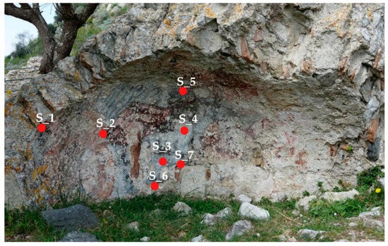
Figure 3.
Mapping of the sampling points on the left part of the apse.
Two cross-sections were prepared according to traditional techniques. Samples S_1 and S_4 were dipped in epoxy resin and let polymerize. Subsequently, the samples were perpendicularly cut and then polished to allow the stratigraphic observation at higher magnification.
Optical Microscopy (OM) was then conducted on the two stratigraphic micro-samples (S_1 and S_4) to identify the main morphological features of both the plaster and the pictorial film. OM analysis was performed using a Zeiss AxioLab microscope equipped with a digital camera to capture images.
In addition, high magnification images were taken on samples S_1 and S_4 by means of the portable digital microscope Dinolite® AM411-FVW, 1.3 Mpx (1280 × 1024), 10–70× and 200× magnification, UV + white led light source connected to a portable computer.
The scanning electron microscope (SEM) coupled with a spectrometer EDS provides information on the morphology and the chemical composition of both the binder and the pigments [25]. For this reason, Ultra-high-resolution SEM (UHR-SEM)—ZEISS CrossBeam 350 equipment, coupled with the spectrometer EDS-EDAX OCTANE Elite Plus-Silicon drift type, was employed on samples S_1 and S_4 coated with a thin and highly conductive graphite film. Instrumental conditions set for EDS analysis were HV: 15 keV and probe current: 100 pA.
Infrared spectroscopy (FT-IR) was used to investigate the nature of organic and inorganic compounds. FT-IR was conducted to characterize the unlaid substance having a resinous appearance, to establish the composition of the whitish patina and to clarify the nature of the binder used to fix the pigments on the painting surface [26,27]. For this purpose, a Perkin Elmer Spectrum 100, equipped with a diamond ATR (attenuated total reflectance), was used, and spectra were collected in the range of 500–4000 cm−1, with a resolution of 4 cm−1.
Ion chromatography (IC) was used in order to quantify cations and anions on the superficial layers. Samples S_6 and S_7 underwent the following protocol: 2 mg of powder was placed in a test tube and treated with 10 mL of MilliQ water; then the solutions were sonicated for 1 h, centrifuged for 30 min and analyzed by mean of HPLC (DIONEX DX120). Cationic (Na+, K+, Ca2+, Mg2+ and NH4+) and anionic (NO3−, SO42−, Cl−) species were determined for each sample.
3. Results and Discussion
3.1. Stratigraphy and Executive Technique of the Painting
As known from medieval treatises and from the investigations carried out on many surviving paintings, Middle Ages paintings employed with a single technique are rather uncommon. As a matter of fact, different wall painting techniques were often used together in the same executive process.
In the first phases, pigments were often dissolved in water only (according to a fresco technique) or mixed with lime water (following the a mezzo fresco technique) and then applied to wet lime plaster. As the plaster dries, the pigments are embedded in the plaster due to the carbonation process (Ca(OH)2 + CO2 → CaCO3 + H2O), thus enhancing the durability of the painting. Retouches or unfinished portions of the composition could be added by combining piments with organic or inorganic binders on the dried plaster (a secco).
The in situ observations, together with the OM, IR and Raman investigation, shed light on the executive process of the apse painting in the church of Santo Niceto.
According to the preliminary in situ observation carried out by restorers and judging from the overlapping correlations between pictorial layers, it was possible to reconstruct the sequence of paint applications.
The painting began with a greyish base layer, applied all over the surface to execute the background. Then, the Christ’s robe was constructed by superimposing darker strokes and highlights. Over the robe, Christ’s mantle was depicted with a pinkish base layer and then defined once again by additional red brushstrokes and white highlights to emphasize the folds and the contours. The mantle brushstrokes appear, to the naked eye, thicker in comparison with those of the Christ’s robe, and their texture is easily recognizable.
Optical microscopy and Dino-Lite observations were conducted on the two polished cross-sections representative of the painting stratigraphy (S_1 and S_4) to better understand the executive procedures involved (Figure 4 and Figure 5).
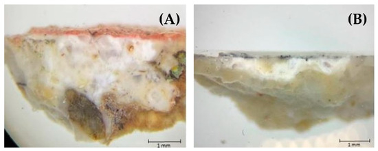
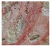
Figure 4.
Images obtained by reflected light microscopy (100× magnification) of the stratigraphic sections of samples S_1 (A) and S_4 (B).
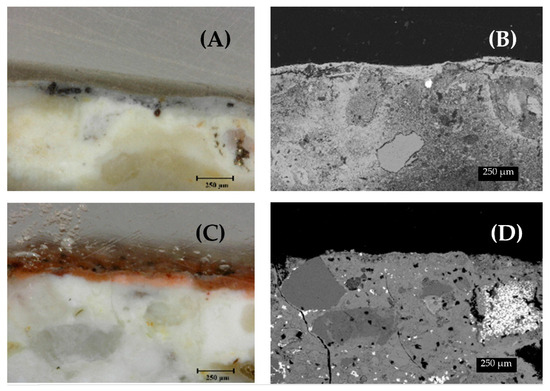
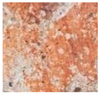
Figure 5.
High-magnification images taken using the portable digital microscope of samples S_1 (C) and S_4 (A); associated images obtained by SEM of sample S_1 (D) and sample S_4 (B).
Sample S_1, taken from the draperies of Christ’s robes, includes both the plaster layer and the pictorial film characterized by pink and white strokes. By first observation under the Optical Microscope, the paint film appears compact and shows a light-red shade. At higher magnifications, the pictorial layer presents a homogeneous coloration, while small reddish particles are sporadically recognizable (Figure 5C). In addition, both under the Optical Microscope and in SEM images, the interface between the paint layer and the underlying plaster appears, in this case, quite clear (Figure 5C,D).
Sample S_4, otherwise, was collected from the background of the painting, and it is characterized by a light greyish tint. By the images obtained from the digital microscope, the pictorial layer appears light-grey and black inclusions can be observed. In addition, differently from the observation employed on sample S_1, the greyish pictorial film seems to have been well absorbed into the plaster and signs of a distinct separation between the paint layer and the plaster are not evident (Figure 5A,B).
Microstratigraphic investigations conducted under OM and DinoLite are coherent with the in situ observations. The greyish pigment of the background, as well as that of the Christ’s robe, was applied directly on the fresh wet plaster, according to the a fresco technique. Subsequently, for the execution of the Christ’s mantle, and thus for the subsequent-applied elements of the composition, pigments were embedded in lime on the already dried and almost carbonated plaster.
Further confirmation comes from the Fourier Transform Infrared Spectroscopy investigations. The analysis carried out on sample S_2, consisting of the red pictorial film and chopped in correspondence with the Christ’s robe, produced exclusively peaks corresponding to calcite (1408, 872 and 712 cm−1), calcium oxalates (3415, 1618, 1316, 779 and 514 cm−1) and silicates/clay minerals or products of their alteration (959 cm−1), thus excluding the use of other pictorial binders (such as organic binders) other than lime to fix pigments on the plaster surface (Figure 6).
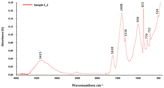
Figure 6.
FT-IR spectrum of sample SP_2 showing calcite (1408, 872 and 712 cm−1), calcium oxalates (3415, 1618, 1316, 779 and 514 cm−1) and silicates/clay minerals or products of their alteration (959 cm−1) peaks.
This result makes it possible to specify the method of using white lime as a binder by distinguishing these brushstrokes from the frescoes. This is an element of the technical process known from the sources [28] and identified in the early medieval painting of the same period as our case study [14], but still little investigated systematically with scientific analyses. Together with the results obtained in relation to the characterization of pigments, this result contributes to enriching the knowledge of medieval wall paintings in terms of execution procedure.
A single plaster layer is distinguishable in both of the two stratigraphic specimens. The latter is characterized by a white color and features a poorly sorted aggregate, with mostly angular grains and a sphericity that goes from high to low.
SEM-EDS analyses carried out on the two stratigraphic samples made it possible to obtain compositional and morphological information on both the binder and the aggregate constituting the plaster. In addition, the SEM images revealed the presence of a single layer of plaster for both samples S_1 and S_4, confirming the in situ observations and the Dino-Light morphological analysis.
The EDS analysis punctually carried out on the binder of the plaster shows a large Ca content, a lower concentration of Si and a lower amount of S, K, Al, Mg, Cl and Na (Table 2).
Table 2.
EDS chemical analyses detected for plastered areas of samples S_1 and S_4. Notes: more abundant elements in bold; minor elements in brackets.
The high percentage of Ca detected in both samples suggests that the lime was obtained starting from the calcination of limestone containing a minor clayey component.
The chemical microanalysis conducted on a lump within sample S_4 confirms the previously advanced assumption, revealing the use of aerial lime (percentage of CaO > 80%) (Figure 7, Table 2) [29].
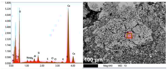
Figure 7.
EDS spectrum and SEM image of the lump observed in sample S_4 (highlighted by the red square).
In addition, the considerable concentrations of Ba and S detected in sample S_1 indicate the presence of barium sulfates within the plaster layer. In particular, barium sulfate (also known as “barite”) and fluorite have a hydrothermal origin, and they can be found associated with carbonate materials. Mineralization processes in limestone generally occur when tectonic activity causes fractures, which allow the ascent of hydrothermal fluids rich in mineralizing components, leading to their crystallization [30,31]. It is reasonable to assume that the limestone used to produce the lime of the plaster had been subjected to mineralization phenomena, giving, as a result, the precipitation of barite.
3.2. Pigments
The non-destructive analysis employed by Raman spectroscopy was addressed to characterize the main pigments and compounds involved in the execution of the mural painting. The data and spectra obtained are reported in Table 3 and in Figure 8.
Table 3.
List of the main colors investigated by Raman spectroscopy, identified compounds and reference bands.
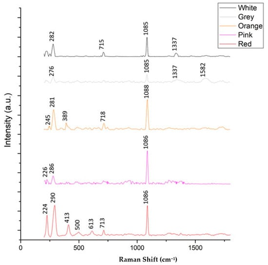
Figure 8.
Raman spectra performed on the white, grey, orange, pinkish and red pictorial layers.
It can be stated that the palette involved in the execution of the wall painting, as is quite usual for paintings of the Medieval period, is not very variegated. It is essentially represented by red, pink, orange, grey and white. At the same time, the presence of colors obtained by mixing several pigments demonstrates the technical and scientific knowledge of painters who were able to create the shades that best suited the aesthetic choices of their time.
It is worth mentioning that calcite typical bands were detected for all the colored areas investigated. This evidence might be explained considering that calcium carbonate was used both in the plaster as a binder to fix pigments on the surface and as a white pigment to lighten pigments.
3.2.1. Red, Pink and Orange
The Raman spectrum of the red pictorial film shows the characteristic bands of hematite, α-Fe2O3 (bands at 224, 290, 413, 500 and 613 cm−1) and calcite (bands at 713 and 1086 cm−1), indicating the use of a hematite-based pigment. Iron-oxide pigments have been widely used since prehistoric times because of their extreme stability and high coloring capacity. Their use was also recorded during the Middle Ages, and their presence was attested in other Calabrian Medieval wall paintings [19,20].
In order to compare the data obtained from Raman Spectroscopy, a chemical investigation of the pictorial film was performed by SEM-EDS.
The results of the compositional micro-analysis of the reddish pictorial layer on sample S_1, performed by the EDS probe combined with SEM, are coherent with those of Raman spectroscopy. EDS investigations revealed the presence of high iron contents and a moderate percentage of silicon and aluminum, which can be attributed to the presence of red ochre pigments, confirming the results of Raman spectroscopy [32,33] (Figure 9, Table 4).
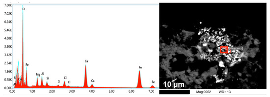
Figure 9.
EDS spectrum punctually acquired on the red pictorial layer (sample S_2) (highlighted by the red square) and related SEM image.
Table 4.
EDS chemical analyses detected for painted layers of samples S_1 and S_4. Notes: more abundant elements in bold; minor elements in brackets.
Bands at 226, 286 (hematite) and 1086 cm−1 (calcite) were detected for the pinkish colored areas of the painting as well. Based on the foregoing, the pink tone was probably obtained by mixing the hematite with a white calcite-based pigment, such as bianco San Giovanni. As a matter of fact, bianco San Giovanni pigment, according to the procedure described by Cennino Cennini, was generally prepared by drying lime and then reducing the newly-formed calcium carbonate to powder [34,35]. On the other hand, it can not be ruled out that the hematite was lightened by adding a significant amount of pure lime to achieve the pinkish hue.
The Raman analysis for the orange pictorial film exhibited bands at 279, 713 and 1088 cm−1, again suggestive of calcite, and bands at 248, 301, 528 cm−1, which can be assigned to iron (III) hydrous oxide, α-FeOOH, also known as “goethite”. The Raman spectrum of goethite exhibits bands at 245 cm−1, 299 cm−1, 387 cm−1, 480 cm−1 and 550 cm−1. The band at 245 cm−1 is due to the symmetric stretching of Fe-O bonds, bands at 480 and 550 cm−1 to asymmetric stretching of Fe-OH bonds, the band at 299 cm−1 is assigned to the symmetric bending of Fe-OH bonds and that at 387 cm−1 to the symmetric stretching of Fe-O-Fe/-OH bonds. In our case, the bands at 245 and 389 cm−1 are consistent with those reported in the literature [36].
3.2.2. Grey
The spectrum for the greyish colorations shows bands at 1337 and 1582 cm−1, suggesting the use of black carbon pigment, and, once again, at 276 and 1085 cm−1 (calcite). The Raman spectrum of amorphous carbons is characterized by a band at 1582 cm−1, given to the G (graphite) band in-plane aromatic ring CC stretching vibrational mode with E2g2 symmetry [37], and an additional D band at around 1350 cm−1, known as a “disorder” band [38,39]. Most likely, the pigment has a vegetable origin and consists of charcoal obtained from the carbonization of wood. This statement is corroborated by the absence of the additional phosphate stretching band at 960 cm−1 related to the presence of bone/ivory black pigment [37]. The bands of calcite detected by Raman indicate that the black pigment was mixed with calcite or lime.
Data obtained from Raman spectroscopy, performed on the greyish pictorial film, are, once again, supported by SEM-EDS analysis on the black particles within the pictorial layer in sample S_4. Peaks of carbon and oxygen were detected, corroborating the presence of the carbon black pigment (Figure 10, Table 4).
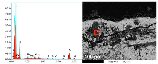
Figure 10.
EDS spectrum punctually acquired on the greyish pictorial layer (sample S_4) (highlighted by the red square) and related SEM image.
EDS analyses punctually performed on areas other than the black inclusions of the greyish pictorial film indicated calcium as the main element, followed by silicon, aluminum, sulfur and potassium. No evidence of copper was found, keeping out the presence of copper-based pigments such as azurite (basic copper(II) carbonate) and Egyptian blue (artificial pigment consisting of cuprorivaite, CaCuSi4O10) [40]. In addition, as evidenced by the autoptic observations made by restorers and the optical microscopy investigations, the greyish pigment was mostly applied to fresh and wet plaster, according to the “fresco” technique. This assumption lets us discard the use of indigo/woad blue. Even if the presence of this pigment is quite rare in wall painting, its use is considered incompatible with strictly alkaline conditions, and its lightfastness is inadequate [41]. Based on the EDS and Raman spectroscopy examinations, it is possible to conclude that blue chromophores have not been unequivocally detected.
As known from the past literature, the formula for the execution of the blue backgrounds consisting of the application of a black carbon layer was widespread after the iconoclastic period. The procedure was followed by the addition of a second layer of pure lime to achieve a lighter tone [42]. It is highly probable that the same executive methodology was applied in this case.
3.2.3. White
The Raman spectrum for the white zones contained bands at 715 and 1085 cm−1, revealing the use of either bianco San Giovanni pigment or pure lime for the application of highlights and details of the robes. The (CO3)−2 group shows four prominent bands at 1440, 1088, 715 and 278 cm−1. Bands recorded at 715 and 1088 cm−1 are due to the plane bending mode and the symmetric stretching vibration of the carbonate groups. In addition, the latter-mentioned two bands are typical of calcite, while for dolomite, characteristic bands in the region between 1069 and 1098 cm−1 and a band at 1393 cm−1 are observed [43]. The band at 1337 cm−1 is indicative of traces of black carbon pigment inside the white pictorial film.
3.3. The State of Conservation of the Painting
From the spectrum obtained by analyzing the S_3 sample by Fourier transform infrared spectroscopy (FT-IR), peaks emerged at 2982, 2952, 1724, 1634, 1446, 1385, 1233, 1138, 1023, 968, 859, 753, 514 and 494 cm−1.
The FT-IR spectrum obtained was then compared to that of Paraloid B72, a commercial acrylic resin often used in the conservation field. The overlapping of the two spectra is clearly visible in Figure 11, confirming the organic nature of the substance.
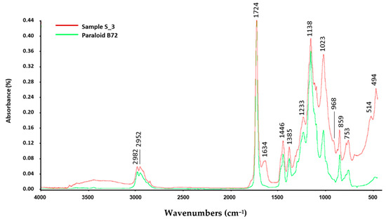
Figure 11.
FT-IR spectrum of the substance upon the pictorial film (red line) compared to that of the Paraloid B72 (green line).
The hypothesis put forward that indicated it was an act of vandalism was denied during the restoration phases by the discovery of the impression of a gauze weave. This made it possible to assume that it was actually an undocumented intervention, carried out in the paste with a conservation purpose to temporarily preserve the pictorial film through the application of a facing.
The carbonatic composition of the diffuse whitish veil on the pictorial surface was confirmed by FT-IR, carried out on sample S_5, taken from a thicker concretion developed at a plaster fracture, which produced a spectrum showing only the peaks of calcite (1415, 873 and 712 cm−1) and silicates/clay minerals or products of their alteration (about 960 cm−1).
Raman spectroscopy, on the other hand, made it possible to confirm the hypothesis put forward for the black-colored coherent deposits present on the pictorial pieces of both trunks, which, from macroscopic and microscopic observation, appeared to consist of burnt material. Bands were detected at 223, 282, 710 and 1085 cm−1, overlapping with calcite, and at 1343 and 1586 cm−1, corresponding to carbon black. In this case, it would not be a pigment, however, but a carbonaceous deposit, presumably due to the prolonged contact of the surface to an open flame. Considering the acts of vandalism to which the painting has been subjected, it was thought that these deposits might be the trace left by a fire, set to damage the painting on purpose. However, the presence of soot is exclusively limited to the internal area of the semi-dome, while there are no traces on the wall structure. It could, therefore, be hypothesized that the dark deposit originated from before the collapse of the building, and it is most likely caused by the use of torches or votive lamps close to the surface that were used in ancient times to illuminate the painting and the environment.
Considering the outdoor environment to which the mural painting is exposed and with the aim to plan the most appropriate conservative strategies, the soluble salt in solution inside the plaster (samples S_6 and S_7) was investigated by ion chromatography (IC). The results of both the cationic and anionic species concentrations (in ppm) are reported in Table 5. The results show that the concentrations detected are not relevant, so it can be stated that the mural painting does not suffer degradation phenomena attributable to salt decay.
Table 5.
Ionic concentrations (in ppm) measured in samples S_6 and S_7.
The concentrations of the cations Na+ and Ca2+ and of the anion SO42−, which are higher in plaster sample S_6, presumably indicate the presence of sodium or calcium sulfates, on the origin of which a variety of hypotheses could be made, given the outdoor conditions to which the artifact is exposed, which allows both capillary rise and percolation of meteoric water, and the presence of biological species capable of depositing salts on the substrates they colonize. However, very small amounts have been detected, for which it is difficult to assume a correlation with the severe forms of degradation observable in the mural painting.
In fact, the development of abrasions and lacunae on the painting surface could instead be the result of previous colonization by lichens, which damaged the substrate, probably also making it more susceptible to other degradation processes due to interaction with the external environment [44]. In particular, carbonic acid, which originates from the reaction between carbon dioxide and water retained in the thallus, on coming into contact with the substrate, reacts with its constitutive minerals, performing a chelating action against the basic cations K+, Na+, Mg2+ and Ca2+ [45].
Oxalic acid, on the other hand, originates from the metabolic activity of the mycobiont that constitutes the lichen and, by coming into contact with the substrate, extracts the cations of its constitutive minerals and causes the precipitation of oxalates, which are stored in the thallus or are deposited on the surface.
Both processes, therefore, cause surface corrosion of the substrate and a loss of original mechanical properties that vary depending on the extent of attack and the species of lichens.
The combination of the chemical actions above described with the mechanical action exerted by the hyphae (which during accretion processes penetrate the interior of the substrate, generating detachments and small holes termed “pitting”) can result in substrate loss and increased susceptibility to external stresses, which can cause further damage of the constitutive material.
Moreover, having already found other traces of undocumented “conservation” intervention, it is not possible to rule out the possibility that the surface over the years has been subjected to further such initiatives and has been mechanically cleaned, removing lichen thalli and exposing the altered substrate to external stresses.
In fact, the resulting spectrum of sample S_2, analyzed by Fourier Transform Infrared Spectroscopy (FT-IR), taken from areas apparently unaffected by biological colonization at the time of sampling, showed peaks at 3349, 1626, 1317, 779, 711 and 515 cm−1, corresponding to calcium oxalates (whewellite), the presence of which could be an indicator of previous biological activity on the substrate by organisms or microorganisms capable of secreting oxalic acid.
The conservative work, carried out in 2021, restored the legibility of the painting and ensured its stability through cleaning, consolidation and grouting operations. The pictorial reintegration intervention, however, will be addressed in the near future when a covering system will be installed to protect the painting.
4. Conclusions
In order to plan the most appropriate conservative procedures, a multi-analytical approach was carried out on the left part of the apse of St. Maria Annunziata’s church (in Motta San Giovanni, RC) in which the figures of Christ and Mary are part of the iconographic theme of the Déesis. The diagnostic campaign was addressed at characterizing the constitutive materials, at defining the execution techniques and at determining the alteration and degradation processes on the painting surface. Following this aim, both non-destructive methodologies and laboratory analysis were employed. The color palette employed in the painting execution was investigated by Raman Spectroscopy, which clarified the use of ochres and carbon black pigments eventually mixed with lime to achieve the red, pink, orange, black, light-grey and white shades. Moreover, microscopic observations, SEM-EDS analysis and FT-IR investigation allowed us to characterize the painting stratigraphy, the composition of the aggregate and the binder and to determine the nature of the pictorial binder. The painting consists of a single plaster layer, in which the aggregate fraction is mixed with a lime-based binder. A combination of different executive techniques has been used: the pigment was started a fresco and then completed a secco, using lime to fix the pigments on the almost carbonated plaster. As evidenced by FT-IR analysis, the unlaid substance having a resinous aspect, probably applied during a previous undocumented intervention, was an acrylic resin and most likely consists of Paraloid. Furthermore, the FT-IR spectra confirmed the carbonatic nature of the whitish patina on the painting surface. Finally, the composition of black-colored coherent deposit was investigated through Raman Spectroscopy, clarifying that the diffuse darkening was most probably caused by soot from torches or votive lamps used in ancient times to illuminate the church.
Author Contributions
Conceptualization, A.A., M.R. and P.P.; methodology, L.R. and M.R.; investigation, A.M., G.S. and L.R.; data curation, G.S., M.A.Z. and S.A.R.; writing—original draft preparation, G.S., A.A. and P.P.; writing—review and editing, M.A.Z. and S.A.R.; supervision, M.F.L.R. All authors have read and agreed to the published version of the manuscript.
Funding
This research received no external funding.
Conflicts of Interest
The authors declare no conflict of interest.
References
- Falla Castelfranchi, M. La decorazione pittorica delle chiese di Santo Niceto. In Santo Niceto Nella Calabria Medievale: Storia, Architettura, Tecniche Edilizie; Martorano, F., Ed.; L’ERMA di BRETSCH-NEIDER: Rome, Italy, 2002; p. 115. [Google Scholar]
- Minuto, D. Le chiese di tradizione bizantina. In Santo Niceto Nella Calabria Medievale: Storia, Architettura, Tecniche Edilizie; Martorano, F., Ed.; L’ERMA di BRETSCHNEIDER: Rome, Italy, 2002; p. 78. [Google Scholar]
- Andaloro, M. Note sui temi iconografici della Déesis e della Haghiosoritissa. Riv. Ist. Arch. 1970, 17, 85–153. [Google Scholar]
- Velmans, T. La pittura murale in Georgia. In L’arte Della Georgia. Affreschi e Architetture; Velmans, T., Alpago Novello, A., Eds.; Jaca Book: Milan, Italy, 1996; pp. 31–56. [Google Scholar]
- Tosatti, S.B. Trattati Medievali di Tecniche Artistiche; Jaka Book: Milan, Italy, 2007; pp. 78–80. [Google Scholar]
- Murat, Z. Wall paintings through the ages: The medieval period (Italy, twelfth to fifteenth century). Archaeol. Anthropol. Sci. 2021, 13, 191. [Google Scholar] [CrossRef]
- Mady, E.; Chartier, C.; Prévot, G.; Vignaud, C.; Garay, H. The colour of ochres explained by their composition. Mater. Sci. Eng. 2006, 127, 70–80. [Google Scholar]
- Zanardi, B.; Arcangeli, L.; Apollonia, L. Della natura del bianco sangiovanni. Un pigmento e la lettura delle fonti. Ric. Stor. Arte 1984, 24, 63–74. [Google Scholar]
- Moretto, L.M.; Orsega, E.F.; Massocchi, G.A. Spectroscopic methods for the analysis of celadonite and glauconite in Roman green wall paintings. J. Cult. Herit. 2011, 12, 384–391. [Google Scholar] [CrossRef]
- Švarcová, S.; Hradil, D.; Hradilová, J.; Čermáková, Z. Pigments—Copper-based greens and blues. Archaeol. Anthropol. Sci. 2021, 13, 210. [Google Scholar] [CrossRef]
- Aceto, M. The palette of organic colourants in wall paintings. Archaeol. Anthropol. Sci. 2021, 13, 159. [Google Scholar] [CrossRef]
- Conti, A. Affresco, pittura a calce, pittura a secco. In Tra Metodo e Ricerca: Contributi di Storia Dell’arte; Poso, R., Galante, L., Eds.; Congedo: Galatina, Italy, 1988; pp. 159–177. [Google Scholar]
- Rinaldi, S. Tecniche di Pittura Murale Dall’alto Medioevo al Quattrocento; Lithos: Rome, Italy, 1998. [Google Scholar]
- Pogliani, P.; Andaloro, M. Materials and Techniques and of Cappadocian Wall Paintings. In Conservation and Painting Techniques of Wall Paintings on the Ancient Silk Road; Aoki, S., Taniguchi, Y., Rickerby, S., Mori, M., Kijima, T., Bomin, S., Kirino, F., Eds.; Springer: Singapore, 2021; pp. 43–88. [Google Scholar]
- Amato, S.R.; Bersani, D.; Lottici, P.P.; Pogliani, P.; Pelosi, C. A multi-analytical approach to the study of the mural paintings in the presbytery of Santa Maria Antiqua al Foro Romano in Rome. Archaeometry 2017, 59, 1050–1064. [Google Scholar] [CrossRef]
- Calia, A.; De Giorgi, E. Le pitture della cripta del Gonfalone a Tricase (Lecce): Problematiche storico-artistiche e contributo alla identificazione dei pigmenti attraverso FRX portatile. In Atti I Convegno Beni Culturali in Puglia; Seite: Bari, Italy, 2021; pp. 125–132. [Google Scholar]
- Taniguchi, Y. Materials and Technologies of the Bamiyan Wall Paintings. In Conservation and Painting Techniques of Wall Paintings on the Ancient Silk Road; Aoki, S., Taniguchi, Y., Rickerby, S., Mori, M., Kijima, T., Bomin, S., Kirino, F., Eds.; Springer: Singapore, 2021; pp. 177–196. [Google Scholar]
- Martorano, F. Santo Niceto, la Storia e il Restauro; Iiriti Edit.: Reggio Calabria, Italy, 2014; p. 48. [Google Scholar]
- Zicarelli, M.A.; La Russa, M.F.; Alberghina, M.F.; Schiavone, S.; Greca, R.; Pogliani, P.; Ricca, M.; Ruffolo, S.A. A Multianalytical Investigation to Preserve Wall Paintings: A Case Study in a Hypogeum Environment. Materials 2023, 16, 1380. [Google Scholar] [CrossRef]
- Ricca, M.; Alberghina, M.F.; Houreh, N.D.; Koca, A.S.; Schiavone, S.; La Russa, M.F.; Randazzo, L.; Ruffolo, S.A. Preliminary Study of the Mural Paintings of Sotterra Church in Paola (Cosenza, Italy). Materials 2022, 15, 3411. [Google Scholar] [CrossRef]
- Giuffrida, D.; Mollica Nardo, V.; Neri, D.; Cucinotta, G.; Calabrò, I.V.; Pace, L.; Ponterio, R.C. A Multi-Analytical Study for the Enhancement and Accessibility of Archaeological Heritage: The Churches of San Nicola and San Basilio in Motta Sant’Agata (RC, Italy). Remote Sens. 2021, 13, 3738. [Google Scholar] [CrossRef]
- Casadio, F.; Daher, C.; Bellot-Gurlet, L. Raman Spectroscopy of cultural heritage Materials: Overview of Applications and New Frontiers in Instrumentation, Sampling Modalities, and Data Processing. Top. Curr. Chem. 2016, 374, 62. [Google Scholar] [CrossRef]
- Jehlička, J.; Culka, A. Critical evaluation of portable Raman spectrometers: From rock outcrops and planetary analogs to cultural heritage—A review. Anal. Chim. Acta 2021, 1209, 339027. [Google Scholar] [CrossRef] [PubMed]
- Matteini, M.; Moles, A. Scienza e Restauro. Metodi D’indagine; Nardini/Panini Edit.: Florence, Italy, 2002; pp. 178–182. [Google Scholar]
- Janssens, K.; Van Grieken, R. Non-Destructive Microanalysis of Cultural Heritage Materials; Elsevier: Amsterdam, The Netherlands, 2005; pp. 80–82. ISBN 9780444559883. [Google Scholar]
- Griffiths, P.; Haseth, J.A. Fourier Transform Infrared Spectrometry; Wiley: Hoboken, NJ, USA, 2007. [Google Scholar]
- Adrover Gracia, I. Applicazioni Della Spettrofotometria IR Allo Studio dei beni Culturali; Il Prato Edit.: Padova, Italy, 2001. [Google Scholar]
- Caffaro, A. Teofilo Monaco. Le Varie Arti. De Diversis Artibus. Manuale di Tecnica Artistica Medievale; Palladio Ed.: Salerno, Italy, 2000. [Google Scholar]
- Barba, L.; Blancas, J.; Manzanilla, L.R.; Ortiz, A.; Barca, D.; Crisci, G.; Miriello, D.; Pecci, A. Provenance of the limestone used in Teotihuacan (Mexico): A methodological approach. Archaeometry 2009, 51, 525–545. [Google Scholar] [CrossRef]
- Speciale, F. Le Mineralizzazioni di Fluorite e Barite nel Territorio di Termini Imerese e Caccamo—Sicilia Aspetti Geologici Esteriori Legati Alla Minerogenesi; Eugenio Maria Falcone Ed.: Bargheria, Italy, 2013. [Google Scholar]
- Bellanca, A.; Censi, P.; Neri, R. Studio isotopico, chimico e tessiturale su materiali carbonatici associati a mineralizzazioni di fluorite e barite nell’area di Termini Imerese (Sicilia). Rend. Soc. Ital. Mineral. Petrol. 1983, 38, 1251–1261. [Google Scholar]
- Bikiaris, D.; Sister, D.; Sotiropoulou, S.; Katsimbiri, O.; Pavlidou, E.; Moutsatsou, A.; Chryssoulakis, Y. Ochre-differentiation through micro-Raman and micro-FTIR spectroscopies: Application on wall paintings at Meteora and Mount Athos, Greece. Spectrochim. Acta Part A Mol. Biomol. Spectrosc. 2000, 56, 3–18. [Google Scholar] [CrossRef] [PubMed]
- Hradil, D.; Hradilová, J.; Bezdicka, P. Clay Minerals in European Painting of the Mediaeval and Baroque Periods. Minerals 2020, 10, 255. [Google Scholar] [CrossRef]
- Cennini, C. Il Libro Dell’arte; Frezzato, F., Ed.; Neri Pozza: Vicenza, Italy, 2012. [Google Scholar]
- Cennini, C. Cennino Cennini’s ‘Il Libro Dell’arte’: A New English Translation and Commentary with Italian Transcription; Archetype: London, UK, 2015. [Google Scholar]
- Faria, D.L.A.; Lopes, F.N. Heated Goethite and Natural Hematite: Can Raman Spectroscopy be Used to Differentiate Them? Vib. Spectrosc. 2007, 45, 117–121. [Google Scholar] [CrossRef]
- Coccato, A.; Jehlicka, J.; Moens, L.; Vandenabeele, P. Raman spectroscopy for the investigation of carbon-based black pigments: Investigation of carbon-based black pigments. J. Raman Spectrosc. 2015, 46, 1003–1015. [Google Scholar] [CrossRef]
- Tomasini, E.P.; Gómez, B.; Halac, E.B.; Reinoso, M.; Di Liscia, E.J.; Siracusano, G.; Maier, M.S. Identification of carbon-based black pigments in four South American polychrome wooden sculptures by Raman microscopy. Herit. Sci. 2015, 3, 19. [Google Scholar] [CrossRef]
- Bokobza, L.; Bruneel, J.-L.; Couzi, M. Raman Spectra of Carbon-Based Materials (from Graphite to Carbon Black) and of Some Silicone Composites. C 2015, 1, 77–94. [Google Scholar] [CrossRef]
- Gaetani, M.C.; Santamaria, U.; Seccaroni, C. The Use of Egyptian Blue and Lapis Lazuli in the Middle Ages: The Wall Paintings of the San Saba Church in Rome. Stud. Conserv. 2004, 49, 13–22. [Google Scholar] [CrossRef]
- West FitzHugh, E. Indigo and woad. In Artists’ Pigments: A Handbook of Their History and Characteristics; National Gallery of Art: Washington, DC, USA, 1997; Volume 3, pp. 81–108. [Google Scholar]
- Mora, P.; Mora, L.; Philippot, P. La Conservazione Delle Pitture Murali; Compositori: Bologna, Italy, 1999; pp. 12–134. [Google Scholar]
- Sun, J.; Wu, Z.; Cheng, H.; Zhang, Z.; Frost, R.L. A Raman spectroscopic comparison of calcite and dolomite. Spectrochim. Acta Part A Mol. Biomol. Spectrosc. 2014, 117, 158–162. [Google Scholar] [CrossRef] [PubMed]
- Municchia, A.C.; Salvadori, O. The Role of Fungi and Lichens in the Biodeterioration of Stone Monuments. Open Conf. Proc. J. 2016, 10, 2210–2892. [Google Scholar] [CrossRef]
- Caneva, G.; Nugari, M.P.; Salvadori, O. La Biologia Nel Restauro; Nardini Ed.: Florence, Italy, 1994; p. 113. [Google Scholar]


Disclaimer/Publisher’s Note: The statements, opinions and data contained in all publications are solely those of the individual author(s) and contributor(s) and not of MDPI and/or the editor(s). MDPI and/or the editor(s) disclaim responsibility for any injury to people or property resulting from any ideas, methods, instructions or products referred to in the content. |
© 2023 by the authors. Licensee MDPI, Basel, Switzerland. This article is an open access article distributed under the terms and conditions of the Creative Commons Attribution (CC BY) license (https://creativecommons.org/licenses/by/4.0/).