
Surface Corrosion from Implant–Abutment Couplings with Different Connection Designs Influences Osteoblasts’ Function: A Novel Technique

Abstract
1. Introduction
2. Materials and Methods
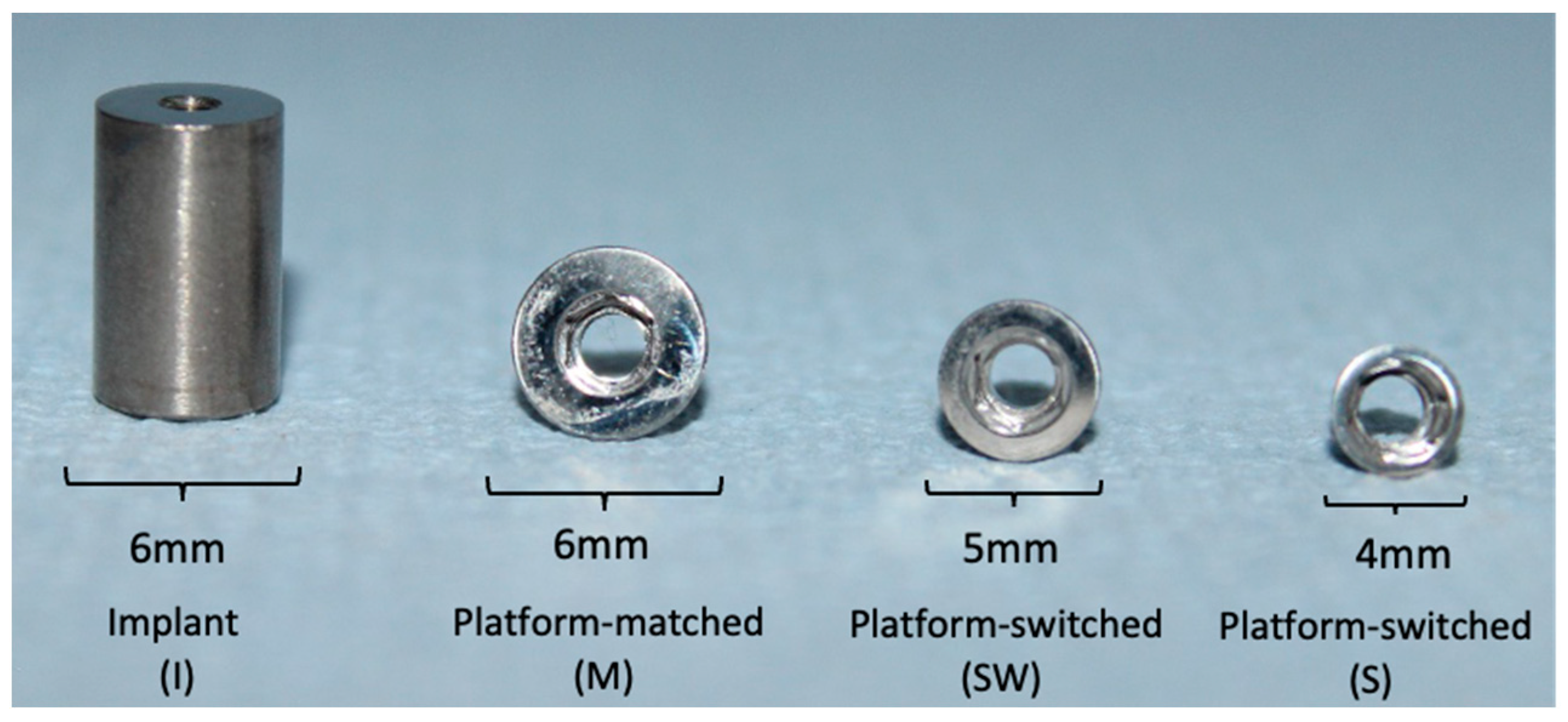
2.1. Preparation of Test Specimens
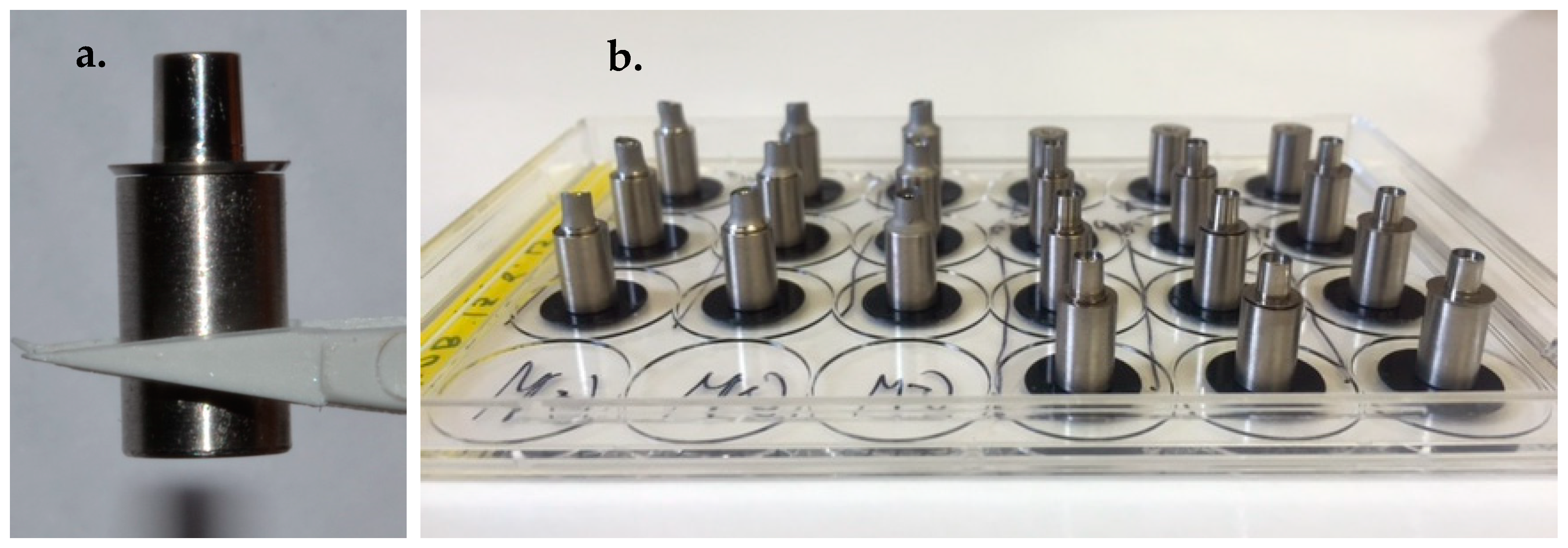
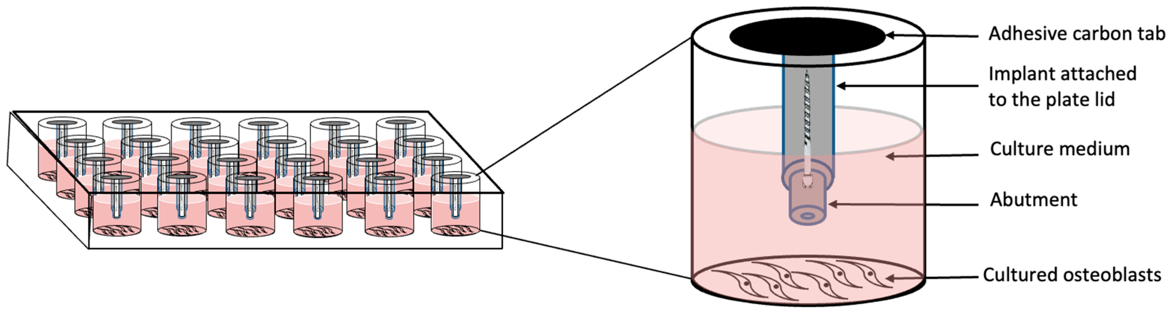
2.2. Assembly of Test Specimens
2.3. Cells and Cell Cultures
2.4. Cell Viability
2.5. Apoptosis
2.6. Gene Expression
2.7. Post-Immersion Observation of Implants and Abutments Interfaces
2.8. Statistical Analysis
3. Results
3.1. Cell Viability
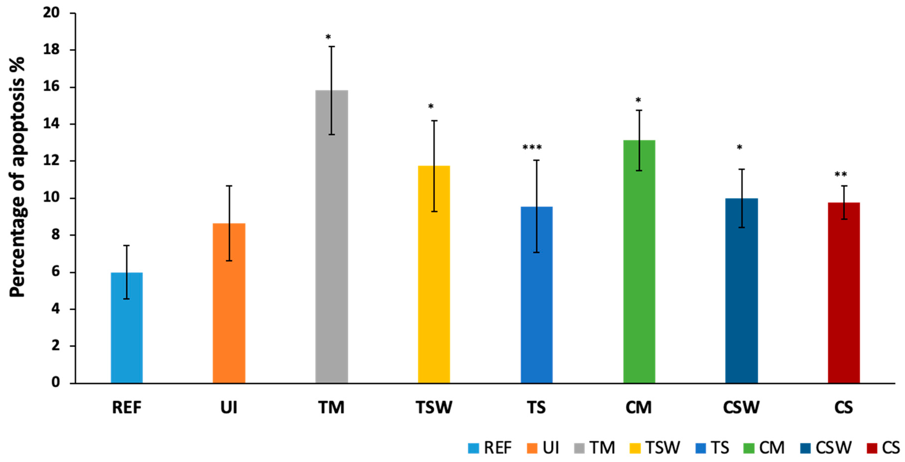
3.2. Apoptosis
3.3. Gene Expression
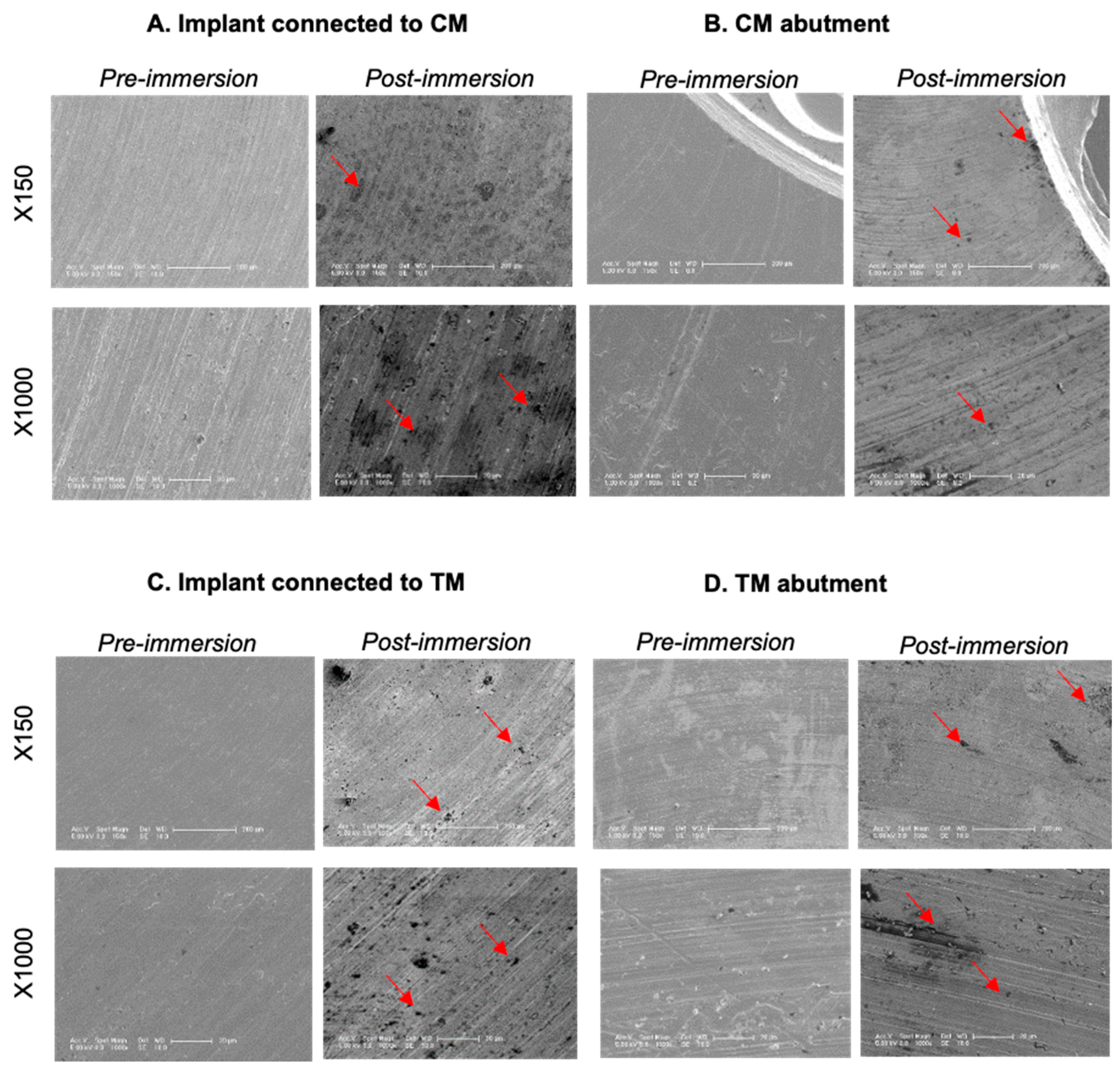
3.4. Post-Immersion SEM Examination
4. Discussion
5. Conclusions
- Osteoblastic cell viability, apoptosis, and regulation of bone-resorbing mediators were significantly altered in the presence of implant–abutment couplings.
- Titanium implants alone did not influence the apoptosis and secretion of the tested cytokines and chemokines, but adversely influenced cell viability up to one week of exposure.
- The adverse biologic responses were more prominent in the platform-matched implant–abutment couplings. Therefore, platform-switching should be considered when restoring dental implants.
- The observed cytotoxic responses in osteoblastic cells could be due to metal ion release from the immersed samples into the surrounding medium as a result of corrosion, suggesting that corrosion products could play a pivotal role in the mediation of crestal bone loss around dental implants.
Author Contributions
Funding
Institutional Review Board Statement
Informed Consent Statement
Data Availability Statement
Acknowledgments
Conflicts of Interest
References
- Derks, J.; Tomasi, C. Peri-implant health and disease. A systematic review of current epidemiology. J. Clin. Periodontol. 2015, 42, S158–S171. [Google Scholar] [CrossRef] [PubMed]
- Thanissorn, C.; Guo, J.; Jing Ying Chan, D.; Koyi, B.; Kujan, O.; Khzam, N.; Miranda, L.A. Success rates and complications associated with single immediate implants: A systematic review. Dent. J. 2022, 10, 31. [Google Scholar] [CrossRef] [PubMed]
- Bragger, U.; Heitz-Mayfield, L. Biological and hardware complications in implant dentistry. In ITI Treatment Guide; Quintessence Publishing Co., Ltd.: Berlin, Germany, 2015; p. 200. [Google Scholar]
- Renvert, S.; Persson, G.R.; Pirih, F.Q.; Camargo, P.M. Peri-implant health, peri-implant mucositis, and peri-implantitis: Case definitions and diagnostic considerations. J. Clin. Periodontol. 2018, 45, S278–S285. [Google Scholar] [CrossRef]
- Albrektsson, T.; Zarb, G.; Worthington, P.; Eriksson, A.R. The long-term efficacy of currently used dental implants: A review and proposed criteria of success. Int. J. Oral Maxillofac. Implants 1986, 1, 11–25. [Google Scholar]
- Hermann, J.S.; Schoolfield, J.D.; Schenk, R.K.; Buser, D.; Cochran, D.L. Influence of the size of the microgap on crestal bone changes around titanium implants. A histometric evaluation of unloaded non-submerged implants in the canine mandible. J. Periodontol. 2001, 72, 1372–1383. [Google Scholar] [CrossRef]
- Lazzara, R.J.; Porter, S.S. Platform switching: A new concept in implant dentistry for controlling postrestorative crestal bone levels. Int. J. Periodontics Restor. Dent. 2006, 26, 9–17. [Google Scholar]
- Alrabeah, G.O.; Knowles, J.C.; Petridis, H. The effect of platform switching on the levels of metal ion release from different implant–abutment couples. Int. J. Oral Sci. 2016, 8, 117–125. [Google Scholar] [CrossRef]
- Alrabeah, G.O.; Knowles, J.C.; Petridis, H. Reduction of tribocorrosion products when using the platform-switching concept. J. Dent. Res. 2018, 97, 995–1002. [Google Scholar] [CrossRef]
- Hallab, N.J.; Jacobs, J.J. Biologic effects of implant debris. Bull. NYU Hosp. Jt. Dis. 2009, 67, 182–188. [Google Scholar]
- Vermes, C.; Glant, T.T.; Hallab, N.J.; Fritz, E.A.; Roebuck, K.A.; Jacobs, J.J. The potential role of the osteoblast in the development of periprosthetic osteolysis: Review of in vitro osteoblast responses to wear debris, corrosion products, and cytokines and growth factors. J. Arthroplast. 2001, 16, 95–100. [Google Scholar] [CrossRef]
- O’neill, S.C.; Queally, J.M.; Devitt, B.M.; Doran, P.P.; O’Byrne, J.M. The role of osteoblasts in peri-prosthetic osteolysis. Bone Jt. J. 2013, 95, 1022–1026. [Google Scholar] [CrossRef]
- Albrektsson, T.; Dahlin, C.; Jemt, T.; Sennerby, L.; Turri, A.; Wennerberg, A. Is marginal bone loss around oral implants the result of a provoked foreign body reaction? Clin. Implant. Dent. Relat. Res. 2014, 16, 155–165. [Google Scholar] [CrossRef] [PubMed]
- Albrektsson, T.; Chrcanovic, B.; Mölne, J.; Wennerberg, A. Foreign body reactions, marginal bone loss and allergies in relation to titanium implants. Eur. J. Oral Implantol. 2018, 11 (Suppl. 1), 537–546. [Google Scholar]
- Mombelli, A.; Hashim, D.; Cionca, N. What is the impact of titanium particles and biocorrosion on implant survival and complications? A critical review. Clin. Oral Implants Res. 2018, 29, 37–53. [Google Scholar] [CrossRef] [PubMed]
- Fretwurst, T.; Nelson, K.; Tarnow, D.P.; Wang, H.L.; Giannobile, W.V. Is metal particle release associated with peri-implant bone destruction? An emerging concept. J. Dent. Res. 2018, 97, 259–265. [Google Scholar] [CrossRef]
- Noronha Oliveira, M.; Schunemann, W.V.; Mathew, M.T.; Henriques, B.; Magini, R.S.; Teughels, W.; Souza, J.C. Can degradation products released from dental implants affect peri-implant tissues? J. Periodont. Res. 2018, 53, 1–11. [Google Scholar] [CrossRef] [PubMed]
- Wilson, T.G., Jr. Bone loss around implants—Is it metallosis? J. Periodontol. 2021, 92, 181–185. [Google Scholar] [CrossRef] [PubMed]
- Xu, A.; Alhamad, M.; Ampadi Ramachandran, R.; Shukla, A.; Barão, V.A.; Sukotjo, C.; Mathew, M.T. Peri-Implantitis in Relation to Titanium Corrosion: Current Status and Future Perspectives. J. Bio- Tribo-Corros. 2022, 8, 46. [Google Scholar] [CrossRef]
- Alrabeah, G.O.; Brett, P.; Knowles, J.C.; Petridis, H. The effect of metal ions released from different dental implant-abutment couples on osteoblast function and secretion of bone resorbing mediators. J. Dent. 2017, 66, 91–101. [Google Scholar] [CrossRef]
- Berglundh, T.; Armitage, G.; Araujo, M.G.; Avila-Ortiz, G.; Blanco, J.; Camargo, P.M.; Chen, S.; Cochran, D.; Derks, J.; Figuero, E.; et al. Peri-implant diseases and conditions: Consensus report of workgroup 4 of the 2017 World Workshop on the Classification of Periodontal and Peri-Implant Diseases and Conditions. J. Periodontol. 2018, 89, S313–S318. [Google Scholar] [CrossRef]
- Gittens, R.A.; Olivares-Navarrete, R.; Tannenbaum, R.; Boyan, B.D.; Schwartz, Z. Electrical implications of corrosion for osseointegration of titanium implants. J. Dent. Res. 2011, 90, 1389–1397. [Google Scholar] [CrossRef]
- Clark, G.C.; Williams, D.F. The effects of proteins on metallic corrosion. J. Biomed. Mater. Res. 1982, 16, 125–134. [Google Scholar] [CrossRef] [PubMed]
- ISO 10993-5: 2009; Biological Evaluation of Medical Devices-Part 5: Tests for In Vitro Cytotoxicity. International Organization for Standardization: Geneva, Switzerland, 2009.
- Livak, K.J.; Schmittgen, T.D. Analysis of relative gene expression data using real-time quantitative PCR and the 2−ΔΔCT method. Methods 2001, 25, 402–408. [Google Scholar] [CrossRef] [PubMed]
- Wataha, J.C. Predicting clinical biological responses to dental materials. Dent. Mater. 2012, 28, 23–40. [Google Scholar] [CrossRef]
- Wennberg, A.; Mjör, I.A.; Hensten-Pettersen, A. Biological evaluation of dental restorative materials—A comparison of different test methods. J. Biomed. Mater. Res. 1983, 17, 23–36. [Google Scholar] [CrossRef] [PubMed]
- Batool, F.; Özçelik, H.; Stutz, C.; Gegout, P.Y.; Benkirane-Jessel, N.; Petit, C.; Huck, O. Modulation of immune-inflammatory responses through surface modifications of biomaterials to promote bone healing and regeneration. J. Tissue Eng. 2021, 12, 20417314211041428. [Google Scholar] [CrossRef] [PubMed]
- Taylor, J.C.; Anderson, G.I.; Sutow, E.J.; Driscoll, C.F.; Mackey, D.C. The effects of the coupling of titanium implants and dissimilar metal abutments on osteoblast differentiation in vitro. Int. J. Oral Maxillofac. Implant. 1999, 14, 785–797. [Google Scholar]
- Hallab, N.J.; Jacobs, J.J.; Skipor, A.; Black, J.; Mikecz, K.; Galante, J.O. Systemic metal–protein binding associated with total joint replacement arthroplasty. J. Biomed. Mater. Res. 2000, 49, 353–361. [Google Scholar] [CrossRef]
- Hjalmarsson, L.; Smedberg, J.I.; Aronsson, G.; Wennerberg, A. Cellular responses to cobalt-chrome and CP titanium--an in vitro comparison of frameworks for implant-retained oral prostheses. Swed. Dent. J. 2011, 35, 177–186. [Google Scholar]
- Hallab, N.J.; Vermes, C.; Messina, C.; Roebuck, K.A.; Glant, T.T.; Jacobs, J.J. Concentration-and composition-dependent effects of metal ions on human MG-63 osteoblasts. J. Biomed. Mater. Res. 2002, 60, 420–433. [Google Scholar] [CrossRef] [PubMed]
- Lochner, K.; Fritsche, A.; Jonitz, A.; Hansmann, D.; Mueller, P.; Mueller-Hilke, B.; Bader, R. The potential role of human osteoblasts for periprosthetic osteolysis following exposure to wear particles. Int. J. Mol. Med. 2011, 28, 1055–1063. [Google Scholar] [CrossRef]
- Dalal, A.; Pawar, V.; McAllister, K.; Weaver, C.; Hallab, N.J. Orthopedic implant cobalt-alloy particles produce greater toxicity and inflammatory cytokines than titanium alloy and zirconium alloy-based particles in vitro, in human osteoblasts, fibroblasts, and macrophages. J. Biomed. Mater. Res. Part A 2012, 100, 2147–2158. [Google Scholar] [CrossRef] [PubMed]
- Jilka, R.L.; Weinstein, R.S.; Bellido, T.; Parfitt, A.M.; Manolagas, S.C. Osteoblast programmed cell death (apoptosis): Modulation by growth factors and cytokines. J. Bone Miner. Res. 1998, 13, 793–802. [Google Scholar] [CrossRef] [PubMed]
- Pioletti, D.P.; Leoni, L.; Genini, D.; Takei, H.; Du, P.; Corbeil, J. Gene expression analysis of osteoblastic cells contacted by orthopedic implant particles. J. Biomed. Mater. Res. 2002, 61, 408–420. [Google Scholar] [CrossRef]
- Shida, J.; Trindade, M.C.; Goodman, S.B.; Schurman, D.J.; Smith, R.L. Induction of interleukin-6 release in human osteoblast-like cells exposed to titanium particles in vitro. Calcif. Tissue Int. 2000, 67, 151–155. [Google Scholar] [CrossRef] [PubMed]
- Zhang, L.; Haddouti, E.M.; Welle, K.; Burger, C.; Wirtz, D.C.; Schildberg, F.A.; Kabir, K. The effects of biomaterial implant wear debris on osteoblasts. Front. Cell Dev. Biol. 2020, 8, 352. [Google Scholar] [CrossRef] [PubMed]
- Kubies, D.; Himmlová, L.; Riedel, T.; Chánová, E.; Balík, K.; Douderova, M.; Bártová, J.; Pesakova, V.J. The interaction of osteoblasts with bone-implant materials: 1. The effect of physicochemical surface properties of implant materials. Physiol. Res. 2011, 60, 95. [Google Scholar] [CrossRef]
- Quabius, E.S.; Ossenkop, L.; Harder, S.; Kern, M. Dental implants stimulate expression of Interleukin-8 and its receptor in human blood—An in vitro approach. J. Biomed. Mater. Res. Part B Appl. Biomater. 2012, 100, 1283–1288. [Google Scholar] [CrossRef]
- Fritz, E.A.; Glant, T.T.; Vermes, C.; Jacobs, J.J.; Roebuck, K.A. Chemokine gene activation in human bone marrow-derived osteoblasts following exposure to particulate wear debris. J. Biomed. Mater. Res. Part A 2006, 77, 192–201. [Google Scholar] [CrossRef]
- Queally, J.M.; Devitt, B.M.; Butler, J.S.; Malizia, A.P.; Murray, D.; Doran, P.P.; O’byrne, J.M. Cobalt ions induce chemokine secretion in primary human osteoblasts. J. Orthop. Res. 2009, 27, 855–864. [Google Scholar] [CrossRef]
- Crofford, L.J. COX-1 and COX-2 tissue expression: Implications and predictions. J. Rheumatol. Suppl. 1997, 49, 15–19. [Google Scholar] [PubMed]
- Wataha, J.C.; Craig, R.G.; Hanks, C.T. The release of elements of dental casting alloys into cell-culture medium. J. Dent. Res. 1991, 70, 1014–1018. [Google Scholar] [CrossRef] [PubMed]
- Wataha, J.C.; Malcolm, C.T.; Hanks, C.T. Correlation between cytotoxicity and the elements released by dental casting alloys. Int. J. Prosthodont. 1995, 8, 9–14. [Google Scholar] [PubMed]
- Hermann, J.S.; Cochran, D.L.; Hermann, J.S.; Buser, D.; Schenk, R.K.; Schoolfield, J.D. Biologic Width around one-and two-piece titanium implants: A histometric evaluation of unloaded nonsubmerged and submerged implants in the canine mandible. Clin. Oral Implant. Res. 2001, 12, 559–571. [Google Scholar] [CrossRef] [PubMed]
- Canullo, L.; Quaranta, A.; Teles, R.P. The microbiota associated with implants restored with platform switching: A preliminary report. J. Periodontol. 2010, 81, 403–411. [Google Scholar] [CrossRef]
- Maeda, Y.; Miura, J.; Taki, I.; Sogo, M. Biomechanical analysis on platform switching: Is there any biomechanical rationale? Clin. Oral Implant. Res. 2007, 18, 581–584. [Google Scholar] [CrossRef]


| Material | Manufacturer | Composition in % by Mass |
|---|---|---|
| Titanium cylinders (Medical grade, Grade II, ASTM F67-13) | Fort Wayne Metals, County Mayo, Ireland | Ti > 99.5%, Fe:0.2%, N:0.03%, C:0.1%, O:0.18%, H:0.015% |
| Ti alloy abutments (Ti-6Al-4V) | Zimmer Dental Inc., Swindon, UK | Ti:91%, V:4%, Al:6% |
| Cobalt–chrome abutments | LaserAbutments, Renishaw, UK | Co:63.1%, Cr:24.7%, Mo:5.4%, Mn < 1%, Si < 1%, Fe < 1% |
| Osteoblast basal medium | OBM™, Clonetics™ OGM™ BulletKit™, Lonza, USA | fetal bovine 10%, Gentamicin Sulfate/Amphotercin-B 0.1%, Ascorbic acid 0.1% |
| Sample Name | Code | Number of Samples |
|---|---|---|
| Sample-free culture medium | REF | 3 |
| Unconnected implan | UI | 3 |
| Implant connected to implant to platform-switched titanium abutment (6 mm) | TM | 3 |
| Implant connected to platform-switched wide titanium abutment (5 mm) | TSW | 3 |
| Implant connected to platform-switched titanium abutment (4 mm) | TS | 3 |
| Implant connected to platform-matched cobalt–chrome abutment (6 mm) | CM | 3 |
| Implant connected to platform-switched wide cobalt–chrome abutment (5 mm) | CSW | 3 |
| Implant connected to platform-matched cobalt–chrome abutment (4 mm) | CS | 3 |
| Total | 24 |
Disclaimer/Publisher’s Note: The statements, opinions and data contained in all publications are solely those of the individual author(s) and contributor(s) and not of MDPI and/or the editor(s). MDPI and/or the editor(s) disclaim responsibility for any injury to people or property resulting from any ideas, methods, instructions or products referred to in the content. |
© 2023 by the authors. Licensee MDPI, Basel, Switzerland. This article is an open access article distributed under the terms and conditions of the Creative Commons Attribution (CC BY) license (https://creativecommons.org/licenses/by/4.0/).
Share and Cite
Alrabeah, G.; Knowles, J.C.; Petridis, H. Surface Corrosion from Implant–Abutment Couplings with Different Connection Designs Influences Osteoblasts’ Function: A Novel Technique. Appl. Sci. 2023, 13, 8957. https://doi.org/10.3390/app13158957
Alrabeah G, Knowles JC, Petridis H. Surface Corrosion from Implant–Abutment Couplings with Different Connection Designs Influences Osteoblasts’ Function: A Novel Technique. Applied Sciences. 2023; 13(15):8957. https://doi.org/10.3390/app13158957
Chicago/Turabian StyleAlrabeah, Ghada, Jonathan C. Knowles, and Haralampos Petridis. 2023. "Surface Corrosion from Implant–Abutment Couplings with Different Connection Designs Influences Osteoblasts’ Function: A Novel Technique" Applied Sciences 13, no. 15: 8957. https://doi.org/10.3390/app13158957
APA StyleAlrabeah, G., Knowles, J. C., & Petridis, H. (2023). Surface Corrosion from Implant–Abutment Couplings with Different Connection Designs Influences Osteoblasts’ Function: A Novel Technique. Applied Sciences, 13(15), 8957. https://doi.org/10.3390/app13158957