Article

Label-Free Detection and Classification of Glaucoma Based on Drop-Coating Deposition Raman Spectroscopy

1
Institute of Biopharmaceutical and Health Engineering, Tsinghua Shenzhen International Graduate School, Tsinghua University, Shenzhen 518055, China
2
State Key Laboratory of Ophthalmology, Guangzhou 510060, China
3
Zhongshan Ophthalmic Center, Sun Yat-sen University, Guangzhou 510060, China
4
Guangdong Provincial Key Laboratory of Ophthalmology and Visual Science, Guangzhou 510060, China
5
Bio-Intelligent Manufacturing and Living Matter Bioprinting Center, Research Institute of Tsinghua University in Shenzhen, Tsinghua University, Shenzhen 518057, China
6
Tsinghua Shenzhen International Graduate School, Tsinghua University, Shenzhen 518055, China
7
Department of Neurosurgery, Sichuan Provincial People’s Hospital, University of Electronic Science and Technology of China, Chengdu 610072, China
*
Authors to whom correspondence should be addressed.
Appl. Sci. 2023, 13(11), 6476; https://doi.org/10.3390/app13116476
Submission received: 7 May 2023 / Revised: 18 May 2023 / Accepted: 22 May 2023 / Published: 25 May 2023

Abstract

:

Featured Application

This study was the first to use drop-coating deposition Raman spectroscopy (DCDRS) for the tear-based detection of glaucoma, including primary open-angle glaucoma (POAG) and primary angle-closure glaucoma (PACG). By using a classification model, POAG, PACG and normal groups can be well distinguished with good accuracy, sensitivity and specificity based on the Raman spectra. The results of this work support DCDRS being a promising tool for the diagnosis of POAG and PACG using human tears as detection samples.

Abstract

Primary open-angle glaucoma (POAG) and primary angle-closure glaucoma (PACG) are prevailing eye diseases that can lead to blindness. In order to provide a non-invasive diagnostic method for glaucoma, we investigated the feasibility of using drop-coating deposition Raman spectroscopy (DCDRS) to discriminate glaucoma patients from healthy individuals based on tear samples. Tears from 27, 19 and 27 POAG patients, PACG patients and normal individuals, respectively, were collected for Raman measurement. For high-dimension data analysis, principal component analysis–linear discriminant analysis (PCA-LDA) was used to discriminate the features of the Raman spectra, followed by a support vector machine (SVM) used to classify samples into three categories, which is called a PCA-LDA-based SVM. The differences in the characteristic peaks of Raman spectra between glaucoma patients and normal people were related to the different contents of various proteins and lipids. For the PCA-LDA-based SVM, the total accuracy reached 93.2%. With the evaluation of 30% test dataset validation, the classification accuracy of the model was 90.9%. The results of this work reveal that tears can be used for Raman detection and discrimination by combining the process with the PCA-LDA-based SVM, supporting DCDRS being a potential method for the diagnosis of glaucoma in the future.

1. Introduction

According to the World Health Organization (WHO), around 1.3 billion people suffered from visual impairment globally due to ocular diseases in 2018 [1]. Among various ocular diseases, glaucoma is the second leading cause of blindness after cataracts worldwide, affecting more than 70 million people worldwide [2,3]. Because there are usually no noticeable symptoms of glaucoma at an early stage, the process may develop gradually to an advanced stage without the awareness of the patients, leading to subsequent visual loss [4,5]. The two most common types of glaucoma—primary open-angle glaucoma (POAG) and primary angle-closure (PACG) glaucoma—have different risk factors [6]. In the region of drainage pathways, aqueous humor outflow through trabecular meshwork brings increasing resistance, which causes POAG. However, for PACG, drainage pathways in human eyes are blocked [7]. Studies have discovered that glaucoma is associated withoptic nerve degeneration and the death of retinal ganglion cells (RGCs) [8,9,10]. Because the level of intraocular pressure (IOP) corresponds to the death of RGCs, both POAG and PACG can be characterized by the raised IOP. Although IOP, age and genetics have been proved to be the factors in glaucoma, the exact mechanism of glaucoma is still not fully understood [11,12,13]. Currently, gonioscopic examination, ultrasound bio-microscopy and optical coherence tomography are commonly used to observe anterior chamber structures in clinical diagnosis of glaucoma. However, the detection process is commonly complicated, and POAG and PACG are difficult to distinguish with a single indicator.
Human tears are the body fluids secreted by lacrimal glands. The functions of tears include protecting the eyes from infection, forming the water-based surface layer on the cornea for light penetration, flushing waste and small particles, and maintaining lubrication and moisture of the eye [14,15]. Tear fluid is a complex aqueous mixture that consists of more than 95% water and some other important proteins, electrolytes, lipids, etc., which correspond to the state of the ocular pathophysiology [16,17]. Several specific components in tears were measured with different techniques. For instance, lactoferrin levels and secretory immunoglobulin A concentrations were evaluated through immunoassays [18,19]. In addition, mass spectrometry was used to analyze proteins and peptides in tear fluid [20,21]. Previous studies proposed that brain-derived neurotrophic factor [22], some differential proteins [23] and autoantibody profiles [24] in tears may provide efficient biomarkers for glaucoma diagnosis.
Raman spectroscopy (RS) is a non-invasive optical technique to determine the existence of certain molecules that can be used in ophthalmology [16,25]. RS can reveal a frequency shift at a specific wavelength caused by the specific vibration mode of the molecular bond, providing a chemical “fingerprint” of the detecting material [26,27]. So far, RS has been used to detect retinal tissues for glaucoma characterization [28,29]. Tears have not been considered as samples with RS for glaucoma detection. However, RS can be utilized to discriminate various diseases via different body fluids, such as classification of glioma with blood serum [30], oral cancer detection with saliva [31] and prostate cancer detection with urine [32]. The total concentration of proteins in tears is about 10 mg/mL, which is much lower than that of blood serum [33]. Therefore, drop-coating deposition Raman spectroscopy (DCDRS) is involved in tear sample detection with RS. The principle of DCDRS is based on the “coffee-ring effect”. When a drop of solution material evaporates on a hydrophobic surface, a ring-shaped deposit forms spontaneously [34]. As the edges of the droplets on the contact surface are fixed and the solvent begins to evaporate, the coffee-ring formation occurs with a net flow of the solution from the center to the edge. The flow can maintain an equilibrium droplet shape and bring solute materials to the periphery of the drop, leading to a ring of excess materials after all the solvent evaporates. A previous study has proved that protein solutions with low concentrations can provide high-quality Raman signals and the dried materials in the ring remain stable for a period of time without significant changes to the Raman spectrum [35]. Furthermore, the protein Raman signal acquired from DCDRS is irrelevant to protein concentration and droplet volume [36]. Based on DCDRS, Choi et al. detected adenoviral conjunctivitis by using human tears without additional tagging or chemical modification [37]. Filik et al. evaluated protein concentration and investigated protein composition of human tears [38,39]. According to previous studies, DCDRS is feasible for diagnosing glaucoma via tear fluids.
Principal component analysis (PCA) is a statistical technique that aims to use the idea of dimensionality reduction to convert multiple indicators into a few comprehensive indicators, extracting the most significant principal components out of original data. PCA has been used in diagnosis of several diseases, such as glioma [30], breast cancer [40] and lung cancer [41]. Sun et al. used tears and RS to classify different ocular diseases with PCA combined with different algorithms [42]. To overcome redundant information more efficiently and to improve the generalization performance, we further used a model called a principal-component-analysis–linear-discriminant-analysis-based support vector machine (PCA-LDA-based SVM) to distinguish features of glaucoma and healthy individuals.
In this work, tears were used as detection samples for the first time in Raman measurement of glaucoma classification. After DCDRS measurements with tears, Raman spectra were processed and analyzed. The PCA-LDA-based SVM model was used for further classification of different tear samples.

2. Materials and Methods

2.1. Tear Samples Collection

In this work, 3 groups were studied with RS, including 27 POAG patients, 19 PACG patients and 27 healthy individuals as control group. All the disposal during experiments with human participants was according to the approval of the Ethics Committee of Tsinghua Shenzhen International Graduate School. PACG and POAG were diagnosed according to the criteria defined by International Society for Geographic and Epidemiological Ophthalmology (ISGEO). Furthermore, members of the control group may have age-related cataract but are without a history of other eye diseases or IOP exceeding 21 mmHg.
Before this study, none of the glaucoma patients had eye drops for at least two hours. In addition, none of the subjects in glaucoma groups and control group had severe systemic diseases, and they did not receive any ocular surgery or other clinical treatment, such as radiotherapy and chemotherapy. Table 1 lists demographic and clinical data of glaucoma patients and normal individuals. For eye type (OD/OS), there were no significant differences in pairwise comparisons between groups (p > 0.5). However, the IOP levels from POAG and PACG groups were significantly higher than those from normal group (p < 0.01). To acquire tear samples, the nontraumatic tear collection proceeded by using sterile glass capillary tubes (Drummond, Birmingham, AL, USA) from the lateral inferior meniscus without local anesthesia or stimulation. Next, 5 μL of the tear samples was collected and promptly transferred to 0.2 mL EP tubes. Then, for subsequent Raman measurements, samples were transported to the laboratory via dry ice within 24 h. All samples were from Zhongshan Ophthalmic Center, Sun Yat-sen University.

2.2. Drop-Coating Deposition Raman SPECTROSCOPIC Measurements

After the tear samples were thawed at room temperature, about 0.8 μL of the sample was taken directly from the capillary tube and moved manually onto the flat stainless-steel sheet for drop-coating deposition. Then, in order to form the tear ring, the sample was dried at about 25 °C in the constant-temperature oven for approximately 15 min.
A confocal Raman micro-spectrometer (Horiba LabRAM HR800, Horiba Jobin Yvon, Japan) was used for Raman spectra acquisition. Under the microscope at 50× magnification, the spectrum grating was 600 lines and hole was 100 μm. The excitation laser wavelength was 785 nm with the power of 25 mW, and the spectral resolution was less than 1 cm−1 in the range of 400–1800 cm−1. Through laser focus adjustment, the field of view was approximately 0.8 μm in diameter. To prevent density effect, for each sample, 3 spectra were recorded to obtain the average spectrum for further data analysis. The exposure time was 20 s, with 5 averaged accumulation measurements.

2.3. Data Processing and Analysis

All the Raman spectral data were smoothed via Savitzky–Golay filter with polynomial order 3 to remove the system noise. The back-ground noises caused by fluorescence were subtracted via linear-fitted lines based on the same points for each spectrum. Then, each spectrum was normalized by dividing by its own maximum value in the range of 400–1800 cm−1, providing accurate spectral shapes and better comparison of various Raman bands from different samples. The smoothing, background subtraction and normalization of the spectra were processed with software Origin 9 (Origin Lab, Northampton, MA, USA). Statistical significance for the Raman intensity of the characteristic peaks was evaluated with one-way analysis of variance (ANOVA) with Tukey’s test, also using the software Origin 9. p values less than 0.05, 0.01, 0.001 and 0.0001 were considered statistically significant.
We proposed PCA-LDA-based SVM, an SVM model based on PCA-LDA dimensionality reduction, to perform classification for the Raman spectral data. PCA and LDA are both important dimensionality-reduction techniques for high-dimensional data. PCA removes the correlation between the eigenvalues corresponding to each wavelength based on the statistical information of data, thus removing redundant information. LDA projects high-resolution, high-dimensional Raman spectral data into the low-dimensional space. The disadvantage of LDA is that it relies heavily on the distribution of data, and it is prone to overfitting. In order to obtain higher classification performance, PCA was realized as a precursor step to LDA classification.
This two-step process combined the benefits of both PCA and LDA. It can reduce the dimensionality while preserving the discriminatory information. However, after reducing the data to two variables using PCA-LDA, it was still difficult to classify the samples in a potentially nonlinear feature space. LDA alone may not be sufficient to handle complex decision boundaries in this reduced feature space. Therefore, we introduced the higher-order SVM as the classifier. SVM is a powerful machine learning algorithm known for its ability to handle nonlinear classification tasks effectively. By utilizing the kernel trick, SVM can map the reduced two-dimensional feature space into a higher-dimensional space where the samples become linearly separable. This can provide more accurate classification and better generalization of unseen data.
In addition, we constructed the model without the whitening operation. Whitening is a technique commonly used in data preprocessing to transform data into a form with zero mean and unit variance. It is also known as a de-correlation operation or spherification operation. The goal of the whitening operation is to remove correlation from the data so that there is no linear correlation between the transformed data features and the variance of each feature is equal to 1. This data-preprocessing technique can help improve the performance of certain machine learning algorithms, especially those that are sensitive to correlation between data features. For PCA, to achieve more accurate classification, 95% of the variance information of the original Raman spectral was retained. For LDA, the number of components after projection was set to 2 to weaken the tendency of overfitting. For SVM, we constructed a nonlinear multidimensional support vector classifier, set the penalty term coefficient of the relaxation coefficient to 1, selected Gaussian radial basis as the kernel and set gamma as the inverse of the Raman spectral feature length.
The training phase consists of three processes: first, redundant information was removed from the Raman spectral data using PCA; second, the LDA was fitted by least squares to project the high-dimensional information into the two-dimensional space; finally, the SVM was trained on the basis of the LDA dimensionality reduction. The steps in the inference process were similar.

3. Results

3.1. The Acquisition and Analysis of Raman Spectra

We confirmed the performance of drop-coating deposition of the tear ring. With the microscope of the confocal Raman micro-spectrometer, a part of the uniform ring for the dried tear sample was imaged, as can be seen in Figure 1a. The Raman spectra at different locations of the tear drop were recorded, including the ring, quartile and central regions. Three spectra were acquired and averaged as one spectrum for each region (Figure 1b). Raman spectra collected from the ring region provided stronger signals and more features of Raman peaks in the range of 400–1800 cm−1. Therefore, the spectra from the ring region for all samples were further analyzed.
After the calculation of the average spectrum for each tear sample, the mean spectra were evaluated and displayed for POAG, PACG and normal group. As shown in Figure 2a–c, the mean spectra of three groups possessed similar profiles, positions and bandwidths for different characteristic peaks. To figure out the specific differences between different spectra, the pairwise difference in the spectral intensities was calculated for all three groups.
Except for 1270 and 1313 cm−1, most other Raman peaks had higher intensities for glaucoma patients than for the normal individuals. The changes in the peaks were basically associated with the different metabolic environment of glaucoma, leading to the changes in chemical components in tear samples. According to statistical analysis with ANOVA, some Raman peaks of the three groups showed significantly different intensities from each other (p < 0.05). The peaks are indicated by black arrows in Figure 2, and the mean intensities of the peaks are displayed in Figure 3. Specifically, peaks at 447, 1003 and 1033 cm−1 of the POAG group had higher intensities than those of the normal group (p < 0.05). For the PACG group, the intensities of Raman peaks at 447, 760, 852, 1003, 1033, 1582 and 1664 cm−1 were higher than those of the normal group, with statistical significance (p < 0.05). The attribution of related Raman peaks in the range of 400–1800 cm−1 for glaucoma and normal tears is listed in Table 2 based on previous reports [30,38,43].

3.2. PCA-LDA-Based SVM

We used PCA-LDA-based SVM to classify three different groups: POAG, PACG and normal groups. Two experiments were conducted to demonstrate the efficiency of our model:
Experiment A: The training and test dataset are the same and consist of all data.
Experiment B: All data are split to generate the training dataset (70%) and test dataset (30%).
Experiment A and Experiment B were completely independent experiments with no influence between the settings and results of the two experiments. We conducted Experiment A to initially verify the validity of our model. However, there was no impact on Experiment B. Experiment B was divided into training and test sets, which strictly avoided data leakage. Therefore, the results of Experiment B can fully reflect the performance of the proposed PCA-LDA-based SVM model.
Figure 4a depicts the classification result of Experiment A with a scatter diagram, which shows that the model can provide an apparent distinction of different groups.
Table 3 lists the related evaluation parameters for the classification results of Experiment A and Experiment B. The classification accuracies of the PCA-LDA-based SVM diagnostic model were 93.2% and 90.9% for Experiment A and Experiment B, respectively. For Experiment A, the correction rates of the model correction for POAG, PACG and normal groups were 88.9% (24/27), 89.5% (17/19) and 100% (27/27), respectively, with a total accuracy of 93.15%. The specificities of the classification for POAG, PACG and normal groups were 97.8% (45/46), 94.4% (51/54) and 97.8% (45/46), respectively. For Experiment-B, the sensitivity and specificity of the differentiating POAG group were 80.0% (4/5) and 100% (17/17); for the PACG group classification, they were 87.5% (7/8) and 92.9% (13/14). The sensitivity and specificity of the normal group classification were 100% (9/9) and 92.3% (12/13).
To further evaluate the model with sensitivity and specificity, the receiver operating characteristic (ROC) curve was generated to illustrate the diagnostic ability of the model (Figure 4b). The area under the curve (AUC) can directly reflect the classification performance based on cross-validation results. The AUCs of POAG, PACG and normal groups were 0.99, 0.97 and 1.0, respectively. Then, micro-average AUC and macro-average AUC were also used for evaluation. Micro-average AUC was defined as the overall AUC related to all categories. First, a series of confidence thresholds t 1 , t n , , t n were determined. Second, the overall false positives and true positives were calculated without categorization at each threshold. This was conducted by taking each category in turn as a positive sample and the other categories as negative samples to obtain a confusion matrix (of size 2 × 2) for that category. The confusion matrix of all categories was superimposed to obtain the overall confusion matrix. Furthermore, the false-positive F P R t and true-positive T P R t under this threshold were calculated based on this confusion matrix. Finally, based on F P R t 1 , T P R t 1 ,   F P R t 2 , T P R t 2 , ,   F P R t n , T P R t n , the ROC curves were drawn and the micro-average AUC was calculated. In addition, macro-average AUC was simply defined as the average of the AUC for each category. The values of both micro-average AUC and macro-average AUC were 0.99, which shows the PCA-LDA model can discriminate POAG, PACG and normal groups from each other through Raman spectra in glaucoma diagnosis.

4. Discussion

RS is a rapid and noninvasive technique, bringing many advantages. It takes relatively less time and does not require complicated operations during the detection, compared with other current detecting techniques. By using tears as detecting subjects, DCDRS can directly provide Raman signal without enhancement methods, which is rather convenient compared to other RS methods, such as surface-enhanced RS. Sun et al. used RS with tear samples to distinguish different ocular diseases, including conjunctivitis, blepharitis, meibomian gland cyst, cataract and glaucoma [42]. However, the detailed analysis and discrimination of glaucoma with RS still remain. In this study, we used DCDRS to obtain Raman spectra for POAG and PCAG tear samples for the first time. According to the results, the characteristics of the spectra showed significant differences between glaucoma patients and normal individuals. Based on the characteristic peaks of Raman spectra, amino acids (tyrosine, tryptophan, phenylalanine and hydroxyproline), proteins (amide I and amide III) and lipids produced strong Raman signals and played important roles in the detection of glaucoma. Because these substances are closely related to the metabolism of the eye, glaucoma may lead to content and structure changes in tears at the molecular level. Based on their rich biochemical composition and easy collection, tears can be a potential option for the detection of glaucoma and its diagnosis through RS.
For the comparison between the glaucoma and the normal groups, all the Raman peaks from amino acids of the glaucoma groups had significantly higher intensities than those from the normal group, revealing that there was more content of amino acids in the glaucoma tears than the normal ones. This is consistent with a previous study, which reported enriched metabolic pathways of amino acids, including phenylalanine, tyrosine and tryptophan [44]. Compared with normal individuals, eight Raman peaks of PACG patients had significant disparities, which can be attributed to various molecular information, including ring torsion mode (447 cm−1), ring breathing mode (760 and 853 cm−1) and twisting mode. In contrast, for the POAG group, five Raman bands, including bands at 447 (ring torsion mode of phenylalanine) and 1313 cm−1 (CH3CH2 twisting mode of collagens and lipids), were statistically different from the normal group. The changes in amide III and proteins/lipids content in tears correspond to the changes in glaucomatous retinal tissues from previous reports [28,29]. Comparing POAG with PACG groups, the intensities of all the different Raman peaks (853, 880, 1270 and 1313 cm−1) of the POAG group were lower than those from the PACG group. For these two sub-groups, the reasons for the different contents of amino acids, proteins and lipids need to be investigated further. Furthermore, the intensity of the Raman peak at 1270 cm−1 showed significantly differences across all three groups. When using tears as detection samples, this peak can be considered as a marker in the discrimination of glaucoma.
Considering the high dimensionality of the Raman spectra, we used the dimension-reduction technique PCA to extract PCs from the Raman spectra data. It not only reduced the dimension of data but also minimized the information loss, reducing sensitivity of LDA to data distribution. LDA was used to build the classifier based on the PCs. The total accuracy of the PCA-LDA-based SVM model reached 93.2% and 90.9% for two different experimental settings, which both perform efficiently. The sensitivities of the classification of POAG, PACG and normal groups were 88.9%, 89.5% and 100%, respectively; the specificity values of the three groups were 97.8%, 94.4% and 97.8%, respectively. We created ROC curves based on the 30% test dataset classification results and calculated AUC values. An AUC value between 0.7 and 0.8 means the classifier is considered acceptable, and a value between 0.8 and 0.9 is considered excellent. The micro-average AUC and macro-average AUC of the model were both more than 0.9, which indicates the PCA-LDA model provided an excellent performance to diagnose glaucoma with Raman spectra based on the tear samples.

5. Conclusions

This study was the first to use DCDRS for the tear-based detection of glaucoma, including POAG and PACG. The measurement results reveal that the features of Raman spectra peaks were capable of reflecting the different chemical information of amino acids, proteins and lipids in tears, which testified that tear samples are feasible for use in glaucoma detection. In combination with the PCA-LDA-based SVM classification model, POAG, PACG and normal groups can be well distinguished with good accuracy, sensitivity and specificity based on the Raman spectra. The results of this work support DCDRS being a promising tool for the diagnosis of POAG and PACG using human tears as detection samples.

Author Contributions

Conceptualization, T.X. and M.L.; methodology, Y.L.; software, Q.H.; validation, Y.L. and H.L.; resources, H.L.; writing—original draft preparation, Y.L.; writing—review and editing, C.Z., T.X. and M.L.; supervision, T.X. and M.L.; funding acquisition, T.X. All authors have read and agreed to the published version of the manuscript.

Funding

This research is supported by National Natural Science Foundation of China (Grant No. 52075285) and Applied Basic Research Project of Sichuan Province (Grant No. 2021YJ0563).

Institutional Review Board Statement

The study was conducted in accordance with the Declaration of Helsinki and approved by the Ethics Committee of Tsinghua Shenzhen International Graduate School.

Informed Consent Statement

Informed consent was obtained from all subjects involved in the study.

Data Availability Statement

The data presented in this study are available on request from the corresponding author.

Conflicts of Interest

The authors declare no conflict of interest.

References

  1. Shi, Y.; Jiang, N.; Bikkannavar, P.; Cordeiro, M.F.; Yetisen, A.K. Ophthalmic sensing technologies for ocular disease diagnostics. Analyst 2021, 146, 6416–6444. [Google Scholar] [CrossRef] [PubMed]
  2. Chan, Y.M.; Ng, E.; Jahmunah, V.; Koh, J.E.W.; Oh, S.L.; Han, W.S.; Yip, L.W.L.; Acharya, U.R. Automated detection of glaucoma using elongated quinary patterns technique with optical coherence tomography angiogram images. Biomed. Signal Process. Control 2021, 69, 102895. [Google Scholar] [CrossRef]
  3. Quigley, H.A.; Broman, A.T. The number of people with glaucoma worldwide in 2010 and 2020. Br. J. Ophthalmol. 2006, 90, 262–267. [Google Scholar] [CrossRef] [PubMed]
  4. Leite, M.T.; Sakata, L.M.; Medeiros, F.A. Managing glaucoma in developing countries. Arq. Bras. Oftalmol. 2011, 74, 83–84. [Google Scholar] [CrossRef]
  5. Sathyamangalam, R.V.; Paul, P.G.; George, R.; Baskaran, M.; Hemamalini, A.; Madan, R.V.; Augustian, J.; Prema, R.; Lingam, V. Determinants of glaucoma awareness and knowledge in urban Chennai. Indian J. Ophthalmol. 2009, 57, 355. [Google Scholar]
  6. Coleman, A.L. Glaucoma. Lancet 1999, 354, 1803–1810. [Google Scholar] [CrossRef]
  7. Weinreb, R.N.; Aung, T.; Medeiros, F.A. The pathophysiology and treatment of glaucoma: A review. JAMA 2014, 311, 1901–1911. [Google Scholar] [CrossRef]
  8. Quigley, H.A.; Nickells, R.W.; Kerrigan, L.A.; Pease, M.E.; Thibault, D.J.; Zack, D.J. Retinal ganglion cell death in experimental glaucoma and after axotomy occurs by apoptosis. Investig. Ophthalmol. Vis. Sci. 1995, 36, 774–786. [Google Scholar]
  9. Morgan, J.E.; Uchida, H.; Caprioli, J. Retinal ganglion cell death in experimental glaucoma. Br. J. Ophthalmol. 2000, 84, 303–310. [Google Scholar] [CrossRef]
  10. Almasieh, M.; Wilson, A.M.; Morquette, B.; Vargas, J.L.C.; Di Polo, A. The molecular basis of retinal ganglion cell death in glaucoma. Prog. Retin. Eye Res. 2012, 31, 152–181. [Google Scholar] [CrossRef]
  11. Doucette, L.P.; Rasnitsyn, A.; Seifi, M.; Walter, M.A. The interactions of genes, age, and environment in glaucoma pathogenesis. Surv. Ophthalmol. 2015, 60, 310–326. [Google Scholar] [CrossRef] [PubMed]
  12. Tsai, T.; Grotegut, P.; Reinehr, S.; Joachim, S.C. Role of heat shock proteins in glaucoma. Int. J. Mol. Sci. 2019, 20, 5160. [Google Scholar] [CrossRef] [PubMed]
  13. Zukerman, R.; Harris, A.; Oddone, F.; Siesky, B.; Verticchio Vercellin, A.; Ciulla, T.A. Glaucoma Heritability: Molecular Mechanisms of Disease. Genes 2021, 12, 1135. [Google Scholar] [CrossRef] [PubMed]
  14. Tong, L.; Zhou, X.Y.; Jylha, A.; Aapola, U.; Liu, D.N.; Koh, S.K.; Tian, D.; Quah, J.; Uusitalo, H.; Beuerman, R.W. Quantitation of 47 human tear proteins using high resolution multiple reaction monitoring (HR-MRM) based-mass spectrometry. J. Proteom. 2015, 115, 36–48. [Google Scholar] [CrossRef] [PubMed]
  15. Posa, A.; Bräuer, L.; Schicht, M.; Garreis, F.; Beileke, S.; Paulsen, F. Schirmer strip vs. capillary tube method: Non-invasive methods of obtaining proteins from tear fluid. Ann. Anat. Anat. Anz. 2013, 195, 137–142. [Google Scholar] [CrossRef]
  16. Lin, C.-C.; Kuo, M.-T.; Chang, H.-C. Raman spectroscopy–a novel tool for noninvasive analysis of ocular surface fluid. J. Med. Biol. Eng. 2010, 30, 343–354. [Google Scholar] [CrossRef]
  17. Leal, L.; Nogueira, M.; Canevari, R.; Carvalho, L. Vibration spectroscopy and body biofluids: Literature review for clinical applications. Photodiagnosis Photodyn. Ther. 2018, 24, 237–244. [Google Scholar] [CrossRef]
  18. Ballow, M.; Donshik, P.C.; Rapacz, P.; Samartino, L. Tear lactoferrin levels in patients with external inflammatory ocular disease. Investig. Ophthalmol. Vis. Sci. 1987, 28, 543–545. [Google Scholar]
  19. Willcox, M.D.; Lan, J. Secretory immunoglobulin A in tears: Functions and changes during contact lens wear. Clin. Exp. Optom. 1999, 82, 1–3. [Google Scholar] [CrossRef]
  20. Grus, F.H.; Podust, V.N.; Bruns, K.; Lackner, K.; Fu, S.; Dalmasso, E.A.; Wirthlin, A.; Pfeiffer, N. SELDI-TOF-MS ProteinChip array profiling of tears from patients with dry eye. Investig. Ophthalmol. Vis. Sci. 2005, 46, 863–876. [Google Scholar] [CrossRef]
  21. Huang, M.-Z.; Hsu, H.-J.; Lee, J.-Y.; Jeng, J.; Shiea, J. Direct protein detection from biological media through electrospray-assisted laser desorption ionization/mass spectrometry. J. Proteome Res. 2006, 5, 1107–1116. [Google Scholar] [CrossRef] [PubMed]
  22. Ghaffariyeh, A.; Honarpisheh, N.; Heidari, M.H.; Puyan, S.; Abasov, F. Brain-derived neurotrophic factor as a biomarker in primary open-angle glaucoma. Optom. Vis. Sci. 2011, 88, 80–85. [Google Scholar] [CrossRef] [PubMed]
  23. Pieragostino, D.; Bucci, S.; Agnifili, L.; Fasanella, V.; D’Aguanno, S.; Mastropasqua, A.; Ciancaglini, M.; Mastropasqua, L.; Di Ilio, C.; Sacchetta, P. Differential protein expression in tears of patients with primary open angle and pseudoexfoliative glaucoma. Mol. Biosyst. 2012, 8, 1017–1028. [Google Scholar] [CrossRef] [PubMed]
  24. Gramlich, O.W.; Bell, K.; Hohenstein-Blaul, N.v.T.U.; Wilding, C.; Beck, S.; Pfeiffer, N.; Grus, F.H. Autoimmune biomarkers in glaucoma patients. Curr. Opin. Pharmacol. 2013, 13, 90–97. [Google Scholar] [CrossRef]
  25. Erckens, R.; Jongsma, F.; Wicksted, J.; Hendrikse, F.; March, W.; Motamedi, M. Raman spectroscopy in ophthalmology: From experimental tool to applications in vivo. Lasers Med. Sci. 2001, 16, 236–252. [Google Scholar] [CrossRef]
  26. Huang, C.-C.; Chen, W. Raman spectroscopic analysis of cataract lens: A compendious review. Appl. Spectrosc. Rev. 2018, 53, 689–702. [Google Scholar] [CrossRef]
  27. Kong, K.; Kendall, C.; Stone, N.; Notingher, I. Raman spectroscopy for medical diagnostics—From in-vitro biofluid assays to in-vivo cancer detection. Adv. Drug Deliv. Rev. 2015, 89, 121–134. [Google Scholar] [CrossRef]
  28. Wang, Q.; Yu, C.; Grozdanic, S.D.; Harper, M.M.; Kecova, H.; Lazic, T.; Hamouche, N. Exploring Raman spectroscopy for the evaluation of glaucomatous retinal changes. J. Biomed. Opt. 2011, 16, 107006. [Google Scholar] [CrossRef]
  29. Wang, Q.; Grozdanic, S.D.; Harper, M.M.; Hamouche, N.; Kecova, H.; Lazic, T.; Hernandez-Merino, E.; Yu, C. Detection and characterization of glaucoma-like canine retinal tissues using Raman spectroscopy. J. Biomed. Opt. 2013, 18, 067008. [Google Scholar] [CrossRef]
  30. Zhang, C.; Han, Y.; Sun, B.; Zhang, W.; Liu, S.; Liu, J.; Lv, H.; Zhang, G.; Kang, X. Label-free serum detection based on Raman spectroscopy for the diagnosis and classification of glioma. J. Raman Spectrosc. 2020, 51, 1977–1985. [Google Scholar] [CrossRef]
  31. Fălămaș, A.; Rotaru, H.; Hedeșiu, M. Surface-enhanced Raman spectroscopy (SERS) investigations of saliva for oral cancer diagnosis. Lasers Med. Sci. 2020, 35, 1393–1401. [Google Scholar] [CrossRef] [PubMed]
  32. Del Mistro, G.; Cervo, S.; Mansutti, E.; Spizzo, R.; Colombatti, A.; Belmonte, P.; Zucconelli, R.; Steffan, A.; Sergo, V.; Bonifacio, A. Surface-enhanced Raman spectroscopy of urine for prostate cancer detection: A preliminary study. Anal. Bioanal. Chem. 2015, 407, 3271–3275. [Google Scholar] [CrossRef] [PubMed]
  33. Ng, V.; Cho, P. The relationship between total tear protein concentrations determined by different methods and standards. Graefe’s Arch. Clin. Exp. Ophthalmol. 2000, 238, 571–576. [Google Scholar] [CrossRef] [PubMed]
  34. Deegan, R.D.; Bakajin, O.; Dupont, T.F.; Huber, G.; Nagel, S.R.; Witten, T.A. Capillary flow as the cause of ring stains from dried liquid drops. Nature 1997, 389, 827–829. [Google Scholar] [CrossRef]
  35. Zhang, D.; Xie, Y.; Mrozek, M.F.; Ortiz, C.; Davisson, V.J.; Ben-Amotz, D. Raman detection of proteomic analytes. Anal. Chem. 2003, 75, 5703–5709. [Google Scholar] [CrossRef]
  36. Ortiz, C.; Zhang, D.; Xie, Y.; Ribbe, A.E.; Ben-Amotz, D. Validation of the drop coating deposition Raman method for protein analysis. Anal. Biochem. 2006, 353, 157–166. [Google Scholar] [CrossRef]
  37. Choi, S.; Moon, S.W.; Shin, J.-H.; Park, H.-K.; Jin, K.-H. Label-free biochemical analytic method for the early detection of adenoviral conjunctivitis using human tear biofluids. Anal. Chem. 2014, 86, 11093–11099. [Google Scholar] [CrossRef]
  38. Filik, J.; Stone, N. Analysis of human tear fluid by Raman spectroscopy. Anal. Chim. Acta 2008, 616, 177–184. [Google Scholar] [CrossRef]
  39. Filik, J.; Stone, N. Investigation into the protein composition of human tear fluid using centrifugal filters and drop coating deposition Raman spectroscopy. J. Raman Spectrosc. 2009, 40, 218–224. [Google Scholar] [CrossRef]
  40. Pichardo-Molina, J.; Frausto-Reyes, C.; Barbosa-García, O.; Huerta-Franco, R.; González-Trujillo, J.; Ramírez-Alvarado, C.; Gutiérrez-Juárez, G.; Medina-Gutiérrez, C. Raman spectroscopy and multivariate analysis of serum samples from breast cancer patients. Lasers Med. Sci. 2007, 22, 229–236. [Google Scholar] [CrossRef]
  41. Oshima, Y.; Shinzawa, H.; Takenaka, T.; Furihata, C.; Sato, H. Discrimination analysis of human lung cancer cells associated with histological type and malignancy using Raman spectroscopy. J. Biomed. Opt. 2010, 15, 017009. [Google Scholar] [CrossRef] [PubMed]
  42. Sun, T.; Xie, X.; Li, H.; Lv, G.; Lv, X.; Tang, J.; Yue, X.; Mo, J. Rapid, noninvasive screening of ocular diseases using tear raman spectroscopy and different classification algorithms. Laser Phys. 2019, 30, 015701. [Google Scholar] [CrossRef]
  43. Talari, A.C.S.; Movasaghi, Z.; Rehman, S.; Rehman, I.U. Raman spectroscopy of biological tissues. Appl. Spectrosc. Rev. 2015, 50, 46–111. [Google Scholar] [CrossRef]
  44. Wang, Y.; Hou, X.-W.; Liang, G.; Pan, C.-W. Metabolomics in glaucoma: A systematic review. Investig. Ophthalmol. Vis. Sci. 2021, 62, 9. [Google Scholar] [CrossRef] [PubMed]
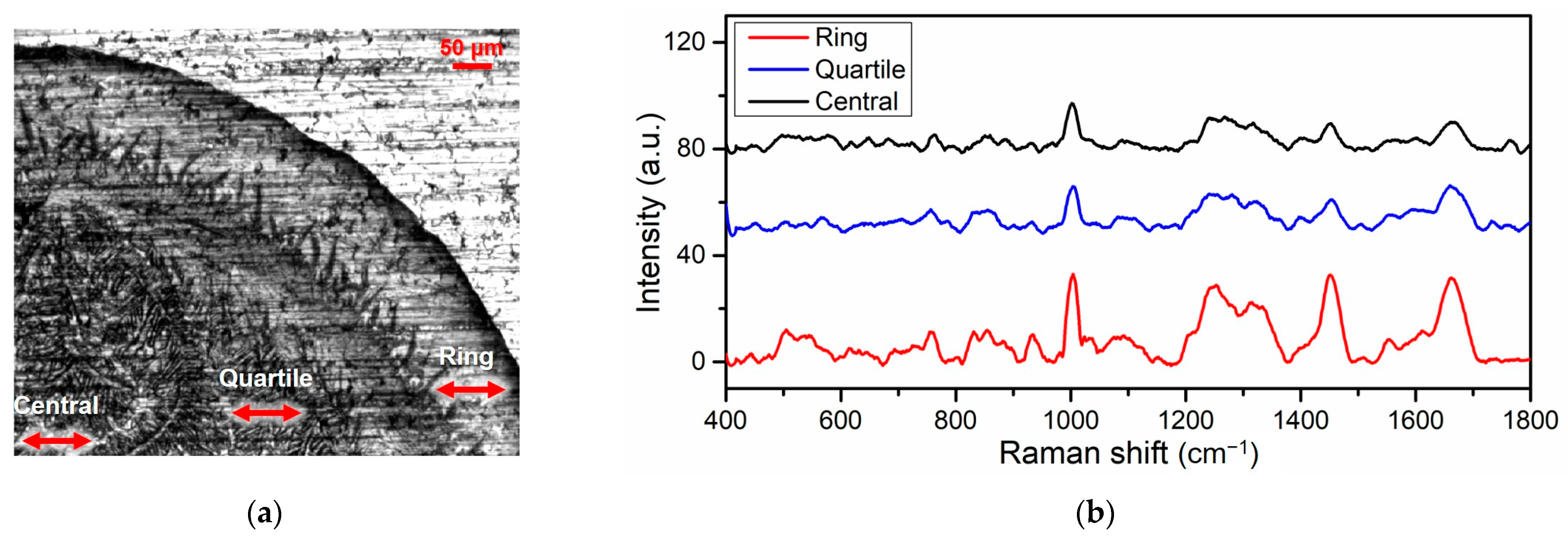
Figure 1. The acquired Raman spectra from different regions of deposited tear drop. (a) A part of the tear sample image after drop-coating deposition. (b) Raman spectra from the ring, quartile and central regions. Scale bar: 50 μm.
Applsci 13 06476 g001
Figure 2. Averaged Raman spectra for POAG, PACG and normal groups. (a) Comparison between POAG and the normal groups. (b) Comparison between PACG and the normal groups. (c) Comparison between POAG and PACG groups. The differences in compared two groups are indicated by black lines. Peaks with significant difference are indicated by black arrows.
Applsci 13 06476 g002
Figure 3. Specific intensity comparisons of Raman peaks from POAG, PACG and normal groups. (a) The spectral range of 620–950 cm−1. (b) The spectral range of 1200–1750 cm−1. The standard deviations were listed for each peak (* p < 0.05).
Applsci 13 06476 g003
Figure 4. Classification results for POAG, PACG and normal groups with the PCA-LDA model. (a) The scatter diagram of the three groups. (b) The receiver operating characteristic (ROC) curves for the classification of three groups.
Applsci 13 06476 g004
Table 1. Demographics and clinical data of glaucoma patients and normal individuals. POAG: primary open-angle glaucoma. PACG: primary angle-closure glaucoma. M: male. F: female. OD: right eye. OS: left eye. IOP: intraocular pressure. SD: standard deviation.
GroupsEye (OD/OS)IOP (mmHg ± SD)
POAG13/1420.14 ± 7.88
PACG9/1023.41 ± 15.78
Normal14/1315.03 ± 2.08
Table 2. Main Raman peak assignments in tears of glaucoma patients and healthy individuals [30,38,43].
Raman Shift (cm−1)Tentative AssignmentMolecular Origin
447Ring torsionPhenylalanine
760Ring breathing modeTryptophan
853Ring breathing modeTyrosine
879 Hydroxyproline, tryptophan
1003 Phenylalanine
1033 Phenylalanine
1270 Amide III
1313CH3CH2 twisting modeCollagens and lipids
1582 Phenylalanine
1664 Amide I
Table 3. Classification results and related evaluation parameters of the PCA-LDA-based SVM model.
Actual GroupPredicted GroupEvaluation Parameters
POAGPACGNormalSensitivitySpecificityAccuracy
Experiment APOAG243088.9%97.8%93.2%
PACG117189.5%94.4%
Normal0027100%97.8%
Experiment BPOAG41080.0%100%90.9%
PACG07187.5%92.9%
Normal009100.00%92.3%
Disclaimer/Publisher’s Note: The statements, opinions and data contained in all publications are solely those of the individual author(s) and contributor(s) and not of MDPI and/or the editor(s). MDPI and/or the editor(s) disclaim responsibility for any injury to people or property resulting from any ideas, methods, instructions or products referred to in the content.