Review of Image Augmentation Used in Deep Learning-Based Material Microscopic Image Segmentation
Abstract
1. Introduction
2. Method Review
2.1. Basic Image Augmentation Methods
2.1.1. Pixel Transformation
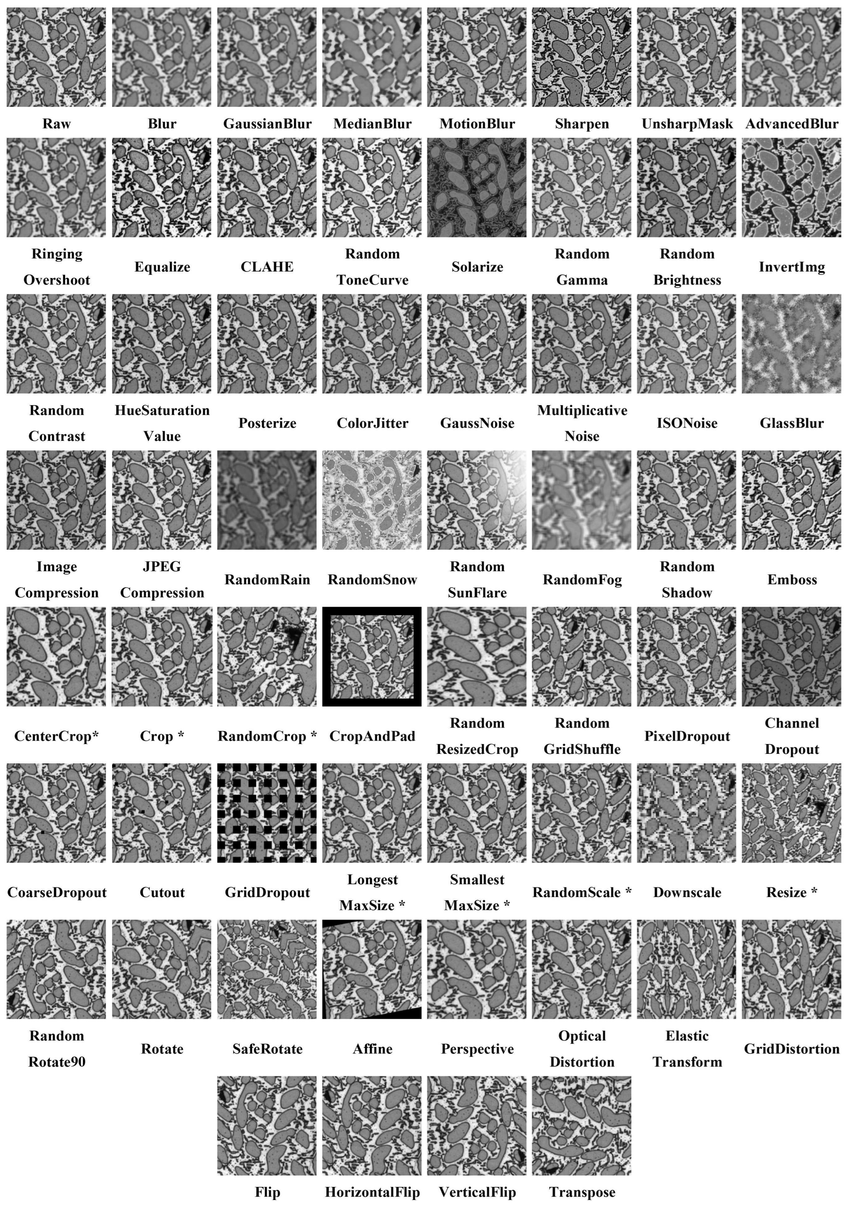
- Filtering: The filtering method modifies the pixel value of the original image based on the weighted sum of those in the surrounding pixel points [20] and then processes the entire image using a sliding window. By adjusting the size and value of the sliding window, it can create various transformation effects such as image blurring, sharpening, and simple deformation. For instance, Gaussian blur convolves the original image with a Gaussian filter of variable size, producing a new image with varying degrees of blurring. Median blurring is a convolution process using a median filter, which sets the gray value of each pixel of the original image to the median of the gray values of all pixels in the neighborhood of that point.
- Histogram equalization: The histogram represents the statistical results of the frequency of different gray values in an image. The gray histogram of an image can visualize the overall gray range, the frequency of different gray values, and the distribution of the image [21]. The contrast of an image is the difference between the highest and lowest brightness in the image, and the greater the difference, the greater the contrast of the image. Histogram equalization refers to the nonlinear stretching of an image with uneven gray distribution so that the frequencies of different gray levels in a certain gray range are approximately equal, and the contrast of the image can be improved by equalization. Ordinary histogram equalization does not fully consider the distribution of gray values in local areas, while contrast limited adaptive histogram equalization, or CLAHE [22], can enhance the local visualization of images while suppressing the effect of noise, and has primarily been applied to the enhancement of medical images.
- Color/brightness/contrast transformation: The color/brightness/contrast transformation method alters the image’s color space, brightness, contrast, and saturation values at random to generate a new, distinct image from the original image [23]. In image processing, color images can be described using a variety of color spaces, including RGB, HSV [24], YUV, and others. The ColorJitter [25] method can change the color distribution of the original image based on custom values of three input parameters. In addition to color space, brightness, contrast, and saturation are three important parameters of an image that can be used to change imaging effect, such as the RandomBrightness (RandomBrightness) and InvertImg (InvertImg) methods for changing image brightness, and the RandomContrast (RandomContrast) method for changing image contrast.
- Noise injection [26]: In image segmentation applications, any information that interferes with the extraction of the target of interest is collectively called noise. By actively introducing noise that may exist in the real scene for the image to achieve the augmentation of training samples, it can introduce interference information to the image segmentation model, and improve the generalization performance of the training model. To achieve data augmentation, there are two basic methods of noise injection: adding randomly distributed noise or adding noise with specific effects. Gaussian noise, uniform noise, gamma noise, pretzel noise, etc., are kinds of randomly distributed noise [27]. For example, by randomly sampling the Gaussian distribution, adding Gaussian noise can alter the intensity of each pixel’s value in an image, simulating the interference that an actual scene can bring. In addition, it can add random algorithmic effects with physical simulations, such as RandomRain, RandomSnow, RandomSunFlare, Emboss, Image Compression, Downscale, and Upscale. Furthermore, the method of using image compression for data augmentation involves generating images of varying qualities by changing compression quality parameters to produce different degrees of compression. This approach can increase the diversity of the data set and improve the robustness and generalization capabilities of neural networks. In the actual image segmentation application, the appropriate noise amplification type should be carefully chosen according to the needs of realistic scenes.
2.1.2. Region Cropping/Padding
2.1.3. Dropout
2.1.4. Geometric Deformation
2.1.5. Domain Adaption
2.2. Deep Learning-Based Image Augmentation Methods
2.2.1. Autoaugment
2.2.2. Image Generation for Image Augmentation
2.2.3. Learned Transformations Method
3. Results
3.1. Data Sets
3.1.1. Austenite
3.1.2. Al-La Alloy
3.2. Experimental Settings
3.3. Evaluation Metrics
3.3.1. Metric for Austenite
3.3.2. Metric for Al-La Alloy
3.4. Discussions
4. Conclusions
Author Contributions
Funding
Institutional Review Board Statement
Informed Consent Statement
Data Availability Statement
Acknowledgments
Conflicts of Interest
References
- Dursun, T.; Soutis, C. Recent developments in advanced aircraft aluminium alloys. Mater. Des. 2014, 56, 862–871. [Google Scholar] [CrossRef]
- Hu, J.; Shi, Y.; Sauvage, X.; Sha, G.; Lu, K. Grain boundary stability governs hardening and softening in extremely fine nanograined metals. Science 2017, 355, 1292–1296. [Google Scholar] [CrossRef] [PubMed]
- LeCun, Y.; Bengio, Y.; Hinton, G. Deep learning. Nature 2015, 521, 436–444. [Google Scholar] [CrossRef] [PubMed]
- Sonka, M.; Hlavac, V.; Boyle, R. Image Processing, Analysis, and Machine Vision; Cengage Learning: Boston, MA, USA, 2014. [Google Scholar]
- Ma, B.; Zhu, Y.; Yin, X.; Ban, X.; Huang, H.; Mukeshimana, M. Sesf-fuse: An unsupervised deep model for multi-focus image fusion. Neural Comput. Appl. 2021, 33, 5793–5804. [Google Scholar] [CrossRef]
- Ma, B.; Yin, X.; Wu, D.; Shen, H.; Ban, X.; Wang, Y. End-to-end learning for simultaneously generating decision map and multi-focus image fusion result. Neurocomputing 2022, 470, 204–216. [Google Scholar] [CrossRef]
- Ma, B.; Ban, X.; Huang, H.; Chen, Y.; Liu, W.; Zhi, Y. Deep learning-based image segmentation for al-la alloy microscopic images. Symmetry 2018, 10, 107. [Google Scholar] [CrossRef]
- Ma, B.; Ma, B.; Gao, M.; Wang, Z.; Ban, X.; Huang, H.; Wu, W. Deep learning-based automatic inpainting for material microscopic images. J. Microsc. 2021, 281, 177–189. [Google Scholar] [CrossRef]
- Chen, L.C.; Papandreou, G.; Kokkinos, I.; Murphy, K.; Yuille, A.L. Deeplab: Semantic image segmentation with deep convolutional nets, atrous convolution, and fully connected crfs. IEEE Trans. Pattern Anal. Mach. Intell. 2017, 40, 834–848. [Google Scholar] [CrossRef]
- Liu, W.; Chen, J.; Liu, C.; Ban, X.; Ma, B.; Wang, H.; Xue, W.; Guo, Y. Boundary learning by using weighted propagation in convolution network. J. Comput. Sci. 2022, 62, 101709. [Google Scholar] [CrossRef]
- Ronneberger, O.; Fischer, P.; Brox, T. U-net: Convolutional networks for biomedical image segmentation. In Proceedings of the International Conference on Medical Image Computing and Computer-Assisted Intervention, Munich, Germany, 5–9 October 2015; pp. 234–241. [Google Scholar]
- Boyuan, M. Research and Application of Few-Shot Image Segmentation Method for Complex 3D Material Microstructure. Ph.D. Thesis, University of Science and Technology Beijing, Beijing, China, 2021. [Google Scholar]
- Ma, B.; Wei, X.; Liu, C.; Ban, X.; Huang, H.; Wang, H.; Xue, W.; Wu, S.; Gao, M.; Shen, Q.; et al. Data augmentation in microscopic images for material data mining. NPJ Comput. Mater. 2020, 6, 125. [Google Scholar] [CrossRef]
- Pan, H.; Guo, Y.; Deng, Q.; Yang, H.; Chen, J.; Chen, Y. Improving fine-tuning of self-supervised models with Contrastive Initialization. Neural Netw. 2023, 159, 198–207. [Google Scholar] [CrossRef] [PubMed]
- Molchanov, D.; Ashukha, A.; Vetrov, D. Variational dropout sparsifies deep neural networks. In Proceedings of the International Conference on Machine Learning, PMLR, Melbourne, Australia, 19–25 August 2017; pp. 2498–2507. [Google Scholar]
- Bjorck, N.; Gomes, C.P.; Selman, B.; Weinberger, K.Q. Understanding batch normalization. In Proceedings of the Advances in Neural Information Processing Systems Conference, Montreal, QC, Canada, 3–8 December 2018; Volume 31. [Google Scholar]
- Zhuang, F.; Qi, Z.; Duan, K.; Xi, D.; Zhu, Y.; Zhu, H.; Xiong, H.; He, Q. A comprehensive survey on transfer learning. Proc. IEEE 2020, 109, 43–76. [Google Scholar] [CrossRef]
- Ma, D.; Tang, P.; Zhao, L.; Zhang, Z. Review of data augmentation for image in deep learning. J. Image Graph. 2021, 26, 487–502. [Google Scholar]
- Xu, M.; Yoon, S.; Fuentes, A.; Park, D.S. A comprehensive survey of image augmentation techniques for deep learning. Pattern Recognit. 2023, 137, 109347. [Google Scholar] [CrossRef]
- Chlap, P.; Min, H.; Vandenberg, N.; Dowling, J.; Holloway, L.; Haworth, A. A review of medical image data augmentation techniques for deep learning applications. J. Med. Imaging Radiat. Oncol. 2021, 65, 545–563. [Google Scholar] [CrossRef]
- Haiqiong, W.; Jiancheng, L. An adaptive threshold image enhancement algorithm based on histogram equalization. China Integrated Circuit 2022, 31, 38–42, 71. [Google Scholar]
- Zuiderveld, K. Contrast limited adaptive histogram equalization. Graph. Gems 1994, 6, 474–485. [Google Scholar]
- Chen, T.; Kornblith, S.; Norouzi, M.; Hinton, G. A simple framework for contrastive learning of visual representations. In Proceedings of the International Conference on Machine Learning, PMLR, Online, 13–18 July 2020; pp. 1597–1607. [Google Scholar]
- Sural, S.; Qian, G.; Pramanik, S. Segmentation and histogram generation using the HSV color space for image retrieval. In Proceedings of the IEEE International Conference on Image Processing, Rochester, NY, USA, 22–25 September 2002; Volume 2, pp. 22–25. [Google Scholar]
- Taylor, L.; Nitschke, G. Improving deep learning with generic data augmentation. In Proceedings of the 2018 IEEE Symposium Series on Computational Intelligence (SSCI), Bengaluru, India, 18–21 November 2018; pp. 1542–1547. [Google Scholar]
- Moreno-Barea, F.J.; Strazzera, F.; Jerez, J.M.; Urda, D.; Franco, L. Forward noise adjustment scheme for data augmentation. In Proceedings of the 2018 IEEE Symposium Series on Computational Intelligence (SSCI), Bengaluru, India, 18–21 November 2018; pp. 728–734. [Google Scholar]
- Shijie, J.; Ping, W.; Peiyi, J.; Siping, H. Research on data augmentation for image classification based on convolution neural networks. In Proceedings of the 2017 Chinese Automation Congress (CAC), Jinan, China, 20–22 October 2017; pp. 4165–4170. [Google Scholar]
- Wang, X.; Wang, K.; Lian, S. A survey on face data augmentation for the training of deep neural networks. Neural Comput. Appl. 2020, 32, 15503–15531. [Google Scholar] [CrossRef]
- Srivastava, N.; Hinton, G.; Krizhevsky, A.; Sutskever, I.; Salakhutdinov, R. Dropout: A simple way to prevent neural networks from overfitting. J. Mach. Learn. Res. 2014, 15, 1929–1958. [Google Scholar]
- DeVries, T.; Taylor, G.W. Improved regularization of convolutional neural networks with cutout. arXiv 2017, arXiv:1708.04552. [Google Scholar]
- Elgendi, M.; Nasir, M.U.; Tang, Q.; Smith, D.; Grenier, J.P.; Batte, C.; Spieler, B.; Leslie, W.D.; Menon, C.; Fletcher, R.R.; et al. The effectiveness of image augmentation in deep learning networks for detecting COVID-19: A geometric transformation perspective. Front. Med. 2021, 8, 629134. [Google Scholar] [CrossRef] [PubMed]
- Farahani, A.; Voghoei, S.; Rasheed, K.; Arabnia, H.R. A brief review of domain adaptation. In Advances in Data Science and Information Engineering; Springer: Berlin/Heidelberg, Germany, 2021; pp. 877–894. [Google Scholar]
- Yang, Y.; Soatto, S. Fda: Fourier domain adaptation for semantic segmentation. In Proceedings of the IEEE/CVF Conference on Computer Vision and Pattern Recognition, Seattle, WA, USA, 13–19 June 2020; pp. 4085–4095. [Google Scholar]
- Yaras, C.; Huang, B.; Bradbury, K.; Malof, J.M. Randomized Histogram Matching: A Simple Augmentation for Unsupervised Domain Adaptation in Overhead Imagery. arXiv 2021, arXiv:2104.14032. [Google Scholar]
- Buslaev, A.; Iglovikov, V.I.; Khvedchenya, E.; Parinov, A.; Druzhinin, M.; Kalinin, A.A. Albumentations: Fast and flexible image augmentations. Information 2020, 11, 125. [Google Scholar] [CrossRef]
- Wold, S.; Esbensen, K.; Geladi, P. Principal component analysis. Chemom. Intell. Lab. Syst. 1987, 2, 37–52. [Google Scholar] [CrossRef]
- Raju, V.G.; Lakshmi, K.P.; Jain, V.M.; Kalidindi, A.; Padma, V. Study the influence of normalization/transformation process on the accuracy of supervised classification. In Proceedings of the 2020 Third International Conference on Smart Systems and Inventive Technology (ICSSIT), Tirunelveli, India, 20–22 August 2020; pp. 729–735. [Google Scholar]
- Shaheen, H.; Agarwal, S.; Ranjan, P. Ensemble Maximum Likelihood Estimation Based Logistic MinMaxScaler Binary PSO for Feature Selection. In Soft Computing: Theories and Applications; Springer: Berlin/Heidelberg, Germany, 2022; pp. 705–717. [Google Scholar]
- Cubuk, E.; Zoph, B.; Mane, D.; Vasudevan, V.; Le, Q. Autoaugment: Learning augmentation policies from data. arXiv 2019, arXiv:1805.09501. [Google Scholar]
- Recht, B.; Roelofs, R.; Schmidt, L.; Shankar, V. Do cifar-10 classifiers generalize to cifar-10? arXiv 2018, arXiv:1806.00451. [Google Scholar]
- Deng, J.; Dong, W.; Socher, R.; Li, L.J.; Li, K.; Fei-Fei, L. Imagenet: A large-scale hierarchical image database. In Proceedings of the 2009 IEEE Conference on Computer Vision and Pattern Recognition, Miami, FL, USA, 20–25 June 2009; pp. 248–255. [Google Scholar]
- Lim, S.; Kim, I.; Kim, T.; Kim, C.; Kim, S. Fast autoaugment. In Advances in Neural Information Processing Systems; Springer: Berlin/Heidelberg, Germany, 2019; Volume 32. [Google Scholar]
- Hataya, R.; Zdenek, J.; Yoshizoe, K.; Nakayama, H. Faster autoaugment: Learning augmentation strategies using backpropagation. In Proceedings of the European Conference on Computer Vision, Glasgow, UK, 23–28 August 2020; pp. 1–16. [Google Scholar]
- Cubuk, E.D.; Zoph, B.; Shlens, J.; Le, Q.V. Randaugment: Practical automated data augmentation with a reduced search space. In Proceedings of the IEEE/CVF Conference on Computer Vision and Pattern Recognition Workshops, Seattle, WA, USA, 13–19 June 2020; pp. 702–703. [Google Scholar]
- Ho, D.; Liang, E.; Chen, X.; Stoica, I.; Abbeel, P. Population based augmentation: Efficient learning of augmentation policy schedules. In Proceedings of the International Conference on Machine Learning, PMLR, Long Beach, CA, USA, 9–15 June 2019; pp. 2731–2741. [Google Scholar]
- Naghizadeh, A.; Abavisani, M.; Metaxas, D.N. Greedy autoaugment. Pattern Recognit. Lett. 2020, 138, 624–630. [Google Scholar] [CrossRef]
- LingChen, T.C.; Khonsari, A.; Lashkari, A.; Nazari, M.R.; Sambee, J.S.; Nascimento, M.A. Uniformaugment: A search-free probabilistic data augmentation approach. arXiv 2020, arXiv:2003.14348. [Google Scholar]
- Gong, C.; Wang, D.; Li, M.; Chandra, V.; Liu, Q. Keepaugment: A simple information-preserving data augmentation approach. In Proceedings of the IEEE/CVF Conference on Computer Vision and Pattern Recognition, Nashville, TN, USA, 20–25 June 2021; pp. 1055–1064. [Google Scholar]
- Zheng, Y.; Zhang, Z.; Yan, S.; Zhang, M. Deep autoaugment. arXiv 2022, arXiv:2203.06172. [Google Scholar]
- Yang, S.; Xiao, W.; Zhang, M.; Guo, S.; Zhao, J.; Shen, F. Image Data Augmentation for Deep Learning: A Survey. arXiv 2022, arXiv:2204.08610. [Google Scholar]
- Goodfellow, I.; Pouget-Abadie, J.; Mirza, M.; Xu, B.; Warde-Farley, D.; Ozair, S.; Courville, A.; Bengio, Y. Generative adversarial networks. Commun. ACM 2020, 63, 139–144. [Google Scholar] [CrossRef]
- Radford, A.; Metz, L.; Chintala, S. Unsupervised representation learning with deep convolutional generative adversarial networks. arXiv 2015, arXiv:1511.06434. [Google Scholar]
- Olaniyi, E.; Chen, D.; Lu, Y.; Huang, Y. Generative adversarial networks for image augmentation in agriculture: A systematic review. arXiv 2022, arXiv:2204.04707. [Google Scholar]
- Mirza, M.; Osindero, S. Conditional generative adversarial nets. arXiv 2014, arXiv:1411.1784. [Google Scholar]
- Isola, P.; Zhu, J.Y.; Zhou, T.; Efros, A.A. Image-to-image translation with conditional adversarial networks. In Proceedings of the IEEE Conference on Computer Vision and Pattern Recognition, Honolulu, HI, USA, 21–26 July 2017; pp. 1125–1134. [Google Scholar]
- Zhu, J.Y.; Park, T.; Isola, P.; Efros, A.A. Unpaired image-to-image translation using cycle-consistent adversarial networks. In Proceedings of the IEEE International Conference on Computer Vision, Venice, Italy, 22–29 October 2017; pp. 2223–2232. [Google Scholar]
- Liu, S.; Zhang, J.; Chen, Y.; Liu, Y.; Qin, Z.; Wan, T. Pixel level data augmentation for semantic image segmentation using generative adversarial networks. In Proceedings of the ICASSP 2019 IEEE International Conference on Acoustics, Speech and Signal Processing (ICASSP), Brighton, UK, 12–17 May 2019; pp. 1902–1906. [Google Scholar]
- Wang, T.C.; Liu, M.Y.; Zhu, J.Y.; Tao, A.; Kautz, J.; Catanzaro, B. High-resolution image synthesis and semantic manipulation with conditional gans. In Proceedings of the IEEE Conference on Computer Vision and Pattern Recognition, Salt Lake City, UT, USA, 18–22 June 2018; pp. 8798–8807. [Google Scholar]
- Pandey, S.; Singh, P.R.; Tian, J. An image augmentation approach using two-stage generative adversarial network for nuclei image segmentation. Biomed. Signal Process. Control. 2020, 57, 101782. [Google Scholar] [CrossRef]
- Li, R.; Bastiani, M.; Auer, D.; Wagner, C.; Chen, X. Image Augmentation Using a Task Guided Generative Adversarial Network for Age Estimation on Brain MRI. In Proceedings of the Annual Conference on Medical Image Understanding and Analysis, Cambridge, UK, 12–14 July 2021; pp. 350–360. [Google Scholar]
- He, X.; Wandt, B.; Rhodin, H. GANSeg: Learning to Segment by Unsupervised Hierarchical Image Generation. In Proceedings of the IEEE/CVF Conference on Computer Vision and Pattern Recognition, New Orleans, LA, USA, 18–24 June 2022; pp. 1225–1235. [Google Scholar]
- Zhao, A.; Balakrishnan, G.; Durand, F.; Guttag, J.V.; Dalca, A.V. Data augmentation using learned transformations for one-shot medical image segmentation. In Proceedings of the IEEE/CVF Conference on Computer Vision and Pattern Recognition, Long Beach, CA, USA, 15–20 June 2019; pp. 8543–8553. [Google Scholar]
- Shaban, A.; Bansal, S.; Liu, Z.; Essa, I.; Boots, B. One-shot learning for semantic segmentation. arXiv 2017, arXiv:1709.03410. [Google Scholar]
- Kingma; Diederik, P.; Jimmy, B. Adam: A method for stochastic optimization. arXiv 2014, arXiv:1412.6980. [Google Scholar]
- Meilă, M. Comparing clusterings—An information based distance. J. Multivar. Anal. 2007, 98, 873–895. [Google Scholar] [CrossRef]
- Long, J.; Shelhamer, E.; Darrell, T. Fully convolutional networks for semantic segmentation. In Proceedings of the IEEE Conference on Computer Vision and Pattern Recognition, Boston, MA, USA, 7–12 June 2015; pp. 3431–3440. [Google Scholar]


| Category | Method | Description | |
|---|---|---|---|
| Pixel transformation | Filtering | Blur | Blurs the input image using a random-sized kernel. |
| GaussianBlur | Blurs the input image using a Gaussian filter with a random kernel size. | ||
| MedianBlur | Blurs the input image using a median filter with a random aperture linear size. | ||
| MotionBlur | Applies motion blur to the input image using a random-sized kernel. | ||
| Sharpen | Sharpens the input image and overlays the result with the original image. | ||
| UnsharpMask | Sharpens the input image using unsharp masking processing and overlays the result with the original image. | ||
| AdvancedBlur | Blurs the input image using a generalized normal filter with randomly selected parameters. | ||
| RingingOvershoot | Creates ringing or overshoot artifacts by convolving image with 2D sinc filter. | ||
| Histogram equalization | Equalize | Equalizes the image histogram. | |
| CLAHE | Applies contrast limited adaptive histogram equalization to the input image. | ||
| Color brightness contrast transformation | RandomToneCurve | Randomly changes the relationship between bright and dark areas of the image by manipulating its tone curve. | |
| Solarize | Inverts all pixel values above a threshold. | ||
| RandomGamma | Adds random Gamma parameters to the image. | ||
| RandomBrightness | Randomly changes brightness of the input image. | ||
| InvertImg | Inverts the input image by subtracting pixel values from 255. | ||
| RandomContrast | Randomly changes contrast of the input image. | ||
| HueSaturationValue | Randomly changes hue, saturation, and value of the input image. | ||
| Posterize | Reduces the number of bits for each color channel. | ||
| ColorJitter | Randomly changes the brightness, contrast, and saturation of an image. | ||
| Noise injection | GaussNoise | Applies Gaussian noise to the input image. | |
| MultiplicativeNoise | Multiplies image to random number or array of numbers. | ||
| ISONoise | Applies camera sensor noise. | ||
| GlassBlur | Applies glass noise to the input image. | ||
| ImageCompression | Decreases image using WebP compression method. | ||
| JepgCompression | Decreases image using jpeg compression method. | ||
| RandomRain | Adds rain effects. | ||
| RandomSnow | Bleaches out some pixel values simulating snow. | ||
| RandomSunFlare | Simulates sun flare for the image. | ||
| RandomFog | Simulates fog for the image. | ||
| RandomShadow | Simulates shadows for the image. | ||
| Emboss | Embosses the input image and overlays the result with the original image. | ||
| Category | Method | Description |
|---|---|---|
| Region | CenterCrop | Crops the central part of the input. |
| Crop | Crops region from image. | |
| RandomCrop | Crops a random part of the input. | |
| CropAndPad | Crops and pads images by pixel amounts or fractions of image sizes. | |
| RandomResizedCrop | Crops a random part of the input and rescales it to some size. | |
| RandomGridShuffle | Randomly shuffles grid’s cells on image. | |
| Dropout | PixelDropout | Sets pixels to 0 with some probability. |
| ChannelDropout | Randomly drops channels in the input image. | |
| CoarseDropout | CoarseDropout of the rectangular regions in the image. | |
| Cutout | CoarseDropout of the square regions in the image. | |
| GridDropout | Drops out rectangular regions of an image and the corresponding mask in a grid fashion. | |
| Geometric deformation | LongestMaxSize | Rescales an image so that the maximum side is equal to max_size. |
| SmallestMaxSize | Rescales an image so that the minimum side is equal to max_size. | |
| RandomScale | Randomly resizes the input. The output image size is different from the input image size. | |
| Downscale | Decreases image quality by downscaling and upscaling back. | |
| Resize | Resizes the input to the given height and width. | |
| RandomRotate90 | Randomly rotates the input by 90 degrees zero or more times. | |
| Rotate | Rotates the input by an angle selected randomly from the uniform distribution. | |
| SafeRotate | Rotates the input inside the input’s frame by a random angle. | |
| Affine | Applies affine transformations, including translation, rotation, scaling, and shear. | |
| Perspective | Performs a random four-point perspective transform of the input. | |
| OpticalDistortion | Random distortion of images. | |
| ElasticTransform | Elastic deformation of images. | |
| GridDistortion | Mesh warping of images. | |
| Flip | Flips the input either horizontally, vertically, or both horizontally and vertically. | |
| HorizontalFlip | Flips the input horizontally around the y-axis. | |
| VerticalFlip | Flips the input vertically around the x-axis. | |
| Transpose | Transposes the input by swapping rows and columns. | |
| Domain adaption | FDA | Fourier domain adaptation. |
| PDA | Pixel distribution adaptation. It fits a simple transform (such as PCA, StandardScaler, or MinMaxScaler) on both the original and reference image. | |
| HistogramMatching | Manipulates the pixels of the input image so that its histogram matches that of the reference image. |
| Category | Method | Description |
|---|---|---|
| AutoAugment | AutoAugment | Searches the optimal data augmentation methods for origin data automatically by using a search algorithm. |
| Fast-AutoAugment | Uses a more effective density matching-based search algorithm. | |
| Faster-AutoAugment | Employs a differentiable search algorithm to further improve the efficiency of automatic search. | |
| RandAugment | Expands the search space, speeds up the ideal augmentation combination approach. | |
| Greedy AutoAugment | Employs a greedy-based search algorithm. | |
| UniformAugment | Avoids a significant number of unwanted searches. | |
| KeepAugment | Detects important regions in the original image and retains this information via saliency map. | |
| Deep AutoAugmentation | Learns an augmentation strategy, even in the absence of default augmentation. | |
| GAN | DCGAN | Led to the explosion of realistic image generation. |
| Pix2Pix | Uses aligned input images to learn the mapping relationship between real and synthetic images. | |
| CycleGAN | Enables training with unpaired samples. | |
| Pix2PixHD | Output can be manually reconstructed, semantically segmented, labeled images. | |
| two-stage GAN | Collaborates creation of paired images and masks. | |
| task-guided GAN | A task-guided branching was integrated into the end of the generator. | |
| GANSeg | Generates images conditional on potential masks. | |
| transfer-based GAN | Creates a bridge for simulated data-assisted predictive analysis of real data. | |
| Learned transformations method | Captures image transformations such as nonlinear deformation and imaging intensity. | |
| No. | Data Set Name | Size | Number |
|---|---|---|---|
| 1 | Austenite | 40 | |
| 2 | Al-La alloy | 50 |
| Category | Method | Austenite | Al-La Alloy | |
|---|---|---|---|---|
| VI (↓) | IoU (↑) | |||
| Original | 0.0853 | 0.9588 | ||
| Pixel transformation | Filtering | Blur | 0.0820 (−0.0033) | 0.9555 (−0.0033) |
| GaussianBlur | 0.0755 (−0.0098) | 0.9591 (+0.0003) | ||
| MedianBlur | 0.0843 (−0.0010) | 0.9631 (+0.0043) | ||
| MotionBlur | 0.0820 (−0.0033) | 0.9591 (+0.0003) | ||
| Sharpen | 0.0856 (+0.0003) | 0.9575 (−0.0013) | ||
| UnsharpMask | 0.0808 (−0.0045) | 0.9601 (+0.0013) | ||
| AdvancedBlur | 0.0792 (−0.0061) | 0.9623 (+0.0035) | ||
| RingingOvershoot | 0.0841 (−0.0012) | 0.9602 (+0.0014) | ||
| Histogram equalization | Equalize | 0.0958 (+0.0105) | 0.9581 (−0.0007) | |
| CLAHE | 0.0818 (−0.0035) | 0.9590 (+0.0002) | ||
| Color brightness contrast transformation | RandomToneCurve | 0.0833 (−0.0020) | 0.9603 (+0.0015) | |
| Solarize | 0.0766 (−0.0087) | 0.9581 (−0.0007) | ||
| RandomGamma | 0.0801 (−0.0052) | 0.9604 (+0.0016) | ||
| RandomBrightness | 0.0778 (−0.0075) | 0.9579 (−0.0009) | ||
| InvertImg | 0.0749 (−0.0104) | 0.9564 (−0.0024) | ||
| RandomContrast | 0.0817 (−0.0036) | 0.9607 (+0.0019) | ||
| HueSaturationValue | 0.0771 (−0.0082) | 0.9603 (+0.0015) | ||
| Posterize | 0.0819 (−0.0034) | 0.9595 (+0.0007) | ||
| ColorJitter | 0.0791 (−0.0062) | 0.9613 (+0.0025) | ||
| Noise Injection | GaussNoise | 0.0754 (−0.0099) | 0.9627 (+0.0039) | |
| MultiplicativeNoise | 0.0829 (−0.0024) | 0.9603 (+0.0015) | ||
| ISONoise | 0.0731 (−0.0122) | 0.9606 (+0.0018) | ||
| GlassBlur | 0.0833 (−0.0020) | 0.9561 (-0.0027) | ||
| ImageCompression | 0.0807 (−0.0046) | 0.9601 (+0.0013) | ||
| JpegCompression | 0.0831 (−0.0022) | 0.9609 (+0.0021) | ||
| RandomRain | 0.0938 (+0.0085) | 0.9518 (−0.0070) | ||
| RandomSnow | 0.0885 (+0.0032) | 0.9565 (−0.0023) | ||
| RandomSunFlare | 0.0855 (+0.0002) | 0.9598 (+0.0010) | ||
| RandomFog | 0.1008 (+0.0155) | 0.9537 (−0.0051) | ||
| Random Shadow | 0.0817 (+0.0036) | 0.9576 (−0.0012) | ||
| Emboss | 0.0721 (−0.0036) | 0.9533 (−0.0055) | ||
| Region cropping padding | CenterCrop | 0.0867 (+0.0014) | 0.9597 (+0.0009) | |
| Crop | 0.0766 (−0.0087) | 0.9586 (−0.0002) | ||
| RandomCrop | 0.0842 (−0.0011) | 0.9615 (+0.0027) | ||
| CropAndPad | 0.0794 (−0.0059) | 0.9601 (+0.0013) | ||
| RandomResizedCrop | 0.0912 (+0.0059) | 0.9580 (−0.0008) | ||
| RandomGridShuffle | 0.0816 (−0.0037) | 0.9594 (+0.0006) | ||
| Category | Method | Austenite | Al-La Alloy |
|---|---|---|---|
| VI (↓) | IoU (↑) | ||
| Original | 0.0853 | 0.9588 | |
| Dropout | PixelDropout | 0.0806 (−0.0047) | 0.9602 (+0.0014) |
| ChannelDropout | 0.0787 (−0.0066) | 0.9586 (−0.0002) | |
| CoarseDropout | 0.0780 (−0.0073) | 0.9601 (+0.0013) | |
| Cutout | 0.0812 (−0.0041) | 0.9613 (+0.0025) | |
| GridDropout | 0.0823 (−0.0030) | 0.9609 (+0.0021) | |
| Geometric deformation | LongestMaxSize | 0.0826 (−0.0027) | 0.9611 (+0.0023) |
| SmallestMaxSize | 0.0891 (+0.0038) | 0.9607 (+0.0019) | |
| RandomScale | 0.0819 (−0.0034) | 0.9573 (−0.0015) | |
| Downscale | 0.0779 (−0.0074) | 0.9541 (−0.0047) | |
| Resize | 0.0891 (+0.0038) | 0.9586 (−0.0002) | |
| RandomRotate90 | 0.0717 (−0.0136) | 0.9607 (+0.0019) | |
| Rotate | 0.0735 (−0.0118) | 0.9641 (+0.0053) | |
| SafeRotate | 0.0850 (−0.0003) | 0.9526 (−0.0062) | |
| Affine | 0.0704 (−0.0149) | 0.9610 (+0.0022) | |
| Perspective | 0.0851 (−0.0002) | 0.9602 (+0.0014) | |
| OpticalDistortion | 0.0797 (−0.0056) | 0.9613 (+0.0025) | |
| ElasticTransform | 0.0834 (−0.0019) | 0.9614 (+0.0026) | |
| GridDistortion | 0.0736 (−0.0117) | 0.9622 (+0.0034) | |
| Flip | 0.0732 (−0.0121) | 0.9594 (+0.0006) | |
| HorizontalFlip | 0.0692 (−0.0161) | 0.9622 (+0.0034) | |
| VerticalFlip | 0.0751 (−0.0102) | 0.9620 (+0.0032) | |
| Transpose | 0.0726 (−0.0127) | 0.9640 (+0.0052) | |
Disclaimer/Publisher’s Note: The statements, opinions and data contained in all publications are solely those of the individual author(s) and contributor(s) and not of MDPI and/or the editor(s). MDPI and/or the editor(s) disclaim responsibility for any injury to people or property resulting from any ideas, methods, instructions or products referred to in the content. |
© 2023 by the authors. Licensee MDPI, Basel, Switzerland. This article is an open access article distributed under the terms and conditions of the Creative Commons Attribution (CC BY) license (https://creativecommons.org/licenses/by/4.0/).
Share and Cite
Ma, J.; Hu, C.; Zhou, P.; Jin, F.; Wang, X.; Huang, H. Review of Image Augmentation Used in Deep Learning-Based Material Microscopic Image Segmentation. Appl. Sci. 2023, 13, 6478. https://doi.org/10.3390/app13116478
Ma J, Hu C, Zhou P, Jin F, Wang X, Huang H. Review of Image Augmentation Used in Deep Learning-Based Material Microscopic Image Segmentation. Applied Sciences. 2023; 13(11):6478. https://doi.org/10.3390/app13116478
Chicago/Turabian StyleMa, Jingchao, Chenfei Hu, Peng Zhou, Fangfang Jin, Xu Wang, and Haiyou Huang. 2023. "Review of Image Augmentation Used in Deep Learning-Based Material Microscopic Image Segmentation" Applied Sciences 13, no. 11: 6478. https://doi.org/10.3390/app13116478
APA StyleMa, J., Hu, C., Zhou, P., Jin, F., Wang, X., & Huang, H. (2023). Review of Image Augmentation Used in Deep Learning-Based Material Microscopic Image Segmentation. Applied Sciences, 13(11), 6478. https://doi.org/10.3390/app13116478


