Review

RNA Modification Related Diseases and Sensing Methods

1
Biological Data Science Research Group, Cellular and Molecular Biotechnology Research Institute, National Institute of Advanced Science and Technology (AIST), Koto 135-0064, Tokyo, Japan
2
Division of Cancer Biology, Research Institute for Biomedical Sciences, Tokyo University of Science, Noda 278-0022, Chiba, Japan
3
Division of Medical Data Science, Research Institute for Science and Technology (RIST), Tokyo University of Science, Noda 278-0022, Chiba, Japan
*
Author to whom correspondence should be addressed.
Appl. Sci. 2023, 13(11), 6376; https://doi.org/10.3390/app13116376
Submission received: 16 March 2023 / Revised: 9 May 2023 / Accepted: 19 May 2023 / Published: 23 May 2023
(This article belongs to the Special Issue Single-Molecule Sensing for Biomedical Applications)

Abstract

:
Epitranscriptomics is the study of RNA base modifications, including functionally relevant transcriptomic changes. Epitranscriptomics has been actively studied in recent years and has been reported to play important roles in development, homeostasis, the immune system, and various life phenomena such as cancer, neurological diseases, and infectious diseases. However, a major problem is the development of sequencing methods to map RNA base modifications throughout the transcriptome. In recent years, various methods for RNA base modification have been actively studied, and we are beginning to successfully measure base modifications that have been difficult to measure in previous years. In this review, we will discuss in detail the biological significance of RNA modifications and the latest techniques for detecting RNA modifications.

1. Introduction

Ribonucleic acid (RNA) can also be modified, just like deoxyribonucleic acid (DNA) and proteins. There are approximately 170 kinds of RNA modifications [1]. This RNA modification is called the “epitranscriptome”. The transcriptome indicates the amount of RNA, while the epitranscriptome indicates qualitative changes in RNA through modifications, which have been widely studied in recent years.
The changed bases are identified by RNA-binding proteins known as “readers”, which control the fate of the RNA, and are rigorously regulated by RNA-modifying enzymes known as “writers” and RNA-de-modifying enzymes known as “erasers”. The regulation of RNA stability, subcellular localization, conformation, splicing, and other functions is known as RNA destiny control [2]. In the 3′-UTR of an mRNA, for instance, the “leader” protein of the m6A alteration, YTH N6-methyladenosine RNA-binding protein 2 (YTHDF2), recognizes the m6A modification. The Carbon Catabolite Repression-Negative on TATA-less (CCR-NOT) protein complex, which YTHDF2 interacts with, promotes mRNA de-adenylation, mRNA instability, and rapid degradation [3]. Similarly, it has been noted that YTH N6-methyladenosine RNA-binding protein 1 (YTHDF1) increases the mobilization of translation factors such as eukaryotic translation initiation factor 3 (eIF3) to resume mRNA translation when it identifies m6A alterations [4]. As already indicated, “writers” and “erasers”, which are identified by “readers”, strictly govern the outcome of RNA alterations. These proteins are collectively known as RNA modification proteins (RMPs), and dysregulation of these proteins has significant negative effects, such as cancer, infertility, obesity, and neurological diseases [5,6,7,8]. The multiple gene duplications of RMPs that take place at the root of eukaryotic, metazoan, vertebrate, and primate lineages have increased the repertoire of RNA modification types and substrate ranges. In the currently indexed human genome, there are 90 RMP “writers” [9,10].
Previously, transfer RNA (tRNA) and ribosomal RNA (rRNA) were the main sources of information about the epitranscriptome. Thin-layer chromatography (TLC), high-performance liquid chromatography (HPLC), and liquid chromatography–tandem mass spectrometry (LC–MS/MS) were used to analyze the quantity and stoichiometry of these RNA molecules [11,12]. Recently, it has been reported that various types of RNA, including mRNA, can also be modified. These modifications have been reported to regulate the stability and other functions of RNAs and to be involved in diseases such as cancer. Therefore, the development of technology to comprehensively detect RNA modifications is an important issue in understanding biological phenomena. Since the invention of next-generation sequencing (NGS), a lot more RNA species have contributed to our understanding of the epitranscriptome. In other words, because various RNA modifications are known to exist in mRNA and non-coding RNA, the introduction of NGS has resulted in an explosion of epitranscriptome knowledge. The biological importance of these RNA alterations and the development of detection strategies for them will be covered in this review, with a focus on single-molecule assay methods.

2. RNA Modification

2.1. 5-Methylcytosine

RNA modifications such as 5-methylcytosine (5mC) are present in rRNA, tRNA, and mRNA (Figure 1A). This modification is abundant in the 3′ untranslated region of mRNA, close to the argonaute (AGO) binding site [13]. Eight 5mC writers have been identified so far for methylated NOP2/Sun RNA Methyltransferase 1-7 (NSUN1-7) and DNA Methyltransferase 2 (DNMT2) mRNAs [14,15,16,17]. NSUN1, 2, 5, and DNMT2 are present in all eukaryotes, while other NSUN members are only found in multicellular organisms, particularly during brain development, and their expression is differential [18,19]. Studies have linked several cranial nerve system diseases, such as Dubowitz-like syndrome [20], autosomal recessive intellectual disability [21], and neurogenic and brain developmental problems, to altered 5mC. Mutations in the NSUN2 gene have been linked to these disorders [16,22]. NSUN7, which is mainly expressed in the testes, is critical for male germination [23].

2.2. N1-Methyladenosine

Alteration of N1-methyladenosine (m1A) (Figure 1B) can be found in mRNA, tRNA, rRNA, and mitochondrial RNA (mtRNA) [24]. Three well-known m1A-writing enzyme types are tRNA Methyltransferase 10C (TRMT10C), TRNA Methyltransferase 61B (TRMT61B), and TRNA Methyltransferase 61A (TRMT61A), while AlkB Homolog 1 (ALKBH1) and AlkB Homolog 3 are two well-known eraser enzyme types (ALKBH3). Three reader proteins, YTHDF1, YTHDF2, YTH N6-Methyladenosine RNA Binding Protein 3 (YTHDF3), and YTH Domain Containing 1 (YTHDC1), have also been identified and strictly regulate post-transcriptional regulation of mRNA and ncRNA through the combined action of a writer, eraser, and reader [25,26,27]. It has been reported that the key modifier that affects translation in mRNA is the 5′ untranslated region (5′-UTR) [28]. Additionally, m1A is thought to prevent reverse transcription [29].

2.3. N6-Methyladenosine

In eukaryotic cells, m6A (Figure 1C) is the most common mRNA modification and accounts for about 80% of mRNA modifications [30]. It is prevalent in the 3′-UTR and around termination codons. m6A facilitates translation by cap-independent 5′-UTR [4]. METTL3 is a common m6A writer, reader, and eraser for mRNA [31,32], while METTL5 is for rRNA [33], and METTL16 is for small nuclear RNA (snRNA) [34]. METTL3 methylates mRNA with m6A and modifies mRNA stability co-transcriptionally [35,36,37]. It also regulates heterochromatin in embryonic stem cells [38] and encourages homologous recombination to repair double-strand breaks [39] by regulating DNA–RNA hybrid accumulation. METTL16 [40] controls expression by retaining an intronic enzyme that makes the methyl donor S-adenosylmethionine [41], which is essential to mouse embryonic development. The addition of m6A to 18S rRNA, mediated by METTL5, is required for the translation mechanism [42], and its absence is linked to heart hypertrophy [43] and neurological impairment [44]. In contrast, LINE1 RNA interacts with the m6A reader YTHDC1 in mouse ESCs and early embryos, changing the scaffold function [45]. Another m6A reader, Proline-Rich Coiled-Coil 2A (PRRC2A), plays a role in oligodendroglia and myelination determination [46]. In male germ cells, the ALKBH5 m6A eraser controls translation [47], as well as the stability and splicing of lengthy 3′UTR mRNAs [48]. FTO Alpha-Ketoglutarate Dependent Dioxygenase (FTO) demethylase regulates correct mRNA splicing [49] and snRNA processing [50] by operating on both m6A and N6, 2′-O-dimethyladenosine (m6Am) [50].

2.4. 2’-O-Methylation

The 2’-O-methylation (Nm) (Figure 1D) pathway is essential in numerous biological processes. Nm modifications are directed by small nucleolar RNAs (snoRNAs) that detect certain sequences and are carried out by RMPs, such as fibrillarin (FBL) and FtsJ RNA 2′-O-Methyltransferases (FTSJ) family members [51]. The overexpression of the 2′-O-methyltransferase FBL enzyme enhances translational interactions and accelerates breast cancer cell proliferation [52], while its lack of activity disrupts translation [53]. 2′-O-methylation is also essential for spliceosome organization and operation and is present in small nuclear RNAs (snRNAs) [54]. Its absence in U6 snRNAs results in aberrant splicing and poor mouse spermatogenesis [55].

3. RNA Modification and Cancer

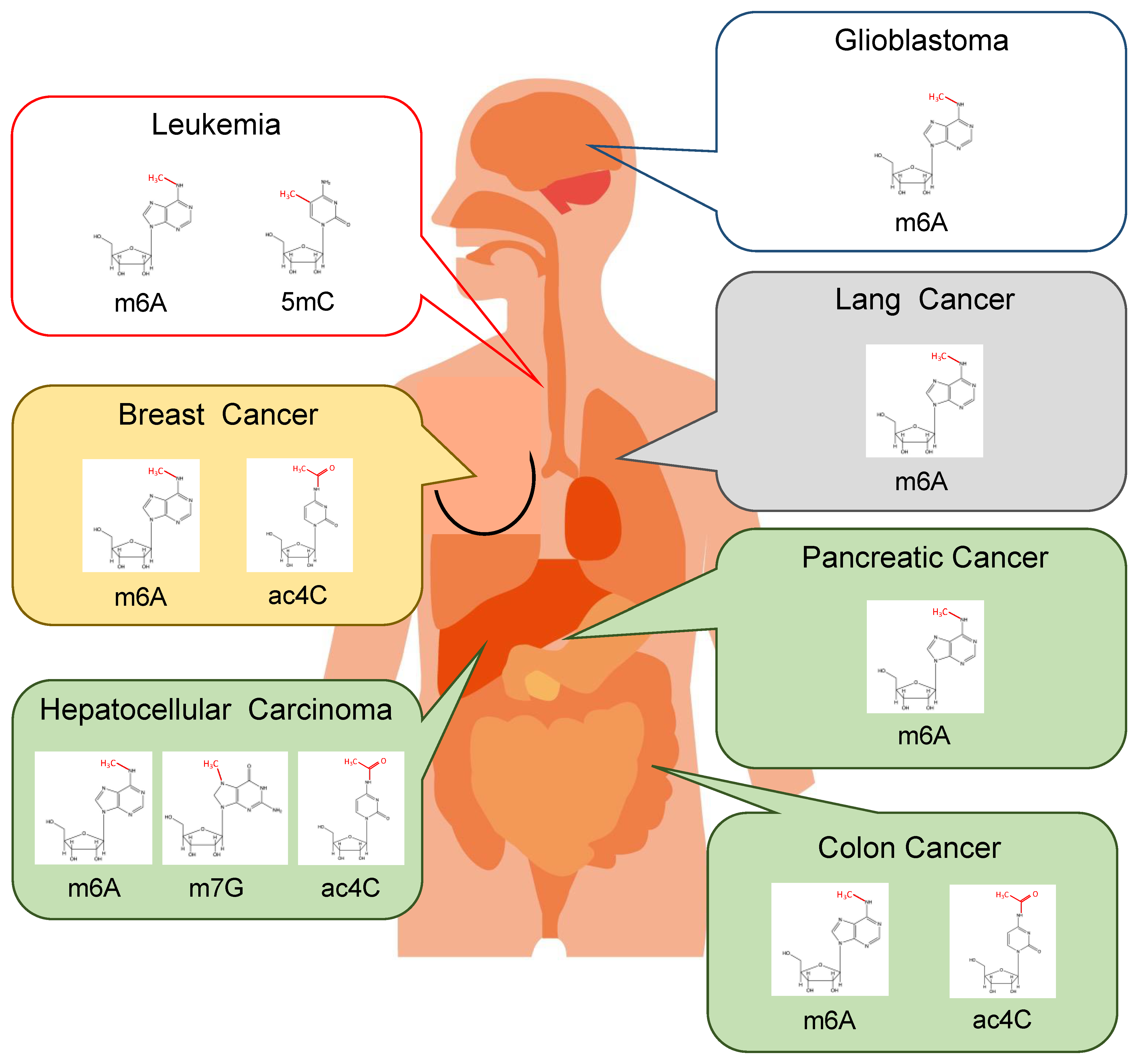
In several forms of cancer, a correlation has been observed between cancer malignancy and RNA modifications. METTL3 increases m6A levels in lung cancer, interacts with the translation initiation machinery to enhance mRNA translation, and activates hippo pathway effector transcription in human cancer cells through binding to the epidermal growth factor receptor and Post-synaptic density protein 95, Disc large homolog 1, and Zonula occludens-1 (PDZ) motifs. Moreover, it has been demonstrated that the mRNAs hippo pathway effector and transcriptional co-activator with PDZ-binding motif are among those that METTL3 helps to translate [56]. Based on this study, METTL3 may be a promising therapeutic target as it promotes the proliferation, survival, and invasion of human lung cancer cells (Figure 2) [56]. Hypoxia stimulates the expression of the m6A demethylase ALKBH5 and the 3′-UTR sequence AAACU, which have been found to decrease NANOG mRNA methylation levels, increase NANOG protein levels, and enhance stem cell populations in studies using human breast cancer cells (Figure 2) [57]. Hypoxia-inducible factor-1 (HIF-1a) and hypoxia-inducible factor-2 (HIF-2)-dependent mechanisms have also been shown to induce NANOG expression [57]. Another study found that, in breast cancer, exposure to hypoxia leads to m6A modification of the mRNAs encoding NANOG and Kruppel-like factor 4 (KLF4) being inhibited in a zinc finger protein 217 (ZNF217)-dependent manner [58]. ZNF217 or ALKBH5 may play a role in controlling the expression of pluripotency factors in breast cancer under hypoxic conditions [58]. Additionally, RNA modifications have been proposed to contribute to the malignant transformation of gastrointestinal malignancies. For example, it has been suggested that m6A modifications play a role in the malignant transformation of colorectal cancer [59]. The oncogene c-myc is implicated in the transcription of the M6A reader YTHDF1 in colorectal cancer, according to epigenetic data obtained by chromatin immunoprecipitation (Figure 2) [59]. YTHDF1 expression has been found to be related to various malignancy behaviors based on immunohistochemistry analysis of YTHDF1 expression and has been discovered to be an independent predictive factor in patients with colorectal cancer [59]. Case-control research on genetic alterations in pancreatic cancer has also revealed a link between FTO mutations and pancreatic cancer risk in Japan (Figure 2) [60]. Additionally, clear links between the FTO rs9939609 mutant polymorphism and endometrial and pancreatic cancer, particularly in Asian populations, have been established, suggesting that these markers could be used for early detection [61].
Malignant pleural mesothelioma risk has also been linked to the FTO rs9939609 mutation [62]. Another investigation into pancreatic cancer revealed that METTL3-deficient cells were more responsive to radiation, gemcitabine, and other anticancer medications such as 5-fluorouracil and cisplatin. Patients with pancreatic cancer may benefit from using METTL3 as a therapeutic agent since it is a potent target that boosts efficacy compared to other therapeutic agents [63]. Additionally, recent studies have demonstrated that m6A levels of miRNAs in the blood can accurately and sensitively identify stage I and stage II pancreatic cancer [64], making m6A levels a potentially novel yet valuable biomarker.
Furthermore, numerous associations between RNA modifications and tumors and epithelial cancers have been reported. FTOs have been linked to acute myeloid leukemia (AML) (Figure 2), including mixed lineage leukemia (MLL) (Figure 2) with t (11q23) rearrangements, t (15; 17), involvement of the retinoic acid receptor-a (RARA), mutations in the FMS-like tyrosine kinase 3 (FLT3) gene with internal tandem duplication (ITD) mutations, and nucleophosmin 1 (NPM1) mutations. In a recent study, FTO was used as a therapeutic method against AML cells. The demethylation from m6A reduce the expression of ankyrin repeat, SOCS box containing 2 (ASB2), and RARA. This led to leukemogenesis (Figure 2). The development of therapeutic medicines and the clarification of disease mechanisms have generally resulted from these results [65].
Moreover, in glioma studies, it has been demonstrated that the knockdown of METTL3 or METTL14 changes the m6A enrichment of mRNA and the mRNA expression of target genes such as ADAM Metallopeptidase Domain 19 (ADAM19), which is crucial for glioblastoma stem cells (Figure 2) [66]. Additionally, blocking the m6A demethylase, FTO, slows tumor growth and lengthens survival in mice receiving glioblastoma stem cell transplants [65]. This study showed that m6A is crucial for the self-renewal and carcinogenesis of glioblastoma stem cells, indicating that m6A alteration is a possible therapeutic target for glioblastoma.
Other modifications besides the modification of m6A have reportedly been linked to cancer. In 5-azacytidine (5-AZA)-resistant leukemia cell lines and clinically in samples from patients with 5-AZA-resistant myelodysplastic syndrome and acute myeloid leukemia, the cytosine-modifying enzyme DNMT2 has been linked to AML [67]. DNMT2 may, therefore, be useful in the management of leukemia. Additionally, the cytosine acetylase NAT10 has been discovered to play a role in the development of numerous cancers and malignancies, such as glioblastoma, HCC, colon cancer, breast cancer, and leukemia.
METTL1 is an enzyme that converts guanine into N7-methylguanine (m7G), which is overexpressed in hepatocellular carcinoma and has carcinogenic activity via the PTEN/AKT serine/threonine pathway (Figure 2). High METTL1 expression has been associated with worse prognosis, larger tumor size, higher serum alpha-fetoprotein levels, and tumor vascular penetration in two separate cohorts of 892 patients with hepatocellular carcinoma [68]. These findings suggest that the METTL1/PTEN axis has therapeutic potential for the treatment of HCC and that METTL1 is a useful predictive biomarker [69]. Additionally, METTL1 has been linked to colorectal cancer drug sensitivity, and through regulation, METTL1 overexpression is connected to the hsa-miR-149-3p/S100A4/p53 axis and sensitizes cisplatin-resistant colorectal cancer cells (Figure 2) [69]. As described above, RNA modifications are altered by various modification species in different cancer types and are associated with malignant transformation. Therefore, accurate measurement of changes in RNA modifications can be useful in detecting these cancers and may have therapeutic applications.

4. RNA Modification and Viral Infection

Numerous viruses, including the Rous sarcoma virus [70], flavivirus [71], Zika virus [72,73], Kaposi’s sarcoma-related herpesvirus [74,75], human immunodeficiency virus-1 (HIV-1) [76], influenza A virus [77], tobacco mosaic virus [78], and severe acute respiratory syndrome coronavirus 2 (SARS-CoV-2) [79], have been shown to affect RNA modifications. Furthermore, the epitranscriptomic state has been linked to viral RNA replication within the host cell nucleus [69,70,71,72,73,74,75,76,77,78]. These viruses use RNA modifications to enhance protein translation and multiply themselves within the host. In addition to utilizing RNA modifications for host growth, viruses also use them to increase their chances of survival within their host. For example, to promote survival and genomic stability in host cells, HIV-1 uses DNMT2’s RNA methylation activity [80]. After HIV-1 infection, DNMT2 relocates from the nucleus to the cytoplasm and methylates HIV-1 RNA. This DNMT2-dependent methylation promotes the post-transcriptional stability of HIV-1 RNA [80]. In addition, DNMT2 overexpression increases HIV-1 viral titer and shedding [80]. HIV-1 also uses NAT10 to boost viral gene expression. NAT10 depletion results in the loss of Ac4C from viral transcripts, reducing viral RNA stability and preventing HIV-1 reproduction [81]. Therefore, DNMT2 and NAT10 are potential targets for the development of antiviral drugs.

5. Detection Methods for RNA Modifications

5.1. Quantitative Methods for Modified Bases

To accurately examine and profile RNA modifications, various challenging methods must be used due to the differences in the position and splicing of adducts as well as the length and structure of the nucleotide strand [2,82]. Several methods have been reported for measuring base modification levels, including dot blotting [83], Northern blotting with antibodies [84], Maz RNA endonuclease [85], high-resolution melting (HRM) [72], RNA photo-crosslinkers and quantitative proteomics (PCL-Proteomics) [86], and silver SiO2-based electrochemical immunosensors (ECI) [87].
RNA modifications are chemical alterations to RNA bases that can occur in various types of RNA molecules. These modifications can regulate RNA structure, stability, and function and play crucial roles in gene expression and disease development. Therefore, detecting and analyzing RNA modifications has become a significant area of research in molecular biology and biomedicine. Several techniques have been developed to detect RNA modifications, including mass spectrometry, high-performance liquid chromatography (HPLC), antibody-based assays, and next-generation sequencing (NGS). Mass spectrometry is a powerful tool for identifying and quantifying RNA modifications with high accuracy and resolution. However, it requires sophisticated instrumentation, sample preparation, and expertise, and it is not suitable for high-throughput analysis [88]. HPLC is another widely used technique for detecting RNA modifications based on the separation and quantification of modified nucleosides using a chromatographic column. HPLC has high sensitivity and specificity, but it is time-consuming, requires specialized equipment, and may not be able to distinguish between closely related modifications [35]. Antibody-based assays, such as enzyme-linked immunosorbent assay (ELISA) and dot blot, use specific antibodies to recognize and quantify RNA modifications. These methods are relatively simple, fast, and cost-effective, but they have limited sensitivity and specificity and may cross-react with other modifications or non-modified nucleotides [89]. NGS-based methods, such as m6A-seq and miCLIP, are emerging as powerful tools for detecting and mapping RNA modifications at a transcriptome-wide scale. These methods use RNA sequencing and specific probes or antibodies to enrich and sequence modified RNA molecules. NGS has high sensitivity, specificity, and throughput, but it requires complex data analysis and may generate false-positive signals due to technical artifacts or sequencing errors [90]. Other emerging techniques for detecting RNA modifications include nanopore sequencing, single-molecule imaging, and chemical probing [91]. These methods have their advantages and limitations and are still under development. In conclusion, the choice of RNA modification detection method depends on the research question, the available resources, and the desired accuracy, sensitivity, and throughput. Therefore, it is essential to evaluate the performance and limitations of each technique carefully and to validate the results using complementary methods. Moreover, methods to determine both the position and the number of base alterations have been published [2,4,92,93,94,95,96].

5.2. Illumina Sequencer

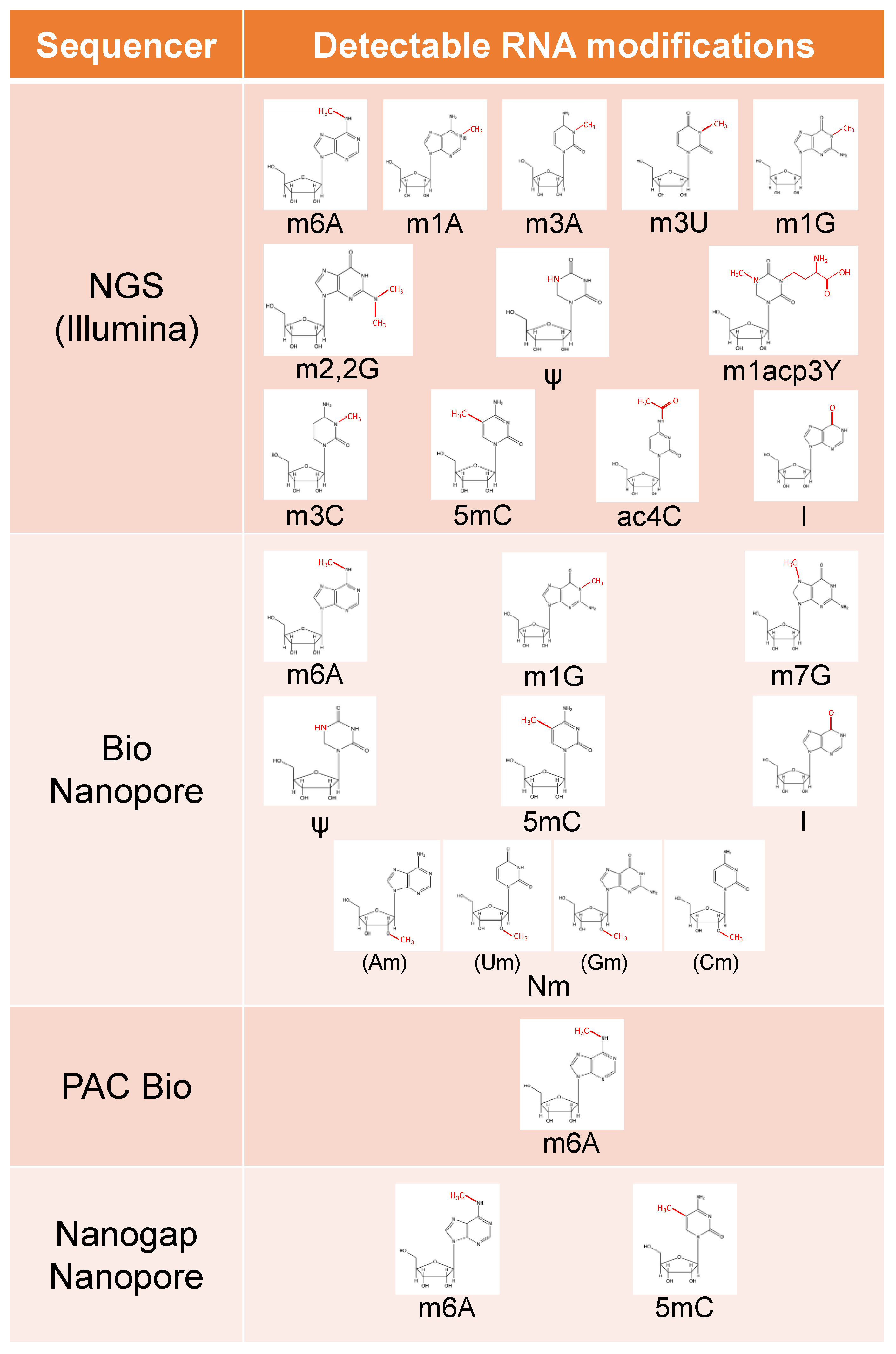
To comprehensively investigate the epitranscriptome, high-throughput techniques are required. In the past, RNA modification mapping was mostly carried out by enriching the modification sites before sequencing and immunoprecipitating the modification sites with modification-specific antibodies [27,97]. However, the m6A modification was initially studied using methylated RNA immunoprecipitation and subsequent sequencing (MeRIP-seq), which revealed unexpectedly high levels and dynamic modulation of the modification in mRNA [27,97]. Reverse transcription (RT) from RNA to cDNA is necessary for NGS, which subsequently relies on synthetic methods for sequencing, such as second-strand synthesis with fluorescently tagged DNA molecules [98]. As a result, NGS methods cannot directly identify the existence of RNA modifications. However, some modifications (m1A, m3C, m3U, m1G, m2,2G, and m1acp3Y) (Figure 3) disrupt Watson–Crick base pairs and cause reverse transcription errors, such as RT omissions and an increase in error frequency [99,100]. This has been shown to be effective in identifying these modifications [101,102]. Reverse transcription can be chemically stopped by treating RNA with substances that react only with modified bases [103], enabling read-end analysis to detect RNA modifications. Examples of such procedures include treatment with CMCT [104], treatment of 5mC with hydrogen sulfite (Figure 3) [105], treatment of ac4C with sodium cyanoborohydride [106], treatment of m3C with hydrazine and/or aniline (Figure 3) [107], and treatment of inosine with glyoxal (Figure 3) [108].

5.3. Bio Nanopore

A more comprehensive answer could be provided by nanopore sequencing technologies, such as those developed by Oxford Nanopore Technologies (ONT) [109,110]. The nanopore sequencing technique measures changes in ionic current that occur as nucleic acids move through protein nanopores encased in the membrane of the flow cell [111]. If this long-read third-generation sequencing technology can directly sequence native RNA and has a modified signal that can be distinguished from that of unmodified nucleotides, it can theoretically detect multiple RNA modifications with a resolution of a single nucleotide and a single molecule [110]. According to a recent groundbreaking study using direct ONT RNA sequencing (Figure 3) [110], the m6A moiety in produced RNA had a different current signal than non-modified moieties at the same site. Shortly after, variations in current intensity were also noticed at the m7G alteration site, which was validated by bacterial direct RNA sequencing of 16S rRNA (Figure 3) [112]. It was later demonstrated in research that base-calling data mismatches, insertions, and deletions were likewise a reflection of variations in electrical current intensity [113]. These findings led to several subsequent studies [113,114,115] that discovered that m6A and other modifications exhibit an “error signature” of base calling. Additionally, several studies used internal unmodified sequences from the same sequence run to discover single modification sites [9,116], paired samples with few or no changes [9,117], or a machine learning strategy to estimate the percentages of the modified molecule in the sample. With the aid of computational tools, both unmodified and modified reads can be clearly distinguished based on their distinctive current signatures, and even minute variations in the proportion of modified molecules, known as modification chemistries, can be found between various conditions and cell types [9,117]. Additionally, a current method termed nano-COP demonstrates that nascent RNA can be identified directly using RNA nanopore sequencing, offering a promising foundation for future research on the interplay between splicing and co-occurring RNA modifications [118]. However, the inability to distinguish between various RNA modification signals within nanopore sequencing signals restricts the ability to increase the repertoire of epitranscriptome changes that may be directly examined using RNA nanopore sequencing. Therefore, the identification of all changes in full-length RNA has become necessary [119]. Signals frequently require sophisticated signal processing and analysis software because they often represent five nucleotides going through a pore at once. Machine learning algorithms are necessary in various situations [9,109,117]. In theory, a standard solution is to build training datasets for nanopore sequencing using synthetic arrays, including all conceivable dimers that are in vitro transcribed with either unmodified (A, C, G, or U) or modified nucleotides [113]. The basic calling signatures for the m6A, I, 5mC, m1G, m7G, and Nm (Figure 3) alterations were found using these and similar data sets [120]. This has been useful for predicting de novo RNA modifications [9,118].

5.4. PacBio

Pacific Biosciences (PacBio) is based on kinetic changes in reverse transcription [120]. Unlike other sequencing systems, the PacBio sequencer is capable of sequencing full-length RNA [120], which allows for the examination of full-length RNA sequences for m6A modifications using this technique (Figure 3) [102,121]. However, there are no further reports on epitranscriptome detection by the PacBio sequencer.

5.5. Nanogap Nanopore

It was reported that nanogap technology-based single molecule detection system can sequence DNA/RNA [122]. The basic conductance value of each nucleotide in DNA/RNA is determined by its molecular energy level, which enables a comprehensive investigation of the epigenome and transcriptome by detecting all known and unknown modified nucleotides [123]. The researchers focused on the 5mC and m6A modification sites, which are common miRNA epigenetic modifications produced by various distinct methyltransferases in nature (Figure 3) [124]. This allows the sequencing of different RNA base modifications in the context of RNA, thus improving our understanding of the epitranscriptome [124].

5.6. Single Molecule Detection Using Raman Spectroscopy

It was reported that the method for detecting queuosine modifications of transfer RNA in single living cells using a plasmonic affinity sandwich assay [125]. In this method, specific antibodies against queuosine modifications are immobilized on affinity-controlled nanoplates and selectively bind to transcriptional RNA in living cells. By using spectroscopic measurements based on plasmon resonance, the queuosine modification of RNA can be detected [125]. This approach has demonstrated high sensitivity for detection at the single-cell level for analyzing RNA modifications within cells. Furthermore, it is possible to simultaneously detect multiple RNA modifications, indicating its potential as a valuable tool for RNA modification analysis in the future.
However, there are still issues that need to be addressed with this method. For example, the current method can only detect a single modification and is not suitable for the simultaneous detection of multiple modifications. The specificity of the antibodies against RNA modifications has not been fully validated either. Once these issues are addressed, this method will become a more widely applicable tool for RNA modification analysis.

6. Software for Analyzing RNA Modification

There are several software tools available for the detection of RNA modifications. For example, RiboMethSeq is a tool that can detect m6A, m5C, and pseudouridine (Ψ) modifications from sequencing data with high accuracy [126]. It uses a two-step approach that first identifies ribosome-protected regions and then analyzes read coverage to detect modifications. RiboMeth-seq can detect a variety of RNA modifications, including m6A, pseudouridine, and inosine. The software has been shown to have high accuracy in detecting RNA modifications, and it can be used to identify the locations and abundance of modifications in specific transcripts. Another tool, exomePeak, can detect m6A modifications with high sensitivity and specificity from m6A-CLIP-seq data [127]. It uses a statistical model to detect significant differences in read coverage between modified and unmodified regions of RNA. The software is optimized for detecting m6A modifications, which are the most common type of RNA modification. However, it can also be used to detect other types of modifications. The tool, exomePeak, has been shown to have high accuracy and sensitivity in detecting m6A modifications. However, these tools also have their limitations. For example, RiboMethSeq cannot distinguish between m5C and Ψ modifications due to their similar chemical properties [126]. Additionally, exomePeak requires high sequencing depth to achieve optimal results, which may not be feasible in certain experimental settings [127]. In the future, it is likely that new software tools will be developed that can overcome these limitations and improve the accuracy and sensitivity of RNA modification detection. For example, a recent study proposed a new method called MAZTER-seq that can detect m6A and m5C modifications simultaneously from RNA sequencing data with high accuracy [128]. This method uses a deep learning algorithm to distinguish between the two modifications based on their distinct sequence contexts and has the potential to overcome the limitations of current RNA modification detection tools. Overall, while there are currently limitations to the available software tools for RNA modification detection, new methods are constantly being developed, and it is likely that the field will continue to progress rapidly in the coming years.

7. Discussion and Future Prospects

The epitranscriptome is known to play a role in the pathogenesis and malignant transformation of various life phenomena and diseases. Therefore, the development of single-molecule technologies to measure the epitranscriptome is crucial for understanding these phenomena. Several equipment-based epitranscriptome measurement methods have been developed, which can be classified into two categories.
The first category includes dot blotting [83], Northern blotting with antibodies [84], Maz RNA endonuclease [85], high-resolution melting (HRM) [72], RNA photo-crosslinkers and quantitative proteomics (PCL-Proteomics) [86], silver SiO2-based electrochemical immunosensors (ECI) [87], Illumina Sequencer [99,100], and PacBio [100]. These methods detect modified RNAs based on their chemical properties. For example, some detection methods use antibodies that bind specifically to modified species [84,97], utilize resistance to RNA-degrading enzymes [85], and use the differences in chemical reactivity with aniline [107], hydrogen sulfate [105], and other compounds [106,108]. However, the weakness of these methods is that they cannot detect multiple modifications simultaneously. There are about 170 types of RNA modifications [1], and RNA functions are regulated by their interactions with each other [11,12]. Therefore, it is difficult to fully understand RNA function with a measurement method that cannot detect multiple modifications at the same time.
The second category includes Bio Nanopore [110,111] and Nanogap Nanopore [124]. These methods measure RNA bases on a molecule-by-molecule basis, allowing for the direct measurement of RNA bases using single-molecule measurement techniques. They can detect multiple types of modifications in a single measurement [124]. However, due to the noise in the signals obtained from these single-molecule measurements, they are less accurate at detecting modified RNA than chemical characterization techniques. The future development of methods for single-molecule measurement of RNA bases and signal analysis is expected to improve the accuracy of detecting multiple types of modifications. This measurement technology is anticipated to reveal detailed mechanisms of life phenomena that have not been understood so far.

Author Contributions

Conceptualization: M.O. and M.K. All authors have read and agreed to the published version of the manuscript.

Funding

This research was funded by Grant-in-Aid for the Japan Society for the Promotion of Science (JSPS) (19K07688 and 22H03686), a Research Grant of the Princess Takamatsu Cancer Research Fund, and Project Mirai Cancer Research Grants.

Institutional Review Board Statement

Not applicable for studies not involving humans or animals.

Informed Consent Statement

Not applicable.

Data Availability Statement

Not applicable.

Acknowledgments

We thank Alan Wright for his English editing.

Conflicts of Interest

The authors declare no conflict of interest. The funders had no role in the design of the study; in the collection, analyses, or interpretation of data; in the writing of the manuscript, or in the decision to publish the results.

References

  1. Sudakov, A.; Knezic, B.; Hengesbach, M.; Fürtig, B.; Stirnal, E.; Schwalbe, H. Site-Specific Labeling of RNAs with Modified and 19F-Labelled Nucleotides by Chemo-Enzymatic Synthesis. Chemistry 2023, 29, e202203368. [Google Scholar] [CrossRef] [PubMed]
  2. Konno, M.; Taniguchi, M.; Ishii, H. Significant epitranscriptomes in heterogeneous cancer. Cancer Sci. 2019, 110, 2318–2327. [Google Scholar] [CrossRef] [PubMed]
  3. Du, H.; Zhao, Y.; He, J.; Zhang, Y.; Xi, H.; Liu, M.; Ma, J.; Wu, L. YTHDF2 destabilizes m6A-containing RNA through direct recruitment of the CCR4–NOT deadenylase complex. Nat. Commun. 2016, 7, 12626. [Google Scholar] [CrossRef]
  4. Meyer, K.D.; Patil, D.P.; Zhou, J.; Zinoviev, A.; Skabkin, M.A.; Elemento, O.; Pestova, T.V.; Qian, S.B.; Jaffrey, S.R. 5′ UTR m6A Promotes Cap-Independent Translation. Cell 2015, 163, 999–1010. [Google Scholar] [CrossRef] [PubMed]
  5. Torres, A.G.; Batlle, E.; de Pouplana, L.R. Role of tRNA modifications in human diseases. Trends Mol. Med. 2014, 20, 306–314. [Google Scholar] [CrossRef] [PubMed]
  6. Jonkhout, N.; Tran, J.; Smith, M.A.; Schonrock, N.; Mattick, J.S.; Novoa, E.M. The RNA modification landscape in human disease. RNA 2017, 23, 1754–1769. [Google Scholar] [CrossRef] [PubMed]
  7. Delaunay, S.; Frye, M. RNA modifications regulating cell fate in cancer. Nat. Cell Biol. 2019, 21, 552–559. [Google Scholar] [CrossRef] [PubMed]
  8. Esteve-Puig, R.; Bueno-Costa, A.; Esteller, M. Writers, readers and erasers of RNA modifications in cancer. Cancer Lett. 2020, 474, 127–137. [Google Scholar] [CrossRef] [PubMed]
  9. Begik, O.; Lucas, M.C.; Pryszcz, L.P.; Ramirez, J.M.; Medina, R.; Milenkovic, I.; Cruciani, S.; Liu, H.; Vieira, H.G.S.; Sas-Chen, A.; et al. Quantitative profiling of pseudouridylation dynamics in native RNAs with nanopore sequencing. Nat. Biotechnol. 2021, 39, 1278–1291. [Google Scholar] [CrossRef]
  10. Boccaletto, P.; Stefaniak, F.; Ray, A.; Cappannini, A.; Mukherjee, S.; Purta, E.; Kurkowska, M.; Shirvanizadeh, N.; Destefanis, E.; Groza, P.; et al. MODOMICS: A database of RNA modification pathways. 2021 update. Nucleic Acids Res. 2021, 50, D231–D235. [Google Scholar] [CrossRef] [PubMed]
  11. Grosjean, H.; Droogmans, L.; Roovers, M.; Keith, G. Detection of enzymatic activity of transfer RNA modification enzymes using radio-labeled tRNA substrates. Methods Enzymol. 2007, 425, 55–101. [Google Scholar] [PubMed]
  12. Jora, M.; Lobue, P.A.; Ross, R.L.; Williams, B.; Addepalli, B. Detection of ribonucleoside modifications by liquid chromatography coupled with mass spectrometry. Biochim. Biophys. Acta Gene Regul. Mech. 2018, 1862, 280–290. [Google Scholar] [CrossRef] [PubMed]
  13. Squires, J.E.; Patel, H.R.; Nousch, M.; Sibbritt, T.D.; Humphreys, D.T.; Parker, B.J.; Suter, C.M.; Preiss, T. Widespread occurrence of 5-methylcytosine in human coding and non-coding RNA. Nucleic Acids Res. 2012, 40, 5023–5033. [Google Scholar] [CrossRef]
  14. Heissenberger, C.; Rollins, J.A.; Krammer, T.L.; Nagelreiter, F.; Stocker, I.; Wacheul, L.; Shpylovyi, A.; Tav, K.; Snow, S.; Grillari, J.; et al. The ribosomal RNA m(5)C methyltransferase NSUN-1 modulates healthspan and oogenesis in Caenorhabditis elegans. eLife 2020, 9, e56205. [Google Scholar] [CrossRef] [PubMed]
  15. Sajini, A.A.; Choudhury, N.R.; Wagner, R.E.; Bornelöv, S.; Selmi, T.; Spanos, C.; Dietmann, S.; Rappsilber, J.; Michlewski, G.; Frye, M. Loss of 5-methylcytosine alters the biogenesis of vault-derived small RNAs to coordinate epidermal differentiation. Nat. Commun. 2019, 10, 2550. [Google Scholar] [CrossRef] [PubMed]
  16. Van Haute, L.; Dietmann, S.; Kremer, L.; Hussain, S.; Pearce, S.F.; Powell, C.A.; Rorbach, J.; Lantaff, R.; Blanco, S.; Sauer, S.; et al. Deficient methylation and formylation of mt-tRNA(Met) wobble cytosine in a patient carrying mutations in NSUN3. Nat. Commun. 2016, 7, 12039. [Google Scholar] [CrossRef]
  17. Selmi, T.; Hussain, S.; Dietmann, S.; Heiß, M.; Borland, K.; Flad, S.; Carter, J.M.; Dennison, R.; Huang, Y.L.; Kellner, S.; et al. Sequence- and structure-specific cytosine-5 mRNA methylation by NSUN6. Nucleic Acids Res. 2021, 49, 1006–1022. [Google Scholar] [CrossRef]
  18. Chi, L.; Delgado-Olguin, P. Expression of NOL1/NOP2/sun domain (Nsun) RNA methyltransferase family genes in early mouse embryogenesis. Gene Expr. Patterns 2013, 13, 319–327. [Google Scholar] [CrossRef]
  19. Flores, J.V.; Cordero-Espinoza, L.; Oeztuerk-Winder, F.; Andersson-Rolf, A.; Selmi, T.; Blanco, S.; Tailor, J.; Dietmann, S.; Frye, M. Cytosine-5 RNA Methylation Regulates Neural Stem Cell Differentiation and Motility. Stem Cell Rep. 2016, 8, 112–124. [Google Scholar] [CrossRef]
  20. Martinez, F.J.; Lee, J.H.; Lee, J.E.; Blanco, S.; Nickerson, E.; Gabriel, S.; Frye, M.; Al-Gazali, L.; Gleeson, J.G. Whole exome sequencing identifies a splicing mutation in NSUN2 as a cause of a Dubowitz-like syndrome. J. Med. Genet. 2012, 49, 380–385. [Google Scholar] [CrossRef]
  21. Khan, M.A.; Rafiq, M.A.; Noor, A.; Hussain, S.; Flores, J.V.; Rupp, V.; Vincent, A.K.; Malli, R.; Ali, G.; Khan, F.S.; et al. Mutation in NSUN2, which Encodes an RNA Methyltransferase, Causes Autosomal-Recessive Intellectual Disability. Am. J. Hum. Genet. 2012, 90, 856–863. [Google Scholar] [CrossRef] [PubMed]
  22. Trixl, L.; Amort, T.; Wille, A.; Zinni, M.; Ebner, S.; Hechenberger, C.; Eichin, F.; Gabriel, H.; Schoberleitner, I.; Huang, A.; et al. RNA cytosine methyltransferase Nsun3 regulates embryonic stem cell differentiation by promoting mitochondrial activity. Cell. Mol. Life Sci. 2017, 75, 1483–1497. [Google Scholar] [CrossRef] [PubMed]
  23. Harris, T.; Marquez, B.; Suarez, S.; Schimenti, J. Sperm Motility Defects and Infertility in Male Mice with a Mutation in Nsun7, a Member of the Sun Domain-Containing Family of Putative RNA Methyltransferases1. Biol. Reprod. 2007, 77, 376–382. [Google Scholar] [CrossRef]
  24. Zhang, C.; Jia, G. Reversible. RNA Modification N1-methyladenosine (m1A) in mRNA and tRNA. Genom. Proteom. Bioinform. 2018, 16, 155–161. [Google Scholar] [CrossRef] [PubMed]
  25. Chujo, T.; Suzuki, T. Trmt61B is a methyltransferase responsible for 1-methyladenosine at position 58 of human mitochondrial tRNAs. RNA 2012, 18, 2269–2276. [Google Scholar] [CrossRef]
  26. Dai, X.; Wang, T.; Gonzalez, G.; Wang, Y. Identification of YTH Domain-Containing Proteins as the Readers for N1-Methyladenosine in RNA. Anal. Chem. 2018, 90, 6380–6384. [Google Scholar] [CrossRef]
  27. Safra, M.; Sas-Chen, A.; Nir, R.; Winkler, R.; Nachshon, A.; Bar-Yaacov, D.; Erlacher, M.; Rossmanith, W.; Stern-Ginossar, N.; Schwartz, S. The m1A landscape on cytosolic and mitochondrial mRNA at single-base resolution. Nature 2017, 551, 251–255. [Google Scholar] [CrossRef]
  28. Dominissini, D.; Nachtergaele, S.; Moshitch-Moshkovitz, S.; Peer, E.; Kol, N.; Ben-Haim, M.S.; Dai, Q.; Di Segni, A.; Salmon-Divon, M.; Clark, W.C.; et al. The dynamic N1-methyladenosine methylome in eukaryotic messenger RNA. Nature 2016, 530, 441–446. [Google Scholar] [CrossRef]
  29. Li, X.; Xiong, X.; Wang, K.; Wang, L.; Shu, X.; Ma, S.; Yi, C. Transcriptome-wide mapping reveals reversible and dynamic N1-methyladenosine methylome. Nat. Chem. Biol. 2016, 12, 311–316. [Google Scholar] [CrossRef]
  30. Meyer, K.D.; Saletore, Y.; Zumbo, P.; Elemento, O.; Mason, C.E.; Jaffrey, S.R. Comprehensive Analysis of mRNA Methylation Reveals Enrichment in 3′ UTRs and Near Stop Codons. Cell 2012, 149, 1635–1646. [Google Scholar] [CrossRef]
  31. Liu, J.; Yue, Y.; Han, D.; Wang, X.; Fu, Y.; Zhang, L.; Jia, G.; Yu, M.; Lu, Z.; Deng, X.; et al. A METTL3–METTL14 complex mediates mammalian nuclear RNA N6-adenosine methylation. Nat. Chem. Biol. 2013, 10, 93–95. [Google Scholar] [CrossRef] [PubMed]
  32. Haussmann, I.U.; Bodi, Z.; Sanchez-Moran, E.; Mongan, N.P.; Archer, N.; Fray, R.G.; Soller, M. m6A potentiates Sxl alternative pre-mRNA splicing for robust Drosophila sex determination. Nature 2016, 540, 301–304. [Google Scholar] [CrossRef] [PubMed]
  33. Van Tran, N.; Ernst, F.G.M.; Hawley, B.R.; Zorbas, C.; Ulryck, N.; Hackert, P.; Bohnsack, K.E.; Bohnsack, M.T.; Jaffrey, S.R.; Graille, M.; et al. The human 18S rRNA m6A methyltransferase METTL5 is stabilized by TRMT112. Nucleic Acids Res. 2019, 47, 7719–7733. [Google Scholar] [CrossRef] [PubMed]
  34. Warda, A.S.; Kretschmer, J.; Hackert, P.; Lenz, C.; Urlaub, H.; Höbartner, C.; Sloan, K.E.; Bohnsack, M.T. Human METTL16 is a N6-methyla-denosine (m6A) methyltransferase that targets pre-mRNAs and various non-coding RNAs. EMBO Rep. 2017, 18, 2004–2014. [Google Scholar] [CrossRef]
  35. Meyer, K.D.; Jaffrey, S.R. Rethinking m6A readers, writers, and erasers. Annu. Rev. Cell Dev. Biol. 2017, 33, 319–342. [Google Scholar] [CrossRef]
  36. Schwartz, S.; Mumbach, M.R.; Jovanovic, M.; Wang, T.; Maciag, K.; Bushkin, G.G.; Mertins, P.; Ter-Ovanesyan, D.; Habib, N.; Cacchiarelli, D.; et al. Perturbation of m6A Writers Reveals Two Distinct Classes of mRNA Methylation at Internal and 5′ Sites. Cell Rep. 2014, 8, 284–296. [Google Scholar] [CrossRef]
  37. Dierks, D.; Garcia-Campos, M.A.; Uzonyi, A.; Safra, M.; Edelheit, S.; Rossi, A.; Sideri, T.; Varier, R.A.; Brandis, A.; Stelzer, Y.; et al. Multiplexed profiling facilitates robust m6A quantification at site, gene and sample resolution. Nat. Methods 2021, 18, 1060–1067. [Google Scholar] [CrossRef]
  38. Xu, W.; Li, J.; He, C.; Wen, J.; Ma, H.; Rong, B.; Diao, J.; Wang, L.; Wang, J.; Wu, F.; et al. METTL3 regulates heterochromatin in mouse embryonic stem cells. Nature 2021, 591, 317–321. [Google Scholar] [CrossRef]
  39. Zhang, C.; Chen, L.; Peng, D.; Jiang, A.; He, Y.; Zeng, Y.; Xie, C.; Zhou, H.; Luo, X.; Liu, H.; et al. METTL3 and N6-Methyladenosine Promote Homologous Recombination-Mediated Repair of DSBs by Modulating DNA-RNA Hybrid Accumulation. Mol. Cell 2020, 79, 425–442.e7. [Google Scholar] [CrossRef]
  40. Mendel, M.; Chen, K.-M.; Homolka, D.; Gos, P.; Pandey, R.R.; McCarthy, A.A.; Pillai, R.S. Methylation of Structured RNA by the m6A Writer METTL16 Is Essential for Mouse Embryonic Development. Mol. Cell 2018, 71, 986–1000.e11. [Google Scholar] [CrossRef]
  41. Pendleton, K.E.; Chen, B.; Liu, K.; Hunter, O.V.; Xie, Y.; Tu, B.P.; Conrad, N.K. The U6 snRNA m 6 A Methyltransferase METTL16 Regulates SAM Synthetase Intron Retention. Cell 2017, 169, 824–835.e14. [Google Scholar] [CrossRef] [PubMed]
  42. Ignatova, V.V.; Stolz, P.; Kaiser, S.; Gustafsson, T.H.; Lastres, P.R.; Sanz-Moreno, A.; Cho, Y.-L.; Amarie, O.V.; Aguilar-Pimentel, A.; Klein-Rodewald, T.; et al. The rRNA m6A methyltransferase METTL5 is involved in pluripotency and developmental programs. Genes Dev. 2020, 34, 715–729. [Google Scholar] [CrossRef] [PubMed]
  43. Han, Y.; Du, T.; Guo, S.; Wang, L.; Dai, G.; Long, T.; Xu, T.; Zhuang, X.; Liu, C.; Li, S.; et al. Loss of m6A Methyltransferase METTL5 Promotes Cardiac Hypertrophy through Epitranscriptomic Control of SUZ12 Expression. Front. Cardiovasc. Med. 2022, 9, 852775. [Google Scholar] [CrossRef] [PubMed]
  44. Wang, L.; Liang, Y.; Lin, R.; Xiong, Q.; Yu, P.; Ma, J.; Cheng, M.; Han, H.; Wang, X.; Wang, G.; et al. Mettl5 mediated 18S rRNA N6-methyladenosine (m6A) modification controls stem cell fate determination and neural function. Genes Dis. 2020, 9, 268–274. [Google Scholar] [CrossRef]
  45. Chen, C.; Liu, W.; Guo, J.; Liu, Y.; Liu, X.; Liu, J.; Dou, X.; Le, R.; Huang, Y.; Li, C.; et al. Nuclear m6A reader YTHDC1 regulates the scaffold function of LINE1 RNA in mouse ESCs and early embryos. Protein Cell 2021, 12, 455–474. [Google Scholar] [CrossRef]
  46. Wu, R.; Li, A.; Sun, B.; Sun, J.G.; Zhang, J.; Zhang, T.; Chen, Y.; Xiao, Y.; Gao, Y.; Zhang, Q.; et al. A novel m6A reader Prrc2a controls oligodendroglial specification and myelination. Cell Res. 2019, 29, 23–41. [Google Scholar] [CrossRef]
  47. Liu, F.; Clark, W.; Luo, G.; Wang, X.; Fu, Y.; Wei, J.; Wang, X.; Hao, Z.; Dai, Q.; Zheng, G.; et al. ALKBH1-mediated tRNA demethylation regulates translation. Cell 2016, 167, 1897. [Google Scholar] [CrossRef]
  48. Tang, C.; Klukovich, R.; Peng, H.; Wang, Z.; Yu, T.; Zhang, Y.; Zheng, H.; Klungland, A.; Yan, W. ALKBH5-dependent m6A demethylation controls splicing and stability of long 3′-UTR mRNAs in male germ cells. Proc. Natl. Acad. Sci. USA 2017, 115, E325–E333. [Google Scholar] [CrossRef]
  49. Merkestein, M.; Laber, S.; McMurray, F.; Andrew, D.; Sachse, G.; Sanderson, J.; Li, M.; Usher, S.; Sellayah, D.; Ashcroft, F.M.; et al. FTO influences adipogenesis by regulating mitotic clonal expansion. Nat. Commun. 2015, 6, 6792. [Google Scholar] [CrossRef]
  50. Mauer, J.; Sindelar, M.; Despic, V.; Guez, T.; Hawley, B.R.; Vasseur, J.-J.; Rentmeister, A.; Gross, S.S.; Pellizzoni, L.; Debart, F.; et al. FTO controls reversible m6Am RNA methylation during snRNA biogenesis. Nat. Chem. Biol. 2019, 15, 340–347. [Google Scholar] [CrossRef]
  51. Dimitrova, D.G.; Teysset, L.; Carré, C. RNA 2′-O-Methylation (Nm) Modification in Human Diseases. Genes 2019, 10, 117. [Google Scholar] [CrossRef] [PubMed]
  52. Marcel, V.; Ghayad, S.E.; Belin, S.; Therizols, G.; Morel, A.-P.; Solano-Gonzàlez, E.; Vendrell, J.A.; Hacot, S.; Mertani, H.C.; Albaret, M.A.; et al. p53 Acts as a Safeguard of Translational Control by Regulating Fibrillarin and rRNA Methylation in Cancer. Cancer Cell 2013, 24, 318–330. [Google Scholar] [CrossRef] [PubMed]
  53. Erales, J.; Marchand, V.; Panthu, B.; Gillot, S.; Belin, S.; Ghayad, S.E.; Garcia, M.; Laforêts, F.; Marcel, V.; Baudin-Baillieu, A.; et al. Evidence for rRNA 2′-O-methylation plasticity: Control of intrinsic translational capabilities of human ribosomes. Proc. Natl. Acad. Sci. USA 2017, 114, 12934–12939. [Google Scholar] [CrossRef]
  54. Karunatilaka, K.S.; Rueda, D. Post-transcriptional modifications modulate conformational dynamics in human U2–U6 snRNA complex. RNA 2013, 20, 16–23. [Google Scholar] [CrossRef] [PubMed]
  55. Wang, X.; Li, Z.-T.; Yan, Y.; Lin, P.; Tang, W.; Hasler, D.; Meduri, R.; Li, Y.; Hua, M.-M.; Qi, H.-T.; et al. LARP7-Mediated U6 snRNA Modification Ensures Splicing Fidelity and Spermatogenesis in Mice. Mol. Cell 2020, 77, 999–1013.e6. [Google Scholar] [CrossRef] [PubMed]
  56. Lin, S.; Choe, J.; Du, P.; Triboulet, R.; Gregory, R.I. The m 6 A Methyltransferase METTL3 Promotes Translation in Human Cancer Cells. Mol. Cell 2016, 62, 335–345. [Google Scholar] [CrossRef] [PubMed]
  57. Zhang, C.; Samanta, D.; Lu, H.; Bullen, J.W.; Zhang, H.; Chen, I.; He, X.; Semenza, G.L. Hypoxia induces the breast cancer stem cell phenotype by HIF-dependent and ALKBH5-mediated m 6 A-demethylation of NANOG mRNA. Proc. Natl. Acad. Sci. USA 2016, 113, E2047–E2056. [Google Scholar] [CrossRef]
  58. Zhang, C.; Zhi, W.I.; Lu, H.; Samanta, D.; Chen, I.; Gabrielson, E.; Semenza, G.L. Hypoxia-inducible factors regulate pluripotency factor expression by ZNF217- and ALKBH5-mediated modulation of RNA methylation in breast cancer cells. Oncotarget 2016, 7, 64527–64542. [Google Scholar] [CrossRef]
  59. Nishizawa, Y.; Konno, M.; Asai, A.; Koseki, J.; Kawamoto, K.; Miyoshi, N.; Takahashi, H.; Nishida, N.; Haraguchi, N.; Sakai, D.; et al. Oncogene c-Myc promotes epitranscriptome m6A reader YTHDF1 expression in colorectal cancer. Oncotarget 2017, 9, 7476–7486. [Google Scholar] [CrossRef]
  60. Lin, Y.; Ueda, J.; Yagyu, K.; Ishii, H.; Ueno, M.; Egawa, N.; Nakao, H.; Mori, M.; Matsuo, K.; Kikuchi, S. Association between variations in the fat mass and obesity-associated gene and pancreatic cancer risk: A case–control study in Japan. BMC Cancer 2013, 13, 337. [Google Scholar] [CrossRef]
  61. Huang, X.; Zhao, J.; Yang, M.; Li, M.; Zheng, J. Association between FTO gene polymorphism (rs9939609 T/A) and cancer risk: A meta-analysis. Eur. J. Cancer Care 2016, 26, e12464. [Google Scholar] [CrossRef] [PubMed]
  62. Khella, M.S.; Salem, A.M.; Abdel-Rahman, O.; Saad, A.S. The Association between the FTO rs9939609 Variant and Malignant Pleural Mesothelioma Risk: A Case–Control Study. Genet. Test. Mol. Biomark. 2018, 22, 79–84. [Google Scholar] [CrossRef] [PubMed]
  63. Taketo, K.; Konno, M.; Asai, A.; Koseki, J.; Toratani, M.; Satoh, T.; Doki, Y.; Mori, M.; Ishii, H.; Ogawa, K. The epitranscriptome m6A writer METTL3 promotes chemo- and radioresistance in pancreatic cancer cells. Int. J. Oncol. 2017, 52, 621–629. [Google Scholar] [CrossRef] [PubMed]
  64. Konno, M.; Koseki, J.; Asai, A.; Yamagata, A.; Shimamura, T.; Motooka, D.; Okuzaki, D.; Kawamoto, K.; Mizushima, T.; Eguchi, H.; et al. Distinct methylation levels of mature microRNAs in gastrointestinal cancers. Nat. Commun. 2019, 10, 3888. [Google Scholar] [CrossRef] [PubMed]
  65. Li, Z.; Weng, H.; Su, R.; Weng, X.; Zuo, Z.; Li, C.; Huang, H.; Nachtergaele, S.; Dong, L.; Hu, C.; et al. FTO Plays an Oncogenic Role in Acute Myeloid Leukemia as a N6-Methyladenosine RNA Demethylase. Cancer Cell 2016, 31, 127–141. [Google Scholar] [CrossRef]
  66. Cui, Q.; Shi, H.; Ye, P.; Li, L.; Qu, Q.; Sun, G.; Sun, G.; Lu, Z.; Huang, Y.; Yang, C.-G.; et al. m6A RNA Methylation Regulates the Self-Renewal and Tumorigenesis of Glioblastoma Stem Cells. Cell Rep. 2017, 18, 2622–2634. [Google Scholar] [CrossRef]
  67. Cheng, J.X.; Chen, L.; Li, Y.; Cloe, A.; Yue, M.; Wei, J.; Watanabe, K.A.; Shammo, J.M.; Anastasi, J.; Shen, Q.J.; et al. RNA cytosine methylation and methyltransferases mediate chromatin organization and 5-azacytidine response and resistance in leukaemia. Nat. Commun. 2019, 9, 1163. [Google Scholar] [CrossRef]
  68. Tian, Q.-H.; Zhang, M.-F.; Zeng, J.-S.; Luo, R.-G.; Wen, Y.; Chen, J.; Gan, L.-G.; Xiong, J.-P. METTL1 overexpression is correlated with poor prognosis and promotes hepatocellular carcinoma via PTEN. J. Mol. Med. 2019, 97, 1535–1545. [Google Scholar] [CrossRef]
  69. Liu, Y.; Yang, C.; Zhao, Y.; Chi, Q.; Wang, Z.; Sun, B. Overexpressed methyltransferase-like 1 (METTL1) increased chemosensitivity of colon cancer cells to cisplatin by regulating miR-149-3p/S100A4/p53 axis. Aging 2019, 11, 12328–12344. [Google Scholar] [CrossRef]
  70. Beemon, K.; Keith, J. Localization of N6-methyladenosine in the Rous sarcoma virus genome. J. Mol. Biol. 1977, 113, 165–179. [Google Scholar] [CrossRef]
  71. Golovina, A.Y.; Dzama, M.M.; Petriukov, K.S.; Zatsepin, T.S.; Sergiev, P.V.; Bogdanov, A.A.; Dontsova, O.A. Method for site-specific detection of m6A nucleoside presence in RNA based on high-resolution melting (HRM) analysis. Nucleic Acids Res. 2013, 42, e27. [Google Scholar] [CrossRef]
  72. Lichinchi, G.; Zhao, B.S.; Wu, Y.; Lu, Z.; Qin, Y.; He, C.; Rana, T.M. Dynamics of Human and Viral RNA Methylation during Zika Virus Infection. Cell Host Microbe 2016, 20, 666–673. [Google Scholar] [CrossRef] [PubMed]
  73. Zhou, H.; Wang, F.; Wang, H.; Chen, C.; Zhang, T.; Han, X.; Wang, D.; Wu, C.; Xie, W.; Wang, Z.; et al. The conformational changes of Zika virus methyltransferase upon converting SAM to SAH. Oncotarget 2017, 8, 14830–14834. [Google Scholar] [CrossRef] [PubMed]
  74. Ye, F.; Chen, E.R.; Nilsen, T.W. Kaposi’s sarcoma-associated herpesvirus utilizes and manipulates RNA N6-adenosine methylation to promote lytic replication. J. Virol. 2017, 91, e00466-17. [Google Scholar] [CrossRef]
  75. Hesser, C.R.; Karijolich, J.; Dominissini, D.; He, C.; Glaunsinger, B.A. N6-methyladenosine modification and the YTHDF2 reader protein play cell type specific roles in lytic viral gene expression during Kaposi’s sarcoma-associated herpesvirus infection. PLoS Pathog. 2018, 14, e1006995. [Google Scholar] [CrossRef] [PubMed]
  76. Lichinchi, G.; Gao, S.; Saletore, Y.; Gonzalez, G.M.; Bansal, V.; Wang, Y.; Mason, C.E.; Rana, T.M. Dynamics of the human and viral m(6)A RNA methylomes during HIV-1 infection of T cells. Nat. Microbiol. 2016, 1, 16011. [Google Scholar] [CrossRef]
  77. Courtney, D.G.; Kennedy, E.M.; Dumm, R.E.; Bogerd, H.P.; Tsai, K.; Heaton, N.S.; Cullen, B.R. Epitranscriptomic Enhancement of Influenza A Virus Gene Expression and Replication. Cell Host Microbe 2017, 22, 377–386.e5. [Google Scholar] [CrossRef]
  78. Li, Z.; Shi, J.; Yu, L.; Zhao, X.; Ran, L.; Hu, D.; Song, B. N6-methyl-adenosine level in Nicotiana tabacum is associated with tobacco mosaic virus. Virol. J. 2018, 15, 87. [Google Scholar] [CrossRef]
  79. Liu, J.; Xu, Y.P.; Li, K.; Ye, Q.; Zhou, H.Y.; Sun, H.; Li, X.; Yu, L.; Deng, Y.Q.; Li, R.T.; et al. The m6A methylome of SARS-CoV-2 in host cells. Cell Res. 2021, 31, 404–414. [Google Scholar] [CrossRef]
  80. Dev, R.R.; Ganji, R.; Singh, S.P.; Mahalingam, S.; Banerjee, S.; Khosla, S. Cytosine methylation by DNMT2 facilitates stability and survival of HIV-1 RNA in the host cell during infection. Biochem. J. 2017, 474, 2009–2026. [Google Scholar] [CrossRef]
  81. Tsai, K.; Vasudevan, A.A.J.; Campos, C.M.; Emery, A.; Swanstrom, R.; Cullen, B.R. Acetylation of Cytidine Residues Boosts HIV-1 Gene Expression by Increasing Viral RNA Stability. Cell Host Microbe 2020, 28, 306–312.e6. [Google Scholar] [CrossRef] [PubMed]
  82. Li, X.; Liang, Q.-X.; Lin, J.-R.; Peng, J.; Yang, J.-H.; Yi, C.; Yu, Y.; Zhang, Q.C.; Zhou, K.-R. Epitranscriptomic technologies and analyses. Sci. China Life Sci. 2020, 63, 501–515. [Google Scholar] [CrossRef] [PubMed]
  83. Li, X.; Xiong, X.; Zhang, M.; Wang, K.; Chen, Y.; Zhou, J.; Mao, Y.; Lv, J.; Yi, D.; Chen, X.-W.; et al. Base-Resolution Mapping Reveals Distinct m1A Methylome in Nuclear- and Mitochondrial-Encoded Transcripts. Mol. Cell 2017, 68, 993–1005.e9. [Google Scholar] [CrossRef] [PubMed]
  84. Mishima, E.; Jinno, D.; Akiyama, Y.; Itoh, K.; Nankumo, S.; Shima, H.; Kikuchi, K.; Takeuchi, Y.; Elkordy, A.; Suzuki, T.; et al. Immuno-Northern Blotting: Detection of RNA Modifications by Using Antibodies against Modified Nucleosides. PLoS ONE 2015, 10, e0143756. [Google Scholar] [CrossRef] [PubMed]
  85. Imanishi, M.; Tsuji, S.; Suda, A.; Futaki, S. Detection ofN6-methyladenosine based on the methyl-sensitivity of MazF RNA endonuclease. Chem. Commun. 2017, 53, 12930–12933. [Google Scholar] [CrossRef] [PubMed]
  86. Arguello, A.E.; DeLiberto, A.N.; Kleiner, R.E. RNA chemical proteomics reveals the N6-methyladenosine (m6A)-regulated protein-RNA interactome. J. Am. Chem. Soc. 2017, 139, 17249–17252. [Google Scholar] [CrossRef]
  87. Yin, H.; Wang, H.; Jiang, W.; Zhou, Y.; Ai, S. Electrochemical immunosensor for N6-methyladenosine detection in human cell lines based on biotin-streptavidin system and silver-SiO2 signal amplification. Biosens. Bioelectron. 2017, 90, 494–500. [Google Scholar] [CrossRef]
  88. Chen, K.; Luo, G.-Z.; He, C. High-Resolution Mapping of N6-Methyladenosine in Transcriptome and Genome Using a Photo-Crosslinking-Assisted Strategy. Methods Enzymol. 2015, 560, 161–185. [Google Scholar] [CrossRef]
  89. Motorin, Y.; Helm, M. RNA nucleotide methylation. Wiley Interdiscip. Rev. RNA 2019, 2, e1515. [Google Scholar] [CrossRef]
  90. Roundtree, I.A.; Evans, M.E.; Pan, T.; He, C. Dynamic RNA Modifications in Gene Expression Regulation. Cell 2017, 169, 1187–1200. [Google Scholar] [CrossRef]
  91. Schwartz, S.; Agarwala, S.D.; Mumbach, M.R.; Jovanovic, M.; Mertins, P.; Shishkin, A.; Tabach, Y.; Mikkelsen, T.S.; Satija, R.; Ruvkun, G.; et al. High-resolution mapping reveals a conserved, widespread, dynamic mRNA methylation program in yeast meiosis. Cell 2013, 155, 1409–1421. [Google Scholar] [CrossRef] [PubMed]
  92. Molinie, B.; Wang, J.; Lim, K.S.; Hillebrand, R.; Lu, Z.X.; Van Wittenberghe, N.; Howard, B.D.; Daneshvar, K.; Mullen, A.C.; Dedon, P.; et al. m6A-LAIC-seq reveals the census and complexity of the m6A epitranscriptome. Nat. Methods 2016, 13, 692–698. [Google Scholar] [CrossRef] [PubMed]
  93. Aschenbrenner, J.; Werner, S.; Marchand, V.; Adam, M.; Motorin, Y.; Helm, M.; Marx, A. Engineering of a DNA Polymerase for Direct m6A Sequencing. Angew. Chem. Int. Ed. 2017, 57, 417–421. [Google Scholar] [CrossRef]
  94. Linder, B.; Grozhik, A.V.; Olarerin-George, A.O.; Meydan, C.; Mason, C.E.; Jaffrey, S.R. Single-nucleotide-resolution mapping of m6A and m6Am throughout the transcriptome. Nat. Methods 2015, 12, 767–772. [Google Scholar] [CrossRef] [PubMed]
  95. Hafner, M.; Landthaler, M.; Burger, L.; Khorshid, M.; Hausser, J.; Berninger, P.; Rothballer, A.; Ascano, M., Jr.; Jungkamp, A.C.; Munschauer, M.; et al. Transcriptome-wide identification of RNA-binding protein and microRNA target sites by PAR-CLIP. Cell 2010, 141, 129–141. [Google Scholar] [CrossRef] [PubMed]
  96. Liu, N.; Parisien, M.; Dai, Q.; Zheng, G.; He, C.; Pan, T. Probing N6-methyladenosine RNA modification status at single nucleotide resolution in mRNA and long noncoding RNA. RNA 2013, 19, 1848–1856. [Google Scholar] [CrossRef]
  97. Dominissini, D.; Moshitch-Moshkovitz, S.; Schwartz, S.; Salmon-Divon, M.; Ungar, L.; Osenberg, S.; Cesarkas, K.; Jacob-Hirsch, J.; Amariglio, N.; Kupiec, M.; et al. Topology of the human and mouse m6A RNA methylomes revealed by m6A-seq. Nature 2012, 485, 201–206. [Google Scholar] [CrossRef]
  98. Slatko, B.E.; Gardner, A.F.; Ausubel, F.M. Overview of Next-Generation Sequencing Technologies. Curr. Protoc. Mol. Biol. 2018, 122, e59. [Google Scholar] [CrossRef]
  99. Mongan, N.P.; Emes, R.D.; Archer, N. Detection and analysis of RNA methylation. F1000Research 2019, 8, 559. [Google Scholar] [CrossRef]
  100. Motorin, Y.; Helm, M. Methods for RNA Modification Mapping Using Deep Sequencing: Established and New Emerging Technologies. Genes 2019, 10, 35. [Google Scholar] [CrossRef]
  101. Tserovski, L.; Marchand, V.; Hauenschild, R.; Blanloeil-Oillo, F.; Helm, M.; Motorin, Y. High-throughput sequencing for 1-methyladenosine (m1A) mapping in RNA. Methods 2016, 107, 110–121. [Google Scholar] [CrossRef] [PubMed]
  102. Potapov, V.; Fu, X.; Dai, N.; Corrêa, I.R., Jr.; Tanner, N.A.; Ong, J.L. Base modifications affecting RNA polymerase and reverse transcriptase fidelity. Nucleic Acids Res. 2018, 46, 5753–5763. [Google Scholar] [CrossRef] [PubMed]
  103. Moshitch-Moshkovitz, S.; Dominissini, D.; Rechavi, G. The epitranscriptome toolbox. Cell 2022, 185, 764–776. [Google Scholar] [CrossRef] [PubMed]
  104. Carlile, T.M.; Rojas-Duran, M.F.; Zinshteyn, B.; Shin, H.; Bartoli, K.M.; Gilbert, W.V. Pseudouridine profiling reveals regulated mRNA pseudouridylation in yeast and human cells. Nature 2014, 515, 143–146. [Google Scholar] [CrossRef] [PubMed]
  105. David, R.; Burgess, A.; Parker, B.; Li, J.; Pulsford, K.; Sibbritt, T.; Preiss, T.; Searle, I.R. Transcriptome-Wide Mapping of RNA 5-Methylcytosine in Arabidopsis mRNAs and Noncoding RNAs. Plant Cell 2017, 29, 445–460. [Google Scholar] [CrossRef]
  106. Gamage, S.T.; Sas-Chen, A.; Schwartz, S.; Meier, J.L. Quantitative nucleotide resolution profiling of RNA cytidine acetylation by ac4C-seq. Nat. Protoc. 2021, 16, 2286–2307. [Google Scholar] [CrossRef]
  107. Cui, J.; Liu, Q.; Sendinc, E.; Shi, Y.; Gregory, R. Nucleotide resolution profiling of m3C RNA modification by HAC-seq. Nucleic Acids Res. 2020, 49, e27. [Google Scholar] [CrossRef]
  108. Cattenoz, P.B.; Taft, R.J.; Westhof, E.; Mattick, J.S. Transcriptome-wide identification of A > I RNA editing sites by inosine specific cleavage. RNA 2012, 19, 257–270. [Google Scholar] [CrossRef]
  109. Novoa, E.M.; Mason, C.E.; Mattick, J.S. Charting the unknown epi-transcriptome. Nat. Rev. Mol. Cell Biol. 2017, 18, 339–340. [Google Scholar] [CrossRef]
  110. Garalde, D.R.; Snell, E.A.; Jachimowicz, D.; Sipos, B.; Lloyd, J.H.; Bruce, M.; Pantic, N.; Admassu, T.; James, P.; Warland, A.; et al. Highly parallel direct RNA sequencing on an array of nanopores. Nat. Methods 2018, 15, 201–206. [Google Scholar] [CrossRef]
  111. Jain, M.; Olsen, H.E.; Paten, B.; Akeson, M. The Oxford Nanopore MinION: Delivery of nanopore sequencing to the genomics community. Genome Biol. 2016, 17, 239. [Google Scholar] [CrossRef] [PubMed]
  112. Smith, A.M.; Jain, M.; Mulroney, L.; Garalde, D.R.; Akeson, M. Reading canonical and modified nucleobases in 16S ribosomal RNA using nanopore native RNA sequencing. PLoS ONE 2019, 14, e0216709. [Google Scholar] [CrossRef] [PubMed]
  113. Liu, H.; Begik, O.; Lucas, M.C.; Ramirez, J.M.; Mason, C.E.; Wiener, D.; Schwartz, S.; Mattick, J.S.; Smith, M.A.; Novoa, E.M. Accurate detection of m6A RNA modifications in native RNA sequences. Nat. Commun. 2019, 10, 4079. [Google Scholar] [CrossRef] [PubMed]
  114. Viehweger, A.; Krautwurst, S.; Lamkiewicz, K.; Madhugiri, R.; Ziebuhr, J.; Hölzer, M.; Marz, M. Direct RNA nanopore sequencing of full-length coronavirus genomes provides novel insights into structural variants and enables modification analysis. Genome Res. 2019, 29, 1545–1554. [Google Scholar] [CrossRef]
  115. Abebe, J.S.; Price, A.M.; Hayer, K.; Mohr, I.; Weitzman, M.D.; Wilson, A.C.; Depledge, D.P. DRUMMER—Rapid detection of RNA modifications through comparative nanopore sequencing. Bioinformatics 2022, 38, 3113–3115. [Google Scholar] [CrossRef]
  116. Ramasamy, S.; Mishra, S.; Sharma, S.; Parimalam, S.S.; Vaijayanthi, T.; Fujita, Y.; Kovi, B.; Sugiyama, H.; Pandian, G.N. An informatics approach to distinguish RNA modifications in nanopore direct RNA sequencing. Genomics 2022, 114, 110372. [Google Scholar] [CrossRef]
  117. Pratanwanich, P.N.; Yao, F.; Chen, Y.; Koh, C.W.Q.; Wan, Y.K.; Hendra, C.; Poon, P.; Goh, Y.T.; Yap, P.M.L.; Chooi, J.Y.; et al. Identification of differential RNA modifications from nanopore direct RNA sequencing with xPore. Nat. Biotechnol. 2021, 39, 1394–1402. [Google Scholar] [CrossRef]
  118. Drexler, H.L.; Choquet, K.; Churchman, L.S. Splicing Kinetics and Coordination Revealed by Direct Nascent RNA Sequencing through Nanopores. Mol. Cell 2020, 77, 985–998.e8. [Google Scholar] [CrossRef]
  119. Alfonzo, J.D.; Brown, J.A.; Byers, P.H.; Cheung, V.G.; Maraia, R.J.; Ross, R.L. A call for direct sequencing of full-length RNAs to identify all modifications. Nat. Genet. 2021, 53, 1113–1116. [Google Scholar] [CrossRef]
  120. Jenjaroenpun, P.; Wongsurawat, T.; Wadley, T.D.; Wassenaar, T.M.; Liu, J.; Dai, Q.; Wanchai, V.; Akel, N.S.; Jamshidi-Parsian, A.; Franco, A.T.; et al. Decoding the epitranscriptional landscape from native RNA sequences. Nucleic Acids Res. 2020, 49, e7. [Google Scholar] [CrossRef]
  121. Check, H.E. Genome sequencing: The third generation. Nature 2009, 457, 768–769. [Google Scholar]
  122. Vilfan, I.D.; Tsai, Y.-C.; Clark, T.; Wegener, J.; Dai, Q.; Yi, C.; Pan, T.; Turner, S.W.; Korlach, J. Analysis of RNA base modification and structural rearrangement by single-molecule real-time detection of reverse transcription. J. Nanobiotechnol. 2013, 11, 8–11. [Google Scholar] [CrossRef] [PubMed]
  123. Di Ventra, M.; Taniguchi, M. Decoding DNA, RNA and peptides with quantum tunnelling. Nat. Nanotechnol. 2016, 11, 117–126. [Google Scholar] [CrossRef] [PubMed]
  124. Ohshiro, T.; Konno, M.; Asai, A.; Komoto, Y.; Yamagata, A.; Doki, Y.; Eguchi, H.; Ofusa, K.; Taniguchi, M.; Ishii, H. Single-molecule RNA sequencing for simultaneous detection of m6A and 5mC. Sci. Rep. 2021, 11, 19304. [Google Scholar] [CrossRef] [PubMed]
  125. Xie, D.; Wen, Y.; Chen, J.; Lu, H.; He, H.; Liu, Z. Probing Queuosine Modifications of Transfer RNA in Single Living Cells via Plasmonic Affinity Sandwich Assay. Anal. Chem. 2022, 94, 12828–12835. [Google Scholar] [CrossRef] [PubMed]
  126. Birkedal, U.; Christensen-Dalsgaard, M.; Krogh, N.; Sabarinathan, R.; Gorodkin, J.; Nielsen, H. Profiling of Ribose Methylations in RNA by High-Throughput Sequencing. Angew. Chem. Int. Ed. 2014, 54, 451–455. [Google Scholar] [CrossRef]
  127. Duan, D.; Tang, W.; Wang, R.; Guo, Z.; Feng, H. Evaluation of epitranscriptome-wide N6-methyladenosine differential analysis methods. Brief. Bioinform. 2023, 24, bbad139. [Google Scholar] [CrossRef]
  128. Wang, H.; Wang, S.; Zhang, Y.; Bi, S.; Zhu, X. A brief review of machine learning methods for RNA methylation sites prediction. Methods 2022, 203, 399–421. [Google Scholar] [CrossRef]
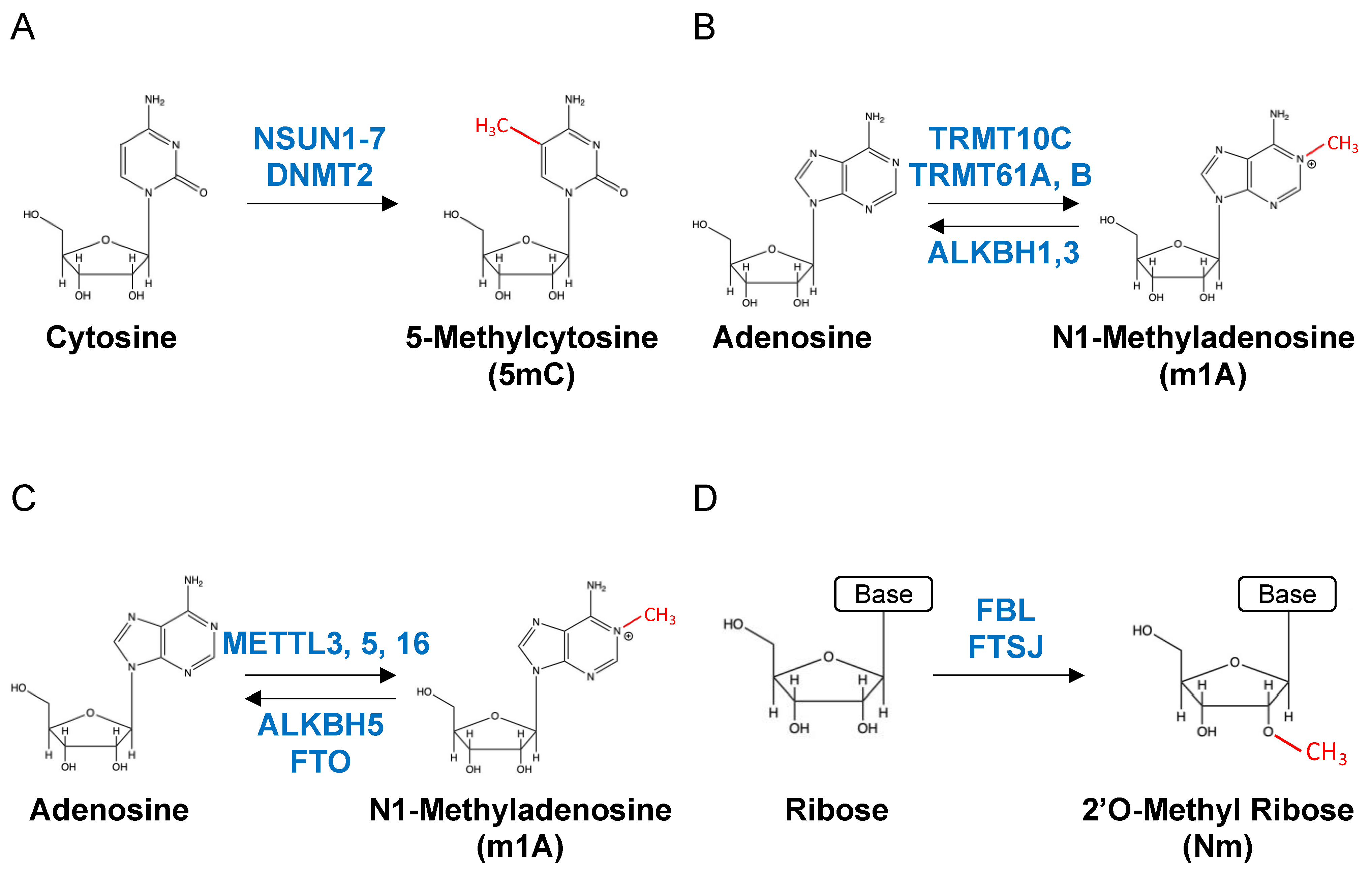
Figure 1. Major RNA modifications and their modification enzymes. (A) Cytosine is methylated by NDUN1-7 or DNMT7 to 5-methylcytosine (5mC). (B) Adenosine is methylated by TRMT10, TRMT61A, or TRMT61B to N1-methyladenosine (m1A). M1A is demethylated by ALKBH1 or ALKBH3 to adenosine. (C) Adenosine is methylated by METTL3, METTL5, or METTL16 to N6-methyladenosine (m6A). M6A is demethylated by ALKBH5 or FTO and returned to adenosine. (D) RNA ribose is methylated to 2′-O-Methyl-Ribose regardless of the base. FBL and FTSJ are known to be methyltransferases.
Applsci 13 06376 g001
Figure 2. RNA modifications associated with cancer malignancy. RNA modifications reported to be associated with malignant transformation of each cancer type (Leukemia, Glioblastoma, Breast cancer, Lung cancer, Hepatocellular carcinoma, Pancreatic cancer, and Colorectal cancer) and their structural formulas are shown. The red part of the structural formula indicates the structure added by the modification.
Applsci 13 06376 g002
Figure 3. Types of sequencers and detectable RNA modifications. Sequencing technologies, RNA modifications reported to be detectable by the technologies, and their structural formulas are shown. The red-colored sites in the nucleotide structure formula indicate the portion added by the modification.
Applsci 13 06376 g003
Disclaimer/Publisher’s Note: The statements, opinions and data contained in all publications are solely those of the individual author(s) and contributor(s) and not of MDPI and/or the editor(s). MDPI and/or the editor(s) disclaim responsibility for any injury to people or property resulting from any ideas, methods, instructions or products referred to in the content.