Article

Microencapsulation of Lactobacillus plantarum 299v Strain with Whey Proteins by Lyophilization and Its Application in Production of Probiotic Apple Juices

1
Department of Bioengineering and Alcoholic Drink Technology, Institute of Food Science and Technology, Hungarian University of Agriculture and Life Sciences, Ménesi út 45, 1118 Budapest, Hungary
2
Department of Livestock and Food Preservation Technology, Institute of Food Science and Technology, Hungarian University of Agriculture and Life Sciences, Ménesi út 45, 1118 Budapest, Hungary
3
Department of Postharvest, Commerce, Supply Chain and Sensory Science, Institute of Food Science and Technology, Hungarian University of Agriculture and Life Sciences, Ménesi út 45, 1118 Budapest, Hungary
*
Authors to whom correspondence should be addressed.
Appl. Sci. 2023, 13(1), 318; https://doi.org/10.3390/app13010318
Submission received: 23 November 2022 / Revised: 24 December 2022 / Accepted: 25 December 2022 / Published: 27 December 2022
(This article belongs to the Special Issue Knowledge-Based Biotechnology for Food, Agriculture and Fisheries)

Abstract

:

Featured Application

Proteins coated probiotic microcapsules can be applied to the apple juice to develop a functional probiotic beverage.

Abstract

The viability of probiotics is strictly influenced by the production, storage, and digestion, while microencapsulation is a technology that can protect them against harsh environments. In this study, the impact of different core-to-wall ratios and wall material formulations on physical properties and the cell number of the microcapsules were investigated. The samples with core-to-wall ratio 1:1 have a significantly higher cell number, encapsulation efficiency, and bulk density than samples with core-to-wall ratio 1:1.5. The yields of the encapsulation method were changes in the opposite direction. Meanwhile, core-to-wall ratios and formulation have a significant effect on the cell number of the microcapsules during the in vitro SGJ test, whereas time, core-to-wall ratios, and formulation have a similar influence in the in vitro SIJ test. Moreover, probiotic apple juices stored at 4 °C for 6 weeks kept the highest cell number at the end. Furthermore, probiotic apple juices fortified by microcapsules coated with WP:DWP 1:1 in core-to-wall ratio 1:1 and stored at 4 °C for 4–8 weeks exhibited a significantly lower pH value. In summary, both whey proteins and denatured whey proteins are as good as coating material for microencapsulation of probiotic bacteria Lactobacillus plantarum 299v strains. These microcapsules have high potential in the production of probiotic apple juice even by fermentation or fortification methods.

1. Introduction

Probiotics are microorganisms that have health benefits for the host when they are administrated in a sufficient amount [1]. They have numerous beneficial characteristics, e.g., control of irritable bowel syndrome, endogenous or exogenous pathogens suppression, lactose tolerance improvement, colon cancer risk reduction, body weight regulation, constipation improvement, tooth decay prevention, etc. [2,3]. Lactobacillus plantarum is a lactic acid bacterium, which consists of various species and is recognized as a potential probiotic bacteria [4]. However, it is challenging for probiotics to survive through the process, not to mention exert their probiotic effect on the host owing to the harsh environments during the manufacturing, storage, and digestion process, e.g., oxygen, heat, low pH, high bile salt content, etc. [5,6,7,8].
Microencapsulation is a talented technology that can protect the probiotics by coating them with wall materials to maintain their viability and functionality during the manufacturing, storage, and digestion process and let them apply their probiotic effect in the gut with an adequate dose level [9,10,11,12,13,14,15,16,17]. Generally, spray-drying and lyophilization (or freeze-drying) are the mainly applied technology, accomplished with other methods, e.g., spray-chilling [17], emulsion [18], extrusion, etc. However, due to the high temperature of the heating process [9,19] through the chamber of spray-drying and the extreme sensitivity of the probiotics to heat, it is not suitable to use that technology on probiotics. As a substrate, lyophilization [20,21,22] is a process that can sublimate the water content in the iced samples to the gaseous state under vacuum conditions to efficiently dehydrate the samples, while having the advantage of minimal heat stress to the probiotic.
No doubt that coating materials play an important role in the microencapsulation technology [12]. Whey proteins (WP) the by-product in dairy industry are sources of valuable biological proteins as well as rich in riboflavin and minerals [4]. The main drawback of WP is environmental issue because of high BOD values. WP are considered an exceptional coating material due to their specific physical and chemical properties, such as excellent emulsification, superb gelation and exquisite fill-forming properties. Denatured whey proteins (DWP) are originated from WP by treatment with acid or heat, which can contribute to some specific properties including high tensile property and low oxygen permeability [7,9,18,23]. Based on the concept of development of new value-added applications of dairy wastes to enlarge the application area and avoid discarding the whey protein, it is appropriate to adopt the practice of using whey proteins as coating materials for the microencapsulation process [24].
Furthermore, those people who improved their health awareness are not fulfilled with the existing probiotic dairy matrices foods anymore. Instead, considerable enthusiasm of consumers prevails in probiotic plant matrix foods with multiple potential health benefits [13,25]. Wherein, fruits are considered nutritious, health-improving, as well as disease-avoiding foods, owing to their nutritional and functional properties [26]. Although these foods are seasonal-specific, it is still possible to make them accessible in plentiful forms for the convenience of consumers all around the year by applying advanced food science technology [27]. Among those fruit products, fruit juice is one of the most favored forms of consumption by consumers [5,28,29]. However, there is a reduction of nutritional value, e.g., bioactive compounds, during the processing of fruit juice. In addition, these juices also contain sufficient sugars, including glucose, fructose, and sucrose [26], which may lead to the over intake of carbohydrates. Reducing the sugar content in these fruit juices may assist in decreasing related diseases, such as diabetes and obesity, which are highly correlated with excessive consumption of carbohydrates [30]. Hence, to compensate for the loss of nutritional compounds and to facilitate the processed fruit juices still as functional food, probiotics can be fortified into it to fulfill the function [27]. Unlike the traditional physical reduction technology of the sugars content in drinks, lactic acid bacteria not only affect the limited taste of the juice, they also increase the nutritional value by producing organic acids [30]. Hence, contributing the characteristics of probiotics together with the nutrition of fruit juice, easily making the food become a functional product.
In this study, whey protein and denatured whey protein are employed as wall materials. In addition, the long-time storage ability of the microcapsules is also examined at different storage temperatures. Moreover, the in vitro gastrointestinal test was carried out by using simulated gastrointestinal juice to explore the cell number of these samples during the digestion process. Furthermore, the application of microencapsulated probiotics with both fermentation and fortification technologies in apple juice was performed.

2. Materials and Methods

2.1. Preparation of Probiotics Culture

A probiotic bacterium L. plantarum 299v strain was from the strain collection of Department of Bioengineering and Alcoholic Drink Technology, Institute of Food Science and Technology, Hungarian University of Agriculture and Life Sciences. It was sub-cultured in de Man, Rogosa and Sharpe (MRS) broth after activation twice in the same medium at 37 °C for 18 h. The cells were obtained by growth in the MRS broth at the same 37 °C for 24 h.

2.2. Preparation of Sample Solutions for Microencapsulation

Cells were collected by centrifugation at 10,000× g rpm at 4 °C for 20 min. Then, they were washed twice with phosphate-buffered saline (PBS) solution. The formulation of the samples for encapsulation was made by mixing the wet pellet of the cells with wall materials based on the ratio, and the ratio between wall materials (Table 1). Whey proteins (Nutriversum®, Budapest, Hungary) were purchased from a local shop. Denatured whey protein was prepared by heating the 20% (w/w) whey protein solution at 90 °C for 20 min. Preparations were placed on the constant temperature shaker with 150 rpm at 25 °C for 1 h. Then, they were stored under −18 °C in a fast refrigerator for 24 h for freezing. All the preparations were conducted in duplicate by using two batches of prepared coating material separately.

2.3. Lyophilization

After obtaining the frozen preparations, they were lyophilized by a laboratory-scale lyophilization machine (Christ Alpha 2-4 Freeze Dryer, Martin Christ, Osterode am Harz, Germany), shown in Figure 1. When the machine reached a constant state, the actual pressure and temperature were 0.250 mbar and 17 °C, respectively. It remained under this temperature and pressure during the whole lyophilization. After 3 days of drying, the dehydrated microcapsules were ground into powder manually under aseptic conditions, followed by transferring into sterilized vials. The microcapsules of each sample were stored under 4 °C and 25 °C, respectively, for future analysis.

2.4. Determination Encapsulation Efficiency

The cells were completely released from microcapsules by the disintegration in 0.85% saline solution. The living cell number was determined by plate-counting method. The samples (0.1 mL) were poured on the MRS agar plate, and the number of colonies was counted after 48–72 h incubation. The enumerations of the colonies between 30–300 were acceptable. The final concentration of the living cells in the liquid form or solid form samples are expressed as Log CFU/mL or Log (CFU/g), respectively. Encapsulation efficiency was evaluated by calculating the percentage of the ratio of cell number after and before lyophilization. The equation was expressed as Equation (1).
EE   % = N t N 0 × 100 %
where Nt and N0 perform the logarithm value of the colony-forming unit after (Log CFU/g) and before (Log CFU/mL) the lyophilization process, respectively.

2.5. Determination of Yield

The yield was investigated by weighting the total amount of solid materials before and after lyophilization. Before lyophilization, the wet weight of probiotics cells and coating materials are considered, after lyophilization, the total solid weight of microcapsules was included. The percentage of the ratio of total solid weight after and before lyophilization was depicted as yield, which can be shown as Equation (2).
Y = m t m 0 × 100 %
where Y refers to the yield (%), m0 and mt means the total solid weight (g) before and after lyophilization, respectively.

2.6. Determination of Bulk Density

Bulk density was typified by measuring the volume of 1 g sample in a 5 mL cylinder after being tapped on a vortex for 2 min. The measurement of the bulk density was revealed by using Equation (3).
ρ = m v
where ρ is the bulk density (kg/m3), m is the mass (kg) of the sample, and v is the volume (m3) occupied in the cylinder.

2.7. Scanning Electron Microscopy

Scanning electron microscopy (SEM) was used to observe the morphological structure of L. plantarum 299v microcapsules with different core-to-wall ratios and formulations. The samples were transferred and stuck on a plate in the vacuum chamber and gradually decreased to 200 Pa. Observation of the samples was carried out in Thermo ScientificTM PrismaTM E (Waltham, MA, USA) SEM under an accelerating voltage of 15 kV. The samples were examined under 1000× and 14,000× magnifications.

2.8. Viability of Microencapsulated Probiotics during Storage

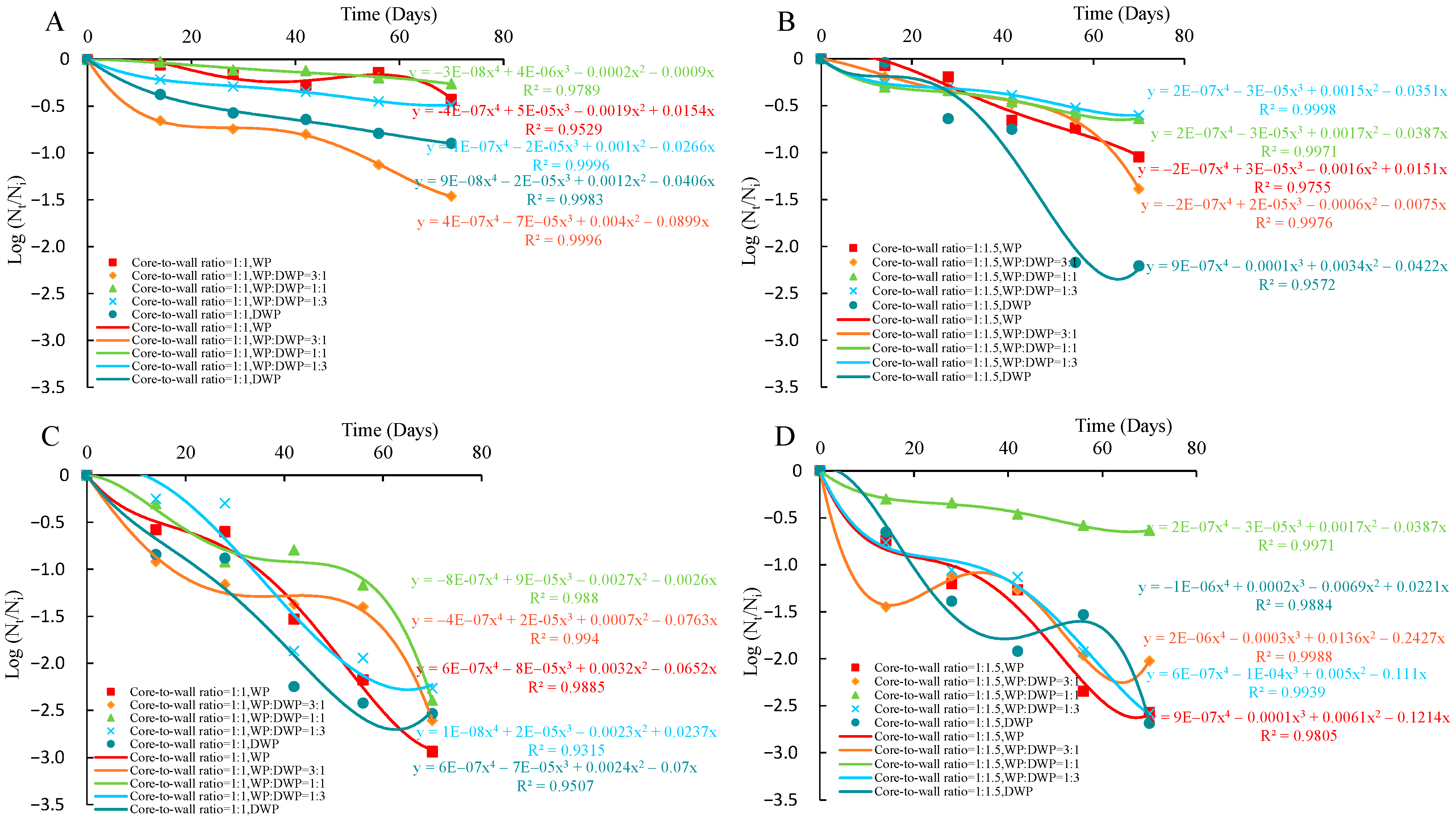
The viability of the microencapsulated probiotics during storage at 4 °C and at 25 °C was determined by enumeration on MRS agar. It was carried out every 2 weeks and the process lasted for 10 weeks. The viability of microencapsulated probiotics during 4 °C and 25 °C storage was outlined by fitting the curve of the logarithmic value of relative cell viability to storage time with a suitable-order reaction kinetics model, where Nt is the enumeration of the living cells at a specific storage time, Ni is the enumeration of the viable cells at the starting point of the storage, t refers to the storage time.

2.9. Viability of Microencapsulated Probiotics In Vitro Digestion Process

Survival of microencapsulated probiotics during the digestion process was carried out in vitro by checking living cell numbers in the samples under simulated gastric juice (SGJ) and simulated intestinal juice (SIJ) with a plate-counting method with slight modification in previous research [9,13,31]. To prepare SGJ, sodium chloride solution (5 g/L) was adjusted to pH 2 by using 6 M HCl and sterilized at 121 °C for 20 min followed by adding 0.3% pepsin (Sigma-Aldrich, St. Louis, MI, USA). To prepare SIJ juice, 0.6% bile salt (Sigma-Aldrich, USA) was added into autoclaved 0.05 M KH2PO4 solution. The sampling process was by adding 0.1 g microcapsules of each sample into 9.9 mL above mentioned SGJ and SIJ. The living cell number was checked by using a plate-counting method by taking samples based on the incubation time 0 h, 0.5 h, 1 h, 2 h, 3 h, and 0 h, 3 h, and 6 h for SGJ and SIJ samples, respectively.

2.10. Application of Microencapsulated Probiotics in Fermented and Fortified Apple Juice

Unfiltered high-quality HAZÁNK Kincsei apple juice was purchased from a local supermarket, and it was applied as the matrix for the application of microencapsulated probiotics samples in the fermentation and fortification process. The pH of apple juice was adjusted to pH 6 by 4 N NaOH solution for future utilization. For the fermented probiotic apple juices, 0.2 g microcapsules of each sample were added to 90 mL apple juice that was mentioned above and incubated at 37 °C for the fermentation process till the pH decreased within 4.0 to 5.0, which means they finished the fermentation process. For the fortified probiotic apple juices, 0.2 g microcapsules of each sample were directly added into 90 mL apple juice where the pH had been adjusted. After the sample preparation, both groups of fermented and fortified samples were stored at 4 °C and 25 °C for future analysis. All the samples were prepared in duplicate. The viability of microencapsulated probiotic samples under fermentation and fortification at 4 °C and 25 °C was characterized by using the plate-counting method. The measurement of the pH of each sample with different treatments under specific temperatures was also checked as an additional reference. These samples were analyzed once every two weeks.

2.11. Statistical Analysis

All experiments were performed in duplicates and the results were presented as means ± standard deviation. ANOVA (analysis of variance), unpaired and paired Student’s t-tests with a significance level of α = 0.05 were used to determine statistical differences among the independent variables by using Minitab 19.

3. Results and Discussion

3.1. Cell Number and Encapsulation Efficiency of Microcapsules

The cell number of microencapsulated L. plantarum 299v samples varies from 7.66 to 11.34 Log CFU/g (Figure 2A). All samples showed numeration above 6 Log CFU/g, the minimal requirement of probiotics as a functional food [1]. Additionally, the cell number was approximately 10 Log CFU/g of most samples except DWP sample. These results are similar to the values published in the studies of Kluyveromyces marxianus VM004 [15] and Bifidobacterium animalis subsp. Lactis INL1 [32] when they microencapsulated microorganisms with whey protein and spray-drying technology. Additionally, the cell number of samples with core-to-wall ratio 1:1 is significantly higher than that of those samples with a core-to-wall ratio 1:1.5. This may be due to the lower ratio of wall materials leading to a higher amount of probiotic bacteria in a certain microcapsules complex, which can result in a higher cell number in truth. In addition, the cell number of the samples with WP and WP:DWP 3:1 is significantly higher than samples with WP:DWP 1:1, WP:DWP 1:3, and DWP; the samples with WP and WP:DWP 1:1 is significantly higher than samples with DWP; the samples with WP:DWP 1:1 and WP:DWP 3:1 is significantly higher than samples with DWP. The reason may be due to the stable membrane stabilization properties of whey proteins, which can avoid cell damage, and with a decrease core-to-wall ratio of whey protein in the microcapsules, more probiotic bacteria lost their viability during the lyophilization process.
The encapsulation efficiency of microencapsulated L. plantarum 299v samples varied between 76.25% and 105.39% (Figure 2B). This agrees with the result published in the study by Vander [15] when they microencapsulated Kluyveromyces marxianus yeast cells using whey protein concentrate as the wall material. Rajam & Anandharamakrishnan in 2015 also reported that both whey protein isolate (WPI) and denatured WPI in combination with fructooligosaccharide (FOS) in 1:1 ratio exhibited higher encapsulation efficiency, lower residual moisture content, and a narrower range of particle size distribution than 1:1.5. In our case, the encapsulation efficiency of samples with ratio 1:1 is significantly higher than that of those samples with ratio 1:1.5. This may also be due to the lower ratio of wall materials will lead to a higher the number of probiotic bacteria in a certain microcapsules complex, which can result in a relatively higher cell number after lyophilization under the same formulation. In addition, the encapsulation efficiency of the samples with WP is significantly higher than samples coated by DWP in the circumstances with ratio 1:1 and 1:1.5. The probable reason may be due to the protecting ability of the cell membrane of WP, which is better than DWP, and results in a relatively higher encapsulation efficiency.
Microencapsulation plays a huge impact in food developments. It can transform essential and conventional foods into fortified processed foods for better nutritional, functional, and controlled delivery. Encapsulation efficiency refers to the percentage of probiotics successfully embedded into the microcapsules, which is one of the most essential criteria for the use of probiotics [13]. Under the same effectiveness of probiotics per unit, based on the quality definition of the products of probiotics, the higher the encapsulation efficiency, the better the product. Due to the definition of encapsulation efficiency, there is some connection between cell number and encapsulation efficiency. In addition, the viable cell number is also a noteworthy indicator for checking the quality of probiotic products [12].

3.2. Yield and Bulk Density of Microcapsules

Yield and bulk density are fundamental parameters during the manufacturing, packaging, and storage process of probiotic microcapsules. The yield and bulk density of encapsulation of L. plantarum 299v strain with five wall materials ratio and two ratios after lyophilization are shown in Figure 3. The yields varied from 49.35% to 68.02% (Figure 3A). Additionally, the samples of 1:1.5 ratio have a significantly higher yield than samples of 1:1 ratio. This is caused by the wetness of the probiotics core. With the same scale of wet probiotics, the higher amount of dry powder will result in a higher total solid amount after the drying process. Meanwhile in the case of ratio 1:1, the yield of the sample with only DWP as coating material was significantly lower than the others, whereas, in the case of ratio 1:1.5, the yield of the sample with only WP was the lowest. This may be due to under ratio 1:1 and ratio 1:1.5, WP and DWP have a poor combination of water molecules together with probiotic bacteria, hence causing more water loss during the lyophilization process.
The bulk density was between 0.23 g/cm3 to 0.27 g/cm3 (Figure 3B), which is lower than the results (approximately 0.5 g/cm3) reported by Rajam & Anandharamakrishnan in 2015. The difference may be due to the use of coating materials and drying technology, the combined whey proteins, and fructooligosaccharide as coating materials and applied spray-drying technology. Probably due to the high-water evaporation ability of spray-drying method, less water content remained in the microcapsules while lyophilization. Furthermore, there is no significant difference in bulk density between different formulations. However, the bulk density of samples with core-to-wall ratio 1:1 is significantly higher than that of samples with core-to-wall ratio 1:1.5. This may be explained by the physical structure state of the WP, while the DWP has no significant difference. The difference ratios of coating materials resulted in a fluffier structure and in a significantly lower bulk density of samples with ratio 1:1.5 than ratio 1:1.

3.3. Morphology of Microcapsules of L. plantarum 299v Strain

The micrographic images of free and microencapsulated L. plantarum 299v strain with whey protein and denatured whey protein are shown in Figure 4. The structure of the microcapsules under 1000× magnification is irregular and rod-shaped cells are clustered together and closely aligned under 14,000× magnification. This may be due to the evaporation of water during the lyophilization resulting in the condensed structure and can be seen clearly with large magnification [33]. Furthermore, the cells of L. plantarum 299v can be found in microcapsules with different coating materials (Figure 4C–V), which are similar to the results by Savedboworn et al. (2020). The thick materials and tight arrangement of the probiotic might work as protective walls and prevent water uptake. However, there are still some free cells that can be found on the surface of the coating materials. This inspection is in accordance with traditional features for matrix-type microcapsules that core molecules are commonly distributed in the coating materials, while some core materials may also present on the surface [6,34]. In addition, several tiny cracks and holes on the microcapsules are visible at higher magnification on the microcapsules on samples with core-to-wall ratio 1:1 and core-to-wall ratio 1:1.5. Halim et al. (2018) revealed that the cracking-like structure was found on the surface of chitosan, while it was not observed on the samples with alginate beads and free cells. This may be due to the specific structure and unique characteristics of whey protein and denatured whey proteins. Furthermore, there is no significant morphology difference as well as in the bulk density between the samples in the group of core-to-wall ratio 1:1 and core-to-wall ratio 1:1.5. Morphology of particles plays important roles in the formation of some physical properties, such as directly influences the bulk density, flowability and rehydration characteristics of the powders [9]. However, in the group of samples with core-to-wall ratio 1:1.5 under 14,000× magnification, the sample coated by DWP with core-to-wall ratio 1:1.5 seems to have more folds and bulges than other samples with core-to-wall ratio 1:1.5, which may be linked to the effect of atomization mechanism and film properties of WP and DWP for the microencapsulation process.

3.4. Viability of Microencapsulated Probiotics during Storage

Viability during the manufacturing or storage is also a fundamental standard for the use of probiotic products. Many researchers have demonstrated that microencapsulated probiotics samples have significantly higher cell density than those samples without microencapsulation [19,22,35,36]. Specifically, the membrane lipid oxidation is the main reason that caused the viability loss [9], thus protecting ability or forming ability of coating materials, temperature, water activity, oxygen content, etc. are the main factors during storage.
The effectiveness of ratio and formulation on the storage stability of microencapsulated L. plantarum 299v were investigated. The storage was done at 4 °C and 25 °C for 70 days and results are demonstrated in Figure 5. In the case of storage at 4 °C with core-to-wall ratio 1:1, the sample coated by WP:DWP 3:1 has significantly higher cell number loss rate than the other samples. The cell number decreased gradually in the first two weeks, then it remained constant till the 6th week. After that the cell number started decreasing again. A similar cell losing pattern was found on samples coated by WP. In this case, the cell number started decreasing sharply after the 8th week. This may be due to the stability of the coating materials WP:DWP 3:1 and WP with core-to-wall ratio 1:1. However, the losing dynamic was quite different for samples coated by WP:DWP 1:1, WP:DWP 1:3, and DWP. The cell number of those three samples started decreasing from the beginning of the storage process and after 4 weeks with almost a constant loss rate. In the case of storage at 4 °C with core-to-wall ratio 1:1.5, samples coated by DWP exhibited significantly higher loss rate in cell number than the other samples. The cell number decreased slowly at the first 4 weeks while then it started decreasing faster. Samples coated by WP:DWP 3:1 have the same losing pattern as samples coated by DWP after storage for 6 weeks. This may be due to the stability of the coating materials DWP and DWP 3:1 with core-to-wall ratio 1:1.5. The cell numbers decreased sharply after storage for 4 and 6 weeks under 4 °C, respectively. Additionally, samples coated by WP, WP:DWP 1:1, and WP:DWP 1:3 shared the same losing dynamic.
In the cases of storage at 25 °C with core-to-wall ratio 1:1 of bacteria to coating material, the cell numbers of samples coated by WP:DWP 3:1 and WP:DWP 1:1 exhibited the same trends, i.e., decreased in the first 4 weeks, then kept constant till the 8th week, and at the end phase they dropped drastically. Generally, the cell numbers of samples with core-to-wall ratio 1:1 stored at 25 °C have a higher losing rate than of samples stored at 4 °C. This may be due to the change of moisture of the microcapsules and thus caused the disruption and deactivation of the cell membrane. The increase of moisture content during storage period may be linked to the absorption of water from the environment [37]. In the case of storage at 25 °C with core-to-wall ratio 1:1.5, the cell number of samples coated by WP:DWP 1:1 had a significantly lower loss rate than by other formulation. This may be due to the relative condense structure. The cell number of samples coated by WP and WP:DWP 1:3 started to decrease in the first two weeks and almost remained constant till the 4th week of storage. After that, it decreased again until the end of the experiment. Moreover, the cell number of samples coated by WP:DWP 3:1 and DWP showed quite a different pattern compared with those two cell number-losing styles. After decreasing for 2 weeks and 6 weeks, they increased until the 6th and 8th week, respectively, and then decreased again until the end. These results indicated that the ratio, formulation and storage temperature play an important role in the cell viability of the microcapsules during the storage. In conclusion, the samples with ratio 1:1 coated by WP:DWP 1:1 and the samples with core-to-wall ratio 1:1.5 coated by WP:DWP 1:1 were the best ones in the storage period at both temperatures 4 °C and 25 °C, respectively.

3.5. Viability of Microencapsulated Probiotics In Vitro Digestion Process

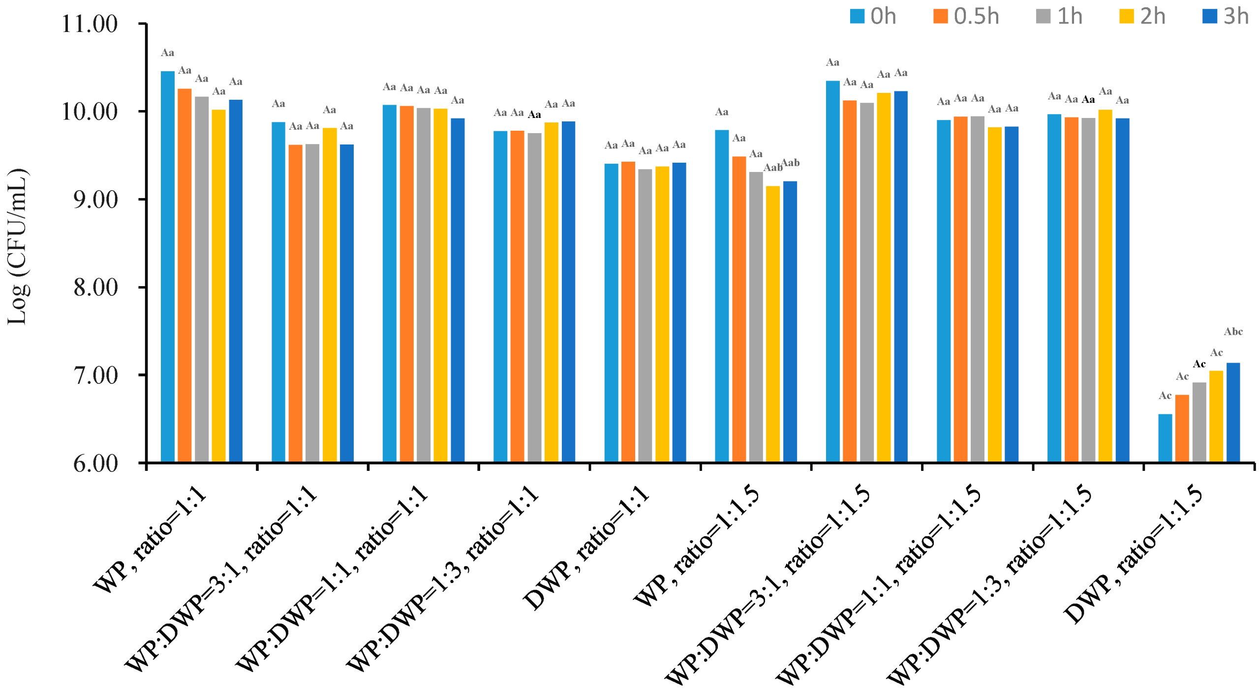
The stomach is the main digestion process that can cause the loss of viability of probiotics due to the low pH and pepsin enzyme. The acidity of the stomach is usually approximately pH 2, which is affected by eating time, quality and quantity of diet [17,19]. The viability of lyophilized L. plantarum 299v strain with different core-to-wall ratios, wall material types and their formulations after being exposed to SGJ at 37 °C for 3 h are shown in Figure 6. Core-to-wall ratios, formulation, and the combination of ratio and formulation have a significant effect on the viability during the in vitro SGJ digestion process.
As can be seen from Figure 6, almost all samples except sample with core-to-wall ratio 1:1.5 coated by DWP did not show any significant differences in viable cells during the SGJ experiment. This is due to the collective effect of the acidity stress [9,13,38] that caused the death of probiotics as well as may release the probiotics from the microcapsules. Hence, it can be as a protecting ability of the coating materials with these formulations and ratios against pepsin and low pH digestion environment. However, the difference in ratios and formulations may affect the releasing characteristics of the microcapsules, for example, the samples with core-to-wall ratio 1:1 coated by WP increased the release of probiotic from microcapsules after 3 h incubation causing suddenly increase in cell number. Or the samples with core-to-wall ratio 1:1.5 and coated by DWP gradually increased the releasing of probiotic from microcapsules during the 3 h of incubation. The samples with core-to-wall ratio 1:1 have a significantly higher cell number than those with ratio 1:1.5. This may be due to the physical properties of the microcapsules, which means under the same circumstance, the higher the viable cells of the probiotics together with properties of the coating materials can exhibit higher protecting ability against pepsin and low pH digestion environment. Formulations of coating by WP, WP:DWP 1:3, WP:DWP 1:1 and WP:DWP = 3:1 have a significantly higher cell number than samples coated by DWP alone. However, there is an interesting phenomenon of samples with ratio 1:1.5 coated by DWP. It showed an increasing cell number during the simulated digestion process, which may indicate that DWP has good protecting and releasing abilities as well as buffering capacity. According to the research of Vanden Braber and co-workers (2020), WP has good buffering capacity that can relieve the digestive stress from gastrointestinal juice and maintain the cell number of the probiotic product. In our study, the buffering capacity of DWP was outstanding, even better than WP. This may explain the reason that in some cases, the cell numbers did not decrease, instead even increase over time of SGJ. Based on our overall results of the lyophilization, storage, and digestion process, it can be stated that probably DWP with core-to-wall ratio 1:1.5 may not good choice, but it is worth doing more research to explore the advantage of DWP as coating material. In addition, the samples with core-to-wall ratio 1:1 coated by WP, WP:DWP 1:1, and WP:DWP 1:3 as well as the samples with core-to-wall ratio 1:1.5 coated by WP:DWP = 3:1, WP:DWP 1:1, and WP:DWP 1:3 have significantly higher cell numbers than others’ ones. Therefore, the physicochemical composition, formulation, and concentration of coating materials need to be taken into consideration when planning to produce probiotic microcapsules with high gastrointestinal juice resistance.
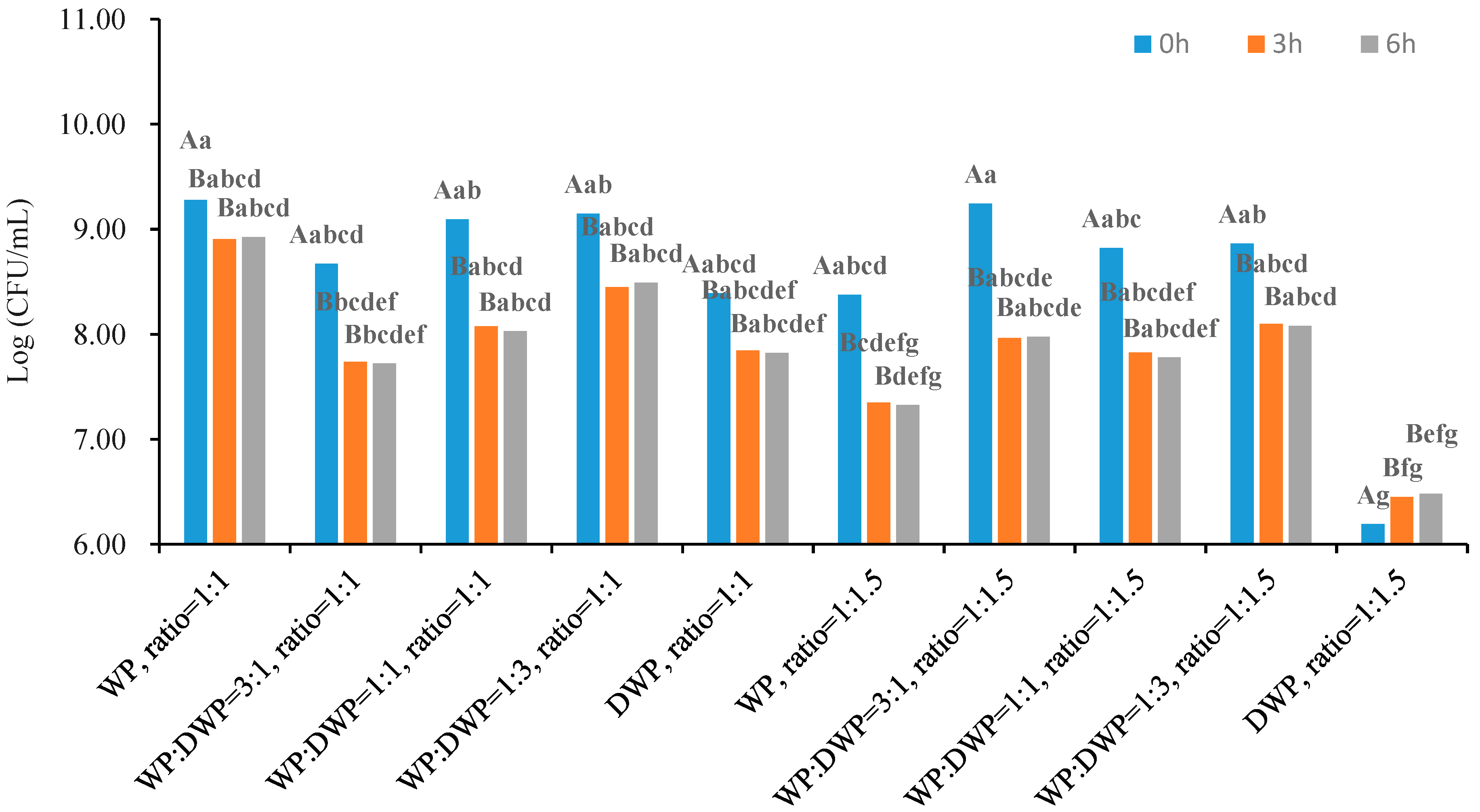
Bile salt is the main component of bile juice that is involved in the digestion and absorption process of fat content. In addition, it can dissolve bacterial cell membranes and results in viability loss [17]; thus, the resistance of probiotic bacteria to bile salt environment is an imperative property [13,19,38]. Therefore, the viability of lyophilized L. plantarum 299v strain with different core-to-wall ratios and wall material types as well as formulations were investigated. Time, core-to-wall ratio, formulation, and the combination of ratio and formulation have a significant effect on the viability during the in vitro SIJ digestion process (Figure 7).
The cell number of each sample significantly decreased in the first 3 h of incubation, and then did not change. Similar results were reported by Arslan-Tontul & Erbas, (2017) of S. boulardii after 180 min incubation. A possible explanation for this phenomenon may be explained by adaptation properties of the cells to the bile salt or synthesizing bile salt hydrolase [19,39].
The core-to-wall ratio is another factor that influences the cell viability in the SGJ experiment. In the case of core-to-wall ratio 1:1 of cell to coating material, the cell numbers were significantly higher than those with core-to-wall ratio 1:1.5. This means the increase in the amount of wall material did not provide any higher protecting effect than core-to-wall 1:1.5. Rajam and Anandharamakrishnan (2015) found that single or double coating materials can take advantage in the elimination of the influence of bile salt on the probiotic bacteria [9]. In addition, capsules coated with WP, WP:DWP 1:3, WP:DWP 1:1 and WP:DWP 3:1 contained a significantly higher cell number than ones coated by DWP. Although the differences of cell numbers of those four formulations were not significant. Meanwhile, the capsules coated by WP with core-to-wall ratio 1:1 exhibited the lowest cell number reduction at approximately 0.35 Log (CFU/g), whereas the highest reduction of cell number (approximately 1.27 Log value) was obtained in the case of capsules coated by WP:DWP 3:1 at the core-to-wall ratio 1:1.5. In the case of DWP as coating material and the core-to-wall ratio was 1:1.5, a slight increase in viable cells was observed suggesting that DWP may weaken the toxicity of bile on cell membrane damage [19,40]. Many more studies are needed to construct in the future to use DWP as wall material in the coating and releasing processes in food application.
After the SGJ and SIJ tests, the samples were stored for 20 weeks. The average cell numbers of samples were 9.34, 9.53, and 7.86 Log (CFU/g), respectively. It reveals that the group of formulation and the ratio had significantly better protecting ability against high acidity but worse protecting properties against high bile salt content. In conclusion, taking advantage of WP and DWP, they are promising materials [10,40] as inner coating materials to form a double layer [17] with better protecting ability against both high acidity and bile salt.

3.6. Application of Microencapsulated Probiotics in Apple Juice

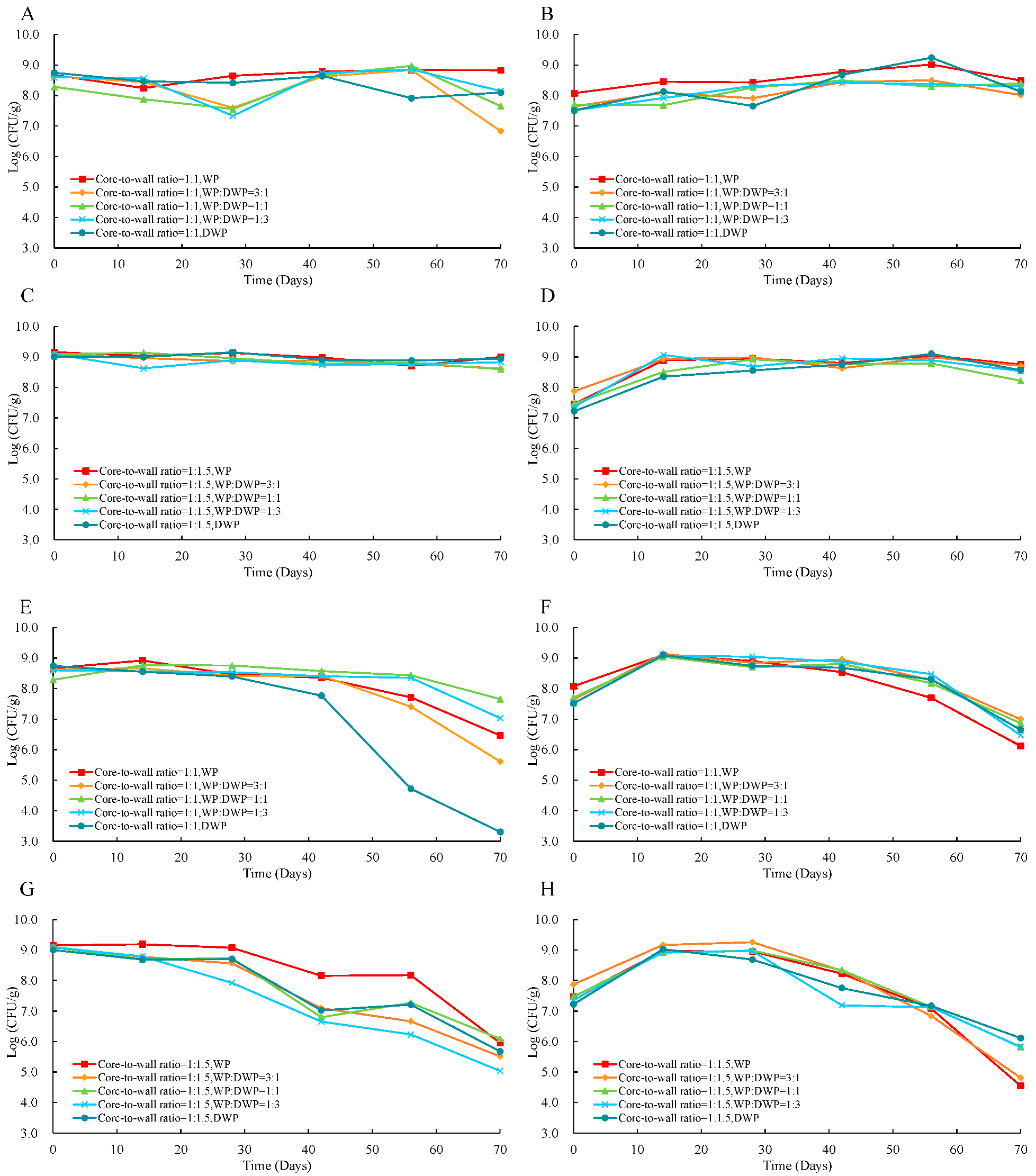
The perception of applying probiotic bacteria into fruit juice can compensate for the nutrition loss due to the manufacturing process. Additionally, the probiotic bacteria also increase the functionality via viable cells that has already been accepted by customers and experts. The cell numbers of fermented and fortified apple juices with different ratios and formulations stored at 4 °C and 25 °C for 10 weeks are shown in Figure 8.
Storage time and storage temperature have a significant effect on the changes of cell numbers of probiotic apple juices during storage. However, the core-to-wall ratio, the formulation and fortification or fermentation methods did not significantly affect the viability of the probiotics.
Storage temperature is a factor that influences the viability of probiotics in apple juice. Meanwhile storage at a high temperature, such as room or higher temperature, may initiate some metabolisms by other microbes especially some pathogens that thus will suppress the probiotics, whereas storage at low temperatures, such as at 4 °C can keep the bacteria alive (Figure 8A–C). Viable cells in all capsules were maintained at the initial levels (approximately 9 Log CFU/g) for a whole storage period (two months). The effect of temperature on the viability of cells can also observed when doing comparison of data in Figure 8C and 8G. Meanwhile the cell number of those samples fermented by microcapsules with core-to-wall ratio 1:1.5 (Figure 8C) was kept unchanged (9 Log CFU/g), whereas it started to decrease from the week 4, and reached 5.04 Log (CFU/g) at the end of storage. It is worth to note that this value is much lower than the viable cell criterium of probiotic products (7 Log CFU/g).
Storage time is also a crucial factor that influences the viability of cells. There was no significant difference in the cell numbers during storage of apple juices for 0, 2, 4, and 6 weeks. However, the cell number started to significantly decrease from the 8th week. This trend was accelerated after week 10 (Figure 8E–H). The cell number apple juice fermented with microcapsules coated by DWP with core-to-wall ratio 1:1 reached even 3.3 Log (CFU/g) at the end of the storage period.
Different ratios of core-to-wall as well as WP to DWP did not affect the trend of change of viable cells of both fermented and fortified apple juices stored at temperature 4 °C. The cell numbers kept constant for the whole storage period (Figure 8A–D). In the case of storage at 25 °C, the cell numbers of fermented samples with microcapsules coated by pure WP or DWP (Figure 8E,G) as coating materials. Instead, they may influence the dynamics of changes of cell numbers. In addition, these changes were also not affected by fortification and fermentation methods, but generally, the viable cell numbers of fermented samples were significantly higher than fortified samples. Meanwhile, in the fermentation process, the bacteria grew, thus resulted the increase in cell number, whereas it was missed in the case of fortification. The same result was also observed by Ying et al., (2013). However, at the end of the storage process for long time (some months), this is no significant difference in the cell number between two methods (fermentation and fortification).
In summary, storage time and temperature are the two main factors that influence the viability of the probiotic cells in apple juice, while the ratio, formulation, fermentation and fortification technologies did not affect it. Storage at 4 °C is suggested for probiotic apple juice. At this temperature, both fermented or fortified probiotic apple juices can maintain their viable cells more than 6 weeks.
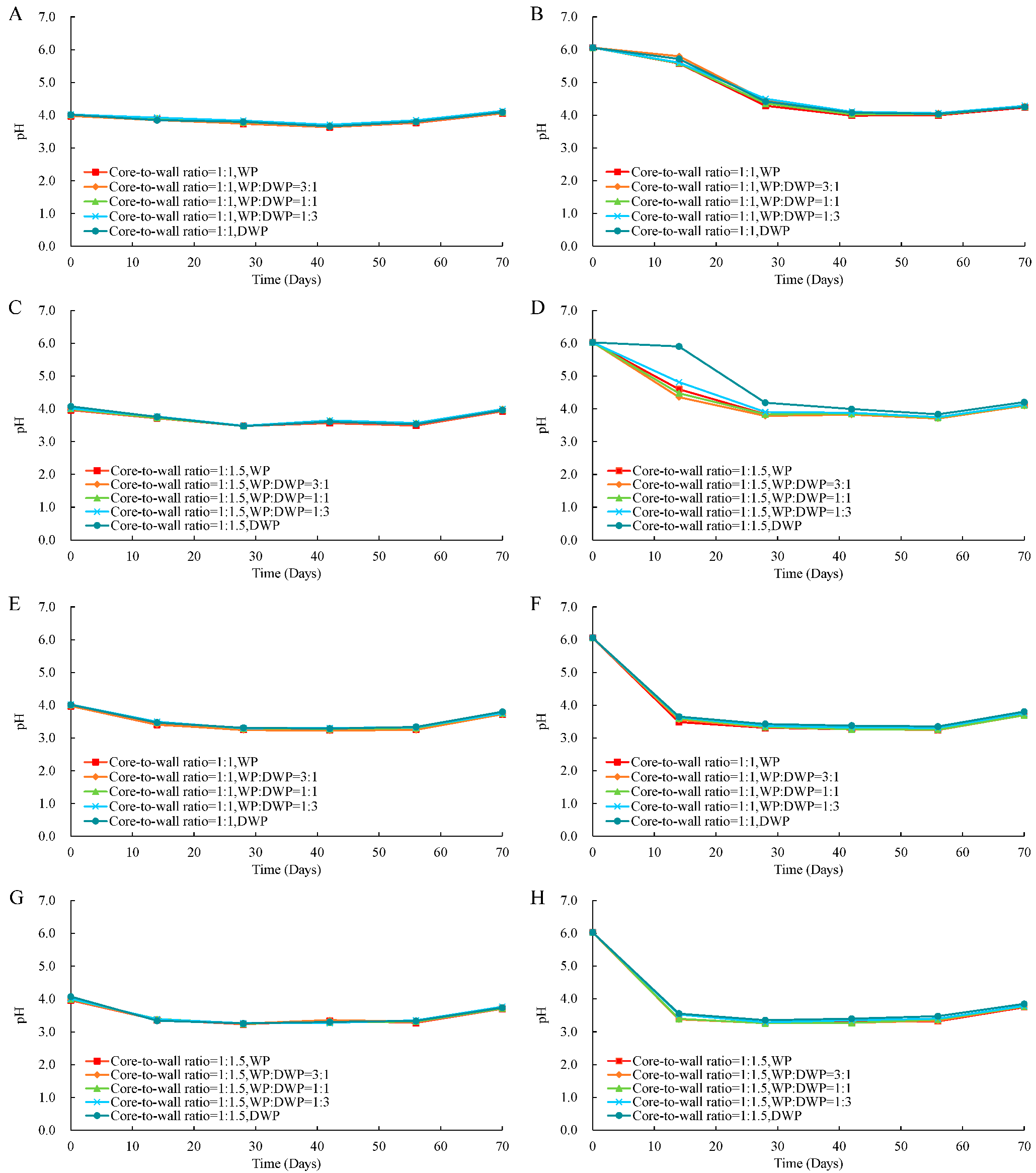
The change of pH can be used as an indicator to monitor both fermentation and storage processes quickly. The pH changes of fermented and fortified apple juices with different ratios and formulations of storage at 4 °C and 25 °C for 10 weeks are illustrated in Figure 9.
Meanwhile the pHs of all fermented samples were approximately pH 4.0 and did not change significantly during storage (Figure 9A,C,E,G), whereas the storage time has significant effect on the changes of pHs of the probiotic apple juice. In the case of fortification, the pHs values dropped to approximately pH 4.0 when storage at 4 °C and 25 °C for 28 days and 14 days, respectively (Figure 9B,D,F,H). Similar results were reported by Sohail et al. in 2012 and Gandomi et al. in 2016. They reported that the orange juices fermented with Lactobacillus rhamnosus GG stored at 4 °C for 30 days exhibited lower acidification compared to one stored at 25 °C for 12 days and it may result better sensory properties.
Summarizing, while the ratios of core-to-wall, storage time and temperature as well as fermentation or fortification methods significantly affected the changes of pH of probiotic apple juices during storage, the formulation of the microcapsules does not have any.

4. Conclusions

The present study demonstrated that whey protein and denatured whey protein are as good as coating materials for microencapsulation of probiotic bacteria L. plantarum 299v strain. After microencapsulation, the bacteria cells were protected during production, storage, digestion processes. The core-to-wall ratio and formulation significantly influenced the cell number, encapsulation efficiency, and yields of the microcapsules after lyophilization. In addition, core-to-wall ratio, formulation, and storage temperature also affect the viable cells of the microcapsules in the long-time storage period. The changes of pH of probiotic fermented or fortified apple juices were influenced by the core-to-wall ratios, storage time and temperature as well as fermentation or fortification methods. Based on these findings, it can be inferred that whey protein and denatured whey protein have high potential applications for encapsulation of probiotics for food industry.

Author Contributions

The manuscript was written with input from all authors. W.S. and E.B. conducted the experiments and collected and analyzed the data; W.S., Q.D.N., G.S., S.R.Z., K.H., L.F., A.V. and G.H. wrote, reviewed, and edited the manuscript; E.B. and A.G. conceptualized and supervised the project and edited the manuscript. All authors have read and agreed to the published version of the manuscript.

Funding

This work is supported by the New Széchenyi Plant Project No. EFOP-3.6.3.-VEKOP-16-2017-00005 and Project No. GINOP-2.2.1-18-2020-00025; by National Research, Development and Innovation Office Project No. TKP2021-NVA-22.

Institutional Review Board Statement

Not applicable.

Informed Consent Statement

Not applicable.

Data Availability Statement

Not applicable.

Acknowledgments

This work is supported by the New Széchenyi Plant Project No. EFOP-3.6.3.-VEKOP-16-2017-00005 and Project No. GINOP-2.2.1-18-2020-00025; by National Research, Development and Innovation Office Project No. TKP2021-NVA-22; by TEMPUS Foundation via Stipendium Hungaricum Program and China Scholarship Council via China-Hungary Exchange Scholarship Program, as well as by the Doctoral School of Food Science. W. Sun, G. Sipiczki, K. Hritovski, S. R. Ziane are students in the Department of Bioengineering and Alcoholic Drink Technology, Institute of Food Science and Technology, Hungarian University of Agriculture and Life Sciences.

Conflicts of Interest

The authors declare no conflict of interest.

References

  1. FAO/WHO. Report of a Joint FAO/WHO Expert Consultation on Evaluation of Health and Nutritional Properties of Probiotics in Food Including Powder Milk with Live Lactic Acid Bacteria; FAO/WHO: Cordoba, Argentina, 2001. [Google Scholar]
  2. Mao, L.; Pan, Q.; Hou, Z.; Yuan, F.; Gao, Y. Development of soy protein isolate-carrageenan conjugates through Maillard reaction for the microencapsulation of Bifidobacterium longum. Food Hydrocoll. 2018, 84, 489–497. [Google Scholar] [CrossRef]
  3. Chen, T.; Wang, Y.; Cai, D.; Zheng, M.; Xiu, L.; Liu, J. Advances in wall materials and methods of probiotic microencapsulation. China Dairy Ind. 2016, 44, 31–37. [Google Scholar]
  4. Eckert, C.; Serpa, V.G.; Felipe dos Santos, A.C.; Marinês da Costa, S.; Dalpubel, V.; Lehn, D.N.; Volken de Souza, C.F. Microencapsulation of Lactobacillus plantarum ATCC 8014 through spray drying and using dairy whey as wall materials. LWT-Food Sci. Technol. 2017, 82, 176–183. [Google Scholar] [CrossRef]
  5. Dias, C.O.; dos Santos Opuski de Almeida, J.; Pinto, S.S.; de Oliveira Santana, F.C.; Verruck, S.; Müller, C.M.O.; Prudêncio, E.S.; de Mello Castanho Amboni, R.D. Development and physico-chemical characterization of microencapsulated bifidobacteria in passion fruit juice: A functional non-dairy product for probiotic delivery. Food Biosci. 2018, 24, 26–36. [Google Scholar] [CrossRef]
  6. Ahmad, M.; Gani, A.; Hamed, F.; Maqsood, S. Comparative study on utilization of micro and nano sized starch particles for encapsulation of camel milk derived probiotics (Pediococcus acidolactici). LWT-Food Sci. Technol. 2019, 110, 231–238. [Google Scholar] [CrossRef]
  7. Ying, D.Y.; Schwander, S.; Weerakkody, R.; Sanguansri, L.; Gantenbein-Demarchi, C.; Augustin, M.A. Microencapsulated Lactobacillus rhamnosus GG in whey protein and resistant starch matrices: Probiotic survival in fruit juice. J. Funct. Foods. 2013, 5, 98–105. [Google Scholar] [CrossRef]
  8. Terpou, A.; Papadaki, A.; Lappa, I.K.; Kachrimanidou, V.; Bosnea, L.A.; Kopsahelis, N. Probiotics in Food Systems: Significance and Emerging Strategies Towards Improved Viability and Delivery of Enhanced Beneficial Value. Nutrients 2019, 11, 1591. [Google Scholar] [CrossRef] [Green Version]
  9. Rajam, R.; Anandharamakrishnan, C. Microencapsulation of Lactobacillus plantarum (MTCC 5422) with fructooligosaccharide as wall material by spray drying. LWT-Food Sci. Technol. 2015, 60, 773–780. [Google Scholar] [CrossRef]
  10. Suryabhan, P.; Lohith, K.; Anu-Appaiah, K.A. Sucrose and sorbitol supplementation on maltodextrin encapsulation enhance the potential probiotic yeast survival by spray drying. LWT-Food Sci. Technol. 2019, 107, 243–248. [Google Scholar] [CrossRef]
  11. Liu, H.; Gong, J.; Chabot, D.; Miller, S.S.; Cui, S.W.; Ma, J.; Zhong, F.; Wang, Q. Incorporation of polysaccharides into sodium caseinate-low melting point fat microparticles improves probiotic bacterial survival during simulated gastrointestinal digestion and storage. Food Hydrocoll. 2016, 54, 328–337. [Google Scholar] [CrossRef]
  12. Kavitake, D.; Kandasamy, S.; Devi, P.B.; Shetty, P.H. Recent developments on encapsulation of lactic acid bacteria as potential starter culture in fermented foods—A review. Food Biosci. 2018, 21, 34–44. [Google Scholar] [CrossRef]
  13. Dimitrellou, D.; Kandylis, P.; Petrović, T.; Dimitrijević-Branković, S.; Lević, S.; Nedović, V.; Kourkoutas, Y. Survival of spray dried microencapsulated Lactobacillus casei ATCC 393 in simulated gastrointestinal conditions and fermented milk. LWT-Food Sci. Technol. 2016, 71, 169–174. [Google Scholar] [CrossRef]
  14. Alfaro-Galarza, O.; López-Villegas, E.O.; Rivero-Perez, N.; Tapia- Maruri, D.; Jiménez-Aparicio, A.R.; Palma-Rodríguez, H.M.; Vargas-Torres, A. Protective effects of the use of taro and rice starch as wall material on the viability of encapsulated Lactobacillus paracasei subsp. Paracasei. LWT-Food Sci. Technol. 2020, 117, 108686. [Google Scholar] [CrossRef]
  15. Vanden Braber, N.L.; Díaz Vergara, L.I.; Rossi, Y.E.; Aminahuel, C.A.; Mauri, A.N.; Cavaglieri, L.R.; Montenegro, M.A. Effect of microencapsulation in whey protein and water-soluble chitosan derivative on the viability of the probiotic Kluyveromyces marxianus VM004 during storage and in simulated gastrointestinal conditions. LWT-Food Sci. Technol. 2020, 118, 108844. [Google Scholar] [CrossRef]
  16. Karrar, E.; Mahdi, A.A.; Sheth, S.; Mohamed Ahmed, I.A.; Manzoor, M.F.; Wei, W.; Wang, X. Effect of maltodextrin combination with gum arabic and whey protein isolate on the microencapsulation of gurum seed oil using a spray-drying method. Int. J. Biol. Macromol. 2021, 171, 208–216. [Google Scholar] [CrossRef]
  17. Arslan-Tontul, S.; Erbas, M. Single and double layered microencapsulation of probiotics by spray drying and spray chilling. LWT-Food Sci. Technol. 2017, 81, 160–169. [Google Scholar] [CrossRef]
  18. Goyal, A.; Sharma, V.; Sihag, M.K.; Tomar, S.K.; Arora, S.; Sabikhi, L.; Singh, A.K. Development and physico-chemical characterization of microencapsulated flaxseed oil powder: A functional ingredient for omega-3 fortification. Powder Technol. 2015, 286, 527–537. [Google Scholar] [CrossRef]
  19. Liao, L.K.; Wei, X.Y.; Gong, X.; Li, J.H.; Huang, T.; Xiong, T. Microencapsulation of Lactobacillus casei LK-1 by spray drying related to its stability and in vitro digestion. LWT-Food Sci. Technol. 2017, 82, 82–89. [Google Scholar] [CrossRef]
  20. Zhang, Z.; Yu, Y.; Wang, Y.; Wei, X.; Liao, M.; Rong, X.; Chen, J. Development of a new protocol for freeze-drying preservation of Pseudoalteromonas nigrifaciens and its protective effect on other marine bacteria. Electron. J. Biotechnol. 2020, 44, 1–5. [Google Scholar] [CrossRef]
  21. Otero, M.C.; Espeche, M.C.; Nader-Macías, M.E. Optimization of the freeze-drying media and survival throughout storage of freeze-dried Lactobacillus gasseri and Lactobacillus delbrueckii subsp. delbrueckii for veterinarian probiotic applications. Process Biochem. 2007, 42, 1406–1411. [Google Scholar] [CrossRef]
  22. Li, K.; Wang, B.; Wang, W.; Liu, G.; Ge, W.; Zhang, M.; Yue, B.; Kong, M. Microencapsulation of Lactobacillus casei BNCC 134415 under lyophilization enhances cell viability during cold storage and pasteurization, and in simulated gastrointestinal fluids. LWT-Food Sci. Technol. 2019, 116, 108521. [Google Scholar] [CrossRef]
  23. Moayyedi, M.; Eskandari, M.H.; Rad, A.H.E.; Ziaee, E.; Khodaparast, M.H.H.; Golmakani, M.T. Effect of drying methods (electrospraying, freeze drying and spray drying) on survival and viability of microencapsulated Lactobacillus rhamnosus ATCC 7469. J. Funct. Foods 2018, 40, 391–399. [Google Scholar] [CrossRef]
  24. Rama, G.R.; Kuhn, D.; Beux, S.; Maciel, M.J.; Volken de Souza, C.F. Potential applications of dairy whey for the production of lactic acid bacteria cultures. Int. Dairy J. 2019, 98, 25–37. [Google Scholar] [CrossRef]
  25. De Bellis, P.; Sisto, A.; Lavermicocca, P. Probiotic bacteria and plant-based matrices: An association with improved health-promoting features. J. Funct. Foods. 2021, 87, 104821. [Google Scholar] [CrossRef]
  26. Ruiz Rodríguez, L.G.; Zamora Gasga, V.M.; Pescuma, M.; Van Nieuwenhove, C.; Mozzi, F.; Sánchez Burgos, J.A. Fruits and fruit by-products as sources of bioactive compounds. Benefits and trends of lactic acid fermentation in the development of novel fruit-based functional beverages. Food Res. Int. 2021, 140, 109854. [Google Scholar] [CrossRef] [PubMed]
  27. Chakkaravarthi, S.; Aravind, S.M. Fruit Juice Added with Prebiotics and Probiotics. In Probiotics and Prebiotics in Foods, 1st ed.; Gomes da Cruz, A., Senaka Ranadheera, C., Nazzaro, F., Mortazavian, A., Eds.; Academic Press: London, UK, 2021; pp. 219–232. [Google Scholar] [CrossRef]
  28. Yang, X.; Zhou, J.; Fan, L.; Qin, Z.; Chen, Q.; Zhao, L. Antioxidant properties of a vegetable–fruit beverage fermented with two Lactobacillus plantarum strains. Food Sci. Biotechnol. 2018, 27, 1719–1726. [Google Scholar] [CrossRef]
  29. Di Cagno, R.; Filannino, P.; Gobbetti, M. Novel Fermented Fruit and Vegetable-Based Products. In Novel Food Fermentation Technologies. Food Engineering Series; Springer: Cham, Switzerland, 2016; pp. 279–291. [Google Scholar] [CrossRef]
  30. Ishii, M.; Matsumoto, Y.; Nishida, S.; Sekimizu, K. Decreased sugar concentration in vegetable and fruit juices by growth of functional lactic acid bacteria. Drug Discov. Ther. 2017, 11, 30–34. [Google Scholar] [CrossRef] [Green Version]
  31. Nguyen, B.T.; Bujna, E.; Fekete, N.; Tran, A.T.M.; Rezessy-Szabo, J.M.; Prasad, R.; Nguyen, Q.D. Probiotic beverage from pineapple juice fermented with Lactobacillus and Bifidobacterium strains. Front. Nutr. 2019, 6, 54. [Google Scholar] [CrossRef] [Green Version]
  32. Loyeau, P.A.; Spotti, M.J.; Vanden Braber, N.L.; Rossi, Y.E.; Montenegro, M.A.; Vinderola, G.; Carrara, C.R. Microencapsulation of Bifidobacterium animalis subsp. lactis INL1 using whey proteins and dextrans conjugates as wall materials. Food Hydrocoll. 2018, 85, 129–135. [Google Scholar] [CrossRef]
  33. Savedboworn, W.; Noisumdang, C.; Arunyakanon, C.; Kongcharoen, P.; Phungamngoen, C.; Rittisak, S.; Charoen, R.; Phattayakorn, K. Potential of protein-prebiotic as protective matrices on the storage stability of vacuum-dried probiotic Lactobacillus casei. LWT-Food Sci. Technol. 2020, 131, 109578. [Google Scholar] [CrossRef]
  34. Halim, M.; Mohd Mustafa, N.A.; Othman, M.; Wasoh, H.; Kapri, M.R.; Ariff, A.B. Effect of encapsulant and cryoprotectant on the viability of probiotic Pediococcus acidilactici ATCC 8042 during freeze-drying and exposure to high acidity, bile salts and heat. LWT-Food Sci. Technol. 2017, 81, 210–216. [Google Scholar] [CrossRef]
  35. Muhammad, Z.; Ramzan, R.; Huo, G.C.; Tian, H.; Bian, X. Integration of polysaccharide-thermoprotectant formulations for microencapsulation of Lactobacillus plantarum, appraisal of survivability and physico-biochemical properties during storage of spray dried powders. Food Hydrocoll. 2017, 66, 286–295. [Google Scholar] [CrossRef]
  36. Guerin, J.; Petit, J.; Burgain, J.; Borges, F.; Bhandari, B.; Perroud, C.; Desobry, S.; Scher, J.; Gaiani, C. Lactobacillus rhamnosus GG encapsulation by spray-drying: Milk proteins clotting control to produce innovative matrices. J. Food Eng. 2017, 193, 10–19. [Google Scholar] [CrossRef] [Green Version]
  37. Minj, S.; Anand, S. Development of a spray-dried conjugated whey protein hydrolysate powder with entrapped probiotics. J. Dairy Sci. 2022, 105, 2038–2048. [Google Scholar] [CrossRef]
  38. Hernández-López, Z.; Rangel-Vargas, E.; Castro-Rosas, J.; Gómez-Aldapa, C.A.; Cadena-Ramírez, A.; Acevedo-Sandoval, O.A.; Gordillo-Martínez, A.J.; Falfán-Cortés, R.N. Optimization of a spray-drying process for the production of maximally viable microencapsulated Lactobacillus pentosus using a mixture of starch-pulque as wall material. LWT-Food Sci. Technol. 2018, 95, 216–222. [Google Scholar] [CrossRef]
  39. Kumar, R.; Grover, S.; Kaushik, J.K.; Batish, V.K. IS30-related transposon mediated insertional inactivation of bile salt hydrolase (bsh1) gene of Lactobacillus plantarum strain Lp20. Microbiol. Res. 2014, 169, 553–560. [Google Scholar] [CrossRef]
  40. Maciel, G.M.; Chaves, K.S.; Grosso, C.R.F.; Gigante, M.L. Microencapsulation of Lactobacillus acidophilus La-5 by spray-drying using sweet whey and skim milk as encapsulating materials. J. Dairy Sci. 2014, 97, 1991–1998. [Google Scholar] [CrossRef]
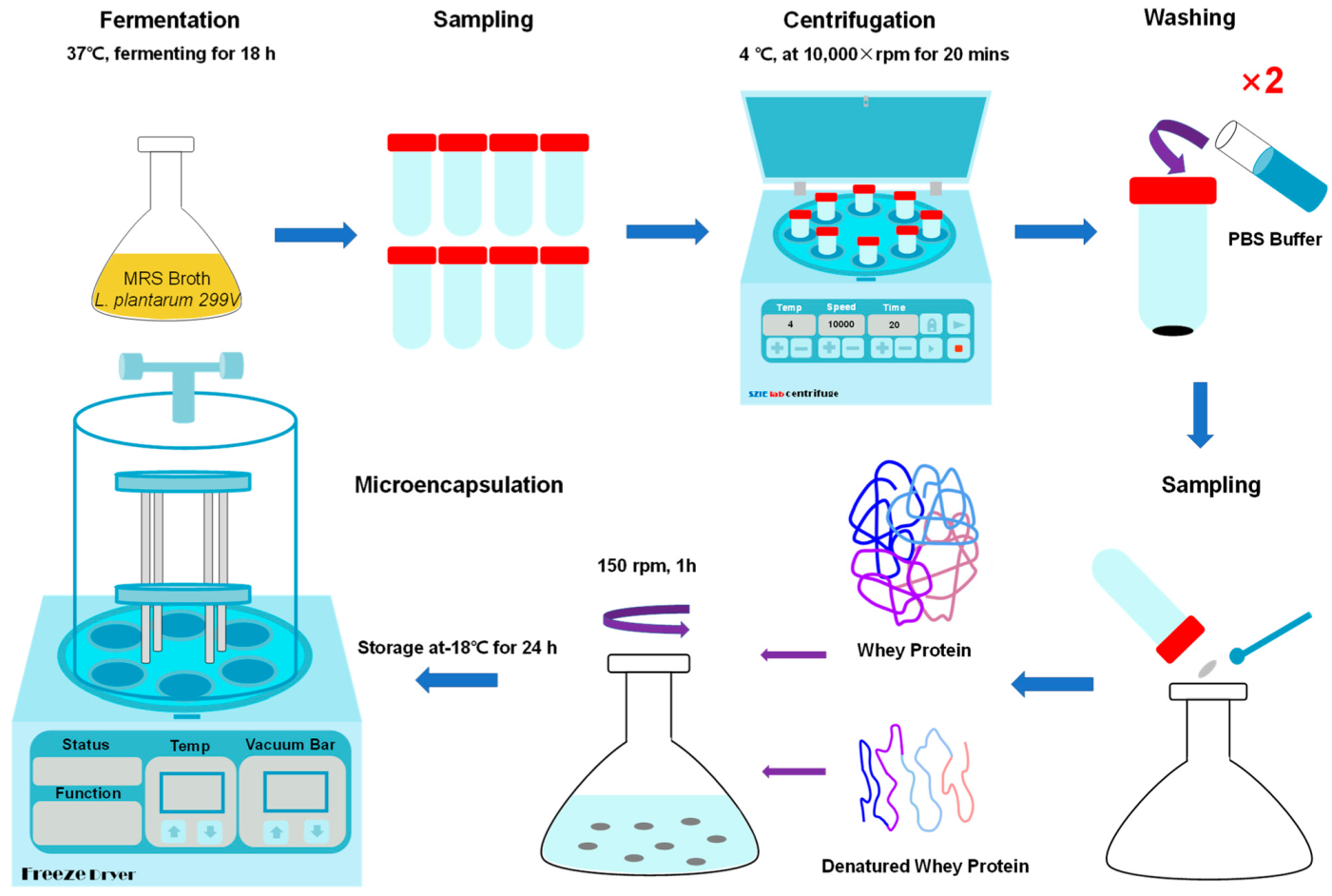
Figure 1. Scheme of microencapsulation process of L. plantarum 299v with proteins by lyophilization.
Applsci 13 00318 g001
Figure 2. Cell number (A) and encapsulation efficiency (B) of microencapsulated L. plantarum 299v strain with different core-to-wall ratios and wall material ratios. WP: whey protein, DWP: denatured whey protein. Results are shown as the means and standard deviations. The superscript letters indicate significant differences (p < 0.05).
Applsci 13 00318 g002
Figure 3. Yield (A) and bulk density (B) of microencapsulated L. plantarum 299v strain with different core-to-wall ratios and wall material ratios. WP: whey protein, DWP: denatured whey protein. Results are shown as the means and standard deviations. Significant differences (p < 0.05) were indicated by superscript letters.
Applsci 13 00318 g003
Figure 4. Scanning electron microscope (SEM) images of L. plantarum 299v cells under 1000× (A,C,E,G,I,K,M,O,Q,S,U) and 14,000× (B,D,F,H,J,L,N,P,R,T,V) magnification. (A,B) are free cells. (C,D) are core-to-wall ratio 1:1, WP; (E,F) are core-to-wall ratio 1:1, WP:DWP is 3:1; (G,H) are core-to-wall ratio 1:1, WP:DWP 1:1; (I,J) are ratio 1:1, WP:DWP 1:3; (K,L) are core-to-wall ratio 1:1, DWP; (M,N) are core-to-wall ratio 1:1.5, WP; (O,P) are core-to-wall ratio 1:1.5, WP:DWP 3:1; (Q,R) are ratio core-to-wall 1:1.5, WP:DWP 1:1; (S,T) are ratio core-to-wall 1:1.5, WP:DWP 1:3; (U,V) are core-to-wall ratio 1:1.5, DWP. WP: whey protein; DWP: denatured whey protein.
Applsci 13 00318 g004
Figure 5. Viability loss of lyophilized L. plantarum 299v strain with core-to-wall ratios and different formulations at 4 °C and 25 °C. (A): 4 °C, core-to-wall ratio 1:1; (B): 4 °C, core-to-wall ratio 1:1.5; (C): 25 °C, core-to-wall ratio 1:1; (D): 25 °C, core-to-wall ratio 1:1.5. WP: whey protein, DWP: denatured whey protein.
Applsci 13 00318 g005
Figure 6. Viability of lyophilized L. plantarum 299v with different core-to-wall ratios and formulations after exposed to SGJ at 37 °C for 3 h. WP: whey protein, DWP: denatured whey protein. Superscript letters indicate significant differences (p < 0.05) among incubation time for a given microcapsule.
Applsci 13 00318 g006
Figure 7. Viability of lyophilized L. plantarum 299v strain with different core-to-wall ratios and formulations after exposure to SIJ for 6 h at 37 °C. WP: whey protein, DWP: denatured whey protein. Superscript letters indicate significant differences (p < 0.05).
Applsci 13 00318 g007
Figure 8. Cell number of fermented and fortified apple juices. (A): 4 °C, fermentation, core-to-wall ratio 1:1; (B): 4 °C, fortification, core-to-wall ratio 1:1; (C): 4 °C, fermentation, core-to-wall ratio 1:1.5; (D): 4 °C, fortification, core-to-wall ratio 1:1.5. (E): 25 °C, fermentation, core-to-wall ratio 1:1; (F): 25 °C, fortification, core-to-wall ratio 1:1; (G): 25 °C, fermentation, core-to-wall ratio 1:1.5; (H): 25 °C, fortification, core-to-wall ratio 1:1.5. WP: whey protein, DWP: denatured whey protein.
Applsci 13 00318 g008
Figure 9. pH of fermented and fortified apple juices. (A): 4 °C, fermentation, core-to-wall ratio 1:1; (B): 4 °C, fortification, core-to-wall ratio 1:1; (C): 4 °C, fermentation, core-to-wall ratio 1:1.5; (D): 4 °C, fortification, core-to-wall ratio 1:1.5. (E): 25 °C, fermentation, core-to-wall ratio 1:1; (F): 25 °C, fortification, core-to-wall ratio 1:1; (G): 25 °C, fermentation, core-to-wall ratio 1:1.5; (H): 25 °C, fortification, core-to-wall ratio 1:1.5. WP: whey protein, DWP: denatured whey protein.
Applsci 13 00318 g009
Table 1. Composition of sample solutions for microencapsulation.
RatioWall Materials
Formulation
Wall Materials
Ratio (w/w)
WP (g)DWP (g)L. plantarum 299v
(Wet Weight, g)
Microcapsule Solution
Concentration (% w/w)
Total Weight (g)
1:1WP-2002020200
WP:DWP3:11552020200
WP:DWP1:110102020200
WP:DWP1:35152020200
DWP-0202020200
1:1.5WP-3002020250
WP:DWP3:122.57.52020250
WP:DWP1:115152020250
WP:DWP1:37.522.52020250
DWP-0302020250
WP—Whey protein; DWP—Denatured whey protein.
Disclaimer/Publisher’s Note: The statements, opinions and data contained in all publications are solely those of the individual author(s) and contributor(s) and not of MDPI and/or the editor(s). MDPI and/or the editor(s) disclaim responsibility for any injury to people or property resulting from any ideas, methods, instructions or products referred to in the content.