Article

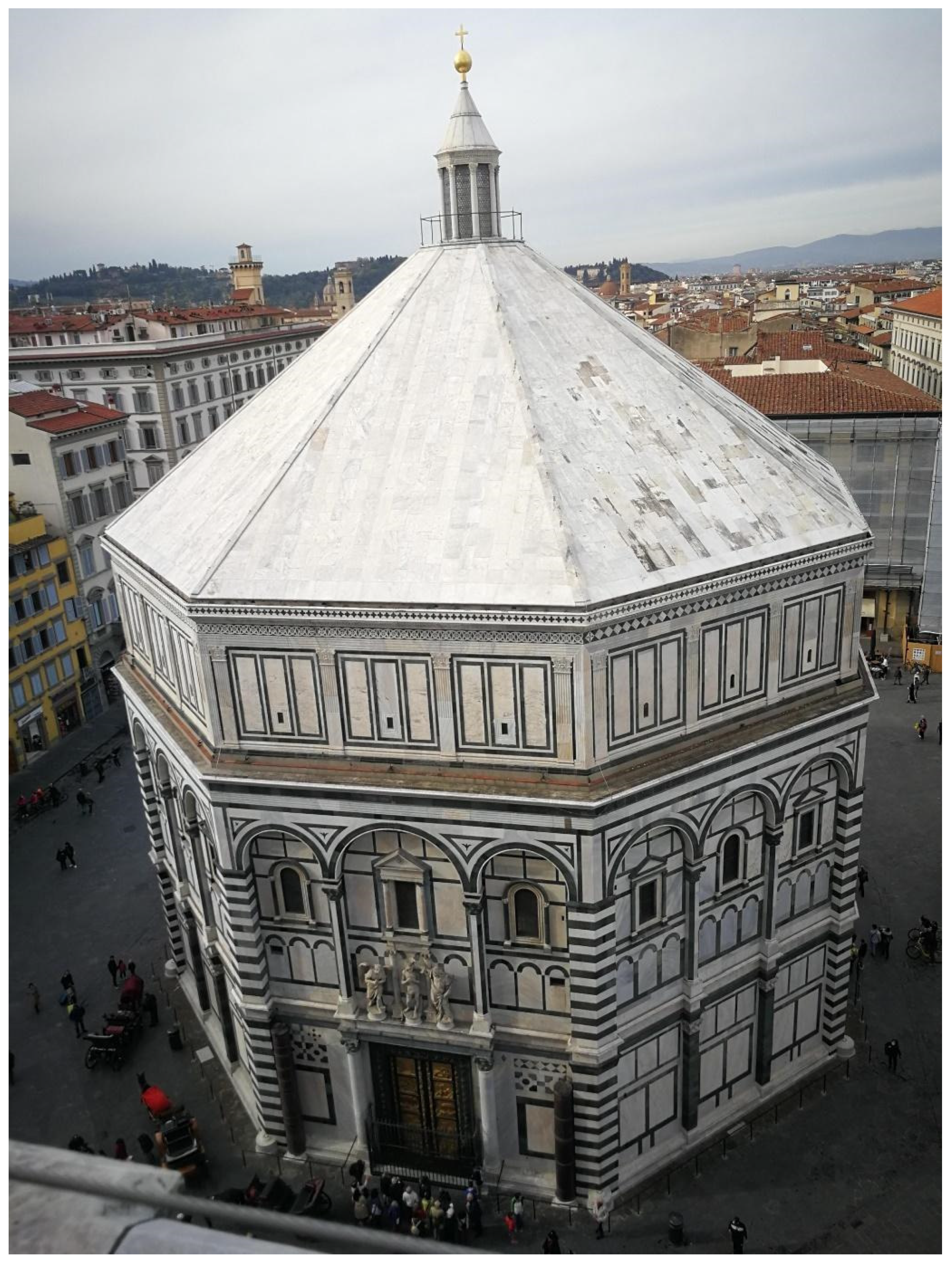
The San Giovanni Baptistery in Florence (Italy): Assessment of the State of Conservation of Surfaces and Characterization of Stone Materials

1
Department of Earth Sciences, University of Florence, 50121 Florence, Italy
2
Chemical Department “Ugo Schiff”, University of Florence, Via della Lastruccia 3-13, Sesto Fiorentino, 50019 Florence, Italy
3
Institute of Heritage Science, National Research Council of Italy, Via Madonna del Piano 10, Sesto Fiorentino, 50019 Florence, Italy
4
Opera del Duomo di Firenze, 50212 Florence, Italy
*
Author to whom correspondence should be addressed.
Appl. Sci. 2022, 12(8), 4050; https://doi.org/10.3390/app12084050
Submission received: 21 February 2022 / Revised: 12 April 2022 / Accepted: 14 April 2022 / Published: 16 April 2022

Abstract

:
During the last restoration campaign of the Baptistery of San Giovanni in Florence (Italy), the assessment of the state of conservation of external surfaces was performed, with a multi-analytical approach, in order to support the conservation and restoration treatments. Black crusts, red staining, sulphation, and organic patinas were identified. Moreover, a complete characterization of marble cladding, bricks, and mortars was carried out. A geochemical and minero-petrographic approach was used in order to classify and assess the provenance of marbles, and define raw materials and technologies of the production for bricks and mortars. Provenances from Italy (Carrara and Lasa marbles) and from Greece (Hymettus and Pentelicum marbles) were identified and attributed to different construction phases, restoration interventions, and re-use of ancient materials. For mortars, the obtained data suggested the use of local materials and traditional technologies for the production. Overall, two different minero-petrographic typologies were identified for the analyzed bricks.

1. Introduction

The Baptistery of San Giovanni, one of the most ancient churches in Florence (Italy), and protected by UNESCO since 1982, sits opposite the city’s cathedral, the church of Santa Maria del Fiore. Octagonal in plan, it is completely clad in slabs of white and green marble, according to the Tuscan dichromic (Figure 1). Raised on the foundations of a Roman building, it is a characteristic example of the Tuscan Romanesque style, dating from around the 11th century (it was consecrated in 1059) [1,2,3,4]. In the early 13th century, the apse was changed from circular to square; the roof was renewed in the 15th century. It is covered by a dome of eight segments resting on perimetral walls, but the dome cannot be detected from the outside because it is concealed by the walls which are raised above the arcade on the second level and crowned by a flattened pyramidal roof. Its maintenance is managed by the Opera di Santa Maria del Fiore (OPA), a private institution established in 1296.
Every conservation project should be based on a sound knowledge of the “object” to be conserved, both from a historical and artistic viewpoint, and from the characteristics of its constituent materials, production technique, and decay conditions [5].
During the last restoration campaign, between March 2014 and September 2015, considering the complexity and heterogeneity of the centuries-old architectural and artistic stratifications of the Baptistery, the OPA promoted a great opportunity for integrated multidisciplinary research and studies to achieve a fuller historical and scientific knowledge about the stone materials used in the Florence Baptistery.
This paper focuses on the characterization of cladding marbles, masonry materials (mortars and bricks), and on the study of their state of conservation. Different kinds of materials (white and veined marbles, mortars, bricks) were analyzed, often belonging to different constructive phases, with the purpose being not only to obtain a complete knowledge of the material, but also to identify the provenance, raw materials, and technologies of production.
The provenancing of white marbles, especially Mediterranean ones, is of great interest for archaeologists and art historians, but it is also one of the most debated problems of petroarchaeometry [6]. In recent years, it has been demonstrated that the application of minero-petrographic and geochemical methods, along with the comparison of data with dataset obtained from quarry samples, allows us to suggest the provenance of white marbles [6,7,8].
In the case of marble slabs of the Baptistery, the research is aimed at identifying reused ancient marbles, the original ones used, and the more recent substitutions.
For the cladding materials, specific research on the main phenomena of alteration/decay and on the presence of traces of previous restoration interventions was carried out in order to address the restoration procedures.
In previous studies, other stone materials were identified, coming from the spolia of Roman ruins in Florence or Fiesole (Porfido Verde Antico, Porfido Rosso Antico, Breccia Greca Antica, Syenite) [9]. Local stones, such as serpentinites (verde Prato) and red marly limestones (“marmi” rossi), were identified and characterized in other Florentine monuments [10,11,12,13].
The paper presents a description of all sampled materials and of the chemical, mineralogical, and petrographic techniques, including optical microscopy (OM), X-ray powder diffraction (XRPD), Fourier-transform infrared spectroscopy (FTIR), thermogravimetric analysis (TGA), micro-Raman, and 13C and 18O isotopic analyzes used to gain a better understanding and characterization of the present materials, as well as their provenance and state of conservation. The results consist of, first of all, an evaluation of the main decay phenomena that affect especially white marble surfaces, an assessment of provenance of white and veined marbles, and a characterization of mortars and bricks.

2. Materials and Methods

During the restoration survey, several powders and micro fragments of 2 cm in size sampled by a scalpel, representative of the different materials (white and veined marbles, bricks, mortars) belonging to different sides of the Baptistery and Lantern, were collected (Figure 2). Figure 2 shows the plan of the Baptistery, and the numbering of the sides assigned during the sampling campaign. Table 1 summarizes the information about the collected samples and the characterization project.
A total of 25 samples of patina and encrustations were investigated in order to characterise the state of conservation and the presence of previous treatments to best address the current restoration campaign; 27 microsamples of marbles were collected from different external sides, on the basis of macroscopic differences, and for the identification of the provenance; 17 core samples of mortars were gathered, from the Attic above the Matroneo, from the external and the internal walls, and from the foundations, at different depths, in relation to the different construction phases, and in order to establish the raw materials and the technologies used; 4 samples of bricks, representative of different sizes, were sampled from side 4. The selection of analyzed bricks was guided by different dates obtained with thermoluminescence technique [14].
All of the samples were first observed under a Zeiss stereomicroscope Stemi 200 C model, with a dedicated camera, and then fragments from each sample were divided and treated according to the protocol described in each technique used for investigating the sample characteristics.
Thin and cross sections were obtained by embedding the sample in epoxy resin (Epofix). In general, cross sections were observed with the optical microscope under reflected light under different magnifications, according to the available microscopy objectives. In this case, a Nikon Eclipse E600 optical microscope was used. Oriented thin sections (30 μm thick) were observed in transmitted light with a ZEISS Axioscope A1 microscope at different magnifications. The image analysis software Axiovision was used for the morphological and morphometric observation, for the evaluation of binder/aggregate ratio in the case of mortars [15,16,17], and of groundmass/ framework for bricks [18]. In the case of marbles, the petrographic observation allowed for the identification of MGS (maximum grain size), the fabric type (homeoblastic or heteroblastic), the main features of microstructure, and the crystal boundaries shapes, identified as the most relevant petrographic parameters for provenance determination [6,8]. For mortars, the petrographic approach allowed for a complete characterization of each component (binder, aggregate, and inorganic additives), as well as the identification of raw materials and technologies used for their production [17,19,20]. In the case of bricks, the optical characteristics of groundmass, the presence and composition of framework, and the quantification of macroporosity could be obtained [21,22]. Optical microscopy was used also for the definition of macroporosity and the characterization of morphological aspects of black crusts and patinas.
X-ray powder diffractometry (XRPD) was employed to determine the mineralogical composition using a powder X-ray diffractometer (Cu anticathode (k = 1.54 Å)), under the following conditions: current intensity of 30 mA, voltage 40 kV, explored 2 h range between 3 and 70°, step size 0.02°, and time to step 50 s. The XRPD analyzes were performed on bulk samples of mortars, bricks, marbles, black crusts, and patinas.
Isotopic analyzes (δ13C, δ18O) were carried out on powders of marbles using a mass spectrometer (Finnigan MAT Delta Plus) equipped with Finnigan Gas-Bench II. Sample and standard mineral powders were treated with phosphoric acid for 1 h in sealed glass containers previously flushed with helium. The CO2 produced was introduced into the mass spectrometer in continuous flow mode, using helium as carrier. The gas from the sample was run along the with reference CO2 gas of a known composition. The experimental values were normalized against international (NBS-18) and internal standards. The mean external reproducibility was better than 0.1 for δ13C and δ18O.
Fourier-transform infrared spectroscopy (FT-IR) was performed for the determination of patina and encrustations through a spectrometer Alpha Bruker. The acquisition of spectra was carried out directly on site by using the instrument equipped with the external total reflectance module for a contactless acquisition and a video camera. Spectra were collected in the range of 7000–400 cm−1 at a resolution of 4 cm−1 and 128 scans in order to optimise the signal-to-noise ratio, on a 4 mm diameter area. FT-IR analysis of powders were conducted, instead, with the ATR (attenuated total reflectance) configuration. The acquisition was carried out in the spectral range between 4000 and 400 cm−1, with a resolution of 4 cm−1 for 24 scans. All of the acquired spectra were elaborated using the OPUS 7.2 software by Bruker.
A micro-chemical analysis of red and yellow patinas was carried out with a Renishaw single-grating (1200 grooves/mm) RM2000 micro-Raman spectrometer and charge-coupled device (CCD) air-cooled (577 × 400 pixels) detector. Spectra were recorded using a near infrared diode laser (785 nm) employed as an excitation source. Laser beam focusing was accomplished through a 50× magnification objective that provided a 1.5 μm laser spot diameter. Neutral density filters were used to attenuate the laser power in order to avoid phase transformations. The laser power at the analyzed samples was within the range 1–2 mW. The spectral range investigated was between 200 and 2000 cm−1. Acquisition times were in the order of 30 s, and the spectral resolution was 4 cm–1 for the diode laser.
Thermo-gravimetric analysis (TGA) was employed for binder characterization of mortars. Some fragments of each sample were disaggregated using a porcelain pestle, and the fraction passing through a sieve with 63 µm openings (ISO R 565 Series) was considered as a binder-enriched specimen. TGA was used to evaluate the presence and the amount of particular volatile compounds (essentially H2O, CO2) in the samples. TGA was conducted in the range 110–1000 °C on approximately 5 mg of sample, and dried (silica gel as drying agent) at room temperature for at least a week under the following experimental conditions: open alumina crucibles, heating rate of 10 °C/min, and 30 mL/min nitrogen gas flow. TGA was used for classifying the studied samples as hydraulic or non-hydraulic mortars, as suggested by most authors [23,24,25]. TGA analyzes were performed using a Perkin Elmer Pyris 6 system on mortar samples.
A list of different types of materials analyzed and of methodologies applied for each material are reported in Table 1.

3. Results and Discussion

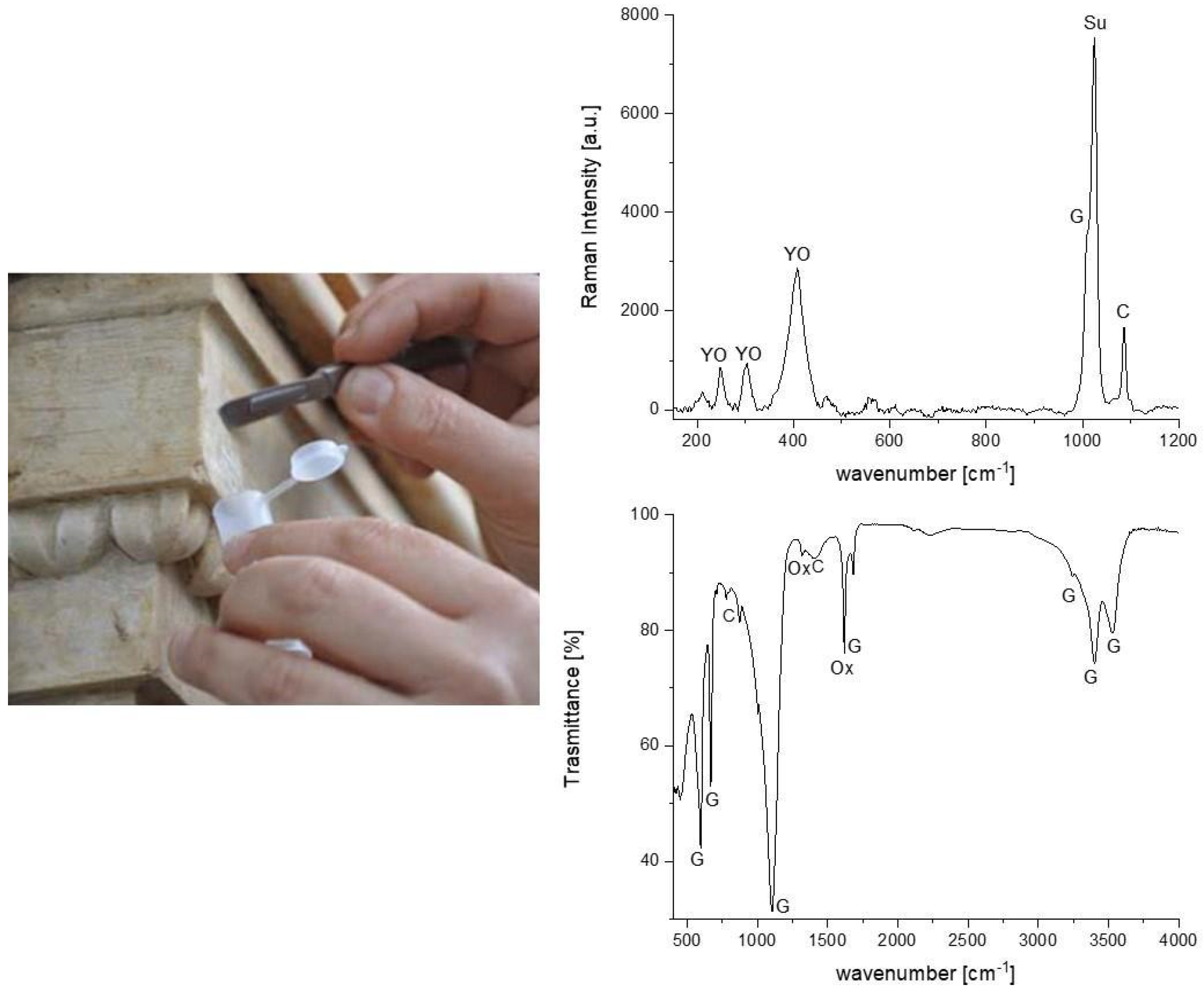
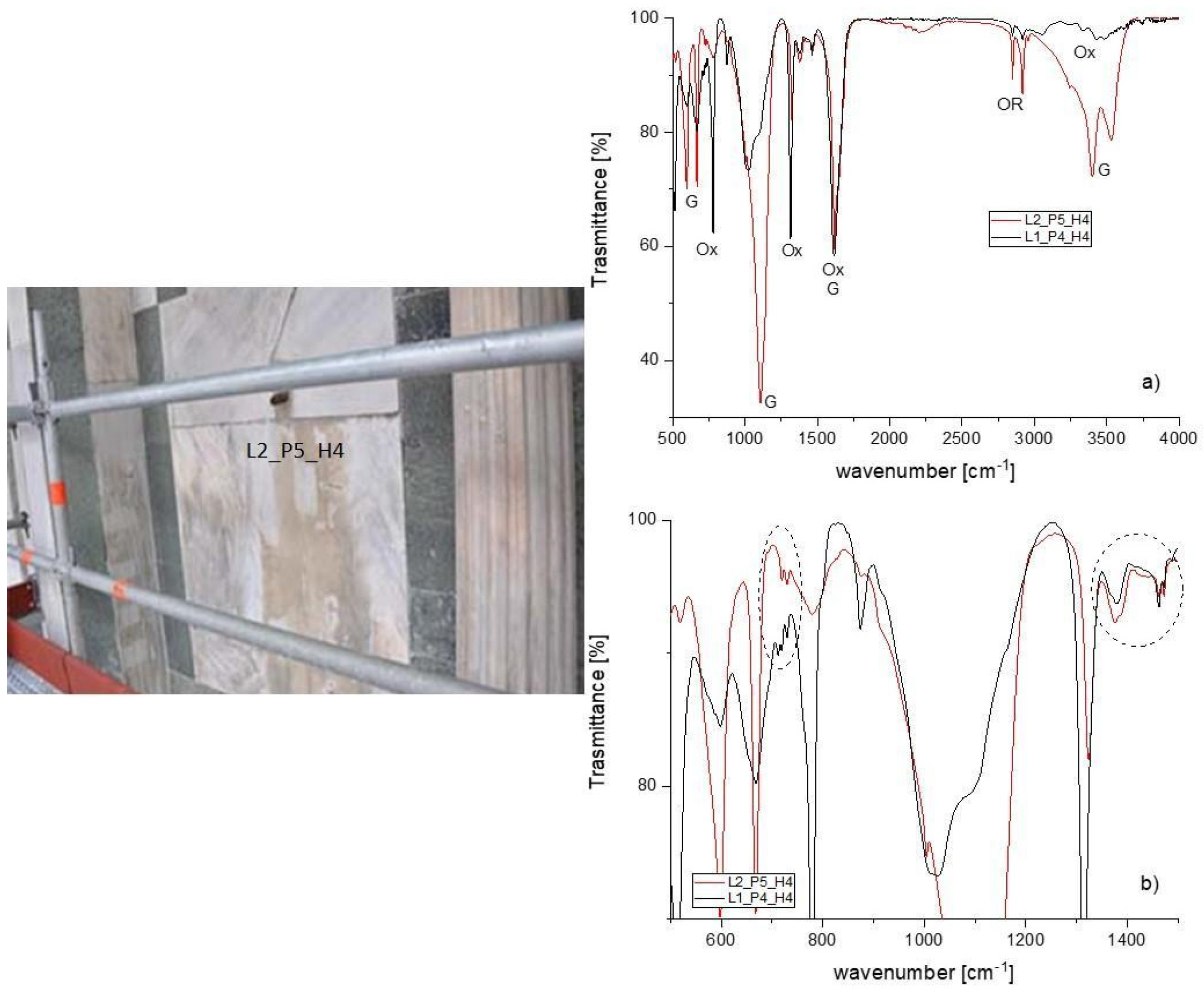
3.1. Evaluation of the State of Conservation: Main Decay Phenomena

An important aspect has been the technical–scientific support to the restoration interventions, mainly aimed at cleaning, through the diagnosis of the altering processes of the materials of the external cladding of the San Giovanni Baptistery, especially white marble.
In Table S1, the main results of state of conservation of external sides of San Giovanni Baptistery (Florence) are reported.
Many capitals, cornices, and decorated cornices of the windows and columns, beyond the intensity of the black crusts or deposits of dirt, were often affected by various decay phenomena, not only chemical–physical (e.g., sulfation), but also physical–mechanical (e.g., abrasion, decohesion, granular disintegration, with potential displacement and detachment, and differences in macro and micro porosity) (Figure 3 and Figure 4).
Laboratory analyzes of the patina and encrustations provided the main operational indications to the restoration technicians for cleaning: these phenomena are mainly attributable to the photochemical smog and to the use of protective organic products, which, with time, have degraded. The films are often decayed and discontinuous, presenting colors from pale pink to yellow, to ochre or brown.
Regarding the identification of the compounds forming the patina, almost identical qualitative results were obtained for all samples analyzed (Figure 5). FT-IR spectra exhibited typical vibration bands of calcium sulphate hydrate, commonly called gypsum (CaSO4·2H2O), centered at 1109, 669, and 596 cm−1, as well as the stretching and deformation vibrations of the O-H bond of water at 3525, 3492, 3401 cm−1, and at 1692 and 1627 cm−1, respectively. In several analyzed samples, a representative band of silicates was present at 1036 and 1006 cm−1 (Si-O stretching vibrations). Gypsum originates from the transformation of calcite in the presence of sulphur oxides, while silicate was subsequently embedded in the crust due to the deposition of wind-borne particles.
Most notably, the prominent bands of calcium oxalate (CaC2O4·nH2O) at 1324 (and 780 cm−1 were present. The features determined in the spectra suggested the presence of the mineralogical phase of calcium oxalate monohydrate, whewellite (CaC2O4·H2O) (Figure 5). This identification was due to the presence of very specific bands at 3487, 3435, 3341, and 3068 (O-H stretching bands), 1624 and 1324 (C=O stretching vibration), and 780 and 674 cm−1 (O-C-O bending vibrations).
Calcium oxalate films are very stable, less soluble, and hardly affected by atmospheric pollution. Wherever the patinas, sometimes caused problematic removal, are discontinuous, especially near the friezes and decorative modules, the underlying marble appears detached, and a sugar-like crumbling can be observed.
In several darker patinas, the presence of organic matter in the FT-IR spectra was highlighted exclusively by the weak bands at 2916 cm−1 and 2841 cm−1 (Figure 6a,b), attributed to the stretching asymmetric and symmetric vibrations, respectively, of methyl and methylene groups, the bands in the region (1470–1375 cm−1) due to CH2 bending vibrations, and the methylene group rocking vibrations near 720 cm−1. Figure 6b shows typical vibrations of the microcrystalline wax used as a protective treatment employed in the previous restorations.
These patinas can be attributed to not only the remains of old protective treatments, but also to substances applied to adjust the color of new marble elements to those which are older. This is the case of the sample in Figure 7, where the Raman analysis shows the presence of a yellow brownish pigment, yellow ochre (iron oxides and hydroxides), calcite (1086 cm−1), gypsum (1007 cm−1), and calcium sulphate (1019 cm−1). The results suggest that the yellow patina is linked to a previous maintenance intervention of color toning of the window frame: in fact, it is assumed that it has been given a coating with lime water and yellow ochre.
Previous research, consisting of a multi-analytical approach for the study of the presence of red discouloration on the white marble of the external cladding of the Bapstistery, pointed out that the red staining is due to the presence of minium (lead tetraoxide) concentrated prevalently in the calcite crystal boundaries [26].

3.2. Marbles

In the Table 2 the mineralogical, petrographic, and isotopic features of white and veined marbles are reported.
Only a multi-analytical approach allows us to identify, with good probability, the origin of a marble: the minero-petrographic analyzes of thin sections and the data from the stable C and O isotope ratio are the most consolidated methods [6,27,28,29,30,31,32].
Mineralogical and petrographic characteristics (microstructure, significant accessory minerals, maximum grain size (MGS) and boundary types of the calcite and/or dolomite crystals) are related to the provenance of marbles, since they are related to the type and degree of metamorphism undergone by the original sedimentary protolith. A first general fabric classification is based upon the equal or different dimensions of the calcite or dolomite crystals, referred to as homeoblastic (HO) or heteroblastic (HE) classification, respectively. The grain size is connected with the metamorphic grade and the MGS (maximum grain size) is reported in the most used database as one of the most diagnostic parameters. Moreover, the shape of the calcite or dolomite crystal boundaries reflects the blastesis/deformation relationships during a tectono-metamorphic event that generated marble crystallization [7,8].
The isotopic ratios of C and O are related to the primary genetic (sedimentation) and evolutionary (diagenesis-metamorphism) processes that have affected the formation of marbles [33].
Combining minero-petrographic features and isotopic data (Table 2), different provenance for marbles used in the San Giovanni Baptistery can be identified:
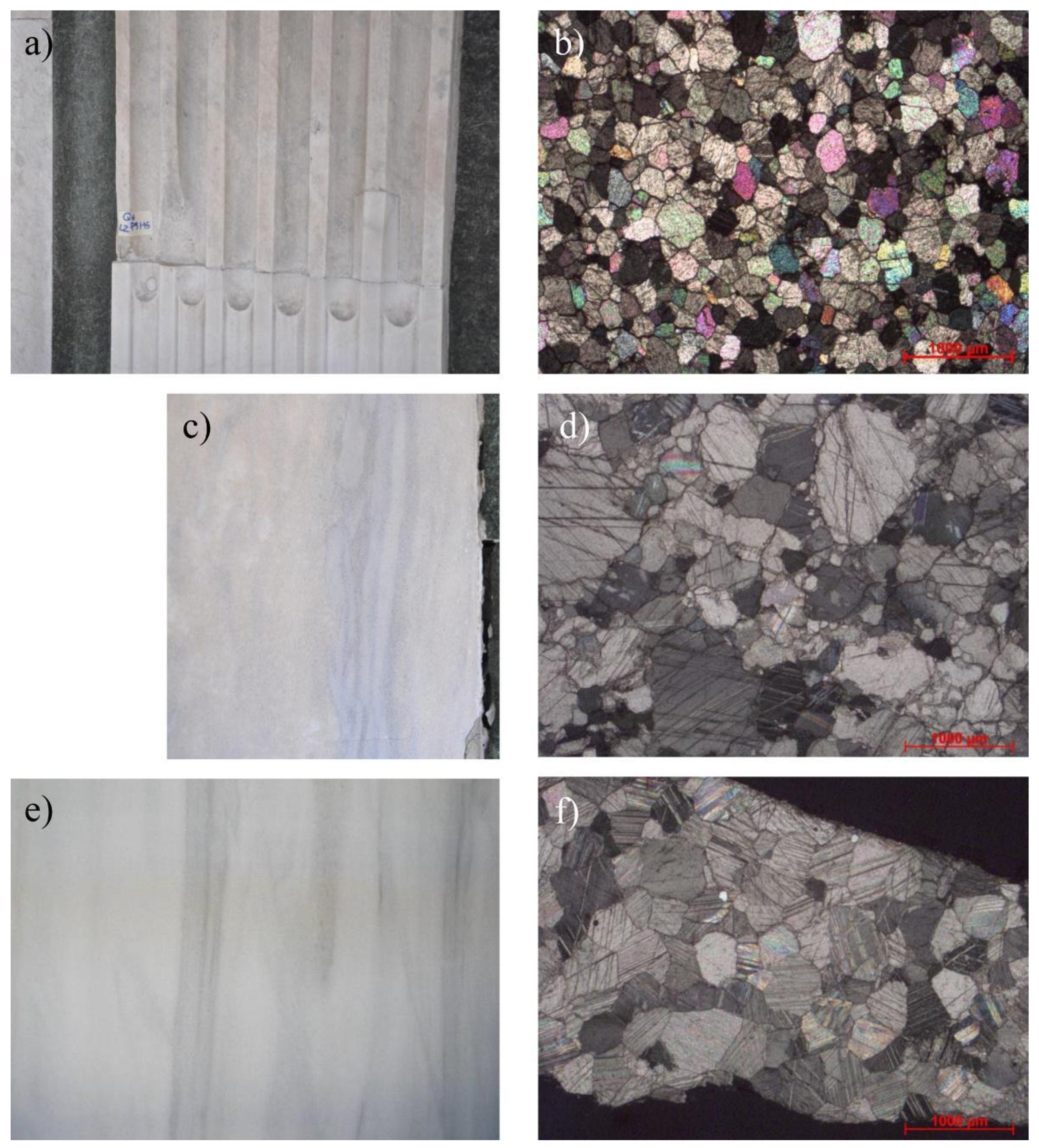
  • Carrara marble (Italy), white and veined, with a typical homeoblastic fabric, a mosaic-polygonal microstructure, with straight grain boundaries, MGS ranging from 150 to 500 micron, and isotopic ratios of C from 1.63 to 2.35 and O from −3.25 to −1.16, is used in the original and substituted portions of monument (Figure 8a,b);
  • Hymettus marble (Greece), likely coming from the spolia of Fiesole Roman Theatre, Capitolium, and Thermae [9] is a white bluish marble characterized by grey/blue veins. The fabric is heteroblastic, with mosaic, lineated microstructure, curved grain boundaries, and an MGS ranging from 1 mm and 2 mm. The isotopic ratio varies from 3.35 to 2.33 for C and from −3.15 to −1.97 for O (Figure 8c,d);
  • Lasa marble, used in the 20th century for the substitution of the most weathered slabs, comes from Laas (Lasa, South Tyrol, Northern Italy), and was selected by restorers for the macroscopic similarities to Hymettus marble (Figure 8e) [34]. It is a pure calcitic marble, containing small amounts of quartz, with a homeoblastic fabric and mosaic microstructure. The grain boundaries are prevalently curved (Figure 8f). The isotopic ratios of C and O fall in the range of data published on Lasa marbles [35];
  • Other ancient marbles are most probably re-used marbles (i.e., Paros, Pentelicum) for some decorative elements (Figure 4).
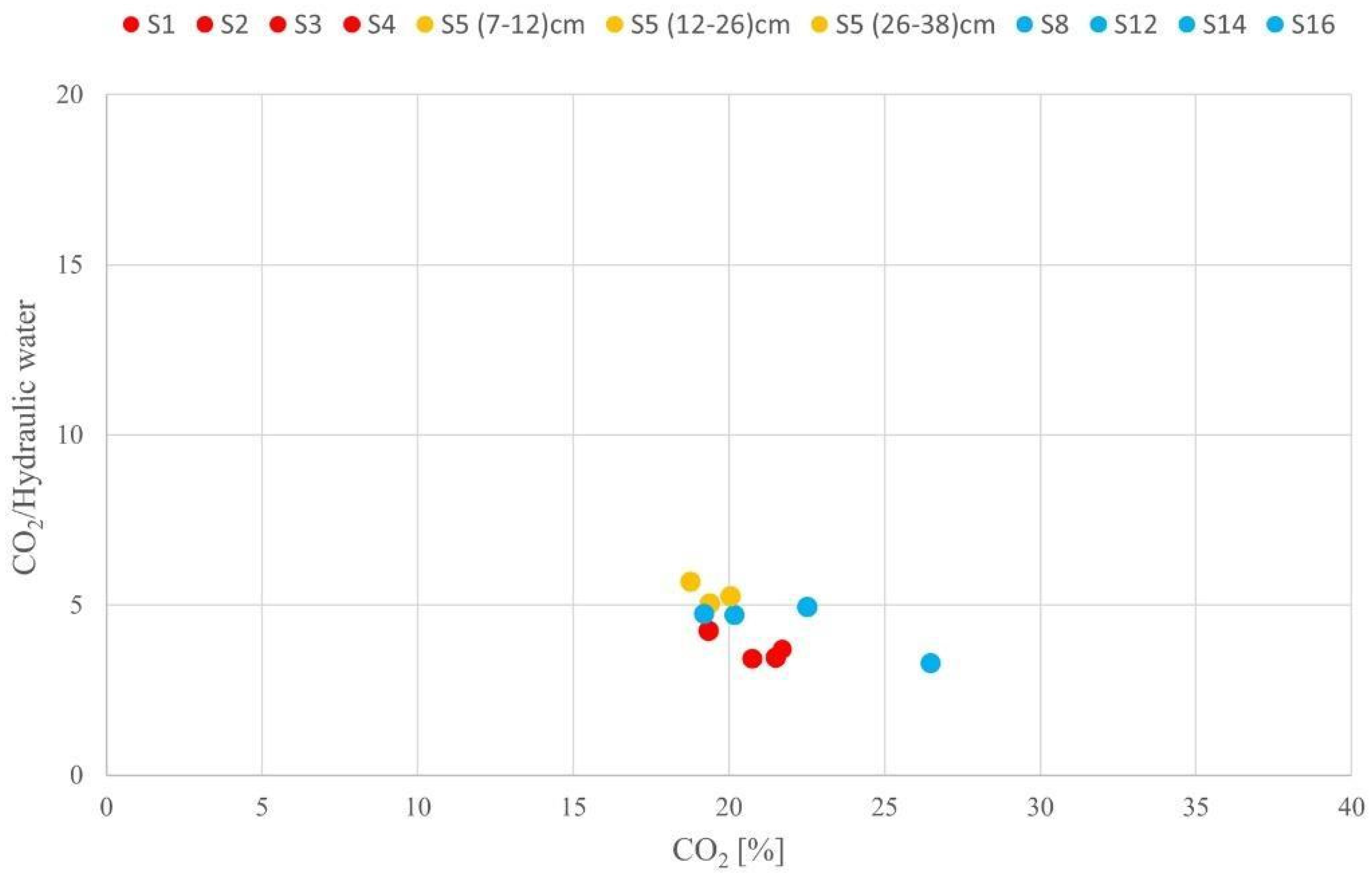
Some uncertainties remain for some samples due to insufficient material available to perform all of the necessary analyzes. In fact, for M3 and M7 samples, obtaining uncontaminated powders for isotopic analyzes was impossible.
The analytical data obtained on provenance of marbles were used to upgrade the database of materials presented in [9,36].

3.3. Mortars

The main mineralogical and petrographic characteristics of analyzed samples are summarized in Table 3 and Table 4, respectively. The analyzed mortar samples belong to: attic, S1, S2, S3, and S4; women’s gallery, S5, and several masonry of basements, from S6 to S17 (summarized in Table 3).
The mineralogical analysis indicates the presence of quartz, calcite, plagioclase, and micas in all samples. Calcite refers to the binder and aggregate fractions; this is due to the presence, in the aggregate, of carbonate fragments of rocks, such as micritic limestone, marly limestone, and single calcite crystals. Semi-quantitative results indicate the presence prevailing of quartz, feldspars (plagioclase and K feldspar), and minor amounts of phyllosilicates (mica-like minerals and chlorites), referred to as the aggregate composition. K feldspars are not present only in the samples S1, S4, S6, S7 (deepest sample), S9 (except in intermediate depth sample), S10 (most superficial sample), S13, and S16; while chlorite is not present in the samples S6, S9 (most superficial sample) S10, S11, S12, and S16. A calcium carbonate polymorphous, vaterite, was individuated in samples S7 (most superficial sample), S12, and S13. Vaterite was typically found in the Florentine mortars, associated with the production of natural hydraulic binders obtained by burning marly limestone [17,19,37]. The presence of portlandite (Ca(OH)2) in the deepest sample of S4 was noted, linked to an incomplete carbonation of binder. Gypsum was registered only in S13, belonging to the basement, most probably due to the sulphation phenomena of the binder.
The petrographic observation (Table 4) performed on Baptistery mortar samples shows, mainly, a binder with a heterogeneous structure and micritic/microsparitic texture, as well as the presence of lumps, both as remnants of the binder not being well mixed and as remnants of overburnt limestone fragments. An exception is represented by S12 sample, which came from the basement, collected from the mosaic masonry, composed of lime hydraulicized with crushed ceramics.
In some cases, the binder of the mortars was subjected to processes of dissolution and recrystallization by aqueous solutions circulating in the masonry, and occurred inside the pore space. The recrystallizations are present in the majority of samples, except in S1, S3, S14, S17, and in the internal samples S5, S7, and S8. Often, the absence of dissolution and recrystallization phenomena is correlated to the homogeneous characteristic of the binder, as shown in samples S3 and S14. The aggregate is, on average, abundant, mainly with homogeneous distribution (except in S5, which is the most superficial sample, and S10, S12, S13, which present heterogeneous grain size distribution) and well selected grain size composed of quartz, feldspars, mica, calcite, and fragments of carbonate rocks.
The core samples extracted from the attic, samples S1, S2, S3, and S4, were obtained by mixing a natural hydraulic binder with an abundant aggregate (B/A 1/3–1/4), characterized by numerous lumps (Figure 9a) and heterogeneous structure and micritic/microsparitic texture (Figure 9b), with the exception of S3, which presents homogeneous structure and micritic texture (Figure 9c), and S4, which is composed by lower B/A, equal to 1/2–1/3. Lumps observed in the binder are useful for identifying the limestone burnt to obtain lime, which appears in the local Alberese limestone (Figure 9a). The aggregate has a homogeneous grain size distribution, ranging from 1 mm for rock fragments to 100 µm for monocrystals. Quartz, plagioclases, rock fragments (quartzite, sandstones, calcarenite, micritic limestone), and few cocciopesto fragments were identified under optical microscopy. The aggregate shape is sub angular, and a medium macroporosity due to pores of irregular shape was observed. In S4 (20–35 cm), the macroporosity is more developed (medium-high) and characterized by a recrystallized binder. Some differences in samples belonging to different core depths have been classified by grain size distribution and composition of aggregate, but the production of the binder has been the same.
For core sample S5, which was obtained from the women’s gallery, three samples extracted from different depths were investigated. Mortars were realized with a natural hydraulic binder, numerous lumps referred to the binder being not well mixed, and remnants of overburnt marly limestones, identified as the local Alberese limestone, were observed (Figure 9d). All mortar samples suffered a strong recrystallization of the binder in the form of spathic calcite. In different depths of S5, some differences in the aggregate characteristics were present: S5 (7–12) cm was composed of abundant binder/aggregate (B/A 1/3) and fine grain size predominant, while S5 (12–26) cm and S5 (26–38) cm were characterized by less binder/aggregate (B/A 1/2–1/3), and a fine-medium size was predominant (Figure 9e). Furthermore, a different amount of carbonate rock fragment was registered, with respect to the attic mortar samples (a higher entity in S5 samples).
For samples S6 to S17, belonging to the basement of the Baptistery, a binder made with natural hydraulic lime with a heterogeneous structure and micritic/microsparitic texture were observed. They show some differences: the binder of the S13 sample has a microsparitic texture (Figure 9f), and S14 has micritic texture (Figure 9g), while the binder of remaining samples has a micritic/microsparitic aspect (Figure 9h). The binder of the major parts of samples are characterized by a relevant recrystallization and heterogeneous structure. Some differences have also been registered in grain size distribution and aggregate composition for the same mortar core. A fair amount of carbonate rock fragments was registered, with abundant presence in S6, S10, S11, and S12 (S6 in Figure 9i). In the mortar basement samples, the aggregate has a homogeneous grain size distribution ranging from 800 µm−1 mm to 100 µm, and in S10 and S13 a heterogeneous grain size distribution was observed (grain size 800–100 µm). The aggregate shape is sub angular/sub rounded, with a high microporosity due to pores of irregular shape observed.
In sample S12, coming from the basement and collected from the mosaic masonry, crushed ceramic (or cocciopesto) was added to the mix consisting of air hardening calcitic lime and fine quartzitic/carbonate aggregate, and the ratio of B/A is 1/2–1/3. The crushed ceramics have a hydraulic function, because the original clay minerals, during firing, become amorphous, and may react with the lime (calcium hydroxide) to form hydrated calcium silicates and hydrated calcium aluminates [17]. Lime hydraulicized with crushed ceramics, a heterogeneous structure, a micritic/microsparitic texture, and an absence of lumps were observed (Figure 9j).
The TGA analyzes were performed on representative samples: the selection was based on petrographic observations. The samples from attic: S1 (10–15) cm, S2 (9–14) cm, S3 (0–4) cm, S4 (4–20) cm; from the women’s gallery: S5 (7–12) cm, S5 (12–26) cm, S5 (26–38) cm; and from the basement S8 (21–38) cm, S12 (0–4) cm, S14 (0–4) cm, and S16 (4–12) cm were analyzed. The variation of hydraulic behavior in the depth were evaluated in samples collected from the women’s gallery (S5). Figure 10 shows the plot of the percentage of weight loss attributed to CO2, as well as the percentage of weight loss attributed to hydraulic water (CO2/H2O ratio), and % CO2, the significant parameters for identifying the type of binder used in the mortar mixture [24]. The presence of hydraulic components is registered through weight loss in the 200–600 °C temperature range, indicated as hydraulic water (%), the values of which range from 3.82% to 8.11%. The parameter %CO2 is observed through weight loss in the 600–900 °C temperature range, and these values vary between 18.77% and 26.50%, resulting a material with a hydraulic behavior. The samples extracted from the same area present consistent hydraulic water and %CO2 values. TGA results show that the samples from the attic, women’s gallery, and basements of the Baptistery fall in the typical cluster of hydraulic mortars, and are characterized by a rather homogeneous hydraulicity. In the same cluster, but away from the other samples, S12, has a different setting reaction of binder (setting and hardening both through carbonation of calcium hydroxide and through reaction of calcium hydroxide itself with the hydraulicizing components), which is present in petrographic observation [17]. The S5 samples (analyzed at three different depths) show small differences among samples selected at different depths, suggesting that there is a slight variation of hydraulic behavior. The TGA results confirm the hydraulic behavior, as suggested by the presence of lumps of unburnt Alberese limestone in petrographic observations. The firing of marly limestone provides natural hydraulic lime, typically produced in the Florentine area [19,37,38,39].
Finally, the mortar samples show, mainly, the presence of lumps which are typical of Alberese limestone, suggesting the use of this stone to produce lime. Moreover, their presence in the mixtures testifies to traditional technologies. This raw material is able to confer to the mortar good performance, durability, and hydraulic behavior, as observed from the TGA results. Through the analysis of core samples at different depths, no substantial differences were found, and incomplete carbonation of the binder was observed in the deepest portion. The mortars of the Baptistery are composed of aggregate consisting of variable compositions (different amounts of monocrystals, carbonate, and silicate rock fragments) and abundance (B/A ranges from 1/1–1/2 to 1/3–1/4). In attic and basement samples, a predominance of carbonate rock fragments was registered. The macroporosity was medium in the attic and women’s gallery mortar samples, and high in the basement samples, due to pores of irregular shape. The high porosity and the position of basement mortars involve a binder characterized by a relevant recrystallization, as revealed in petrographic observations. The sample, collected from the mosaic masonry, is a hydraulic lime mortar used for the addition of cocciopesto. This artificial material, obtained by crushing bricks or ceramic, reacts with the air hardening lime binder, developing amorphous hydrated calcium silicates and hydrated calcium aluminates which are able to set under conditions of high humidity or under water, thereby allowing the preparation of mortars with good mechanical characteristics and durability [19,20]. In the past, cocciopesto was mainly used for the preparation of mortars with different functions (floors, plasters, tanks, aqueducts) when natural pozzolans were unavailable, and has also been found in the preparation of mosaic layers [40].

3.4. Bricks

The mineralogical and petrographic characteristics of the brick samples coming from side 4 of the inner dome are summarized in Table 5. XRPD data show the presence of quartz, calcite, and plagioclase in all of the examined samples; muscovite is identified in samples 1A and 1D, traces of diopside were identified in sample 1B, and K feldspar in sample 1A. All of the detected minerals can be ascribed to raw materials used for the production of bricks, with the exception of diopside, which can nucleate for the reaction between carbonates and clay materials [21,41,42]. The coexistence of this newly formed mineral and calcite is quite a common occurrence, although the latter is not stable at high temperatures. Its presence can be explained by considering a secondary recrystallization due to the movement of Ca-rich solutions in the walls.
Concerning the petrographic characteristics, two typologies have been identified: the first one (samples 1A and 1B) is characterized by a red colored paste (Figure 11a). The groundmass has a low birefringent with a medium amount of framework (ratio groundmass/framework 1/2). The framework is composed of quartz, plagioclase, and K feldspar. The maximum grain size of the framework’s components is 300 μm, the texture is not oriented, and secondary recrystallization of calcite in macroporosity is observed. The second typology (samples 1C and 1D) has a light red colored paste, and more abundant framework (ratio groundmass/framework 1/3) made of quartz, micas, plagioclase, and carbonate rock fragments (micritic limestones) (Figure 11b). The framework is not well sorted and selected. A bimodal grain size distribution was observed for sample 1D. A medium macroporosity was registered.
Thermoluminescence (TL) dating [14,43] of these samples revealed two different ages: 4th–5th century, and around the 20th century.
The first date is probably linked to the re-use of most ancient bricks, while the 20th century must be considered a post quem term for the building of the structure. The limited number of samples analyzed and dated cannot be considered exhaustive of the resolution of the chronological problematics in such a complex building.
A specific research project devoted to these materials would be desirable.

4. Conclusions

This research confirms the importance played by the mineralogical–petrographic and chemical characterisation of the stone materials utilized in historical architecture in order to understand their use, the role played in the urban development, and the durability towards atmospheric agents from the perspective of conservation. The results obtained represent a further milestone for better management of future restoration interventions, especially in terms of choosing of the best cleaning procedures for historical and monumental complexes.
To design the restoration interventions and maintenance of the Baptistery of San Giovanni, one of the most important churches in Florence (Italy) and protected by UNESCO since 1982, requires a complete characterization of the state of conservation and natural and artificial materials. The results are relevant for the knowledge of: (i) main phenomena of cladding alteration/decay, (ii) provenance of marbles, and (iii) the identification of raw materials and technologies used for their production of mortars and bricks.
The identification of altering processes in the materials provided the main operational indications to the restoration technicians for cleaning. The external surface presents patina and encrustations due to the photochemical smog, as well as the use of old protective treatments products which, with time, have degraded. The eight sides of the Baptistery have different states of conservation, both in terms of the characteristics of the materials, and of the different exposures to atmospheric agents. A conservative intervention aimed to remove the layers of black encrustations and the deposits of pollutants, as well as to consolidate the marble elements that have deteriorated over time, has been performed.
The different provenance of white and veined marble allows us to identify the construction phases, the restoration interventions, and the re-use of ancient materials. In this way, it was shown that the workshop originally used marble from Carrara (Carrara marble), and from Greece (Hymettus and Pentelicum marbles), the latter of which are most probably re-used marbles from the spolia of ancient buildings. After that, marble from Carrara and Laas (Lasa marble) were used for replacement interventions.
The mortars of the Baptistery consist of a natural hydraulic lime binder, obtained through the burning of a marly limestone, testified to be under-burnt fragments of Pietra Alberese (Monte Morello formation). This raw material is able to confer to the mortar good performance, durability, and hydraulic behavior, as observed from the TGA results. The aggregate is composed of silicate and carbonate components. Although all of the core samples were obtained from quite similar raw materials, different technological characteristics were observed: B/A ratio, sieving and mean grain size of aggregates, and macroporosity.
The two sets of brick samples, attributed to different ages (4th–5th and 20th century) through the thermoluminescence method, have different minero–petrographic features.

Supplementary Materials

The following supporting information can be downloaded at: https://www.mdpi.com/article/10.3390/app12084050/s1, Table S1: Main results, obtained with a multi-analytical approach, on state of conservation of external sides of San Giovanni Baptistery (Florence).

Author Contributions

Conceptualization, E.C., C.A.G. and B.A.; methodology, E.C. and S.V.; investigation, S.C., E.C., S.V. and M.R.; data curation, S.C., E.C., S.V. and M.R.; writing—original draft preparation, S.C., E.C., S.V. and M.R.; writing—review and editing, S.C., E.C., S.V. and M.R.; supervision, C.A.G. All authors have read and agreed to the published version of the manuscript.

Funding

This research did not receive any specific grant from funding agencies in the public, commercial, or not-for-profit sectors.

Institutional Review Board Statement

Not applicable.

Informed Consent Statement

Not applicable.

Data Availability Statement

Not applicable.

Acknowledgments

The authors are grateful to Opera del Duomo for giving them the opportunity to study the stone materials of the San Giovanni baptistery.

Conflicts of Interest

The authors declare no conflict of interest.

References

  1. Morolli, G. L’architettura del Battistero e l’ordine antico. In Il Battistero di San Giovanni; Paolucci, A., Ed.; Franco Cosimo Panini: Modena, Italy, 1994; pp. 33–132. [Google Scholar]
  2. Cardini, M. L’ipotesi tardo antica del Battistero. In Il bel San Giovanni e Santa Maria del Fiore; Cardini, D., Ed.; Le Lettere: Firenze, Italy, 1996; pp. 62–93. [Google Scholar]
  3. Rocchi Coopmans de Yoldi, G. Il Battistero di San Giovanni: Lo svolgimento della fabbrica. In Santa Maria del Fiore—Piazza, Battistero, Campanile; Rocchi Coopmans de Yoldi, G., Ed.; Università Degli Studi di Firenze, Dipartimento di Architettura, Il Torchio: Firenze, Italy, 1996; pp. 27–72. [Google Scholar]
  4. Degl’Innocenti, P. Misurare, disegnare, conoscere: Dai rilievi del San Giovanni alle ipotesi storico-costruttive. In Il Battistero di San Giovanni, Conoscenza, Diagnostica, Conservazione; Gurrieri, F., Ed.; Mandragora: Firenze, Italy, 2017; pp. 87–103. [Google Scholar]
  5. Siegesmund, S.; Torok, A. Building stones. In Stone in Architectures. Properties, Durability; Siegesmund, E., Snethlage, R., Eds.; Springer: Berlin/Heidelberg, Germany; New York, NY, USA; Dordrecht, The Netherlands; London, UK, 2014. [Google Scholar]
  6. Antonelli, F.; Lazzarini, L. An updated petrographic and isotopic reference database for white marbles used in antiquity. Rend. Fis. Acc. Lincei 2015, 26, 399–413. [Google Scholar] [CrossRef]
  7. Moens, L.; Roos, P.; Rudder, J.D.; Paepe, P.D.; Hende, J.V.; Waelkens, M. A multi-method approach to the identification of white marbles used in antique artifacts. In Classical Marble: Geochemistry, Technology, Trade; Herz, N., Waelkens, M., Eds.; Kluwer Academic Publishers: Dordrecht, The Netherlands, 1988; pp. 243–250. [Google Scholar] [CrossRef]
  8. Lazzarini, L. Archaeometric aspects of white and coloured marbles used in antiquity: The state of the art. Per. Mineral. 2004, 73, 113–125. [Google Scholar]
  9. Coli, M.; Ciuffreda, A.L.; Donigaglia, T.; Bencaster, A.; Caciagli, S.; Agostini, B.; Iandelli, N. Saint John Baptistery in Florence (Italy): Studies for Conservation of the External Marble Cladding. Appl. Sci. 2021, 11, 6329. [Google Scholar] [CrossRef]
  10. Fratini, F.; Rescic, S.; Pittaluga, D. Serpentinite and ophicalcite in the architecture of eastern Liguria and as decoration of Tuscan religious buildings. Resour. Polic 2022, 75, 102505. [Google Scholar] [CrossRef]
  11. Pecchioni, E.; Magrini, D.; Cantisani, E.; Fratini, F.; Garzonio, C.A.; Nosengo, C.; Santo, A.P.; Vettori, S.A. Non-Invasive Approach for the Identification of “Red Marbles” from Santa Maria Del Fiore Cathedral (Firenze, Italy). Int. J. Archit. Herit. 2021, 15, 494–504. [Google Scholar] [CrossRef]
  12. Pecchioni, E.; Cantisani, E.; Fratini, F. La città di Firenze: Un museo di litologia all’aperto. In Il Museo di Storia Naturale dell’Università degli Studi di Firenze; Le collezioni mineralogiche e litologiche; Firenze University Press: Firenze, Italy, 2012; Volume IV, pp. 245–269. [Google Scholar]
  13. Malesani, P.; Pecchioni, E.; Cantisani, E.; Fratini, F. Geolithology and provenance of materials of some historical buildings and monuments in the centre of Florence (Italy). Episodes 2003, 26, 250–255. [Google Scholar] [CrossRef]
  14. Garzonio, C.A.; Cantisani, E.; Coli, M.; Cuzman, O.; Del Luca, D.; Lubrito, C.; Ricci, M.; Vettori, S.; Sibilia, E. I materiali costitutivi del Battistero. In Il Battistero di San Giovanni, Conoscenza, Diagnostica, Conservazione; Gurrieri, F., Ed.; Mandragora: Firenze, Italy, 2017; pp. 179–191. [Google Scholar]
  15. Elsen, J. Microscopy of historic mortars—A review. Cem. Concr. Res. 2006, 36, 1416–1424. [Google Scholar] [CrossRef]
  16. Ingham, J.P. Geomaterials under the Microscope. A Colour Guide; CRC Press: London, UK, 2010. [Google Scholar]
  17. Pecchioni, E.; Fratini, F.; Cantisani, E. Atlas of the Ancient Mortars in Thin Section under Optical Microscope, 2nd ed.; Nardini: Florence, Italy, 2020. [Google Scholar]
  18. Reedy, C.L. Thin-Section Petrography of Stone and Ceramic Cultural Materials; Archetype Publications: London, UK, 2008; p. 256. ISBN 978-1-9049-8233-3. [Google Scholar]
  19. Cantisani, E.; Fratini, F.; Pecchioni, E. Optical and Electronic Microscope for Minero-Petrographic and Microchemical Studies of Lime Binders of Ancient Mortars. Minerals 2022, 12, 41. [Google Scholar] [CrossRef]
  20. Arizzi, A.; Cultrone, G. Mortars and plasters-how to characterize hydraulic mortars. Archaeol. Anthropol. Sci. 2021, 13, 2–22. [Google Scholar] [CrossRef]
  21. Maggetti, M. Phase analysis and its significance for technology and origin. In Archaeological Ceramics; Olin, J.S., Franklin, A., Eds.; Smithsonian Institution Press: Washigton, DC, USA, 1982; pp. 121–133. [Google Scholar]
  22. Pavía, S. The determination of brick provenance and technology using analytical techniques from the physical sciences. Archaeometry 2006, 48, 201–218. [Google Scholar] [CrossRef] [Green Version]
  23. Moropoulou, A.; Bakolas, A.; Bisbikou, K. Characterization of ancient, byzantine and later historic mortars by thermal and X-ray diffraction techniques. Thermochim. Acta 1995, 269–270, 779–795. [Google Scholar] [CrossRef]
  24. Bakolas, A.; Biscontin, G.; Contardi, V.; Franceschi, E.; Moropoulou, A.; Palazzi, D.; Zendri, E. Thermoanalytical research on traditional mortars in Venice. Thermochim. Acta 1995, 269–270, 817–828. [Google Scholar] [CrossRef]
  25. Maravelaki-Kalaitzaki, P.; Bakolas, A.; Moropoulou, A. Physico-chemical study of Cretan ancient mortars. Cem. Concr. Res. 2003, 33, 651–661. [Google Scholar] [CrossRef]
  26. Cantisani, E.; Cuzman, O.A.; Vettori, S.; Chelazzi, L.; Ciattini, S.; Ricci, M.; Garzonio, C.A. A multi-analytical approach for the study of red stains on heritage marble. Analyst 2019, 144, 2375–2386. [Google Scholar] [CrossRef]
  27. Pensabene, P.; Antonelli, F.; Lazzarini, L.; Cancelliere, S. Provenance of marble sculptures and artifacts from the so-called Canopus and other buildings of ‘‘Villa Adriana’’ (Hadrian’s villa—Tivoli, Italy). J. Archaeol. Sci. 2012, 37, 994–1005. [Google Scholar] [CrossRef]
  28. Columbu, S.; Antonelli, F.; Lezzerini, M.; Miriello, D.; Adembri, B.; Blanco, A. Provenance of marbles used in the Heliocaminus Baths of Hadrian’s Villa (Tivoli, Italy). J. Archaeol. Sci. 2014, 49, 332–342. [Google Scholar] [CrossRef]
  29. Antonelli, F.; Columbu, S.; Lezzerini, M.; Miriello, D. Petrographic characterization and provenance determination of the white marbles used in the Roman sculptures of Forum Sempronii (Fossombrone, Marche, Italy). Appl. Phys. A 2014, 115, 1033–1040. [Google Scholar] [CrossRef]
  30. Antonelli, F.; Lazzarini, L. The use of white marble in the central and upper adriatic between Greece and Rome: Hellenistic Stelae from the necropolis of Ancona (Italy). Camb. Archaeol. J. 2013, 23, 149–162. [Google Scholar] [CrossRef]
  31. Attanasio, D.; Boschi, C.; Bracci, S.; Cantisani, E.; Paolucci, F. Provenance Studies of the Marble of Ancient Sculptures in the Tribune of the Uffizi Gallery, Florence. Archaeometry 2015, 57, 74–89. [Google Scholar] [CrossRef]
  32. Attanasio, D.; Boschi, C.; Bracci, S.; Cantisani, E.; Paolucci, F. The Greek and Asiatic marbles of the Florentine Niobids. J. Archaeol. Sci. 2016, 66, 103–111. [Google Scholar] [CrossRef]
  33. Attanasio, D.; Brilli, M.; Ogle, N. The Isotopic Signatures of Classical Marbles; L’erma di Bretschneider: Rome, Italy, 2006; p. 279. [Google Scholar]
  34. Bianchini, P. I Paramenti esterni. I materiali, i restauri degli anni 1938–1944 e cenni sullo stato di conservazione attuale. In Santa Maria del Fiore—Piazza, Battistero, Campanile; Rocchi Coopmans de Yoldi, G., Ed.; Università Degli Studi di Firenze, Dipartimento di Architettura; Il Torchio: Firenze, Italy, 1996; pp. 97–98. [Google Scholar]
  35. Unterwurzacher, M.; Obojes, U. White marble from Laas (Lasa), south Tyrol-its occurrence, use and petrographic-isotopical characterisation. Austrian J. Earth Sci. 2012, 105, 26–37. [Google Scholar]
  36. Iandelli, N.; Coli, M.; Donigaglia, T.; Ciuffreda, A.L. An Unconventional Field Mapping Application: A Complete Opensource Workflow Solution Applied to Lithological Mapping of the Coatings of Cultural Heritage. ISPRS Int. J. Geo. Inf. 2021, 10, 357. [Google Scholar] [CrossRef]
  37. Cantisani, E.; Calandra, S.; Barone, S.; Caciagli, S.; Fedi, M.; Garzonio, C.A.; Liccioli, L.; Salvadori, B.; Salvatici, T.; Vettori, S. The mortars of Giotto’s Bell Tower (Florence, Italy): Raw materials and technologies. Constr. Build. Mater 2021, 267, 120801. [Google Scholar] [CrossRef]
  38. Cantisani, E.; Falabella, A.; Fratini, F.; Pecchioni, E.; Vettori, S.; Antonelli, F.; Giamello, M.; Lezzerini, M. Production of the Roman Cement in Italy: Characterization of a raw material used in Tuscany between 19th and 20th century and its comparison with a commercialized French stone material. Int. J. Archit. Herit. 2018, 12, 1038–1050. [Google Scholar] [CrossRef]
  39. Lezzerini, M.; Ramacciotti, M.; Cantini, F.; Fatighenti, B.; Antonelli, F.; Pecchioni, E.; Fratini, F.; Cantisani, E.; Giamello, M. Archaeometric study of natural hydraulic mortars: The case of the Late Roman Villa dell’Oratorio (Florence, Italy). Archaeol. Anthrop. Sci. 2017, 9, 603–615. [Google Scholar] [CrossRef]
  40. Megna, B.; Rizzo, G.; Ercoli, L. The mortars and plasters under the mosaics and the wall paintings of the Roman villa at Piazza Armerina, Sicily. In Proceedings of the 2nd Conference on Historic Mortars—HMC 2010 and RILEM TC 203-RHM Final Workshop, Prague, Czech Republic, 22–24 September 2010; pp. 275–283. [Google Scholar]
  41. Riccardi, M.P.; Messiga, B.; Duminuco, P. An approach to the dynamics of clay firing. Appl. Clay Sci. 1999, 15, 393–409. [Google Scholar] [CrossRef]
  42. Cultrone, G.; Rodriguez-Navarro, C.; Sebastian, E.; Cazalla, O.; De la Torre, M.J. Carbonate and silicate phase reactions during ceramic firing. Eur. J. Mineral. 2001, 13, 621–634. [Google Scholar] [CrossRef] [Green Version]
  43. Galli, A.; Martini, M.; Maspero, F.; Panzeri, L.; Sibilia, E. Surface dating of bricks, an application of luminescence techniques. Eur. Phys. J. Plus 2014, 129, 101. [Google Scholar] [CrossRef]
Figure 1. San Giovanni Baptistery in Florence (photo taken from the terrace of the Santa Maria del Fiore Cathedral).
Applsci 12 04050 g001
Figure 2. The octagonal plan of San Giovanni Baptistery in Florence. The numbering of the sides is reported.
Applsci 12 04050 g002
Figure 3. Scarsella, south corner. (a) Angular-shaped human head in marble: loss of molded and black crusts; (b) a detail of the black crust (red square); (c) a cross section of a micro fragment of a black crust.
Applsci 12 04050 g003
Figure 4. (a) An example of a widespread granular disintegration phenomenon (see detail in (b)).
Applsci 12 04050 g004
Figure 5. Photographic documentation of yellow-brown patinas on marble cladding. FT-IR spectra of two analyzed patinas, samples L1_P4_H4 and L8_P1_H5: G, gypsum; Ox, calcium oxalate; S, silicate.
Applsci 12 04050 g005
Figure 6. Photographic documentation of dark brown patinas on marble. FTIR spectra of two samples are reported showing the presence of: (a) Ox, calcium oxalate; G, gypsum; OR, organic compound, microcrystalline wax. (b) Detail of the spectra between 500 to 1500 cm−1: the dashed circles highlight the vibrational bands of microcrystalline wax.
Applsci 12 04050 g006
Figure 7. Raman and FT-IR spectra of the yellow-brownish patina: YO, yellow ochre; G, gypsum; Su, calcium sulphate; C, calcite; Ox, calcium oxalate.
Applsci 12 04050 g007
Figure 8. Macroscopic appearance and microphotograph of thin sections of marbles (by polarized optical microscopy; crossed nicols) of external cladding of the Baptistery (a,b) Cararra; (c,d) Hymettus; (e,f) Lasa.
Applsci 12 04050 g008
Figure 9. Microphotograph of thin sections of some selected mortar samples: (a) S1, (b) S2, (c) S3, (d) S5 (12–26) cm, (e) S5 (26–38) cm, (f) S13, (g) S14, (h) S7, (i) S6 and (j) S12 (by polarized optical microscopy; crossed nicols).
Applsci 12 04050 g009
Figure 10. Plot of CO2/H2O ratio vs. %CO2 measured on the enriched binders of mortar samples by TGA analysis.
Applsci 12 04050 g010
Figure 11. Microphotograph of thin sections of brick samples 1A and 1D, (a) and (b) respectively (through polarized optical microscopy; crossed nicols).
Applsci 12 04050 g011
Table 1. List of the collected stone materials samples, their localization on the monument, and the analytical methodologies applied in the investigation.
Type of MaterialsSamples NumberType of AnalyzesPositionNotes
Patina, encrustations, and other decay phenomena25XRPD, FT-IR, OM, Ramansides 1, 3, 5, 6, 8, 9, 10for the evaluation of the state of conservation and the identification of past treatments
White and veined marbles27XRPD, OM, C and O Isotopesside 2 (5 samples)
side 3 (2 samples)
side 4 (5 samples)
side 6 (9 samples)
side 7 (2 samples)
side 8 (1 sample)
side 9 (2 samples)
for provenance assessment
Mortars17
(core samples)
XRPD, OM, TGAside 1 (1 core samples)
side 4 (2 core sample)
side 10 (2 core samples)
basements (12 core samples)
for identification of raw materials and technologies of production
Bricks4XRPD, OMside 4for identification of differences related to different size and dating
XRPD = X-ray powder diffraction; OM = optical microscopy; TGA = thermo-gravimetric analysis; FT-IR = Fourier-transform infrared spectroscopy.
Table 2. Summary of the main minero-petrographic and isotopic features of analyzed marble samples.
Id SamplePositionMineralogical CompositionMGSType of FabricMain Features of MicrostructureGrain Boundariesδ13Cδ18OProvenance
M1Side 6Calcite1 mmHEMosaic, lineated with deformed crystalsCurved3.35−2.56Hymettus
M2Side 6Calcite, quartz *650 μmHOMosaicCurved1.46−5.19Lasa
M3Side 6Calcite, quartz *800 μmHEMosaic, lineatedStraight/curved--Uncertain attribution
M4Side 6Calcite350 μmHOMosaic, polygonalStraight--Carrara
M5Side 2Calcite1.5 mmHEMosaic, fine-grained areas and strained crystalsCurved2.77−0.46Greek marble (Paros II)
M6Side 8Calcite400 μmHOMosaic, polygonalStraight--Carrara
M7Side 8Calcite, mica *, quartz *550 μmHEMortar, lineatedStraight/curved--Uncertain attribution
M8Side 6Calcite, quartz *150 μmHOMosaic, polygonalStraight1.63−3.25Carrara
M9Side 7Calcite500 μmHOMosaic, polygonalStraight2.01−1.73Carrara
M10Side 8Calcite, mica *, quartz *550 μmHEMosaicStraight/curved1.32−7.77Pentelicum
M11Side 9Calcite, dolomite, mica *, quartz *550 μmHEMosaicStraight/curved0.15−6.56Pentelicum
M12Side 4Calcite, mica *, quartz *350 μmHOMosaic, polygonalStraight2.10−1.51Carrara
M13Side 4Calcite, quartz *350 μmHOMosaic, polygonalStraight2.15−1.67Carrara
M14Side 3Calcite, dolomite *, quartz *1 mmHELineatedCurved/embayed1.81−8.12Pentelicum
M15Side 2Calcite, quartz *500 μmHOMosaic, polygonalStraight2.00−1.51Carrara
M16Side 2Calcite, quartz *350 μmHOMosaic, polygonalStraight2.10−1.51Carrara
M17Side 2Calcite, mica *, quartz *500 μmHOMosaic, polygonalStraight2.15−1.67Carrara
M18Side 6Calcite, mica *, quartz *500 μmHOMosaic, polygonalStraight2.35−1.16Carrara
M19Side 6Calcite, mica *, dolomite *, quartz *500 μmHOMosaic, polygonalStraight2.11−1.76Carrara
M20Side 4Calcite, Kfeld *, quartz *1.5 mmHEMortarStraight/curved3.07−1.97Hymettus
M21Side 1Calcite, dolomite, mica *, quartz *2 mmHEMortar, with deformed crystalsCurved/sutured2.33−3.15Hymettus
M22Side 6Calcite, quartz *500 μmHOMosaic, polygonalStraight2.07−2.22Carrara
M23Side 4Calcite, quartz *500 μmHOMosaic, polygonalStraight2.00−2.08Carrara
M24Side 4Calcite, dolomite *, quartz *500 μmHOMosaic, polygonalStraight2.21−2.02Carrara
M25Side 9Calcite, dolomite *, quartz *2 mmHEMortar, bimodal distributionCurved/sutured4.16−1.60Thassos/Proconneso
M26Side 6Calcite, dolomite *, mica *600 μmHELineatedCurved/embayed2.61−7.23Pentelicum
M27Side 3Calcite, mica *700 μmHELineatedCurved/embayed2.54−7.03Pentelicum
* Accessory minerals; MGS = maximum grain size; HO: homeoblastic; HE: heteroblastic.
Table 3. Mineralogical composition (semiquantitative data) of mortars.
Mortar SamplesPositionDepthQuartzCalcitePlagioclaseK FeldsparOther
S1Attic, side 1010–15 cm+++++-Muscovite (*), chlorite (*)
S2Attic, side 109–14 cm+++++++Muscovite (+), chlorite (*)
S3Attic, side 4–59–25 cm++++++Muscovite (*), chlorite (*)
25–30 cm++++++Muscovite (*), chlorite (*)
S4Attic, side 44–20 cm+++++-Muscovite (*), chlorite (*)
20–35 cm+++++++*Muscovite (*), chlorite (*), portlandite (*)
S5Women’s gallery, side 17–12 cm+++++++Muscovite (+), chlorite (*)
12–26 cm+++++++Muscovite (*), chlorite (*)
26–38 cm++++++++Muscovite (*), chlorite (*)
S6Basements, side 9–100–10 cm++++++-Muscovite (*)
10–32 cm+++++*-Muscovite (*)
S7Basements, side 9–100–11 cm+++++**Muscovite (*), chlorite (*), vaterite (*)
11–32 cm+++++-Muscovite (*), chlorite (*), gypsum (*)
S8Basements, side 80–12 cm++++++++Muscovite (+), chlorite (*)
12–38 cm+++++*Muscovite (*), chlorite (*)
S9Basements, side 90–11 cm++++*-Muscovite (*)
11–21 cm++++++Muscovite (*), chlorite (*)
21–38 cm++++++-Muscovite (*), chlorite (*)
S10Basements, side 9–100–15 cm+++++*-Muscovite (*)
15–38 cm+++++**Muscovite (*)
S11Basements, side 90–5 cm+++++++Muscovite (*)
S12Basements, central part0–4 cm+++++++Muscovite (*), vaterite (+)
S13Basements, central part0–5 cm+++++-Muscovite (*), chlorite (*), gypsum (*), vaterite (+)
S14Basements, central part0–4 cm++++++*Muscovite (*), chlorite (*)
S15Basements, central part0–2 cm++++++*Muscovite (*), chlorite (*)
S16Basements, side 74–12 cm++++*-Muscovite (*)
S17Basements, side 60–2 cm++++++Muscovite (*), chlorite (*)
+++: very abundant; ++: abundant; +: present; *: traces; -: below detection limit.
Table 4. Petrographic description of core samples (B/A = binder/aggregate ratio).
Mortar SamplesDepthBinderAggregateB/AMacroporosity
S1S1
(10–15) cm
Natural hydraulic lime and heterogeneous structure and micritic/microsparitic texture.
Numerous lumps
Composition: quartz (monocrystalline), feldspar, rock fragments (quartzite, sandstone, calcarenite, micritic limestone) and few cocciopesto grains
Homogeneous grain size distribution
Grain size: 100 μm–700 μm
Shape: sub rounded
1/3–1/4Medium amount due to pores of irregular shape
S2S2
(9–14) cm
Natural hydraulic lime and heterogeneous structure and micritic/microsparitic texture. Recrystallized binder.
Numerous lumps
Composition: quartz (monocrystalline), feldspar, calcite, rock fragments (quartzite, sandstone, calcarenite, micritic limestone)
Homogeneous grain size distribution
Grain size 100 μm–>1 mm
Shape: sub angular/sub rounded
1/3–1/4Medium amount due to pores of irregular shape
S3S3
(9–25) cm
Natural hydraulic lime and homogeneous structure and micritic texture.
Some lumps
Composition: quartz (monocrystalline), feldspar, calcite, quartzite, few carbonates rock fragments and cocciopesto
Homogeneous grain size distribution
Mean grain size 150–200 μm, few mm grains
Shape: sub angular
1/3Medium amount due to pores elongated and sub rounded shape
S3
(25–30) cm
Natural hydraulic lime and homogeneous structure and micritic texture.
Numerous lumps
Composition: quartz (monocrystalline), feldspar, rock fragments (quartzite, calcarenite, micritic limestone, flint) and few cocciopesto grains
Homogeneous grain size distribution
Grain size 100 μm–>1 mm
Shape: sub angular
1/3Medium amount due to pores of sub rounded shape
S4S4
(4–20) cm
Natural hydraulic lime and heterogeneous structure and micritic/microsparitic texture. Slight recrystallized binder
Some lumps
Composition: quartz (monocrystalline), feldspar, rock fragments (quartzite, sandstone, calcarenite, micritic limestone) and few cocciopesto grains
Homogeneous grain size distribution
Grain size: 100 μm–700 μm
Shape: sub angular
1/3Medium amount due to microcracks and pores of sub rounded shape
S4
(20–35) cm
Natural hydraulic lime and heterogeneous structure and micritic/microsparitic texture. Recrystallized binder
Some lumps
Composition: quartz (monocrystalline), feldspar, rock fragments (quartzite, sandstone, calcarenite, micritic limestone) and few cocciopesto grains
Homogeneous grain size distribution
Grain size 100 μm–1 mm
Shape: sub angular/sub rounded
1/2–1/3Medium/high amount due to pores of irregular and sub rounded shape
S5S5
(7–12) cm
Natural hydraulic lime and heterogeneous structure and micritic/microsparitic texture. Recrystallized binder.
Numerous lumps
Composition: quartz (monocrystalline), feldspar, rock fragments (quartzite, sandstone, micritic limestone)
Heterogeneous grain size distribution
Grain size 100 μm–600 μm
Shape: sub angular/sub rounded
1/3Medium amount due to microcracks and pores of irregular shape
S5
(12–26) cm
Natural hydraulic lime and heterogeneous structure and micritic texture. Recrystallized binder.
Numerous lumps
Composition: quartz (monocrystalline), feldspar, several fragments of carbonate rocks and quartzite, sandstone rocks
Homogeneous grain size distribution
Grain size 100 μm–600 μm
Shape: sub angular/ sub rounded
1/2–1/3Medium amount due to pores of irregular and sub rounded shape
S5
(26–38) cm
Natural hydraulic lime and heterogeneous structure and micritic/microsparitic texture. Recrystallized binder.
Numerous lumps
Composition: quartz (monocrystalline), feldspar, rock fragments (quartzite, calcarenite, micritic limestone, marble) and few cocciopesto grains
Homogeneous grain size distribution
Grain size 100 μm–600 μm
Shape: sub angular/sub rounded
1/2–1/3Medium amount due to microcracks and pores of sub rounded and irregular shape
S6S6
(0–10) cm
Natural hydraulic lime and heterogeneous structure and micritic/microsparitic texture. Recrystallized binder.
Some lumps
Composition: quartz (monocrystalline), feldspar, abundant fragments of carbonate rocks
Homogeneous grain size distribution
Grain size 100 μm–1 mm
Shape: sub angular/sub rounded
1/3–1/4High amount due to pores of irregular
S6
(10–32) cm
Natural hydraulic lime and heterogeneous structure and micritic/microsparitic texture. Recrystallized binder.
Some lumps
Composition: quartz (monocrystalline), feldspar, abundant fragments of carbonate rocks
Homogeneous grain size distribution
Grain size 100 μm–1 mm
Shape: sub angular/sub rounded
1/3–1/4High amount due to pores of irregular shape
S7S7
(0–11) cm
Natural hydraulic lime and heterogeneous structure and micritic/microsparitic texture. Recrystallized binder.
Some lumps
Composition: quartz (monocrystalline), feldspar, rock fragments (micritic limestone, flint, quartzite, calcarenite)
Homogeneous grain size distribution
Grain size 100 μm–1 mm
Shape: sub angular/sub rounded
1/3–1/4High amount due to pores of irregular shape
S7
(11–32) cm
Natural hydraulic lime and heterogeneous structure and micritic/microsparitic texture.
Some lumps.
Composition: quartz (monocrystalline), feldspar, abundant fragments of carbonate rocks, fragments of sandstone
Homogeneous grain size distribution
Grain size 100 μm–1 mm
Shape: sub angular/sub rounded
1/3–1/4High amount due to pores of irregular shape
S8S8
(0–12) cm
Natural hydraulic lime and heterogeneous structure and micritic/microsparitic texture. Recrystallized binder.
Some lumps
Composition: quartz (monocrystalline), feldspar, quartzite, few fragments of carbonate rocks and flint
Homogeneous grain size distribution
Grain size 100 μm–1 mm
Shape: sub angular/sub rounded
1/4Medium/ high amount due to pores of irregular shape
S8
(12–38) cm
Natural hydraulic lime and heterogeneous structure and micritic/microsparitic texture.
Numerous lumps
Composition: quartz (monocrystalline), feldspar, quartzite, few fragments of carbonate rocks
Homogeneous grain size distribution
Grain size 100 μm–1 mm
Shape: sub angular/sub rounded
1/3–1/4Low amount due to pores of irregular shape
S9S9
(0–11) cm
Natural hydraulic lime and heterogeneous structure and micritic/microsparitic texture. Recrystallized binder.
Some lumps
Composition: quartz (monocrystalline), feldspar, mica, quartzite, few fragments of carbonate rocks
Homogeneous grain size distribution
Grain size 100 μm–500 μm
Shape: sub angular/sub rounded
1/3–1/4High amount due to pores of irregular shape
S9
(11–21) cm
Natural hydraulic lime and heterogeneous structure and micritic/microsparitic texture. Recrystallized binder.
Some lumps
Composition: quartz (monocrystalline), feldspar, mica, quartzite, few fragments of carbonate rocks and cocciopesto
Homogeneous grain size distribution
Grain size 100 μm–700 μm
Shape: sub angular/sub rounded
1/3–1/4High amount due to pores of irregular shape
S9
(21–38) cm
Natural hydraulic lime and heterogeneous structure and micritic/microsparitic texture. Recrystallized binder.
Some lumps
Composition: quartz (monocrystalline), feldspar, mica, quartzite, few fragments of carbonate rocks (with fossil, calcarenite)
Homogeneous grain size distribution
Grain size 100 μm–500 μm
Shape: sub angular/sub rounded
1/3High amount due to pores of irregular shape
S10S10
(0–15) cm
Natural hydraulic lime and heterogeneous structure and micritic/microsparitic texture. Recrystallized binder.
Some lumps
Composition: quartz (monocrystalline), feldspar, mica, quartzite, large and abundant fragments of carbonate rocks (with fossil, calcarenite)
Heterogeneous grain size distribution
Grain size 100 μm–700 μm
Shape: sub angular
1/3High amount due to pores of irregular shape
S10
(15–38) cm
Natural hydraulic lime and heterogeneous structure and micritic/microsparitic texture. Recrystallized binder.
Few lumps
Composition: quartz (monocrystalline), feldspar, mica, quartzite, calcite, abundant fragments of carbonate rocks (with fossil, calcarenite) and fragments of cocciopesto
Heterogeneous grain size distribution
Grain size 100 μm–700 μm
Shape: sub angular
1/3High amount due to pores of irregular shape
S11S11
(0–21) cm
Natural hydraulic lime and heterogeneous structure and micritic/microsparitic texture. Recrystallized binder.
Few lumps
Composition: quartz (monocrystalline), feldspar, quartzite, flint, calcite, abundant fragments of carbonate rocks (with fossil, calcarenite) and fragments of cocciopesto
Homogeneous grain size distribution
Grain size 100 μm–800 μm
Shape: sub angular
1/3–1/4High amount due to pores of irregular shape
S12 Lime hydraulicized with crushed ceramics and heterogeneous structure and micritic/microsparitic texture. Recrystallized binder.
Absent lumps
Composition: cocciopesto, calcite, carbonate fragments and some fragments of sandstone, abundant fragments of carbonate rocks
Heterogeneous grain size distribution (bimodal)
Grain size 50 μm–1 mm
Shape: sub angular/sub rounded
1/2–1/3Medium amount due to pores of irregular shape
S13 Natural hydraulic lime and homogeneous structure and microsparitic texture. Recrystallized binder.
Some lumps
Composition: quartz (monocrystalline), feldspar, rock fragments (micritic limestone, quartzite) and few fragments of cocciopesto
Heterogeneous grain size distribution
Grain size 100 μm–800 μm
Shape: sub angular
1/2High amount due to pores of irregular shape
S14 Natural hydraulic lime and homogeneous structure and micritic texture.
Some lumps
Composition: quartz (monocrystalline), feldspar, sandstone few fragments of carbonate and rare fragments of cocciopesto
Homogeneous grain size distribution
Grain size 100 μm–500 μm
Shape: sub angular
1/1–1/2Medium amount due to pores of irregular shape
S15 Natural hydraulic lime and heterogeneous structure and micritic/microsparitic texture. Recrystallized binder.
Some lumps
Composition: quartz (monocrystalline), feldspar, few fragments of carbonate rocks and cocciopesto
Homogeneous grain size distribution
Grain size 100 μm–500 μm
Shape: sub angular/sub rounded
1/3High amount due to pores of irregular shape
S16 Natural hydraulic lime and heterogeneous structure and micritic/microsparitic texture. Recrystallized binder.
Some lumps
Composition: quartz (monocrystalline), feldspar, fragments of sandstone, few fragments of carbonate rocks and cocciopesto
Homogeneous grain size distribution
Grain size 100 μm–500 μm
Shape: sub angular
1/3–1/4Medium amount due to pores of irregular shape
S17 Natural hydraulic lime and heterogeneous structure and micritic/microsparitic texture.
Some lumps
Composition: quartz (monocrystalline), feldspar, few fragments of carbonate rocks and cocciopesto
Homogeneous grain size distribution
Grain size 100 μm–800 μm
Shape: sub angular
1/3High amount due to pores of irregular shape
Table 5. Mineralogical composition (semiquantitative data) and petrographic description of bricks.
Brick SamplesGroundmassFrameworkMacroporosityXRD Data
1ALow birefringenceComposition: quartz, micas, plagioclase and k feldspar
Well sorted
Grain size 150–300 μm
Shape: sub rounded/sub angular
Low, presence inside the pores of recrystallization of calciteQuartz +++
Calcite *
Plagioclase +
K feldspar *
Muscovite *
1BLow birefringenceComposition: quartz, micas and plagioclase
Well sorted
Grain size 150–300 μm
Shape: sub angular
Low, presence inside the pores of recrystallization of calciteQuartz +++
Calcite *
Plagioclase +
Diopside *
1CLow birefringenceComposition: quartz, plagioclase, carbonate rock fragments
Medium sorted
Grain size 150–500 μm
Shape: sub angular
MediumQuartz +++
Calcite +
Plagioclase +
1DLow birefringenceComposition: quartz, micas, plagioclase, carbonate rock fragment
Grain size distribution (bimodal)
150–200 μm 1–2 mm
Shape: sub rounded/sub angular
MediumQuartz +++
Calcite *
Plagioclase +
Muscovite *
+++: very abundant; ++: abundant; +: present; *: traces.
Publisher’s Note: MDPI stays neutral with regard to jurisdictional claims in published maps and institutional affiliations.

