Article

Distinctive Handwriting Signs in Early Parkinson’s Disease

1
Department of Electrical and Information Engineering and Applied Mathematics, Università degli Studi di Salerno, 84084 Fisciano, Italy
2
Natural Intelligent Technologies, Ltd., 84084 Fisciano, Italy
3
Department of Advanced Medical and Surgical Sciences, University of Campania “Luigi Vanvitelli”, 80138 Naples, Italy
4
NeuroScript LLC, Tempe, AZ 85282, USA
*
Author to whom correspondence should be addressed.
Appl. Sci. 2022, 12(23), 12338; https://doi.org/10.3390/app122312338
Submission received: 16 October 2022 / Revised: 22 November 2022 / Accepted: 28 November 2022 / Published: 2 December 2022
(This article belongs to the Special Issue Movement Analysis for Health and Biometrics)

Abstract

:

Featured Application

This work identifies a set of distinctive signs in handwriting movement performed by patients in the early stage of PD. The analysis here performed can be used for the design of a diagnostic protocol, comprising specific handwriting motor tasks. The analysis of specific features, depending on the task examined, could provide clinicians with a useful tool for supporting the early clinical diagnosis of the disease.

Abstract

Background: The analysis of handwriting movements to quantify motor and cognitive impairments in neurodegenerative diseases is increasingly attracting interest. Non-invasive and quick-to-administer tools using handwriting movement analysis can be used in early screening of Parkinson’s disease (PD) and maybe in the diagnosis of other neurodegenerative disease. Theaim of this work is to identify the distinctive signs characterizing handwriting in the early stage of PD, in order to provide a diagnostic tool for the early detection of the disease. Compared to previous studies, here, we analyzed handwriting movements of patients on which the disease affects the contralateral side with respect to the one used for writing. Methods: We collected and analyzed a set of handwriting samples by PD patients and healthy subjects. Participants were asked to follow a novel protocol, containing handwriting patterns of various levels of complexity, using both familiar and unfamiliar movements. Results: We found that the signs characterizing the early stage of PD differ from those appearing in later stages. Our work provides evidence that early detection of PD, even when the disease affects mainly the contralateral side with respect to the one used for writing, could be achieved by analyzing specific features measured during the execution of specific handwriting tasks. Eventually, we found that patients’ performance benefits from the execution of handwriting in specific conditions. Conclusions: The analysis provides the guidelines for the design of a diagnostic tool for the early detection of PD and some suggestions for reducing motor impairments in PD patients.

1. Introduction

Parkinson’s disease (PD) is a neurodegenerative disease caused by the degeneration of the dopaminergic neurons in the basal ganglia. The disease affects both motor and cognitive abilities [1]. Especially, fine motor tasks such as handwriting are impaired in PD patients. Many studies investigated the deficits underlying the poor performance of handwriting by PD patients to improve rehabilitation strategies and pharmacological treatments. Handwriting is slower, less fluent, progressively smaller than normal (i.e., micrographia), and is characterized by abrupt changes of pen-stroke direction [2,3,4,5,6,7]. PD patients show also impaired coordination of fingers and wrist [8,9], impairment of force-modulation [10,11], and impaired control of movement speed and amplitude [6,7,12]. The observation of these impairments has led other researchers to develop AI-based classification approaches, aimed at providing supporting tools for the automatic diagnosis of the disease through the analysis of the handwriting features [13]. Such approaches may provide an effective tool to aid to the clinicians, as they allow to analyze a large amount of patients data and provide accurate prediction about patients’ health. However, they need large amount of manually annotated data, whose production is tedious, attention-demanding and costly, and, probably more important, it is very difficult, if not impossible, the make explicit the criteria used in the decision process. To improve the trustworthiness, and consequently to favour their acceptance by the clinicians, our recent work has aimed at developing accurate yet explainable system to support the clinical diagnosis [14,15].
In this study, we analyzed handwriting movements performed by PD patients in the early stage of the disease in order to detect a set of distinctive signs that can be used by clinicians for the early diagnosis. Indeed, both the protocol proposed here and the analysis performed has been designed to provide a set of explicit criteria for the diagnosis of the disease, by identifying the specific set of handwriting tasks and features that have to be investigated, especially when other motor symptoms associated to PD (such as tremor) are not still present. These criteria could by associated with those already used by the clinician (related to other clinical signs) and used for obtaining the final diagnosis.
In our previous work, we used a novel paradigm where we elicited the early learning stage of non-automated handwriting movements in healthy (H) subjects [16]. Participants wrote a separate cursive ‘l-loops’ in two ways: in counterclockwise fashion, which is the usual stroke sequence, and in clockwise fashion, which yields a known pattern but with an unusual stroke sequence. We used prerecorded handwriting movements of PD patients from a public database and added our recordings of handwriting by healthy subjects. We compared the handwriting patterns performed by the PD patients with the same patterns performed by the healthy participants who performed the patterns in an unfamiliar fashion. We observed striking similarities suggesting that PD patients generate handwriting movements as if they were unfamiliar with execution sequence of the movement patterns. This suggests that movement patterns in PD patients will deteriorate more in the complex movement patterns as compared to the simpler movement patterns. However, as PD patients rely predominantly on visual feedback to produce writing patterns, we would predict that increasing stroke complexity would not affect significantly their performance [17]. On the other hand, as increased complexity should affect the performance in the healthy writers, especially when they need to write loops in the unfamiliar, clockwise fashion, we would predict that increasing complexity will affect the performance in the healthy participants more than in the PD patients. Furthermore, since redundant and repetitive tasks can be conducted automatically, we would predict that drawing multiple l-loops will show more signs of micrographia in the PD patients compared to single loops. Conversely, since writing unfamiliar stroke sequences requires more attention, we would predict that PD patients will show less effects of micrographia in writing unfamiliar stroke sequence compared to familiar stroke sequences. To evaluate these contrasting predictions we applied the same paradigm as in our previous work with handwriting tasks of different complexity and measure the performance of PD patients and age-matched H subjects. To be able to quantify and compare these effects we not only included the familiar counter-clockwise l-loops [8,18,19,20], but also the less familiar clockwise loops. In addition to the isolated l-loops we added more complex writing tasks consisting of multiple, connected l-loops and l–loop sequences of alternating sizes. Both for simple and complex tasks, we added a condition where participants received visual cues to help them with their execution. According to previous studies, PD patients benefit from the use of external cues, such as reference points for the execution of the task and augmented feedback, i.e., extrinsic feedback from an outside source [21]. Consequently, we expect that cues will help PD patients, especially in the execution of unfamiliar clockwise tasks, as they are used to rely on visual feedback [17]. On the other hand, we expect that visual cues would represent a disturbing factor for the healthy subjects, as their movements are highly automated. Consequently, they may have greater trouble than PD patients as they tend to ignore visual feedback. Eventually, we asked participants to write multiple l-loops (both in clockwise and counterclockwise condition) twice bigger than normal by using a set of provided visual cues. This condition was introduced for evaluating if and to which extent the isochrony principle, i.e., the increase of the average movement velocity with the linear extent of the trajectory, ref. [22] hold in handwriting of PD patients.

2. Materials and Methods

2.1. Participants

Thirty-three volunteers, 16 PD patients and 17 healthy subjects were initially recruited at the Movement Disorders Unit of the First Division of Neurology at the University of Campania “Luigi Vanvitelli” in Naples, Italy. All patients were clinically diagnosed with idiopathic PD according to the diagnostic criteria of the UK Parkinson’s Disease Society Brain Bank Diagnostic Criteria [23]. Inclusion criteria were: (a) disease duration <5 years from first PD symptoms; (b) modified Hoehn and Yahr (mH&Y) stage ≤2 [24], meaning that they were mostly unilaterally affected by PD; (c) non-dominant side as the most affected side by the disease, in order to minimize the impact of PD-related motor impairment on the handwriting. Indeed, the patients included in the study with mH&Y stage equals to 2 (indicating bilateral involvement), were mostly affected in the non-dominant side of the body, with a slight involvement of the dominant side. Exclusion criteria were: (a) PD onset before the age of 40 years; (b) cognitive impairment associated with PD according to consensus criteria [25,26]; (c) major depression or minor depression, dysthymic disorder according to DSM-IV criteria; (d) history of or current psychiatric illness (i.e., hypomanic or manic episodes, psychosis, substance abuse, and attention deficit hyperactivity disorder) (e) any other neurological disorder or clinically significant medical condition. Moreover, we enrolled a group of age and gender-matched subjects, with no known history of PD nor parkinsonism, no history or current psychiatric illness as for DSM-IV criteria, no presence or history of muscular and neurological diseases. All participants had normal or corrected to normal vision. This study was carried out in accordance with the Code of Ethics of the World Medical Association (Declaration of Helsinki). All participants were informed about the goal of the experiment and signed informed consent. They received no financial compensation for participating.

2.1.1. Clinical Assessments

Patients underwent a neurological examination consisting of the Unified Parkinson’s Disease Rating Scale part III (UPDRS III) [27] and the modified H&Y stages [24] to measure the severity of motor symptoms. Global cognition in both patients and healthy subjects was assessed by means of Montreal Cognitive Assessment (MoCA) [26,28] and the Mini-Mental State Examination (MMSE) [29], respectively. MoCA consists of 12 subtasks exploring the following cognitive domains: (1) memory (score range 0–5), assessed by means of delayed recall of five nouns, after two verbal presentations; (2) visuospatial abilities (score range 0–4), assessed by a clock-drawing task (3 points) and by copying of a cube (1 point); (3) executive functions (score range 0–4), assessed by means of a brief version of the Trail Making B task (1 point). Moreover, all subjects underwent the Beck depression inventory (BDI) to measure severity of depressive symptoms [30]. Clinical and experimental assessments were conducted in the “ON state” (as the patients taking their normal daily medications in the optimally medicated state, as determined by both the patient and the neurologist).

2.1.2. Demographic and Clinical Features

After the clinical evaluation twenty-seven participants (13 PD patients and 14 H subjects) were selected for performing the handwriting tasks. All participants were right-handed (according to the Edinburg Handedness Inventory [31]; handedness score > 0.5 ), and use the right hand for performing handwriting. Their first language was Italian and they have completed at least 8 years (i.e., the compulsory period) of education. Since handwriting proficiency reaches a stable level during the first years of school [32,33], we assumed that all participants were proficient in performing handwriting. The PD patients were in their early stage with mean ± SD mH&Y score being 1.5 ± 0.1 . One healthy subject was excluded because we observed clinically significant depressive symptoms signs (according to the BDI scores). One PD patient was excluded because the disease duration was longer than 5 years, and one PD patient was excluded because she was in the OFF state at the time of handwriting assessment. Eventually, two young healthy subjects and one elderly PD patient were omitted from the study to improve age matching. Table 1 summarizes demographic features (gender, age, education and handedness) and the results of the clinical evaluation of both groups. PD patients and healthy subjects were sufficiently sex and age matched ( t = 1.80 , d f = 25 , p > 0.05 ) and showed no significant differences in terms of education level ( p > 0.3 ) and handedness ( p > 0.6 ). Furthermore, we found no significant difference of global cognitive ( p > 0.6 ) and depression symptoms assessment ( p > 0.5 ) between PD patients and H subjects.

2.2. Procedure

All participants performed the handwriting tasks while they were comfortable seated in front of a desk. Handwriting samples were collected through a digitizer tablet (WACOM Intuos 29 × 12, active area 24.06 cm × 30.48 cm, manufacturer: Wacom, Saitama, Japan) that recorded the pen tip movements (Intuos2 Ink Pen XP-110 with a ballpoint refill, manufacturer: Wacom, Saitama, Japan). This tablet does not display the pen trace, but was overlaid with a paper on which participants performed the handwriting tasks. Using the inking pen enabled participants to write naturally while seeing the ink trace produced. Pen tip position (x and y coordinates) and pressure were recorded by the table with 0.001 cm of spatial resolution and 100 Hz sampling rate. We used the software MovAlyzeR (NeuroScript LLC, Tempe, AZ, USA) [34] for setting-up the entire experimental procedures. Participants used their dominant hand to perform handwriting tasks and were instructed to write at their more comfortable speed. Subjects were asked to perform the writing tasks according to the instructions by the experimenter and shown by a brief animated movie of the task. The writing tasks had to be performed in 10 conditions, each task in a separate box. The dimensions were 2 cm × 12 cm for Tasks 1–8 and 3 cm × 12 cm for Tasks 9 and 10. The tasks were printed on a horizontally oriented A4 sheet (Figure 1). In previous work [16], we asked healthy subjects to write separated l-loops in two fashions: in the normal counterclockwise fashion and clockwise fashion (denoted as Tasks 1 and 3 in Table 2), i.e., as they are used to and in the opposite direction, respectively (Figure 2 reports an example of the tasks and the direction of the pen tip).
Hereinafter we refer to these conditions as habitual and novel, respectively. Ten handwriting tasks composed the protocol, which is based on the same paradigm used in [16], and includes other handwriting modalities: free and guided conditions, which consist of drawing the loops freely (Tasks 1, 3, 5, 7 in Table 2) or following a set of visual cues (Tasks 2, 4, 6, 8 in Table 2); simple and complex (multiple) conditions, which consist of drawing single l-loops (Tasks 1, 2, 3, 4 in Table 2) or a set of multiple, connected l-loops with different heights (the word “elle”, Tasks 5, 6, 7, 8 in Table 2); bigger and guided condition, which consists of drawing a set of multiple, connected l-loops, twice bigger than the other tasks, following a sequence of dots (Tasks 9, 10 in Table 2).
Boxes related to the first eight tasks had a straight baseline at 0.8 cm from the bottom, those related to the guided tasks contained blue dots located at the starting and ending points, crossing points, and maximum curvature points of the l-loops. Table 2 reports for each task the instructions provided and an example of a subject’s execution. For free tasks, we provided no constraints on the number of repetitions.

2.3. Analyzed Features

We measured and compared handwriting features at different levels. At the global level we analyzed average pen velocity, acceleration, and pressure across the entire task. We also estimated the reaction time (RT), i.e., the time interval between the start signal by the experimenter and the starting of the task execution. At the stroke level we evaluated normalized jerk and size of the up and down strokes, respectively. Normalized jerk evaluate the rate of change of movement acceleration. High normalized jerk values indicate deficiency in movement coordination and force control, which causes reduced fluency in handwriting movements (for more details, see [8]). In [16], we observed that drawing loops in clockwise fashion, i.e., in a non-habitual way, corresponding to a novel handwriting task, yielded high values of normalized jerk. Eventually, as in [16], we found that in novel tasks (i.e., loops drawn in clockwise fashion) the ink traces were characterized by the appearance of novel segmentation points, representing the beginning/end of the additional strokes appearing in the ink trace of clockwise tasks respect to habitual (counterclockwise) tasks (See Figure 2). Therefore, here, we analyzed the acceleration measured at the novel segmentation points appearing in novel tasks.
For each measured feature we evaluated, through correlation analysis, whether there were relationships with clinical and demographic data. We report only significant correlations.

2.4. Data Analysis

The signals recorded by the tablet were preprocessed according to the procedure reported in [35] and briefly reported: (a) to reduce noise, recorded pen tip trajectory signals were low pass filtered at 7 Hz; (b) the filtered signal was segmented into strokes, trough the identification of the segmentation points, corresponding to the points in which the vertical velocity of the pen tip crosses the zero; (c) Segmentation points distant less than 0.05 cm in space or 0.04 s in time were removed. Statistical analysis was carried out through paired t-tests corrected with the procedure of FDR (false discovery rate) [36], and correlation analysis was performed by computing the Pearson correlation coefficient.

3. Results

3.1. Pen Speed

In accordance with [12], we observed that PD patients performed handwriting tasks slower than healthy participants. Absolute velocity across PD patients and H subjects for each condition have been plotted in Figure 3. In both groups we found that handwriting velocity decreases with age as expressed by negative correlations between writing velocity and age (PD patients: r ( 11 ) = 0.31 , p < 0.005 ; H subjects: r ( 12 ) = 0.16 , p = 0.074 ).

3.1.1. Free vs. Guided Conditions

For tasks performed using the habitual motor plan, both groups reduced writing speed in the guided condition compared to the free condition (Figure 3A,C). However, while difference in writing velocity between groups in the guided condition is significant for simple tasks ( p < 0.01 ), we found no significant difference between groups for complex tasks, both in the normal handwriting size condition ( p = 0.19 , Figure 3C) and in the twice bigger than normal size condition ( p = 0.12 , Figure 3E). For tasks performed using a novel motor plan (Figure 3B,D) we found no significant differences between free and guided conditions for both groups.

3.1.2. Simple vs. Complex Shape Conditions

The performance of patients with PD does not change significantly from simple to complex shape task, regardless writing condition (free or guided) and automaticity of the motor plan involved (habitual or novel) ( p > 0.1 for all conditions). In contrast, H subjects significantly slow down their pen velocity as shape complexity increases, both in the habitual motor plans ( p < 0.01 in the free condition, p < 0.005 in the guided condition) and a novel motor plan in the free condition ( p < 0.05 ) but not in the guided condition ( p > 0.1 ) (See Figure 3A–D).

3.1.3. Habitual vs. Novel Motor Plan

Writing velocity decreases for both groups as the motor plan complexity of the task increases (i.e., when using a novel motor plan), although we observed that speed reduction in the PD group is greater than that measured for the H group, with the exception of that measured for simple tasks written in guided condition.

3.1.4. Normal vs. Twice as Large Handwriting Size

Both H and PD groups almost double their pen speeds when performing a novel motor plan at twice the size ( 80 % and 81 % in the H and PD groups, resp. Figure 3F) but not in the habitual motor plans ( p > 0.1 ; Figure 3E).

3.2. Acceleration

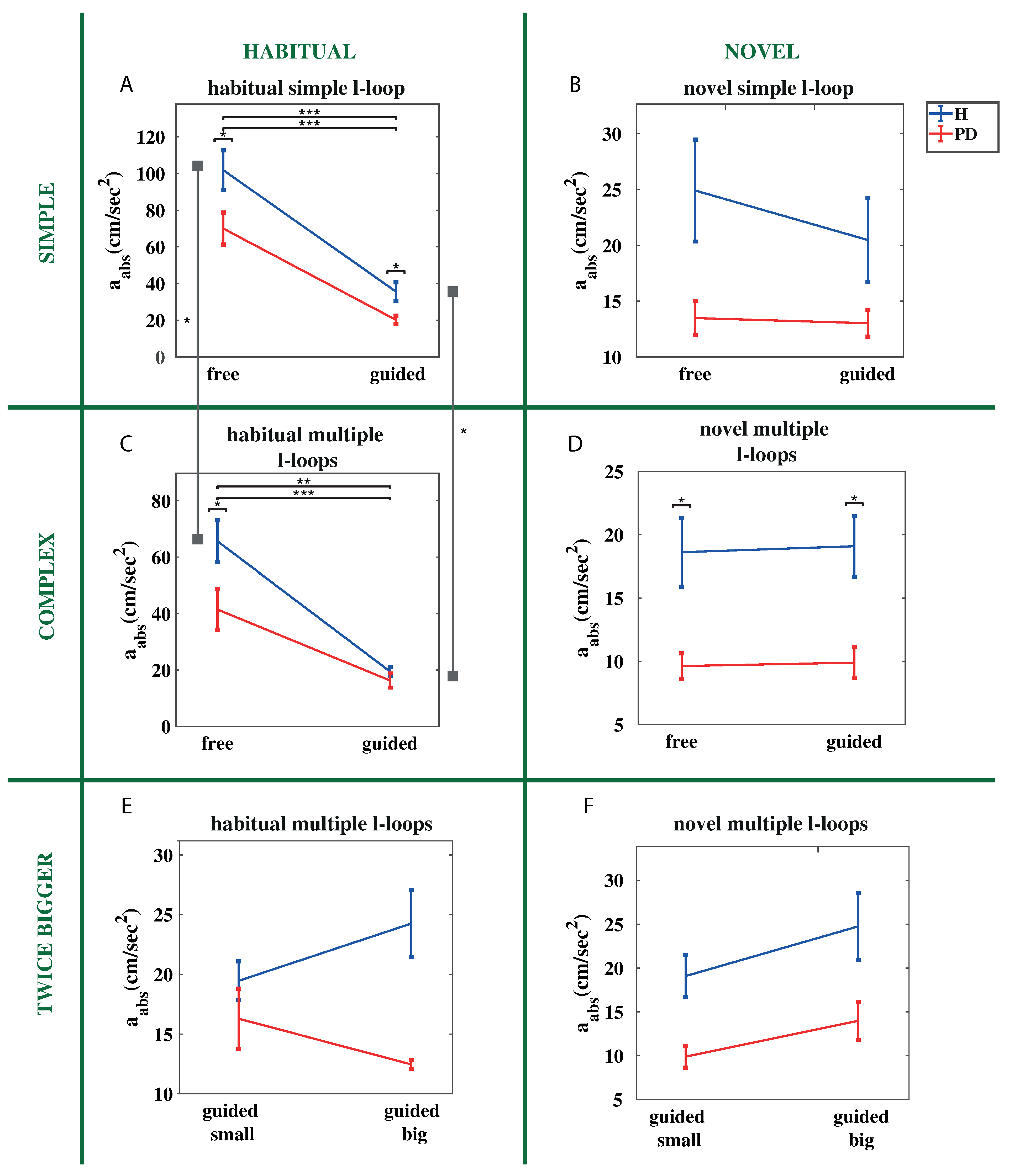
The difference between groups becomes smaller in the guided condition compared to the free one, both for simple and complex tasks. Similarly as for velocity, we found that exerted acceleration decays with age for both groups as shown by significant, negative correlations between age and acceleration (In the PD group: r ( 24 ) = 0.24 , p < 0.05 ; in the H group: r ( 26 ) = 0.17 , p = 0.058 ).

3.2.1. Free vs. Guided Conditions

Both groups significantly reduced acceleration in the guided condition compared to the free one when performing habitual tasks (Figure 4A,C). The difference between groups in the simple tasks (Figure 4A) is significant both in the free and the guided conditions ( p < 0.05 ). In the complex tasks (Figure 4C,D), PD patients show lower acceleration than H subjects in the free condition ( p < 0.05 both for habitual and novel tasks).
No significant differences were found between free and guided conditions for both groups when performing novel tasks (Figure 4B,D). In the complex novel task (Figure 4D) mean acceleration in the guided condition is similar to that measured in the free condition for both groups, with PD patients showing significantly lower acceleration than H subjects both in the free and the guided conditions ( p < 0.05 ).

3.2.2. Simple vs. Complex Shape Conditions

Acceleration decreases for both groups as the shape complexity of the task increases, although the difference in performance is significant only for H subjects when using the habitual motor plan ( p = 0.03 , both in free and guided conditions).

3.2.3. Habitual vs. Novel Motor Plan

Acceleration decreases for both groups in executing novel tasks compared to habitual ones, although acceleration reduction observed for the PD group is greater than that of the H group, with the exception of the simple tasks written in the guided condition.

3.2.4. Increased Handwriting Size

Contrary to writing velocity, the two groups show a different trend of acceleration when they were required to write in habitual condition and twice bigger than normal. Indeed, acceleration measured from the H group increases ( 25 % ) in bigger condition compared to normal one, whereas decreases ( 24 % ) for the PD group (Figure 4E). This result is in accordance with previous work [9], showing that patients with PD modulates acceleration inefficiently compared to H subjects when increasing their handwriting size. However, we found no significant difference between groups when writing twice bigger than normal, both in executing habitual ( p > 0.18 , Figure 4E) and novel tasks ( p > 0.22 , Figure 4F).

3.2.5. Acceleration at Novel Segmentation Points

In previous work [16], we observed that ink traces of l-loops drawn in clockwise fashion were characterized by the appearance of novel strokes, and thus novel segmentation points in the proximity of the loop, where the writer had to modify the habitual order of movements. Furthermore, we observed no significant differences between the mean absolute acceleration measured at the novel segmentation points in H subjects handwriting and that measured at the regular segmentation points (i.e., those normally appearing when drawing a loop in counterclockwise fashion) in PD handwriting. Here, we found no significant difference between the absolute acceleration measured at the novel segmentation points of PD and H handwriting in all novel tasks, regardless writing condition (free or guided) and shape complexity (simple or complex) ( p > 0.2 ).

3.3. Pressure

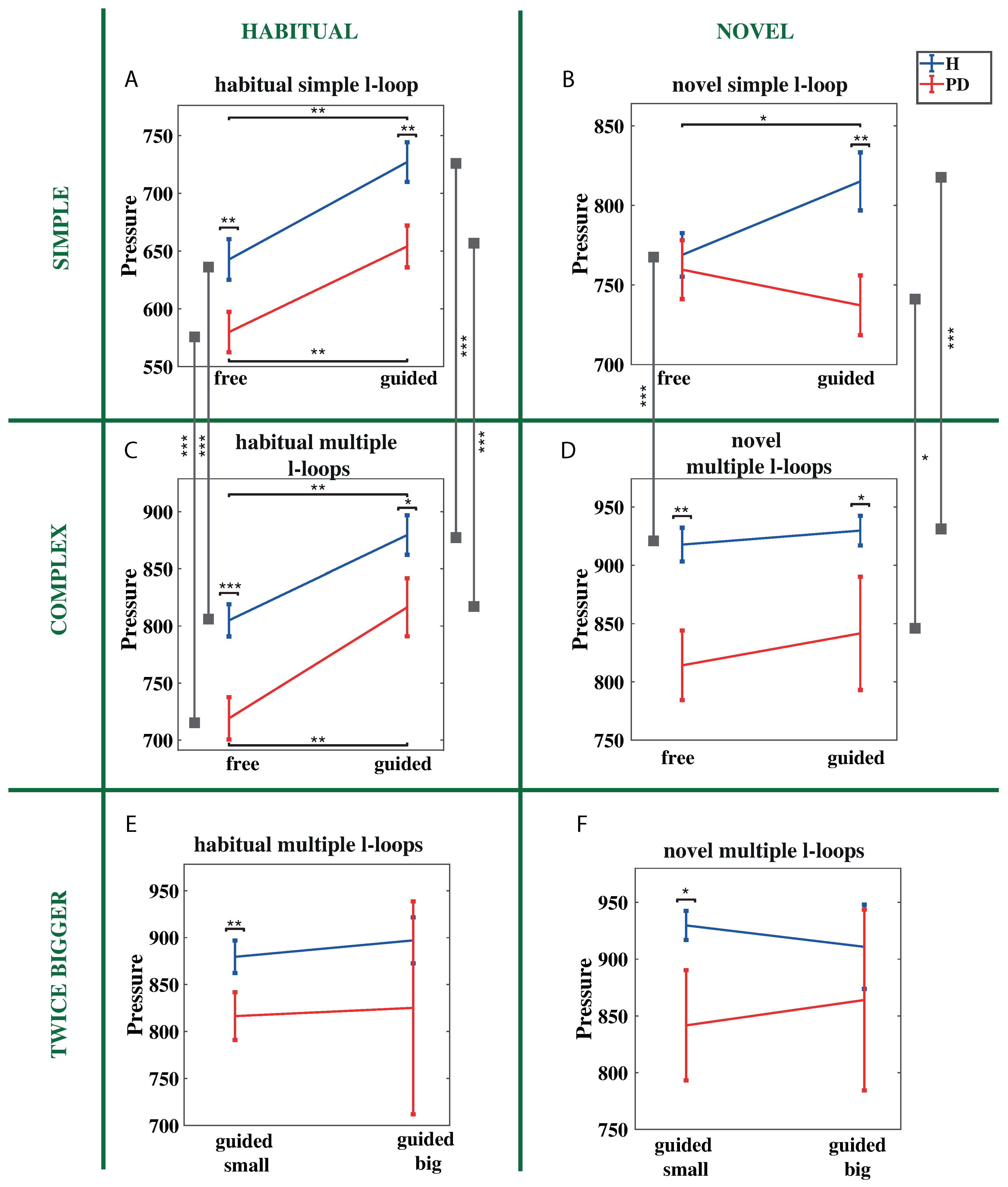
PD group exerted lower pressure than H group in executing all the tasks (Figure 5).

3.3.1. Free vs. Guided Conditions

For habitual tasks, both for simple and complex shape (Figure 5A,C) we observed that writing pressure significantly increases in the guided condition compared to the free one for both groups ( p < 0.01 ) and that PD patients exert lower pressure than H subjects both in the free ( p < 0.01 ) and the guided ( p < 0.05 ) conditions.
For novel tasks, we found that in the guided condition compared to the free condition, pressure significantly increased for H subjects ( p < 0.05 ) and slightly decreased for patients with PD. Indeed, we found that for simple shape in the free condition (Figure 5B) pressure exerted in the PD patients and the H subjects was not significantly different ( p > 0.69 ), whereas in the guided condition PD patients exert lower pressure than H subjects ( p < 0.01 ). As regarding novel complex tasks (Figure 5D) pressure does not change significantly in guided condition compared to free one for both groups ( p > 0.62 for both groups).

3.3.2. Simple vs. Complex Shape Conditions

Pressure significantly increases as the shape complexity increases for the H group in all conditions ( p < 0.001 ) and for the PD group in executing habitual motor tasks ( p < 0.0001 both in the free and guided conditions) and novel motor tasks in the guided condition ( p < 0.05 ). No significant increase was found for the PD group in executing the novel motor task in the free condition ( p > 0.1 ).

3.3.3. Habitual vs. Novel Motor Plan

Both groups increased pressure in a novel motor plan compared to the habitual one. The percentage of increase of the exerted pressure is similar for H and PD group when performing complex tasks ( 13 % in the free condition for both groups and 5 % vs. 3 % in guided condition for H and PD, respectively) and simple tasks in the guided condition ( 12 % for both groups). The percentage of increase of exerted pressure when performing simple tasks in the free condition is higher for PD than H group ( 31 % vs. 19 % , respectively).

3.3.4. Increased Handwriting Size

We observed no significant differences of exerted pressure between groups when writing twice bigger than normal, both in habitual ( p > 0.7 ; Figure 5E) and novel conditions ( p > 0.7 ; Figure 5F).

3.4. Normalized Jerk

Generally, PD patients show increased handwriting dysfluency compared to H subjects [8,12]. A measure for dysfluency is normalized jerk. We found indeed that normalized jerk of PD group is higher than H group in most of the tasks (Figure 6), although differences were significant only for simple tasks in the guided condition ( p < 0.01 ; Figure 6A,B).

3.4.1. Free vs. Guided Conditions

Normalized jerk significantly increases in the guided condition compared to the free condition for both groups in the habitual motor plan ( p < 0.001 ; Figure 6A,C). As regarding novel tasks, no significant changes were found between free and guided conditions for both groups (Figure 6B,D).

3.4.2. Simple vs. Complex Shape Conditions

Handwriting dysfluency increases as the shape complexity increases for both groups when they were asked to use the habitual motor plan, both in the free and guided conditions ( p < 0.05 for PD and p < 0.05 for H). Conversely, when using a novel motor plan, we found no difference in handwriting dysfluency between simple and complex tasks, both in free and guided conditions for PD patients ( p > 0.9 in the free condition; p > 0.4 in the guided condition) and in the free condition for the H subjects ( p > 0.6 ). Significant increase in handwriting dysfluency as the shape complexity increases is observed only for the H group in performing novel tasks in the guided condition ( p = 0.01 ).

3.4.3. Habitual vs. Novel Motor Plan

Normalized jerk measured when executing novel motor plan is higher than that measured in executing habitual motor plan for both groups, regardless the shape complexity and writing condition (free or guided) ( p < 0.001 ).

3.4.4. Increased Handwriting Size

We observed a significant increase of normalized jerk for both groups when writing twice bigger than normal, both in executing habitual ( p < 0.001 and p < 0.01 for PD and H group, respectively; Figure 6E) and novel motor plan ( p < 0.001 and p < 0.05 for PD and H group, respectively; Figure 6F).

3.5. Stroke Size

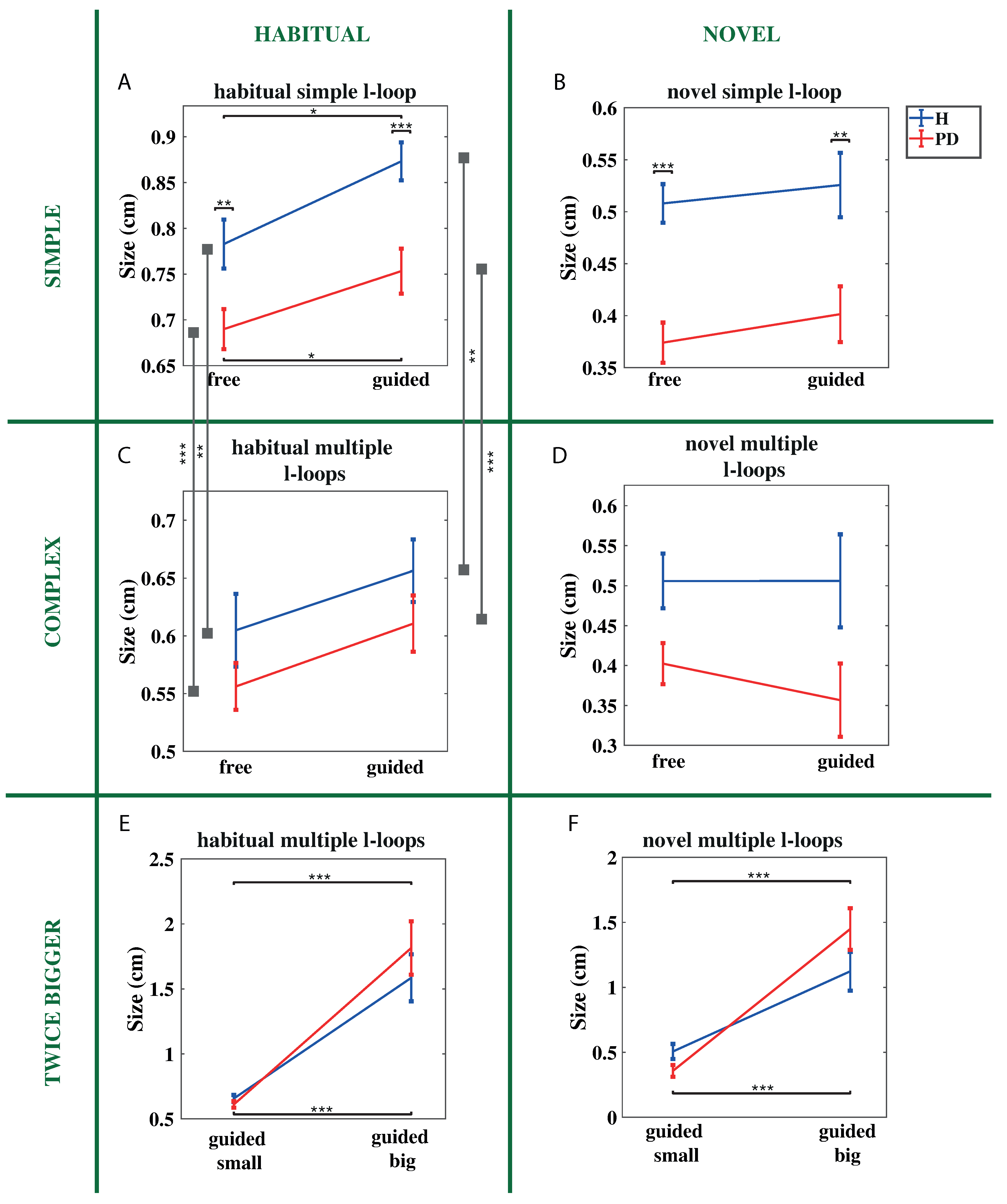
Strokes drawn by PD patients are smaller than those drawn by H subjects in most of the tasks. We found that stroke size of PD patients is significantly smaller than stroke size of H subjects for simple tasks (Figure 7A,B) regardless the writing condition (free or guided) and the motor plan complexity (habitual or novel) ( p < 0.001 ).
Instead, for complex tasks (Figure 7C,D) stroke size of PD patients was not significantly smaller than stroke size of H subjects.

3.5.1. Free vs. Guided Conditions

Both groups tend to increase stroke size in the guided condition compared to the free condition, especially when using the habitual motor plan (Figure 7A,C). However, size increases significantly only when performing simple tasks using the habitual motor plan ( p < 0.05 for both groups). Conversely, stroke size does not change significantly between free and guided conditions for complex tasks, executed through the habitual motor plan ( p > 0.2 for H group, p > 0.1 for PD group) or a novel motor plan (Figure 7B,D) both for PD patients ( p > 0.4 for simple tasks, p > 0.3 for complex tasks) and H subjects ( p > 0.6 for simple tasks, p > 0.9 for complex tasks).

3.5.2. Simple vs. Complex Shape Conditions

As the shape complexity increases we observed that stroke size decreases for habitual motor tasks, both for the PD ( p < 0.0001 ) and the H group ( p < 0.01 ). Conversely, stroke size does not change significantly when performing novel tasks, both for PD patients ( p > 0.9 ) and H subjects ( p > 0.4 ).

3.5.3. Habitual vs. Novel Motor Plan

We observed that strokes become smaller in novel tasks compared to habitual tasks for both groups, regardless the writing condition and the shape complexity of the task ( p < 0.001 ).

3.5.4. Increased Handwriting Size

As expected, when participants from both groups were asked to write twice bigger than normal (Figure 7E,F) they increased their stroke size ( p < 0.001 ). However, we found that PD patients increased their stroke size more than H subjects, both for habitual tasks (three times bigger for PD and two times bigger for H, Figure 7E) and novel tasks (four times bigger for PD and two times bigger for H, Figure 7F). Indeed, as shown in Figure 7F, PD patients drew strokes bigger than H subjects (although size difference was not significant; p > 0.18 ).

3.6. Reaction Time

We observed that the PD group took more time than the H group for starting most of the tasks (Figure 8).

3.6.1. Free vs. Guided Conditions

For habitual tasks (Figure 8A,C) both groups took more time for starting the tasks in guided than in the free condition ( p < 0.001 ). Conversely, for simple novel tasks (Figure 8B), PD patients needed less time in the guided than in the free condition. Indeed, we found that PD patients took less time than H subjects in starting the task in the guided condition (although we found no significant difference between groups; p > 0.1 ). For complex novel tasks (Figure 8D) we observed no significant differences in reaction time between free and guided conditions for both groups ( p > 0.2 ).

3.6.2. Simple vs. Complex Shape Conditions

Time needed for starting the task significantly increases as the complexity of the shape increases, regardless the writing condition (free or guided) and the motor plan complexity, both for H subjects ( p < 0.001 ) and PD patients ( p < 0.0001 ).

3.6.3. Habitual vs. Novel Motor Plan

Time needed for starting the task significantly increases in novel compared to habitual tasks for both groups ( p < 0.001 ).

3.6.4. Increased Handwriting Size

When participants from both groups were asked to write twice bigger than normal (Figure 8E,F) they took more time for starting the task (although the difference is significant only for H subjects: p < 0.05 ).

3.7. In-Air and On-Paper Parameters

We also computed and analyzed other handwriting features, such as (a) the ratio between in-air and on-paper times, (b) the ratio between in-air and on-paper trajectory length [37] and (c) the number of components drawn for producing a task, that is the number of pen-up executed by the participants in drawing the simple or the multiple l-loop shapes. We found that both time and trajectory length ratios were higher in PD patients compared to H subjects ( p < 0.05 ) and that PD patients tend to produce more handwriting components per trial than H subjects ( 1.47 vs. 1.18 components per trial for patients with PD and H subjects, respectively). In other words, PD patients tend to lift the pen from the paper more than H subjects when drawing loop shapes.

4. Discussion

In recent years, handwriting analysis has become widely used for investigating motor impairments caused by neurodegenerative diseases and as an additional instrument for supporting the diagnosis [13]. Here, we used a novel handwriting protocol, in which we asked Parkinson’s disease patients and healthy subjects (matched for age, handedness, and education) to perform handwriting tasks under different conditions, and compared performance obtained by the two groups. The proposed protocol consists of 10 handwriting tasks, including simple tasks (i.e., separate l-loops) and complex tasks (i.e., a sequence of connected l-loops with alternating heights). We asked participants to perform each task in 2 conditions: freely and guided (i.e., following a sequence of dots). Furthermore, participants were asked to draw loops in counterclockwise (i.e., highly automated, habitual motor plan) or clockwise order (i.e., novel motor plan) [16]. Eventually, we asked participants to perform complex guided tasks, drawing the shape of the loops twice bigger than normal using both the habitual and novel motor plans. Compared to previous studies, here, we focused on handwriting produced by patients in the early stage of PD, for which the disease affects predominantly the non-dominant hand (i.e., the one that is not used for performing handwriting). The healthy group significantly outperformed the PD group in most of the investigated tasks, making these handwriting tasks useful for the diagnosis of PD, especially in the early stage of the disease. Our analysis highlights that the signs during the early stage of PD differ from the signs uncovered previously during the later stage of PD. Indeed, we found that isochrony and fluency is spared in PD patients during the early stage of the disease, whereas previous work reports that these features are impaired during the later stage of the disease [8,12,38]. Furthermore, this study provides insights in which tasks and features are suited for detecting the presence of the disease, and provides criteria for developing a handwriting-based diagnostic protocol for the early diagnosis of PD. The results support our previous work [16] where we concluded that the clockwise handwritten loops form a novel, non-automated sequence of movements and that PD patients show impaired fine tuning of motor parameters.

4.1. PD Patients Rely More on Visual Feedback with Increasing Shape Complexity

Previous studies reported that PD patients benefit from the use of external cues [21] since their movements rely on visual feedback [17].
Regarding the tasks performed using a habitual motor plan, we found that:
  • For simple tasks, the performance difference between the H and PD groups observed in the free condition does not become smaller in the guided condition. In contrast, the discrepancy between groups in terms of handwriting fluency (normalized jerk) increases in the guided condition.
  • For complex tasks, the difference of performance (in terms of velocity and acceleration) between groups in the guided condition is reduced compared to free condition. Indeed, we observed that, in the guided condition, the performance of H subjects has a larger decay compared to that of PD patients, and that the performance of patients becomes not significantly different from that of healthy subjects.
Visual cues represent a disturbing factor for the execution of automated movements, whereas they are beneficial for movements based on the visual feedback [17]. Obtained results suggest that, at least in the early stage of the disease, movements performed by patients for producing simple habitual shapes, still show some automatisms, and slightly rely on visual feedback. Indeed, we observed that performance decay from free to guided conditions of PD patients is similar to that observed in H subjects. Conversely, for more complex tasks, performance decay is worse in H subjects than PD patients, suggesting that, as the complexity of the task increases (in terms of shape), PD patients tend to rely more on the visual feedback. As regarding tasks performed through a novel motor plan, we found no significant difference in performance between free and guided conditions for both groups. This result was expected, since the sequence of movements involved in this task is mainly novel (non-automated) for both groups (although at stroke level movements can be considered skilled), and therefore requires more the use of visual and proprioceptive feedback both for PD patients and H subjects. However, the H subjects seem to have more troubles in contrasting the automatisms, especially for simple handwriting patterns, where the difference between groups in the guided condition is reduced compared to the free condition.

4.2. Increasing Task Shape Complexity Affects the Performance of Healthy Participants More than the Performance of PD Patients

Our protocol contains tasks at different level of complexity, both in terms of shape of the drawing pattern and the automaticity of the drawing movement. We assumed that performing loops of alternating sizes increases control complexity compared to performing separate loops of equal sizes.
Analysis of the measured parameters showed that, when using the habitual motor plan, the performance of healthy participants significantly decreased both in terms of writing velocity and acceleration as the shape complexity increases, regardless of whether the task is performed freely or through guiding dots. In contrast, the performance of PD patients does not change significantly when increasing shape complexity, both in the free and in the guided conditions. Our results are in accordance with the observations in [17], showing that performance of PD patients was only slightly influenced by the difficulty of the task, which consisted of movements based on visual feedback, and therefore depended only little on the complexity of the shape of the writing pattern. On the other hand, performance obtained by H subjects, that had to retrieve and instantiate a more complex motor program, was significantly affected. Our results suggest that using a handwriting test that includes different levels of shape complexity, could provide clues about the presence of PD, even in its early stage, e.g., when significant changes in velocity and acceleration are not observed with increased shape complexity of the task.
As regarding different level of complexity in terms of automaticity of motor plans involved, we asked participants to perform handwriting by using the habitual or a novel motor plan. Conversely to what has been observed when increasing shape complexity, we found that increasing motor plan complexity induces a worsening of performance (in terms of writing velocity, acceleration, and normalized jerk) greater in the PD group than in the H group.

4.3. Isochrony Is Preserved in the Early Stage of PD

The previous work reported that isochrony is impaired in PD patients [38]. Here, we observed that, when drawing bigger complex shapes (according to a sequence of guiding dots) both groups increased handwriting velocity and that, especially in performing a novel motor plan, the trends of the two groups are similar. This suggests that, at least in the early stage of the disease, handwriting performed by PD patients follows the isochrony principle commonly observed in healthy subjects. However, we observed that when PD patients have to increase their handwriting size, they show a broad variation in exerted pressure, both in performing habitual and novel motor tasks. Furthermore, PD patients tend to increase stroke size more than healthy subjects when they were asked to increase handwriting size by following visual cues. These observations suggest that, in analyzing handwriting performed by increasing shape size through guiding cues, the percentage of increase in stroke size is more meaningful than other parameters, such as pressure and velocity, for evaluating whether the subjects is affected by PD, especially in the early stage of the disease.

4.4. Handwriting Fluency in PD Patients at the Early Stage Is Slightly Affected by the Disease

Other works report that fluency is impaired in handwriting performed by patients with PD [8,12]. Here, we observed that, at least in the early stage of the disease, this feature is not very informative for the diagnosis, since we found that the differences measured between groups were not significant in most of the analyzed conditions, except for simple tasks performed in guided condition.

4.5. Performance of PD Patients Improves in the Execution of Novel Motor Plan Tasks in Terms of Reaction Time and Exerted Handwriting Pressure

The analysis of handwriting parameters measured when performing a novel motor plan shows that the generation of non-automated sequence of movements, compared to the execution of habitual sequence of movements, induces a performance decay in terms of writing velocity, acceleration, and normalized jerk for both groups. Although we observed that H subjects outperform patients with PD for most of the analyzed handwriting parameters, our analysis shows that executing a novel motor plan for drawing a simple loop shape improves two handwriting features in patients with PD: the exerted pressure and the response time. Previous work has shown that one of the main features for the diagnosis of PD through handwriting is the pressure exerted by the writer for producing the ink trace [39], since PD patients tend to exert lower pressure than healthy subjects. Results we obtained for habitual tasks are in accordance with previous studies, since we observed that the H group exerted significantly higher pressure than PD patients. Noteworthy we found that, when PD patients freely drew simple loop shapes by using a novel motor plan the pressure they exerted increased compared to that exerted when using the habitual motor plan. We found that also H subjects increase exerted pressure, but the rate of increase is lower than that observed for PD patients, so that both group exerted similar pressure when writing simple loops through a novel motor plan in free condition.
Slowness in initiation of movements (akinesia) is one of the main symptoms characterizing PD [40,41]. In line with previous works, here, we observed that the reaction time of patients with PD is longer than that of H subjects. However, when patients with PD were asked to write simple loop shapes through a novel motor plan, by following a sequence of dots, their response time slightly decreases compared to freely writing condition, showing an opposing trend compared to H subjects, for which response time increases from free to guided conditions. Indeed, we observed that response time of PD became similar to that of H subjects. These results suggest that the comparison of exerted pressure and response time measured in executing simple shape task by using the habitual and a novel sequence of movements would provide some indications on the presence of the disease.

4.6. Micrographia in Early PD Is More Associated to Shape Complexity than Motor Plan Complexity

Previous studies report that handwriting produced by PD patients is affected by micrographia, which occurs especially in tasks that escape attention, such as redundant and repetitive handwriting tasks [4]. Accordingly, we expected that tasks involving drawing of repetitive loops should have been more affected by PD micrographia compared to single loops. Conversely, since writing in clockwise condition requires more attention, we expected that micrographia should not occur in executing a novel motor plan.
Here, we observed that, for simple tasks, the size of strokes drawn by PD patients is significantly smaller than that produced by H subjects, both in free and guided conditions, regardless whether the loops were performed through the habitual or the novel motor plan. Conversely, increasing the shape complexity of the task reduces the difference in stroke size between groups compared to simple tasks (measured differences are not significant), suggesting that it tends to reduce the effect of micrographia on PD patients. This result could be due to the characteristic of the shape we used for the complex task, containing repetitive loops, but with different heights. Indeed, we also observed that complex shape tasks led the PD patients to rely more on visual feedback, therefore increasing the attention in executing the task. Noteworthy, increasing the complexity of motor plan involved (from habitual to novel) seems to have small influence on the stroke size in terms of differences between groups, that depends more on the shape complexity of the task. Furthermore, when asking participants to draw the complex shape twice bigger than normal, following a sequence of dots, PD patients tend to increase their stroke size more than H subjects, both for habitual and novel tasks. As a consequence, while on average the strokes drawn by PD patients are smaller than those drawn by H subjects for normal size shapes, strokes drawn by PD patients result bigger than those drawn by H subjects for twice bigger size shapes (although difference in stroke size between groups is not significant). This analysis suggests that the greater increase of stroke size in patients’ handwriting is due to the additional attention required for drawing loop shapes twice bigger than normal while following the guiding dots, rather than just the use of guiding dots. Indeed, we did not observe the same phenomenon for guided tasks in other conditions. Taken together, our results suggest that using more complex task (in terms of shapes) and asking patients to force themselves to write bigger than normal (providing also visual cues) could allow to trigger the attentional mechanisms that can be effective for contrasting micrographia. However, further experiments are needed for supporting this claim.

4.7. Criteria for the Early Diagnosis of the Disease through the Analysis of Handwriting

The analysis carried in this work suggests that a specific designed protocol, and the analysis of specific features, depending on the characteristics of the handwriting tasks, would provide clinicians with a useful tool for supporting the early clinical diagnosis of the disease. Our analysis suggests that a diagnostic protocol should:
  • Contain tasks characterized by different level of shape complexity, as those analyzed in this work. Indeed, reduced differences of performance (in terms of writing speed and acceleration) between simple and complex shape tasks would indicate the presence of the disease.
  • Consider task performed with different writing conditions, such as freely and guided performed tasks, since the performance decay observed from free to guided conditions is informative for the early diagnosis. Here, we observed that, for patients with PD, performance decay between free and guided condition is greater for simple shape tasks than for complex shape tasks. For healthy subjects we observed the opposite, that is performance decay is greater for complex shape tasks than for simple shape tasks.
  • Contain tasks aimed at testing the ability of patients in increasing handwriting size through the use of guiding dots. However, since we have observed that isochrony is preserved in the early stage of PD, the analysis has to be focused more on the percentage of stroke size increase, rather than other parameters, such as velocity and pressure, for identifying the signs of the disease in the early stage.
  • Take into account that handwriting fluency is slightly affected by the disease in the early stage, and that normalized jerk values falling into a “healthy range” do not exclude the presence of the disease.
  • Contain tasks involving the use of novel motor plan for drawing a simple known shape in different writing conditions, as those analyzed in this work. Indeed, since PD patients benefits from the execution of novel motor plan tasks in terms of response time and exerted handwriting pressure, a higher increase of exerted pressure when performing simple novel tasks in free condition would suggest the presence of the disease. Similarly, response time decrease or similar response time between free and guided conditions when drawing simple shapes by using a novel motor plan would suggest the presence of the disease.

4.8. PD Patients Perform Handwriting as They Got Stuck on the Early Stages of the Learning Process

Here, we provide further evidence to previous work [16], sustaining that the clockwise tasks elicit the execution of a novel, non-automated sequence of movements. Indeed, here, we observed a decrease of performance (in terms of velocity, acceleration, normalized jerk and response time) for both groups when performing clockwise (novel) tasks compared to the counterclockwise (habitual) tasks, and no significant difference in performance between free and guided conditions for both groups when performing novel motor plan. From a neuro-physiological point of view, it is known that motor and cognitive impairments affecting patients with PD are mainly due to the loss of dopaminergic neurons in the Basal Ganglia. There are several lines of evidence that a reduced amount of dopamine impairs the initial learning [42,43,44] and correlates with reduced performance in the acquisition and expression of a behavior during the initial stage of learning [45,46,47]. As in [16], here, we found that the execution of novel tasks gives rise to novel elementary movements, different from those used in the execution of habitual tasks (Figure 2). The novel movements correspond mainly to those performed for drawing the initial and the final trace of the upper part of the loop. In order to evaluate the difference of performance related only to non-automated movements, we analyzed the acceleration in the proximity of the beginning/ending of novel movements. No differences were found between PD and H groups, regardless the shape complexity and the writing condition (free or guided). This further supports our claim that the fine tuning of the motor plan parameters involved in the handwriting production is impaired in PD patients [16]. In other words, our results suggest that PD patients perform handwriting as they got stuck on the early stages of the learning process.

5. Conclusions

In this work, we investigated the presence of distinctive signs in handwriting performed by patients in the early stage of PD, with the aim of providing a set of diagnostic criteria for the early detection of the disease. Our analysis shows that even when the disease affects the contralateral side with respect to the one used for writing, the execution of specific tasks and the evaluation of specific features provide many insights on the presence of the disease. We found that PD patients still carry out some automatisms when drawing simple shapes, whereas they tend to rely more on the visual feedback as the shape complexity of the task increases. Consequently, reduced performance decrease with increased shape complexity of the task and reduced difference of performance when performing complex shape tasks in free and guided writing condition would indicate the presence of the disease. We also observed that fluency in PD patients is slightly affected by the disease and that the isochrony principle still holds for handwriting movements. Furthermore, we have observed that the performance of PD patients benefits from the execution of specific tasks. In particular, the execution of simple shape tasks by using novel motor plan improves the exerted pressure, which becomes similar to that exerted by healthy subjects. Similarly, response time of PD patients decreases, becoming similar to that of healthy subjects, when drawing simple shapes by using a novel motor plan. Furthermore, when drawing simple shapes by using a novel motor plan, we have observed that the pressure and the reaction time of patients become similar to that of healthy subjects. Eventually, according to the performance obtained by PD patients in drawing complex shape by forcing themselves to increase the handwriting size through the use of guiding dots, our analysis suggests that such task triggers the attentional mechanisms that can be effective for contrasting PD micrographia.
According to these findings, we proposed a set of criteria for the design of a diagnostic protocol, based on handwriting analysis, that can help the physicians in the early diagnosis of PD. Future work will be aimed at collecting further data from patients and apply a classification approach for the automatic diagnosis of the disease based on the proposed criteria. Further experiments will be aimed at evaluating whether a training phase on novel simple handwriting tasks, which improves both the response time and exerted pressure of patients, would lead to better performance also in performing habitual tasks. Similarly, further work will be aimed at evaluating whether extended training on complex shape task, forcing the patients to increase handwriting size through the use of guiding dots, would be beneficial for contrasting micrographia in normal handwriting.

Author Contributions

Conceptualization, R.S., A.M. and H.-L.T.; methodology, R.S.; software, R.S.; formal analysis, R.S.; investigation, R.S. and R.D.M.; data curation, R.S. and R.D.M.; validation, A.M. and A.T.; visualization, R.S.; writing—original draft preparation, R.S.; writing—review and editing, A.M., R.D.M., A.T. and H.-L.T.; resources, A.T. and A.M.; supervision, A.M.; project administration, A.M.; funding acquisition, A.M. All authors have read and agreed to the published version of the manuscript.

Funding

This work is part of the research program HAND (Handwriting Analysis against Neuromuscular Disease) funded by the grant PRIN—20154C9M5P-002 from the Italian Ministero dell’Istruzione, dell’Università e della Ricerca.

Institutional Review Board Statement

The study was conducted according to the guidelines of the Declaration of Helsinki, and approved by the Ethics Committee of the University of Campania ”Luigi Vanvitelli”, Napoli, Italy (protocol code 590/2018, date of approval 1 August 2018).

Informed Consent Statement

Informed consent was obtained from all subjects involved in the study. Written informed consent has been obtained from the patients to publish this paper.

Data Availability Statement

The data presented in this study are available on request from the corresponding author.

Conflicts of Interest

The authors declare no conflict of interest. The funders had no role in the design of the study; in the collection, analyses, or interpretation of data; in the writing of the manuscript, or in the decision to publish the results.

References

  1. Jankovic, J. Parkinson’s disease: Clinical features and diagnosis. J. Neurol. Neurosurg. Physiatry 2008, 79, 368–376. [Google Scholar] [CrossRef] [PubMed] [Green Version]
  2. Flash, T.; Inzelberg, R.; Schechtman, E.; Korczyn, A.D. Kinematic analysis of upper limb trajectories in Parkinson’s disease. Exp. Neurol. 1992, 118, 215–226. [Google Scholar] [CrossRef] [PubMed]
  3. Marsden, C.D. Slowness of movement in Parkinson’s disease. Mov. Disord. 1989, 4, S26–S37. [Google Scholar] [CrossRef] [PubMed]
  4. McLennan, J.E.; Nakano, K.; Tyler, H.R.; Schwab, R.S. Micrographia in Parkinson’s disease. J. Neurol. Sci. 1972, 15, 141–152. [Google Scholar] [CrossRef]
  5. Sheridan, M.R.; Flowers, K.A.; Hurrell, J. Programming and execution of movement in parkinson’s disease. Brain 1987, 110, 1247–1271. [Google Scholar] [CrossRef]
  6. Teulings, H.L.; Stelmach, G.E. Control of stroke size, peak acceleration, and stroke duration in Parkinsonian handwriting. Hum. Mov. Sci. 1991, 10, 315–334. [Google Scholar] [CrossRef]
  7. Gemmert Van, A.W.A.; Teulings, H.L.; Contreras-Vidal, J.L.; Stelmach, G.E. Parkinson’s disease and the control of size and speed in handwriting. Neuropsychologia 1999, 37, 685–694. [Google Scholar] [CrossRef]
  8. Teulings, H.; Contreras-Vidal, J.; Stelmach, G.; Adler, C. Parkinsonism reduces coordination of fingers, wrist, and arm in fine motor control. Exp. Neurol. 1997, 146, 159–170. [Google Scholar] [CrossRef]
  9. Van Gemmert, A.; Adler, C.H.; Stelmach, G.E. Parkinson’s disease patients undershoot target size in handwriting and similar tasks. J. Neurol. Neurosurg. Psychiatry 2003, 74, 1502–1508. [Google Scholar] [CrossRef] [Green Version]
  10. Stelmach, G.E.; Teasdale, N.; Phillips, J.; Worringham, C.J. Force production characteristics in Parkinson’s disease. Exp. Brain Res. 1989, 76, 165–172. [Google Scholar] [CrossRef]
  11. Stelmach, G.E.; Worringham, C.J. The preparation and production of isometric force in Parkinson’s disease. Neuropsychologia 1988, 26, 93–103. [Google Scholar] [CrossRef] [PubMed]
  12. Broderick, M.P.; Van Gemmert, A.W.A.; Shill, H.A.; Stelmach, G.E. Hypometria and bradykinesia during drawing movements in individuals with Parkinson’s disease. Exp. Brain Res. 2009, 197, 223–233. [Google Scholar] [CrossRef] [PubMed]
  13. Vessio, G. Dynamic Handwriting Analysis for Neurodegenerative Disease Assessment: A Literary Review. Appl. Sci. 2019, 9, 4666. [Google Scholar] [CrossRef] [Green Version]
  14. Senatore, R.; Della Cioppa, A.; Marcelli, A. Automatic Diagnosis of Neurodegenerative Diseases: An evolutionary approach for facing the interpretability problem. Information 2019, 10, 30. [Google Scholar] [CrossRef] [Green Version]
  15. Senatore, R.; Della Cioppa, A.; Marcelli, A. Automatic diagnosis of Parkinson disease through handwriting analysis: A cartesian genetic programming approach. In Proceedings of the IEEE Symposium on Computer-Based Medical Systems, Cordoba, Spain, 5–7 June 2019; Volume 2019, pp. 312–317. [Google Scholar] [CrossRef]
  16. Senatore, R.; Marcelli, A. A paradigm for emulating the early learning stage of handwriting: Performance comparison between healthy controls and Parkinson’s disease patients in drawing loop shapes. Hum. Mov. Sci. 2019, 65, 89–101. [Google Scholar] [CrossRef]
  17. Teulings, H.L.; Contreras-Vidal, J.L.; Stelmach, G.E.; Adler, C.H. Adaptation of handwriting size under distorted visual feedback in patients with Parkinson’s disease and elderly and young controls. J. Neurol. Neurosurg. Psychiatry 2002, 72, 315–324. [Google Scholar] [CrossRef] [Green Version]
  18. Parisi, R.; Parziale, A.; Marcelli, A. Some observations on lognormality and motor control in handwriting. In Proceedings of the 1st International Conference on Pattern Recognition and Artificial Intelligence, Kolkata, India, 20–22 December 2013; pp. 732–736. [Google Scholar]
  19. Smits, E.J.; Tolonen, A.J.; Cluitmans, L.; Van Gils, M.; Conway, B.A.; Zietsma, R.C.; Leenders, K.L.; Maurits, N.M. Standardized handwriting to assess bradykinesia, micrographia and tremor in Parkinson’s disease. PLoS ONE 2014, 9, e97614. [Google Scholar] [CrossRef]
  20. Van Gemmert, A.W.; Teulings, H.L.; Stelmach, G.E. Parkinsonian patients reduce their stroke size with increased processing demands. Brain Cogn. 2001, 47, 504–512. [Google Scholar] [CrossRef] [Green Version]
  21. Nackaerts, E.; Vervoort, G.; Heremans, E.; Smits-Engelsman, B.C.M.; Swinnen, S.P.; Nieuwboer, A. Relearning of writing skills in Parkinson’s disease: A literature review on influential factors and optimal strategies. Neurosci. Biobehav. Rev. 2013, 37, 349–357. [Google Scholar] [CrossRef]
  22. Viviani, P.; Schneider, R. A Developmental Study of the Relationship Between Geometry and Kinematics in Drawing Movements. J. Exp. Psychol. Hum. Percept. Perform. 1991, 17, 198–218. [Google Scholar] [CrossRef]
  23. Gibb, W.R.; Lees, A.J. A comparison of clinical and pathological features of young- and old-onset parkinson’s disease. Neurology 1988, 38, 1402–1406. [Google Scholar] [CrossRef] [PubMed]
  24. Hoehn, M.M.; Yahr, M.D. Parkinsonism: Onset, progression, and mortality. Neurology 2001, 57, 318. [Google Scholar] [CrossRef]
  25. Emre, M.; Aarsland, D.; Brown, R.; Burn, D.J.; Duyckaerts, C.; Mizuno, Y.; Broe, G.A.; Cummings, J.; Dickson, D.W.; Gauthier, S.; et al. Clinical diagnostic criteria for dementia associated with Parkinson’s disease. Mov. Disord. 2007, 22, 1689–1707. [Google Scholar] [CrossRef] [PubMed]
  26. Santangelo, G.; Siciliano, M.; Pedone, R.; Vitale, C.; Falco, F.; Bisogno, R.; Siano, P.; Barone, P.; Grossi, D.; Santangelo, F.; et al. Normative data for the Montreal Cognitive Assessment in an Italian population sample. Neurol. Sci. 2015, 36, 585–591. [Google Scholar] [CrossRef] [Green Version]
  27. Fahn, S.; Elton, R. Unified Parkinsons Disease Rating Scale. In Recent Developments in Parkinson’s Disease; Macmillan Health Care Information: Florham Park, NJ, USA, 1987; pp. 153–163. [Google Scholar]
  28. Nasreddine, Z.S.; Phillips, N.A.; Bédirian, V.; Charbonneau, S.; Whitehead, V.; Collin, I.; Cummings, J.L.; Chertkow, H. The Montreal Cognitive Assessment, MoCA: A brief screening tool for mild cognitive impairment. J. Am. Geriatr. Soc. 2005, 53, 695–699. [Google Scholar] [CrossRef]
  29. Folstein, M.F.; Folstein, S.E.; McHugh, P.R. “Mini-mental state”. A practical method for grading the cognitive state of patients for the clinician. J. Psychiatr. Res. 1975, 12, 189–198. [Google Scholar] [CrossRef] [PubMed]
  30. Beck, A.T.; Ward, C.H.; Mendelson, M.; Mock, J.; Erbaugh, J. An Inventory for Measuring Depression. Arch. Gen. Psychiatry 1961, 4, 561–571. [Google Scholar] [CrossRef] [Green Version]
  31. Oldfield, R.C. The assessment and analysis of handedness: The Edinburgh inventory. Neuropsychologia 1971, 9, 97–113. [Google Scholar] [CrossRef]
  32. Maeland, A.F.; Karlsdottir, R. Development of reading, spelling, and writing skills from third to sixth grade in normal and dysgraphic school children. In Development of Graphic Skills: Research Perspectives and Educational Implications; Academic Press: Cambridge, MA, USA, 1991; pp. 179–189. [Google Scholar]
  33. Marr, D.; Cermak, S. Consistency of handwriting in early elementary students. Am. J. Occup. Ther. 2003, 57, 161–167. [Google Scholar] [CrossRef]
  34. Teulings, H. MovAlyzeR. 2010. Available online: www.neuroscript.net (accessed on 1 January 2022).
  35. Teulings, H.; Maarse, F. Digital recording and processing of handwriting movements. Hum. Mov. Sci. 1984, 3, 193–217. [Google Scholar] [CrossRef]
  36. Benjamini, Y.; Hochberg, Y. Controlling the false discovery rate: A practical and powerful approach to multiple testing. J. R. Stat. Soc. B 1995, 57, 289–300. [Google Scholar] [CrossRef]
  37. Rosenblum, S.; Livneh-Zirinski, M. Handwriting process and product characteristics of children diagnosed with developmental coordination disorder. Hum. Mov. Sci. 2008, 27, 200–214. [Google Scholar] [CrossRef] [PubMed]
  38. Viviani, P.; Burkhard, P.R.; Chiuvé, S.C.; Dell’Acqua, C.C.; Vindras, P. Velocity control in Parkinson’s disease: A quantitative analysis of isochrony in scribbling movements. Exp. Brain Res. 2009, 194, 259–283. [Google Scholar] [CrossRef] [PubMed] [Green Version]
  39. Rosenblum, S.; Samuel, M.; Zlotnik, S.; Erikh, I.; Schlesinger, I. Handwriting as an objective tool for Parkinson’s disease diagnosis. J. Neurol. 2013, 260, 2357–2361. [Google Scholar] [CrossRef]
  40. Bloxham, C.A.; Mindel, T.A.; Frith, C.D. Initiation and execution of predictable and unpredictable movements in parkinson’s disease. Brain 1984, 107, 371–384. [Google Scholar] [CrossRef]
  41. Hallett, M. Clinical neurophysiology of akinesia. Rev. Neurol. 1990, 146, 585–590. [Google Scholar]
  42. Doyon, J.; Ungerleider, L.G. Functional anatomy of motor skill learning. In Neuropsychology of Memory, 3rd ed.; The Guilford Press: New York, NY, USA, 2002; pp. 225–238. [Google Scholar]
  43. Krebs, H.; Hogan, N.; Hening, W.; Adamovich, S.; Poizner, H. Procedural motor learning in parkinson’s disease. Exp. Brain Res. 2001, 141, 425–437. [Google Scholar] [CrossRef]
  44. Packard, M.G.; Knowlton, B.J. Learning and Memory Functions of the Basal Ganglia. Annu. Rev. Neurosci. 2002, 25, 563–593. [Google Scholar] [CrossRef] [Green Version]
  45. Horvitz, J.C.; Won, Y.C.; Morvan, C.; Eyny, Y.; Balsam, P.D. A “good parent” function of dopamine: Transient modulation of learning and performance during early stages of training. Ann. N. Y. Acad. Sci. 2007, 1104, 270–288. [Google Scholar] [CrossRef] [Green Version]
  46. Senatore, R.; Marcelli, A. A Neural Scheme for Procedural Motor Learning of Handwriting. In Proceedings of the 2012 International Conference on Frontiers in Handwriting Recognition, Bari, Italy, 18–20 September 2012; pp. 659–664. [Google Scholar] [CrossRef]
  47. Smith-Roe, S.L.; Kelley, A.E. Coincident activation of NMDA and dopamine D1 receptors within the nucleus accumbens core is required for appetitive instrumental learning. J. Neurosci. Off. J. Soc. Neurosci. 2000, 20, 7737–7742. [Google Scholar] [CrossRef]
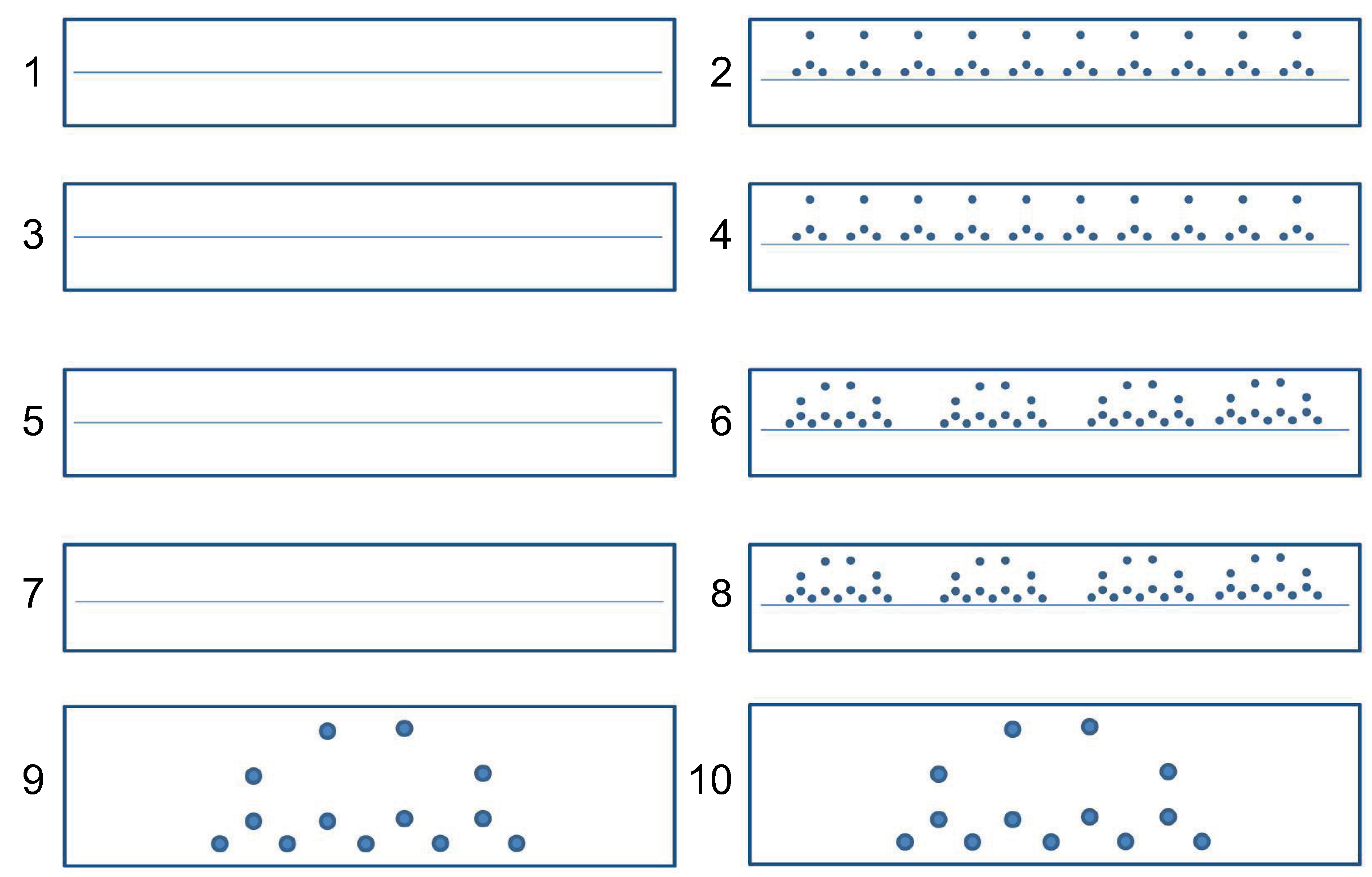
Figure 1. Horizontally oriented A4 sheet containing the boxes (dimensions 2 cm × 12 cm for 1–8 boxes, 3 cm × 12 cm for boxes 9 and 10) on which the participants had to perform the ten tasks. In 1–8 boxes the participants had to write on the central lines. Blue circles represent the visual cues that have to be reached for performing the task.
Applsci 12 12338 g001
Figure 2. Exampleof ink traces of simple handwritten l-loops drawn with (A) the habitual motor plan and (B) a novel motor plan. Arrows indicate the directions followed by the pen tip for drawing the trace. Green circles indicate the segmentation points that appear when drawing the l-loop using both the habitual and the novel motor plan and red circles indicate the additional segmentation points that appear when drawing the l-loop using the novel motor plan. Different strokes have been indicated using different colors.
Applsci 12 12338 g002
Figure 3. Absolute velocity (mean ± SEM) across PD patients (red lines) and H subjects (blue lines) for all conditions: (A) Habitual simple l-loop, (B) Novel simple l-loop, (C) Habitual multiple l-loops, (D) Novel multiple l-loops, (E) Habitual multiple l-loops, (F) Novel multiple l-loops. Grey and black lines indicate the results of t-tests that gave significant differences. Stars indicate the results of one-tailed t-tests corrected with the FDR procedure (* p < 0.05 ; ** p < 0.01 ; *** p < 0.001 ).
Applsci 12 12338 g003
Figure 4. Absolute acceleration (mean ± SEM) across PD patients (red lines) and H subjects (blue lines) for all conditions: (A) Habitual simple l-loop, (B) Novel simple l-loop, (C) Habitual multiple l-loops, (D) Novel multiple l-loops, (E) Habitual multiple l-loops, (F) Novel multiple l-loops. Grey and black lines indicate the results of t-tests that gave significant differences. Stars indicate the results of one-tailed t-tests corrected with the FDR procedure (* p < 0.05 ; ** p < 0.01 ; *** p < 0.001 ).
Applsci 12 12338 g004
Figure 5. Pressure (mean ± SEM) across PD patients (red lines) and H subjects (blue lines) for all conditions: (A) Habitual simple l-loop, (B) Novel simple l-loop, (C) Habitual multiple l-loops, (D) Novel multiple l-loops, (E) Habitual multiple l-loops, (F) Novel multiple l-loops. Grey and black lines indicate the results of t-tests that gave significant differences. Stars indicate the results one-tailed t-tests corrected with FDR (* p < 0.05 ; ** p < 0.01 ; *** p < 0.001 ).
Applsci 12 12338 g005
Figure 6. Normalized Jerk (mean ± SEM) across PD patients (red lines) and H subjects (blue lines) for all conditions: (A) Habitual simple l-loop, (B) Novel simple l-loop, (C) Habitual multiple l-loops, (D) Novel multiple l-loops, (E) Habitual multiple l-loops, (F) Novel multiple l-loops. Grey and black lines indicate the results of t-tests that gave significant differences. Stars indicate the results of one-tailed t-tests corrected with the FDR procedure (* p < 0.05 ; ** p < 0.01 ; *** p < 0.001 ).
Applsci 12 12338 g006
Figure 7. Stroke size (mean ± SEM) across PD patients (red lines) and H subjects (blue lines) for all conditions: (A) Habitual simple l-loop, (B) Novel simple l-loop, (C) Habitual multiple l-loops, (D) Novel multiple l-loops, (E) Habitual multiple l-loops, (F) Novel multiple l-loops. Grey and black lines indicate the results of t-tests that gave significant differences. Stars indicate the results of one-tailed t-tests corrected with the FDR procedure (* p < 0.05 ; ** p < 0.01 ; *** p < 0.001 ).
Applsci 12 12338 g007
Figure 8. Reaction Time (mean ± SEM) across PD patients (red lines) and H subjects (blue lines) for all conditions: (A) Habitual simple l-loop, (B) Novel simple l-loop, (C) Habitual multiple l-loops, (D) Novel multiple l-loops, (E) Habitual multiple l-loops, (F) Novel multiple l-loops. Grey and black lines indicate the results of t-tests that gave significant differences. Stars indicate the results of one-tailed t-tests corrected with the FDR procedure (* p < 0.05 ; ** p < 0.01 ; *** p < 0.001 ).
Applsci 12 12338 g008
Table 1. Demographic characteristics and data about clinical evaluation of the subjects participating in the experiments. Features are reported in terms of mean ± SEM.
PD
N = 13
Healthy
N = 14
Two-Tailed t-Test
df = 25
Gender7 men and 6 women8 men and 6 womenp > 0.05
Age (years)64.4 ± 1.560 ± 1.9p > 0.05
Education (years)9.4 ± 1.211.3 ± 1.3p > 0.3
Handedness score76.7 ± 12.269.4 ± 12.1p > 0.6
MoCA corrected21.6 ± 0.722.1 ± 0.8p > 0.6
BDI
II score
3.2 ± 0.72.5 ± 0.8p > 0.5
Disease Duration
(months)
44.1 ± 5.6
Modified
Hoehn & Yahr score
1.5 ± 0.1
UPDRS III20.6 ± 2.6
Table 2. Handwriting tasks: the first column lists the task number, the second column reports the instructions provided to the participant by the experimenter, and the third column shows an example of the performed task.
Task
InstructionsExample
1Write a sequence of separated l-loops.Applsci 12 12338 i001
2Write a sequence of 10 separated l-loops following the dotsApplsci 12 12338 i002
3Write a sequence of clockwise separated l-loops.Applsci 12 12338 i003
4Write a sequence of 10 clockwise separated l-loops following the dotsApplsci 12 12338 i004
5Write a sequence of words “elle”.Applsci 12 12338 i005
6Write a sequence of 4 words “elle” following the dotsApplsci 12 12338 i006
7Write a sequence of word “elle” drawing loop clockwise.Applsci 12 12338 i007
8Write a sequence of 4 words “elle”, drawing loop clockwise and following the dotsApplsci 12 12338 i008
9Write the word “elle” bigger following the dotsApplsci 12 12338 i009
10Write the word “elle” drawing the loop clockwise, following the dotsApplsci 12 12338 i010
Publisher’s Note: MDPI stays neutral with regard to jurisdictional claims in published maps and institutional affiliations.