Article

Carum carvi Modulates Acetaminophen-Induced Hepatotoxicity: Effects on TNF-α, NF-κB, and Caspases

1
Section of Molecular Carcinogenesis and Chemoprevention, Department of Medical Elementology and Toxicology, School of Chemical and Life Science, Jamia Hamdard, Hamdard Nagar, New Delhi 110062, India
2
National Centre for Natural Products Research, University of Mississippi, University, MS 38677, USA
3
Department of Clinical Pharmacy, College of Pharmacy, King Saud University, Riyadh 11451, Saudi Arabia
4
Department of Pharmacology and Toxicology, College of Pharmacy, King Saud University, Riyadh 11451, Saudi Arabia
5
Department of Nano-Therapeutics, Institute of Nano Science & Technology, Habitat Centre, Phase 10, Sector 64, Mohali 160062, India
6
Department of Clinical Biochemistry, School of Biological Sciences, University of Kashmir, Hazratbal, Srinagar 190006, India
7
Department of Pharmaceutics, College of Pharmacy, King Saud University, Riyadh 11451, Saudi Arabia
*
Authors to whom correspondence should be addressed.
Appl. Sci. 2022, 12(21), 11010; https://doi.org/10.3390/app122111010
Submission received: 21 September 2022 / Revised: 19 October 2022 / Accepted: 26 October 2022 / Published: 30 October 2022
(This article belongs to the Special Issue Bioactive Potential of Plants)

Abstract

:
Carum carvi is a well-known herb traditionally used as a spice in Asian countries. Acetaminophen is a known marketed drug mainly used as an analgesic. It has been scientifically proven that consumption of acetaminophen (paracetamol) is associated with liver toxicity if taken in high doses without medical supervision. The present study evaluated the in vivo antioxidant and hepatoprotective efficacy of Carum carvi against acetaminophen-induced hepatotoxicity in Wistar rats. Our results demonstrate that Carum carvi, at doses (mg/kg) of 100 (D1) and 200 (D2), showed inhibitory properties for DNA-sugar damage, lipid peroxidation, DPPH scavenging, and increased reducing potential in a concentration-dependent manner. Our results also confirm that liver toxicity associated with paracetamol, such as depletion of reduced glutathione and antioxidant enzyme levels, as well as induction of cytochrome P450, oxidative stress, apoptosis, and inflammatory cytokines, was efficiently restored by Carum carvi treatment in rats. Moreover, the expression of redox-sensitive transcription factors, namely, NF-κB and TNF-α levels, was also modulated by Carum carvi in the rats. In summary, our study confirms that Carum carvi inhibits inflammation and oxidative stress, thereby protecting liver cells from paracetamol prompted hepatotoxicity.

1. Introduction

Liver toxicity mediated by drug metabolism is the most serious problem [1,2]. In the body, the principal organ involved in drug and xenobiotics metabolism is the liver [3,4]. Although most of the drugs at therapeutic dose are usually metabolized by the liver without causing liver injury, over-administration of drugs can be a point of concern for liver toxicity [5,6,7]. The ability of the liver to metabolize xenobiotics (toxins) is interrupted when the harmful entities enter the bloodstream at a more rapid frequency than its capability to process them. The damage to the liver is frequently caused by drug-prompted toxicity. Acetaminophen (paracetamol) is a popular analgesic and antipyretic medication that is widely used. The metabolic pathway for acetaminophen involves three main phases including glucuronidation, sulfation, and cytochrome P450 2E1 oxidation. The mechanism of acetaminophen metabolism involves sulfate conjugation and glucuronidation resulting in the production of inert metabolites, usually expelled in the urine in sulfate and glucuronide conjugate form [8]. Consumption of paracetamol in overdoses generates reactive intermediates or metabolites in the liver, which causes disruption of cellular biomolecules leading to hepatic toxicity [9,10]. It has been assumed that the most important factors that facilitate acetaminophen-induced liver injury involve the generation of free radicals (ROS) [11], nitric oxides, peroxides [12], and transcription factor NF-κB [13]. However, depending on dose, usually at a higher dose of acetaminophen, glutathione content in the body is depleted and a fraction of acetaminophen is metabolized to a highly toxic byproduct (N-acetyl-p-benzoquinone imine, NAPQI) by cytochrome P450 2E1 (CYP2E1). NAPQI has the potential to covalently bind to cellular molecules, thereby causing disruption of nuclear and mitochondrial functions, resulting in generation of ROS and RNS by hepatic non-parenchymal cells and infiltration of phagocytes causing centrilobular necrosis in the liver [14]. Moreover, depletion of glutathione leads to the inhibition/reduction in the biotransformation of NAPQI to mercapturic acid, which is a harmless excretable water-soluble product. Thus, any strategy that results in restoring the glutathione levels will ameliorate the acetaminophen-induced liver toxicity. In addition to oxidative damage, various transcription factors, NF-κB, and activator protein-1 (AP-1) participate in signal transduction pathways [6]. These are known to modulate inflammatory mediating genes including TNF-α, COX-2, nitric oxide synthase II, and IL-1β, which are all involved in acetaminophen-induced hepatotoxicity [15]. Medicinal plants have been widely used for therapeutic purposes with relatively less knowledge about their mode of action. Therefore, evaluation of pharmacological activities of medicinal plants has becoming a growing field of research. Several drugs from natural sources are known to protect the free radical-induced organ damage. Carum carvi L. is a well-known herb, traditionally used as spice and medicinally as a carminative, for gastrointestinal complaints, for lack of appetite, and in relieving infant flatulent colic disease [16]. Moreover, aqueous extract of Carum carvi has been used as an aphrodisiac, aperitif, diuretic, tranquilizer, gastric stimulant, antiulcerogenic, antitumor, antiproliferative, antihyperglycemic, and antimicrobial [17]. On the basis of these considerations and available literature, the current study aimed to evaluate the role of Carum carvi in the amelioration of hepatotoxicity induced by acetaminophen and elucidating its role in modulating the NF-κB, TNF-α, glutathione content, and caspases expression in rat models.

2. Materials and Methods

2.1. Chemicals

Reduced glutathione (GSH), oxidized glutathione (GSSG), glutathione reductase (GR), bovine serum albumin (BSA), 1, 2-dithio-bis-nitrobenzoic acid (DTNB), oxidized and reduced nicotinamide adenine dinucleotide phosphate (NADP and NADPH), thiobarbituric acid (TBA), 1-chloro-2,4-dinitobenzene (CDNB), flavin adenine dinucleotide (FAD), and 2,6-dichlorophenolindophenol were procured from Sigma-Aldrich, USA. Ferric nitrate, perchloric acid, sodium hydroxide, and trichloroacetic acid (TCA) were obtained from CDH, New Delhi, India. Carum carvi seed extract was obtained from CCRUM Hyderabad, India.

2.2. Animals

In this study, the experimental female Wistar rats were purchased from Central Animal Housing Facility of Hamdard University. The experimental rats were 6–8 weeks old and weighed about 150–200 g. The experimental rats were exposed to a 12 h light/dark cycle (LD). The rats used throughout the study were given a standard nutrient diet and free access to water, and they were kept in a temperature-controlled room (25 °C). Prior to experimentation, all the rats were allowed to acclimatize and monitored closely for 7 days. The rats were treated humanely in accordance with the recommendations of the Government of India’s Committee for the Purpose of Control and Supervision of Experiments on Animals (CPCSEA). The Institutional Animal Ethics Committee (IAEC) approved this research study via No. 173/CPCSEA, 28 January 2000.

2.3. Experimental Procedure

Doses of Carum carvi were prepared as a suspension in water with the help of pestle and mortar.

2.3.1. Experiment 1: In Vitro Evaluation of Antioxidant Properties of Carum carvi

In this study, the antioxidant potential of Carum carvi was assessed in terms of its potential to inhibit lipid peroxidation and DNA-sugar damage. In addition, ferric reducing potential, DPPH scavenging activity, and total phenolic content were evaluated. The extract of Carum carvi was given in different concentrations (µg/mL) of 20, 40, 60, 80, and 100 to evaluate its in-vitro antioxidant potential.

2.3.2. Experiment 2: To Study Effects of Carum carvi Extract on Acetaminophen-Induced Hepatotoxicity, Oxidative Stress, TNF-α, NF-κB, Caspases 3, 7, and 9, and Histopathological Changes

In this study, female Wistar rats were arranged into five groups (n = 6). Group I was given distilled water only and served as a control. Groups III and IV were treated with Carum carvi extract at doses (mg/kg) of 100 (D1) and 200 (D2) for 7 days by oral gavage only once a day. Acetaminophen was given as 1 g/kg body weight to experimental rats in groups II, III, and IV by oral gavage from day 5 to day 7 once daily. Group V was given a higher dosage (200 mg/kg) of Carum carvi for 7 days.
The rats were slaughtered by cervical dislocation underneath local anesthesia on the eighth day following the completion of the drug regimen. Samples of the liver tissues and blood were collected for determining molecular and biochemical parameters. A portion of liver tissue was stored in formalin for histopathology.

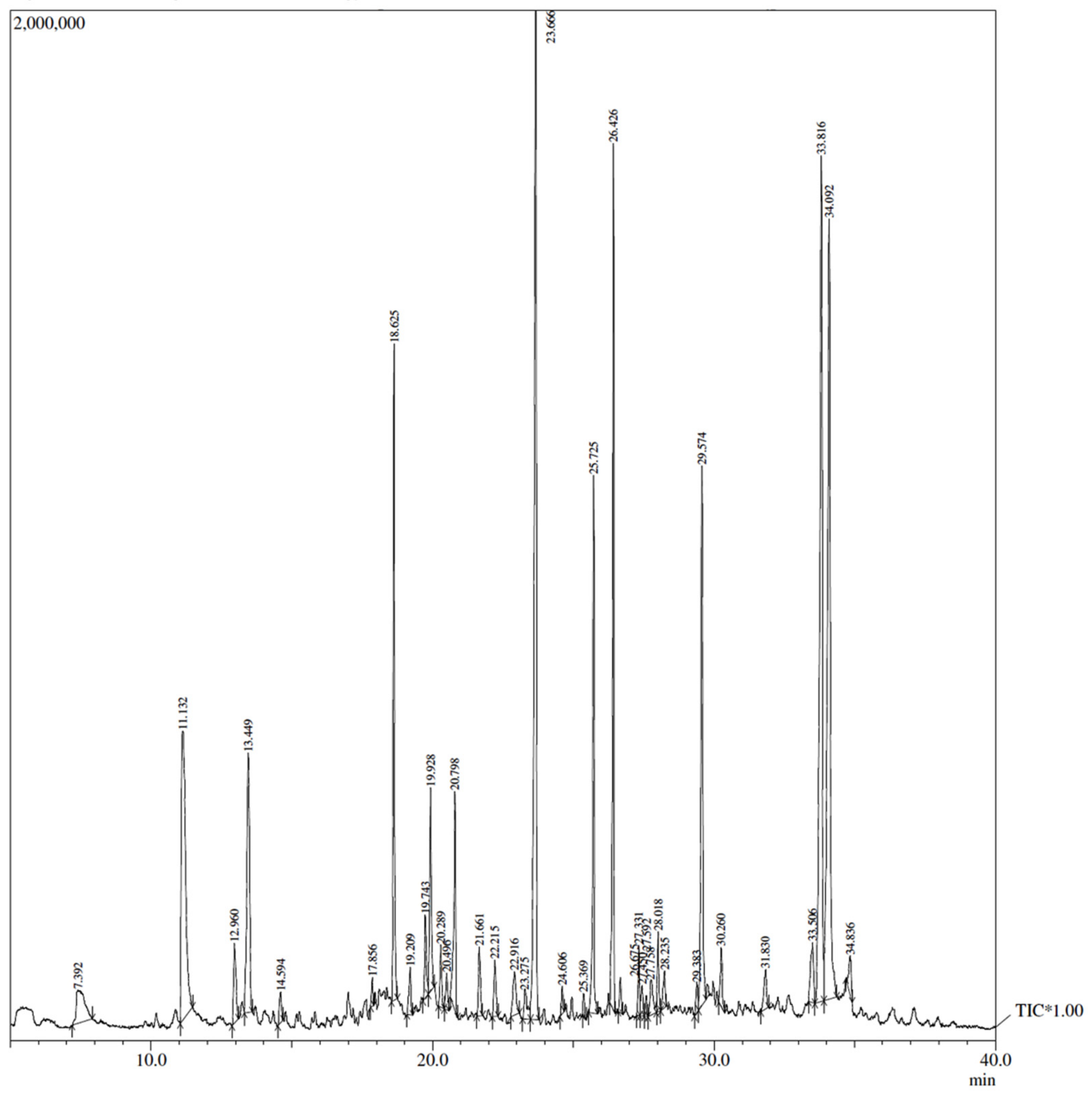
2.3.3. Analysis of the Carum carvi by GC (Gas Chromatography) Coupled with Mass Spectrometry

The Claurus 500 GC from Perkin Elmer (Shelton, CT, USA) coupled to a mass spectrometer was used to conduct the GC–MS evaluation of the extracts. An electron ionization device with an ionization energy of 70 eV was employed for GC–MS detection. With an injection volume of 2 Pl and a constant flow rate of 1 mL/min, helium (99.999%) served as the carrier gas (split ratio of 10:1). The injection temperature was 270 °C, and the ion-source temperature was 280 °C. The temperature of the oven was initially set to begin for 2 min at 110° C, rise to 200 °C at a rate of 10 °C/min, followed by decrease in rate (5 °C/min) to 280 °C, and terminate with an isothermal period of 9 min at 280 °C. At 70 eV, with a 0.5 s scan interval, fragments were used in the mass spectra. The duration of the GC was 30 min. By comparing each component’s regular area of peak to total peak area, the relative percentage elevation of each constituent was computed. Turbo Mass Version 5.2.0 software (Perkin Elmer) was used to decipher mass spectra and chromatograms. The retention time and molecular mass values from Wiley Library were interpreted.

2.4. Post-Mitochondrial Supernatant (PMS) and Microsome Preparation

A specific quantity of tissue was chopped and homogenized in cold saline phosphate buffer (0.1 M) with pH 7.4 and KCl (1.17%) by utilizing a polytron homogenizer for biochemical tests (Kinematica AGPT 3000, Kinematica, Eschbach, Germany). To remove the nuclear debris, the homogenate was subjected to centrifugation (88× g) at 4 °C for 15 min in an Eltek refrigerated centrifuge (RC 4100D, Eltek, Palghar, India) after being filtered using a muslin cloth. The resulting supernatant served as an enzyme source. Some of the PMS was put forth to ultracentrifugation (Beckman L7-55, Beckman, Brea, CA, USA) for 60 min at 4 °C at 100,500 g (34,000 rpm). In phosphate buffer, the pellet was suspended and was believed to be the microsomal fraction.

2.5. In Vitro Parameters

2.5.1. Total Phenolic Composition

The amount of total phenolics was measured by spectrometry method as mentioned by Singleton et al. (1999) [18] using gallic acid as standard phenol. About 0.5 mL of the aqueous extract of Carum carvi was added to 2.5 mL of Folin–Ciocâlteau’s reagent (10%, v/v) and 2.0 mL of sodium carbonate (7.5%). The incubation of the mixture was conducted for 40 min at a temperature of 45 °C, and a spectrometer (Perkin Elmer Lamda Ez 201) was used to calculate the absorbance at 765 nm. The content of total phenolics was estimated and expressed as milligrams of gallic acid equivalent/g of extract.

2.5.2. Estimation of Lipid Peroxidation

The evaluation of lipid peroxidation was performed using the procedure outlined by Write et al. (1981) [19]. The solution mixture was composed of 0.1 M phosphate buffer (0.58 mL) with a pH of 7.4, 100 mM ascorbic acid (0.2 mL), a 0.2 mL microsome fraction, and 100 mM ferric chloride (0.02 mL). At a temperature of 37 °C, the solution was placed in a water bath and heated for 1 h. After adding 10% TCA (1 mL), the reaction was terminated. In all the tubes, 0.67% of TBA with a concentration of 1 mL was added, and, for a duration of 20 min, the tubes were submerged in a steaming water bath. Consequently, all tubes were kept on ice and subjected to centrifugation (2500× g) for about 10 min. At a wavelength of 535 nm, the supernatant optical density was estimated to measure the amount of formed malondialdehyde (MDA) in each tube. Lastly, the molar extinction coefficient (1.56 × 10⁻⁵ M−1·cm−1) was used, and the results were expressed as nmol MDA formed/h/g tissue at 37 °C.

2.5.3. Assay of DNA-Sugar Damage

The method of Halliwell et al. (1981) [20] was used to quantify DNA-sugar damage. About 0.5 mL of calf thymus DNA (1 mg/mL of 0.15 M NaCl) and a mixture composed of phosphate buffer (0.450 mL), and 0.05 mL of ferric chloride (FeCl3) in a final concentration of 100 M were used. In a shaking water bath, the solution was kept at a temperature of 37 °C for 1 h. The addition of 10% TCA (1 mL) was used to stop the reaction. Furthermore, 0.67% TBA (1 mL) was put on, and the liquid solution was heated for 25 min in a steaming water bath. After the reaction material had cooled down, the absorbance was assessed using a Perkin Elmer Lamda Ez 201.

2.5.4. DPPH-Scavenging Activity

The Carum carvi extract’s ability to scavenge DPPH was evaluated using the method described in Fenglin et al. (2004) [21]. The mixture was composed of a 3 mL extract of Carum carvi (1 mg/mL) and DPPH (1 mL of 0.mM in 95% ethanol). After being vortexed, the solution was left for 30 min. The absorbance was calculated with a spectrophotometer at 517 nm (Perkin elmer Lamda Ez 201).
DPPH scavenging activity was calculated as a percentage according to %DPPH scavenging activity = Ab (control) − Ab (sample) × 100, where Ab is the absorbance. A low absorbance of test samples implies high activity of DPPH scavenging, indicating potential free-radical scavenging properties of the test sample.

2.5.5. Ferric Reducing Potential

The method of Oyaizu and Makoto (1986) [22] was used to assess the Carum carvi extract’s reducing capability. About 2.5 mL of potassium ferricyanide (1%), and 0.2 M phosphate buffer (pH 6.6) were used to make up the chemical mixture. The reaction mixture was incubated at 50 °C for 20 min. After adding 2.5 mL of TCA and centrifuging the mixture for 10 min at 4000 rpm, the reaction was stopped. The supernatant (2.5 mL) was collected in a tube to which distilled water (2.5 mL) and 1% FeCl3 (0.5 mL) were then added. At 700 nm, the absorbance was assessed using a Perkin Elmer Lamda Ez 201. Greater reducing potential is indicated by a test sample with a higher absorbance.

2.6. In Vivo Parameters

2.6.1. In Vivo Enzyme Assays and Cytotoxicity Parameters

Xanthine oxidase (XO) activity, glutathione reduced (GSH) content, catalase (CAT), glutathione peroxidase (GPX) and reductase (GR), and lactate dehydrogenase (LDH) activities, serum alanine aminotransferase (ALT) and aspartate aminotransferase (AST) levels, and protein estimation were evaluated as described by Tahir and Sultana (2011) [23].

2.6.2. Cytochrome P450 Content

The total content of cytochrome P450 was estimated using the method of Omufca et al. (1964) [24]. The reaction mixture consisted of a 0.2 mL microsomal fraction and 0.8 mL of phosphate buffer, along with a pinch of sodium dithionite. CO2 was passed through the mixture for 30 s. The optical density was recorded at 450 nm and 490 nm. The total cytochrome content was determined with a molar extinction coefficient of 0.091 M−1·cm−1 and expressed as nmol cytochrome per mg protein.

2.6.3. Quinone Reductase Activity

The approach described by Benson et al. (1980) [25] was used to assess quinone reductase activity. In preparation of the reaction mixture, 25 mM Tris-HCl buffer (2.13 mL) with a pH of 7.4, BSA (0.07 mL), FAD (0.10 mL), 10% of 50 AL PMS, and 0.1 mM of NADPH were used to make up the final volume (3 mL). Reduced dichlorophenolindophenol (DCPIP) was determined by calorimetry (600 nm) with a molar extinction coefficient of 2.1 × 10⁴ M−1·cm−1; the enzymatic activity was estimated as nmol of DCPIP reduced/min/mg protein.

2.6.4. Expression of Caspases 3, 7, and 9

The activity of caspases 3, 7, and 9 was measured using an ELISA kit (CasPASE-3, Assay kit, G-Bioscience, Cat.No:786-202B) according to the instructions of the manufacturer. The data were given in units of µg/mg protein.

2.6.5. Tumor Necrosis Factor-Alpha (TNF-α)

The quantity of TNF-α was determined using an ELISA kit (Rat TNF-α ELISA Ready Set Go, E-bioscience, in San Diego, CA, USA, Cat. No. 887340-22). The samples were prepared in a protease inhibitor cocktail containing phosphate buffer saline (1× PBS) and performed as per the instructions of the manufacturer. The findings were presented as pg/mg tissue protein.

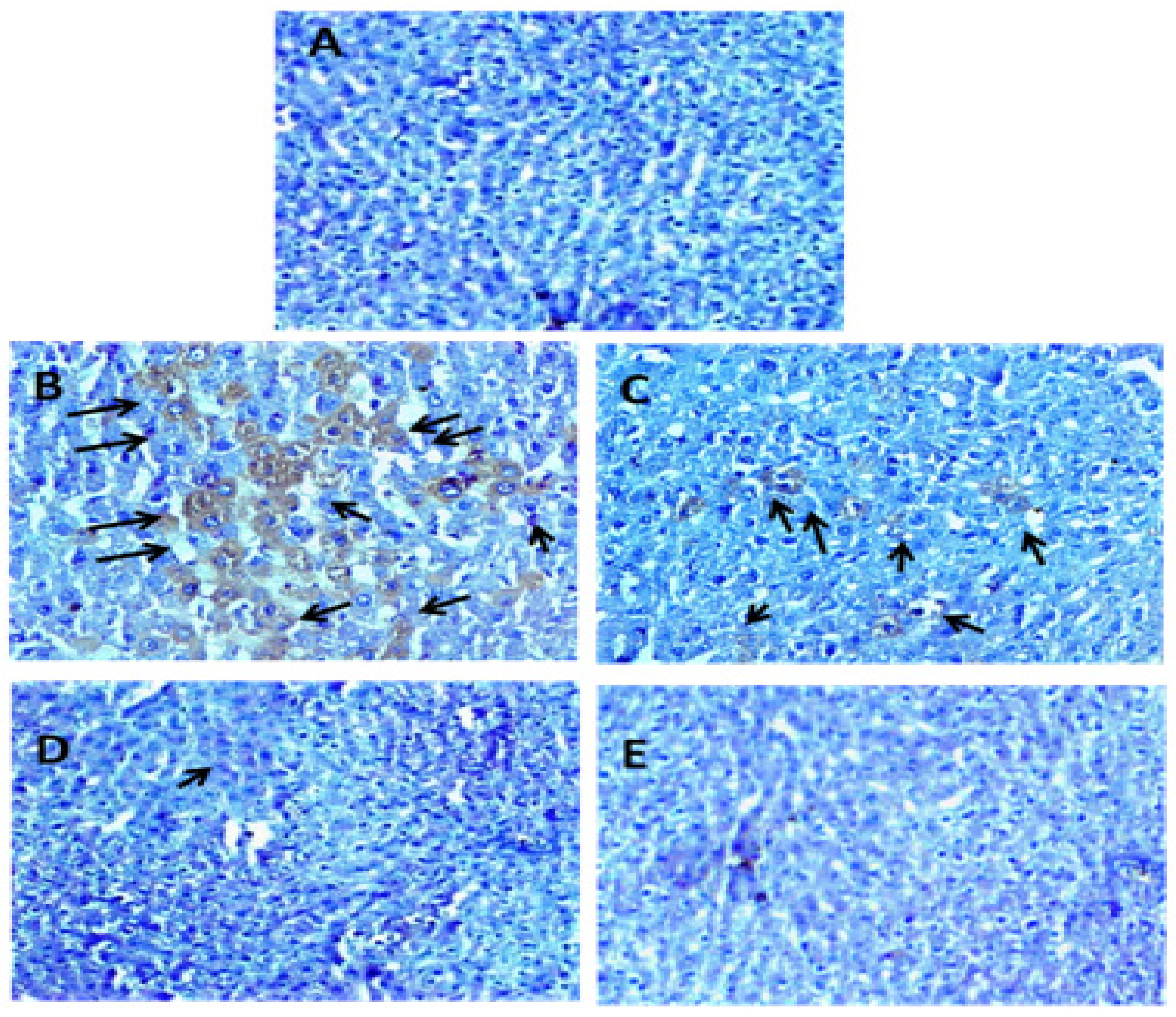
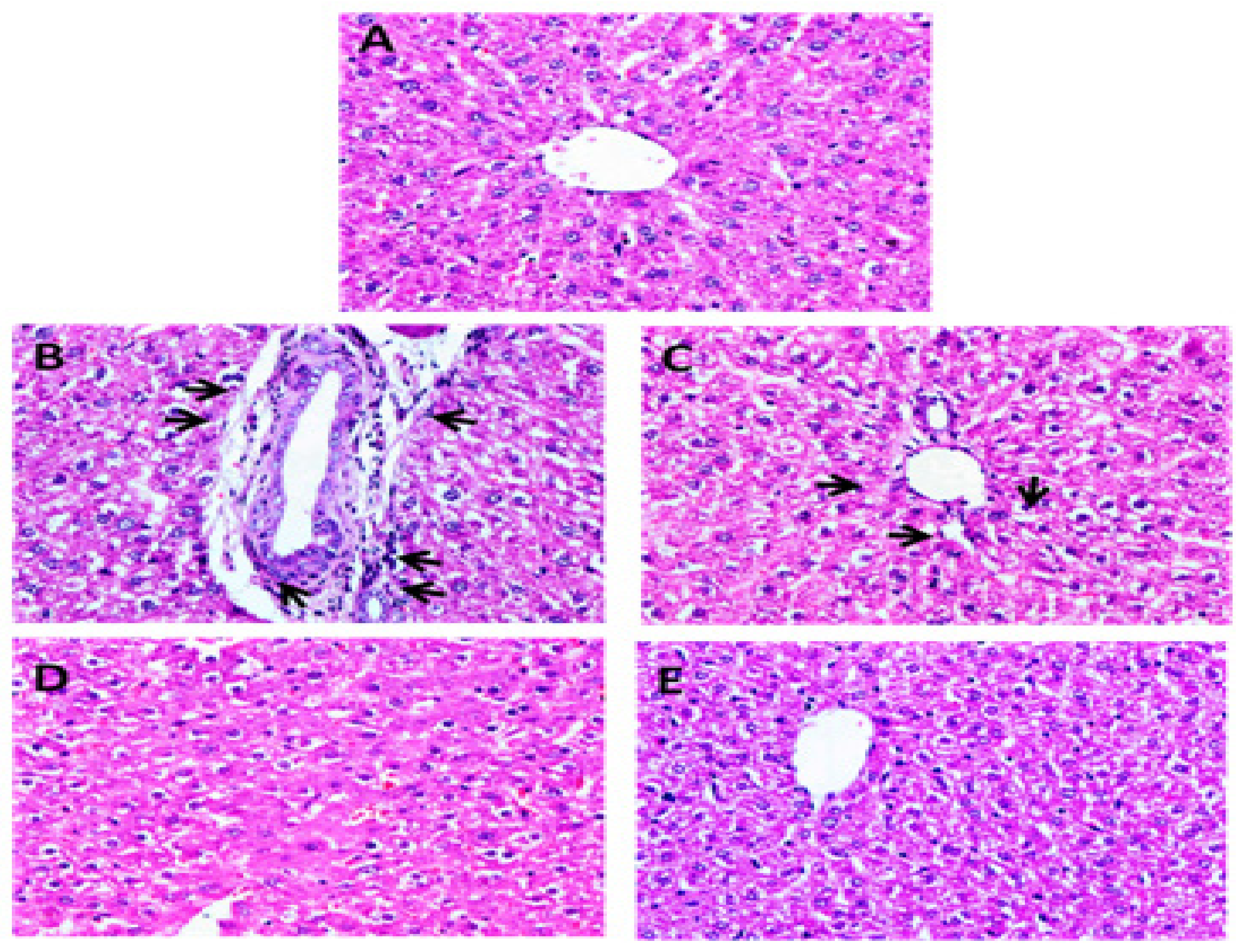
2.6.6. Immunohistochemical Staining of NF-κB and Histopathology

The methods for immunohistochemistry and histopathology were described in our earlier studies [26].

2.7. Statistical Analysis

The mean ± standard error (SE) was used to present the data from every group. The differences among the groups were examined by ANOVA (one-way analysis of variance). The Tukey–Kramer test was used for multiple data comparisons across the groups with a statistical significance of p < 0.05.

3. Results

3.1. In Vitro Antioxidant Potential of Carum carvi

3.1.1. Total Phenolic Content and GC–MS Analysis

Figure 1 depicts the GC–MS analysis of the methanolic extract of Carum carvi. The individual components of the Carum carvi extract are shown in Table 1. The total phenol content in Carum carvi extract was found to be 56 mg GAE/g extract, indicating the potent antioxidant potential of Carum carvi (standard curve equation: y = 0.019x + 0.060; R2 = 0.997).

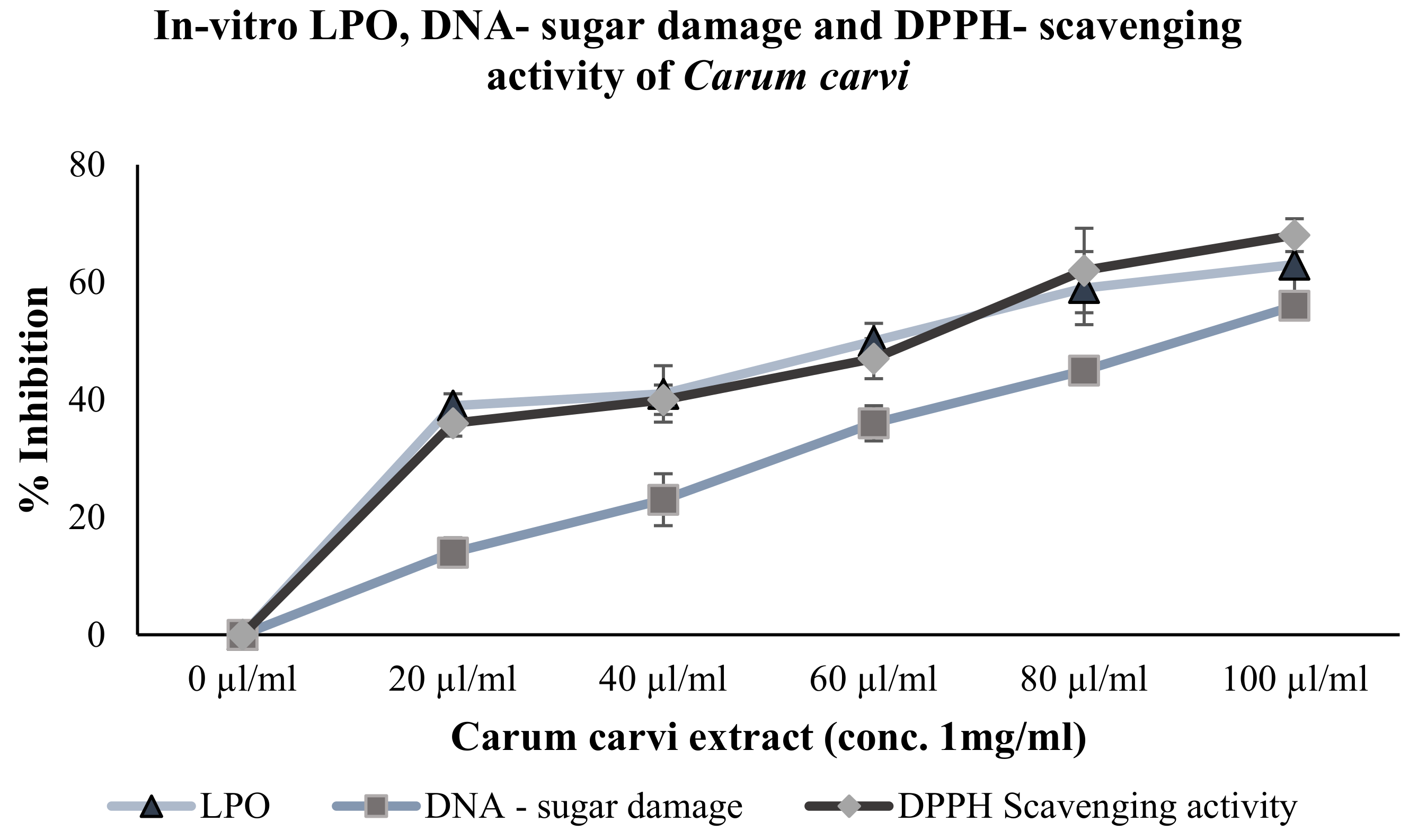
3.1.2. Inhibition of Lipid Peroxidation and DNA-Sugar Damage (In Vitro)

Carum carvi showed concentration-dependent inhibition of LPO and DNA-sugar damage. With increasing concentration (20, 40, 60, 80, and 100 µL) of Carum carvi extract, there was an increase in the inhibition of LPO and in DNA damage by 39%, 41%, 50%, 59%, and 63%, and by 14%, 23%, 36%, 45%, and 56%, respectively (Figure 2).

3.1.3. DPPH-Scavenging Activity

Figure 2 shows the DPPH scavenging activity of Carum carvi extract. Increased DPPH scavenging activity indicates higher antioxidant potential. In this study, the methanolic extract of Carum carvi exhibited concentration-dependent free-radical scavenging activity of 36%, 40%, 47%, 62%, and 68% at 20, 40, 60, 80, and 100 µg/mL, respectively.

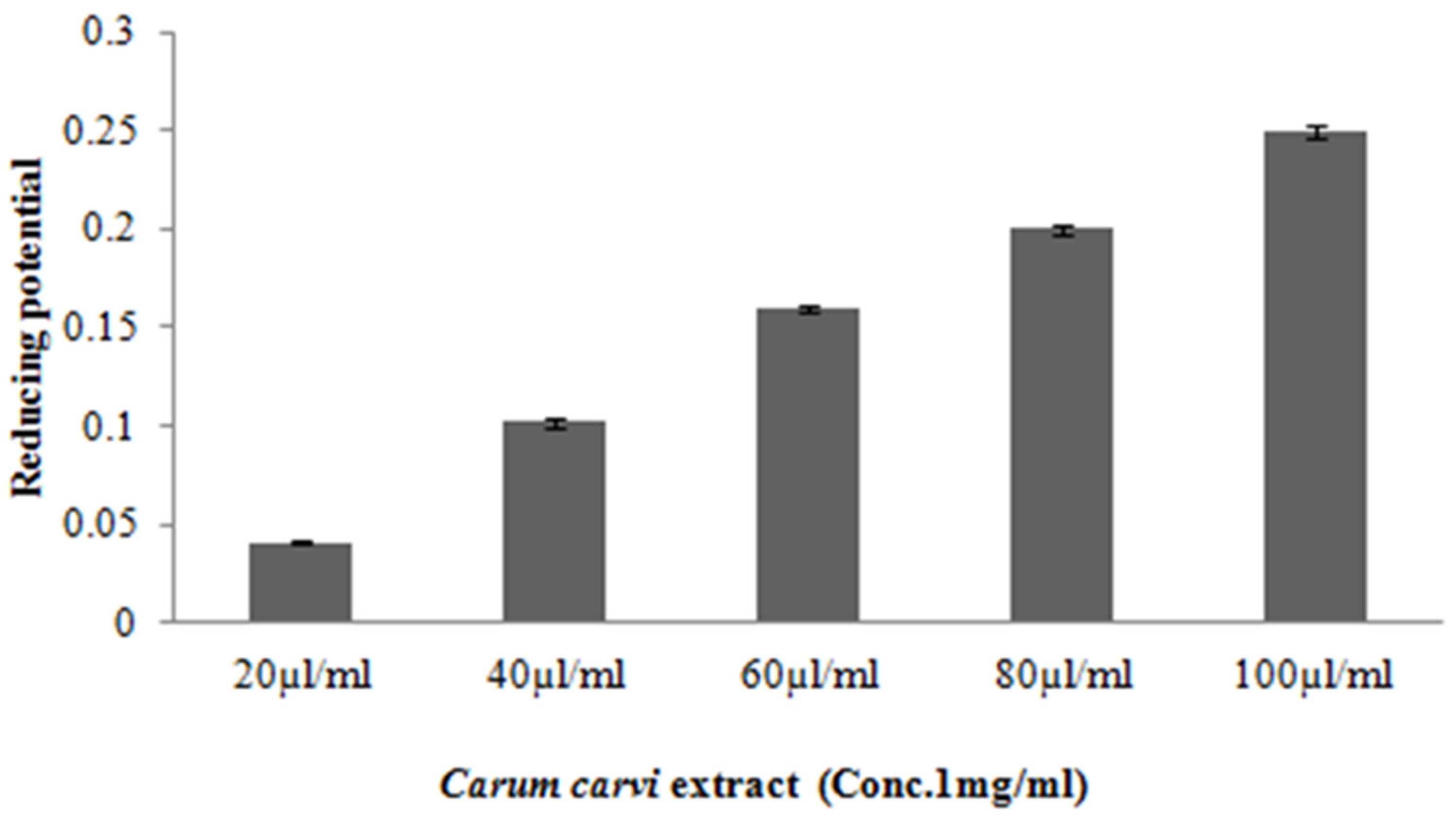
3.1.4. Ferric Reducing Potential

In the present study, it was found that the reducing potential of Carum Carvi increased from 0.041 (±0.001) to 0.25 (±0.003). Figure 3 shows a concentration-dependent increase in the reducing potential of Carum carvi extract, indicating its antioxidant potential.

3.2. Effect of Carum carvi on In Vivo Antioxidant and Hepatoprotective Activities

3.2.1. Effect of Carum carvi on Liver Glutathione (Reduced) Levels, Lipid Peroxidation, and Cytochrome P450

From the data (Table 2), it can be observed that acetaminophen treatment resulted in elevated levels of CYP 450 content as compared with normal control rats. However, Carum carvi (D1: p < 0.01; D2: p < 0.001) treatment restored the levels of cytochrome P450 levels in the liver of rats.
In comparison with control group animals, MDA adduct generation was significantly increased by acetaminophen treatment (14.03 ± 1.15, p < 0.001). Both doses of Carum carvi (D1: p < 0.01; D2: p < 0.001) were found to be significantly effective in lowering lipid peroxidation induced by acetaminophen (Table 2).
GSH levels presented a significant decline (p < 0.001) in acetaminophen group (0.031 ± 0.004) in comparison with control group animals (0.056 ± 0.01). Carum. carvi treatment restored the acetaminophen-induced depletion in GSH levels (D1: 0.046 ± 0.004, p < 0.01; D2: 0.052 ± 0.005, p < 0.001, (Table 3). Animals in Group V, i.e., only Carum carvi, did not show any noteworthy variation in any of the parameters in comparison to the control rats.

3.2.2. Effect of Carum carvi on Intracellular Antioxidant and Phase II Drug-Metabolizing Enzymes such as Quinone Reductase (QR), Glutathione Reductase (GR), and Glutathione S-Transferase (GST) Activity

Increased resistance against acetaminophen-induced hepatotoxicity can be attributed to phase II enzymes such as QR and GST. Our results showed (Table 2) that administration of Carum carvi at D1 and D2 doses to rats led to a substantial increase (p < 0.001) in QR activity in comparison with rats treated with acetaminophen. Hepatic tissues from acetaminophen treatment groups exhibited a substantial elevation (p < 0.001) in GR activity (Table 3) as compared with control rats. Both doses of Carum carvi treatment resulted in significant restoration (D1: p < 0.01; D2: p < 0.001) of GR activity in rat liver tissues.
A significant decrease (p < 0.001) in GST activity was observed in acetaminophen-treated rats in comparison to the control animals. A substantial elevation in activity of GSH was found in animals with Carum carvi treatment (D1: p < 0.01; D2: p < 0.001) in contrast to animals treated with acetaminophen (see Table 3).

3.2.3. Carum carvi Administration Mediates Alteration of Antioxidant/Oxidant Enzymatic Activities in Liver Tissue

A substantial (p < 0.001) decline in catalase activity was found in the acetaminophen-treated group (29.41 ± 2.74) as compared with the liver samples of control animals (42.27 ± 1.29). A significant enhancement (D1: p < 0.05; D2: p < 0.01) in CAT (liver tissues) was found in the Carum carvi treatment group. No differences were observed in CAT activity in rats administered with Carum carvi alone in comparison to the control animal group (Table 2). XO reflected a significant increase (0.21 ± 0.08, p < 0.001) in the hepatic activity of enzymes (Table 2) in tissues of the paracetamol treatment groups in contrast to the control animal group (0.11 ± 0.06). The concentration of XO activity was significantly restored in liver tissue by Carum carvi administration at dosages D1 and D2 (0.15 ± 0.04, p < 0.01 and 0.12 ± 0.03, p < 0.001 respectively). The D2 group, as compared with animals in the control group (Table 2), showed no significant change. Animals with acetaminophen treatment presented substantial depletion (p < 0.001) of glutathione peroxidase (GPx) activity (138.4 ± 16.8) as compared with rats in the control group. GPx activity was significantly (D1 and D2: p < 0.01) restored in rats treated with Carum carvi. The Carum carvi only group did not show any significant differences in GPx activity in comparison with the control group (Table 3).

3.2.4. Levels of Serum Toxicity Markers in Rats Treated with Carum carvi

In this study, acetaminophen was found to cause a substantial increase (p < 0.001) in the levels of AST, ALT, and LHD by 61%, 86%, and 63% respectively. Treatment of Carum carvi showed a significant effect on reversing the increased levels of serum toxicity markers by 30%, 45%, and 36% at dose D1 and by 55%, 75%, and 57% at dose D2 of Carum carvi in comparison with the acetaminophen-treated animal group, indicating antioxidant properties and a membrane-stabilizing effect of Carum carvi (Table 4).

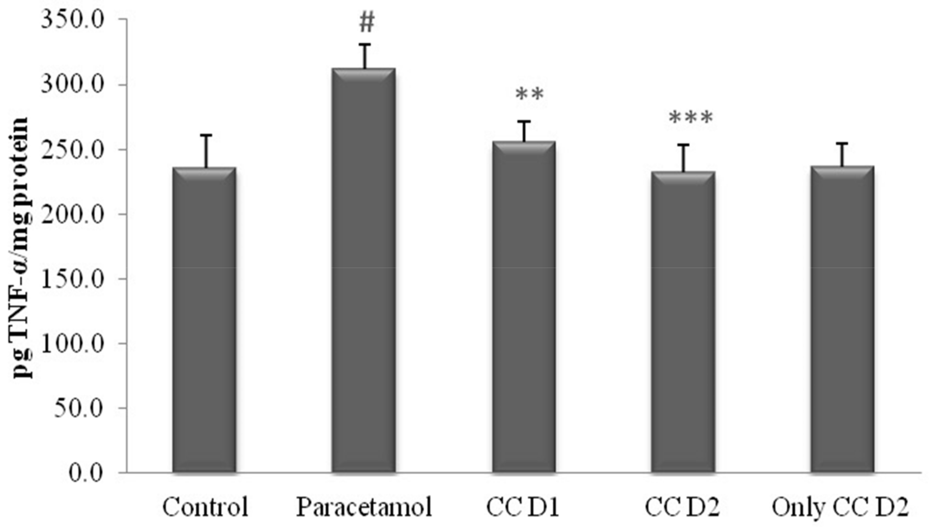
3.2.5. Effect of Carum carvi on Acetaminophen-Induced TNF-α Levels

The concentration of TNF-alpha, a cytokine marker of systemic inflammation, was monitored in rat hepatic tissues treated with acetaminophen for 1 week. Figure 4 indicates that acetaminophen caused an increase in hepatic TNF-alpha levels. Treatment for the same period with the extract of Carum carvi tangibly decreased the TNF-alpha levels compared to those obtained in the acetaminophen-treated group.

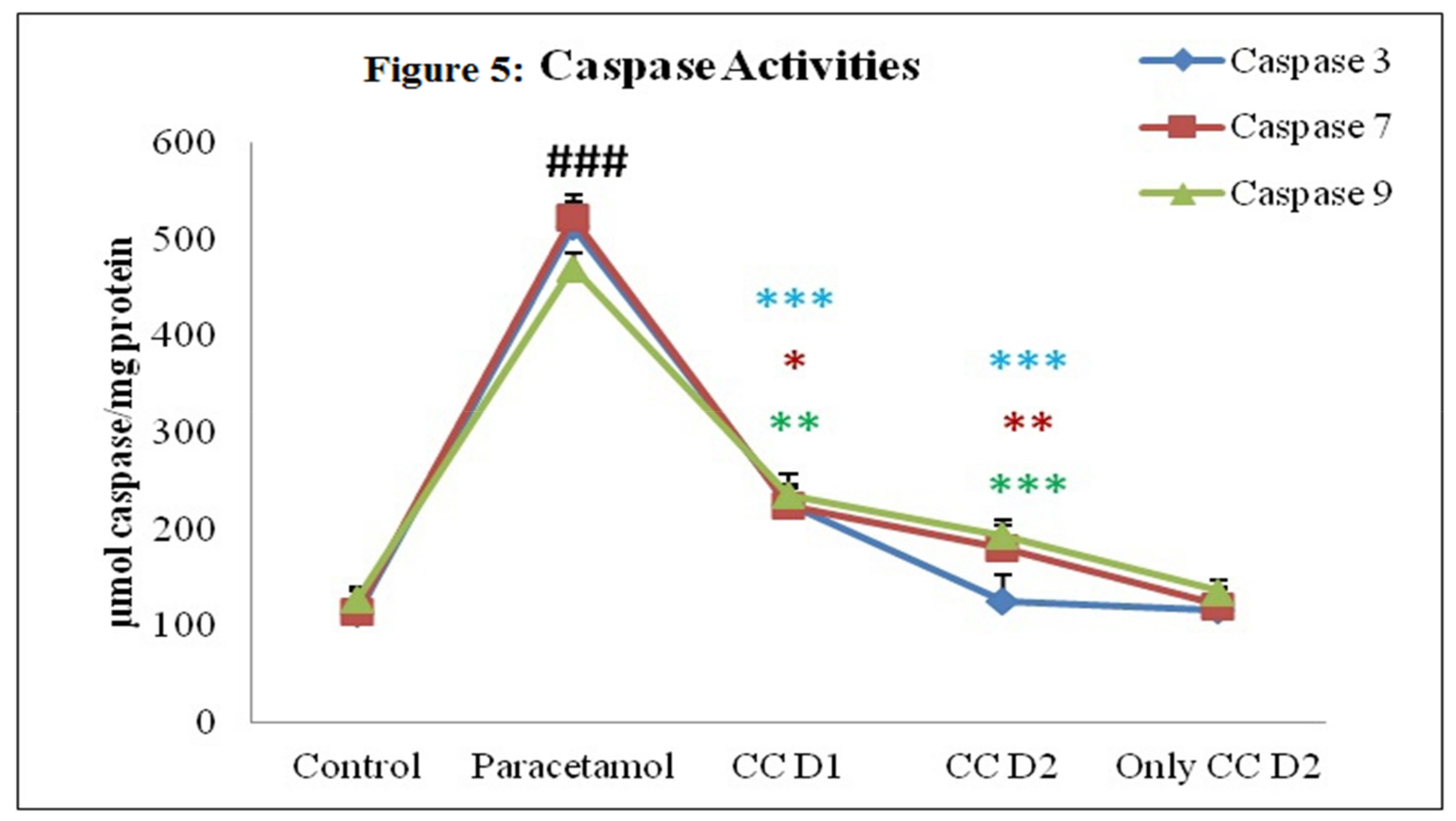
3.2.6. Effect of Carum carvi Extract on Caspase Activity

Our results revealed a significant increase (p < 0.001) in caspase activity in liver tissues of rats with acetaminophen treatment in contrast to control group rats. A significant restoration of caspases was found in rat liver tissues treated with C. carvi extract (Figure 5).

3.2.7. Effect of Carum carvi Extract on Regulation of NF-κB as a Result of Paracetamol Treatment

In this study, we determined the expression of NF-κB protein (Figure 6), and it was found that control rats showed almost no expression of NF-κB (Figure 6A). The acetaminophen-treated group showed maximum nuclear translocation of NF-κB (Figure 6B), and the animals treated with Carum carvi (at dose D1) showed less expression of NF-κB (Figure 6C). Animals of group IV (D2 + acetaminophen) (Figure 6D) and V (only D2) (Figure 6E) did not show any difference in expression of NF-κB compared to the control group.

3.3. Histopathology

Liver micrographs (Figure 7) of the present study showed that centrilobular spaces presented with mild hepatocellular necrosis and sinusoidal space congestion. Treatment with Carum carvi in acetaminophen-treated rats showed a potential protective effect.

4. Discussion

Plants synthesize aromatic substances such as phenols or their oxygenated derivatives. Most of these are recognized as flavoring compounds, while some herbs and spices that are typically used to season food also carry compounds that hold medicinal value.
Generally, extracts that have high phenolic content exhibit good antioxidant activity. Our results suggest that Carum carvi possesses strong potential to inhibit oxidative stress-induced lipid peroxidation and in vitro DNA damage. Our results were also in accordance with the DPPH-scavenging property of Carum carvi, indicating the manifestation of potent antioxidant activity [27]. Another significant indicator of antioxidant potential lies in the plant extract’s reducing capacity [28]. Carum carvi extract transformed the Fe3+/ferricyanide complex to a ferrous state. Carum carvi extract was found to have a dose-dependent increase in reducing power at concentrations between 20 and 100 µL/ml. In cases of paracetamol overdose, a toxic metabolic product, i.e., N-acetyl-p-benzo-quinone-imine (NAPQI), is produced and metabolized via CYP 450 [29]. This highly reactive byproduct in turn alkylates and oxidizes intracellular GSH, resulting in GSH depletion in the liver, which ultimately leads to increased peroxidative activity. Elevation in peroxidation of lipids results in disruption of mitochondrial and nuclear functions, leading to oxidative stress [30]. Any agent that has the potential to suppress the levels of CYP 450 can protect the tissues from the deleterious effect of acetaminophen toxicity, such as depletion in reduced glutathione and peroxidation of membrane lipids, leading to cellular toxicity. Lipid peroxidation, measured by MDA formation is an important marker to study cellular toxicity. It has been observed that enzymes such as QR and GST may promote resistance to hepatotoxicity induced by acetaminophen [31]. We observed that acetaminophen toxicity led to lipid peroxidation, resulting in the membrane destabilization of hepatocytes, which in turn resulted in the depletion of phase II enzymes produced by hepatocytes. In the present study, we observed that GST levels were depleted by the acetaminophen administration to rats, which may have been due to the damage caused by acetaminophen to the liver cells, resulting in the poor functioning of hepatocytes. However, high levels of oxidative species production during acetaminophen toxicity can attack biological macromolecules, leading to peroxidation of lipids and exhaustion of antioxidant enzymes (CAT, GPx, etc.) and, thus, resulting in further stressed conditions.
In cells with excessive oxidative stress, the levels of xanthine oxidase (a non-cytochrome oxidative enzyme) are significantly higher, resulting from the interaction of cellular macromolecules with free radicals. It has been observed that xanthine oxidase is actively involved in the metabolism of acetaminophen. In determining liver toxicity induced by acetaminophen, elevated activities of xanthine oxidase have been reported [32]. Thus, depletion of antioxidant enzymes could be the result of a decrease in glutathione content caused by acetaminophen toxicity [33]. A significant increase in enzymatic activities was observed when the animals were treated with Carum carvi, thus indicating protection against acetaminophen-induced liver toxicity by upregulation of the antioxidant defense system in liver cells. This restoration of phase II enzymes by Carum carvi was also supported by [34].
It has been depicted that, in acetaminophen-induced liver toxicity, apoptosis (programmed cell death) plays a significant role [35]. Thus, for the prevention of acetaminophen-induced liver toxicity, interventions to prevent apoptosis can serve as one of the promising tools. In this study, evaluation of effector caspase (3 and 7) and initiator caspase (caspase 9) activities was carried out in acetaminophen-induced liver toxicity in rats. Fodrin, poly (ADP-ribose) polymerase (PARP), and nuclear lamin proteins are degraded or cleaved by effector caspases 3 and 7; therefore, these caspases are considered to be among the key executioners of apoptosis. An attempt was made to determine if Carum carvi extract could reduce DNA damage and apoptosis in the liver of acetaminophen-treated rats. The decline in caspase activity by Carum carvi action indicates the mechanism via which Carum carvi alleviated acetaminophen-induced apoptosis and toxicity.
Oxidative stress promotes the activation of proinflammatory genes, which eventually stimulate inflammatory cells to overproduce ROS. This vicious cycle leads to the onset and progression of numerous diseases. As a result, the liver’s detoxification system is crucial when the body is exposed to toxic substances, and the inflammatory process can exacerbate tissue damage and result in poor healing [36]. There is growing confirmation that paracetamol intoxication can raise several proinflammatory cytokine levels in the blood. Oxidative stress activates signal transduction pathways involving NF-κB and activator protein-1 (AP-1) [37]. A vital signaling pathway is NF-κB, which facilitates the synthesis of molecules associated with inflammation, such as by incorporating NF-κB-binding motifs in their promoters. When paracetamol is administered, the NF-κB protein is activated, which increases the activity of iNOS, TNF-α, and COX-2 [38]. Inflammation plays a crucial role in acetaminophen overdose-induced liver toxicity, which is mediated by the activation of transcription factor NF-κB and the secretion of several cytokines such as TNF-α and IL-1β. Activated NF-κB is then translocated into the nucleus, resulting in the induction of chemokines and proinflammatory enzymes, including iNOS and COX-2. It has been reported that acetaminophen overdose leads to an inflammatory reaction and oxidative stress, resulting in the production of iNOS and reactive nitrogen species. Both iNOS and COX-2 exert their activities in a similar way by inducing inflammatory response [39]. Furthermore, iNOS expression causes an excessive amount of NO to be produced, which heightens the immune response by triggering inflammatory cell signaling transduction. Therefore, it is assumed that decreasing NO levels by blocking iNOS expression is a desirable first step in assessing the effectiveness of novel drugs in the management of inflammatory conditions [40]. Our results completely coincide with those reported by Jaeschke et al., whereby acetaminophen administration to rats resulted in higher expressions on NF-κB and TNF-α. Carum carvi exhibited potential efficacy in alleviating acetaminophen-induced expression of NF-κB and TNF-α. The anti-inflammatory properties of Carum carvi have been reported by researchers in experiments including colitis [41]. Similar findings of anti-inflammatory properties have been reported by various research studies [42,43,44]. Moreover, studies have confirmed that Carum carvi possesses effects in wound healing, liver protection, antioxidant, pain management, antifungal, and diuretic properties [42,44,45,46].
Therefore, the findings postulate that Carum carvi could prove to be a vital molecule to suppress the drug-mediated hepatotoxicity by protecting liver cells; however, further studies are needed.

5. Conclusions

The food spice, Carum carvi, used in the present study, is a regular food component of large populations of Asia and other regions of the world. Our results provide strong evidence that Carum carvi is a dietary constituent which possesses powerful antioxidant potential, hepatoprotective efficacy, and an inhibitory effect toward acetaminophen-induced oxidative stress, liver toxicity, apoptosis, and expression of inflammatory mediators. Pharmacokinetic and pharmacodynamic studies are needed in the future to investigate Carum carvi for clinical trials. More studies are required to strongly recommend the use of Carum carvi so as to establish mechanisms that can decipher the active constituents that are really responsible for its pharmacological properties.

Author Contributions

Conceptualization, T.M.M. and S.S.; Writing—Original Draft Preparation, T.M.M. and M.U.R., Investigation, T.M.M., W.Q. and R.K., Project Administration, S.S. and T.M.M., Writing—Reviewing and Editing, M.K.A., A.A., S.S., Resource, M.A., A.A., W.Q., R.K., Funding acquisition, S.A., M.A. All authors have read and agreed to the published version of the manuscript.

Funding

This research was funded by the Researchers Supporting Project Number (RSP-2021/115) at King Saud University, Riyadh, Saudi Arabia.

Institutional Review Board Statement

All relevant procedures related to animal health in this study were performed in compliance with the aforementioned laws and guidelines of the Committee for the Purpose of Control and Supervision of Experiments on Animals (CPCSEA), Government of India, and prior permission was sought from the Institutional Animal Ethics Committee (IAEC No: 173/CPCSEA, 28 January 2000).

Informed Consent Statement

Not applicable.

Data Availability Statement

Available on request to corresponding author.

Acknowledgments

Authors are thankful to Researchers Supporting Project (number RSP–2021/115) at King Saud University, Riyadh, Saudi Arabia. The authors acknowledge the help and support provided by the Central Council for Research in Unani Medicine, Ministry of Health and Family Welfare, Government of India. All authors are thankful to Mohammad Khalid Ashfaq for checking the manuscript for English language.

Conflicts of Interest

The authors declare no conflict of interest.

References

  1. Tujios, S.; Fontana, R.J. Mechanisms of drug-induced liver injury: From bedside to bench. Nat. Rev. Gastroenterol. Hepatol. 2011, 8, 202–211. [Google Scholar] [CrossRef] [PubMed]
  2. Chen, S.; Zhao, X.; Ran, L.; Wan, J.; Wang, X.; Qin, Y.; Shu, F.; Gao, Y.; Yuan, Q.; Zhang, Q.; et al. Resveratrol improves insulin resistance, glucose and lipid metabolism in patients with non-alcoholic fatty liver disease: A randomized controlled trial. Dig. Liver Dis. 2015, 47, 226–232. [Google Scholar] [CrossRef] [PubMed]
  3. Singh, D.; Cho, W.C.; Upadhyay, G. Drug-Induced Liver Toxicity and Prevention by Herbal Antioxidants: An Overview. Front. Physiol. 2016, 6, 363. [Google Scholar] [CrossRef] [Green Version]
  4. Grattagliano, I.; Bonfrate, L.; Diogo, C.V.; Wang, H.H.; Wang, D.Q.; Portincasa, P. Biochemical mechanisms in drug-induced liver injury: Certainties and doubts. World J. Gastroenterol. WJG 2009, 15, 4865. [Google Scholar] [CrossRef]
  5. De Martin, E.; Michot, J.-M.; Rosmorduc, O.; Guettier, C.; Samuel, D. Liver toxicity as a limiting factor to the increasing use of immune checkpoint inhibitors. JHEP Rep. 2020, 2, 100170. [Google Scholar] [CrossRef] [PubMed]
  6. Chiu, H.; Gardner, C.R.; Dambach, D.M.; Brittingham, J.A.; Durham, S.K.; Laskin, J.D.; Laskin, D.L. Role of p55 tumor necrosis factor receptor 1 in acetaminophen-induced antioxidant defense. Am. J. Physiol.-Gastrointest. Liver Physiol. 2003, 285, G959–G966. [Google Scholar] [CrossRef] [Green Version]
  7. Fisher, E.S.; Curry, S.C. Evaluation and treatment of acetaminophen toxicity. Adv. Pharmacol. 2019, 85, 263–272. [Google Scholar]
  8. Wong, L.T.; Whitehouse, L.W.; Solomonraj, G.; Paul, C.J. Pathways of disposition of acetaminophen conjugates in the mouse. Toxicol. Lett. 1981, 9, 145–151. [Google Scholar] [CrossRef]
  9. Bessems, J.G.M.; Vermeulen, N.P.E. Paracetamol (acetaminophen)-induced toxicity: Molecular and biochemical mechanisms, analogues and protective approaches. Crit. Rev. Toxicol. 2001, 31, 55–138. [Google Scholar] [CrossRef]
  10. Mazaleuskaya, L.L.; Sangkuhl, K.; Thorn, C.F.; FitzGerald, G.A.; Altman, R.B.; Klein, T.E. PharmGKB summary: Pathways of acetaminophen metabolism at the therapeutic versus toxic doses. Pharm. Genom. 2015, 25, 416–426. [Google Scholar] [CrossRef] [Green Version]
  11. Adamson, G.M.; Harman, A.W. Oxidative stress in cultured hepatocytes exposed to acetaminophen. Biochem. Pharmacol. 1993, 45, 2289–2294. [Google Scholar] [CrossRef]
  12. Kamiyama, T.; Sato, C.; Liu, J.; Tajiri, K.; Miyakawa, H.; Marumo, F. Role of lipid peroxidation in acetaminophen-induced hepatotoxicity: Comparison with carbon tetrachloride. Toxicol. Lett. 1993, 66, 7–12. [Google Scholar] [CrossRef]
  13. Blazka, M.E.; Germolec, D.R.; Simeonova, P.; Bruccoleri, A.; Pennypacker, K.R.; Luster, M.I. Acetaminophen-induced hepatotoxicity is associated with early changes in NF-kB and NF-IL6 DNA binding activity. J. Inflamm. 1995, 47, 138–150. [Google Scholar]
  14. Laskin, D.L.; Gardner, C.R. Nonparenchymal cells, inflammatory macrophages, and hepatotoxicity. In Drug-Induced Liver Diseases; Marcel Dekker: New York, NY, USA, 2003; pp. 183–212. [Google Scholar]
  15. Laskin, D.L.; Pilaro, A.M.; Ji, S. Potential role of activated macrophages in acetaminophen hepatotoxicity: II. Mechanism of macrophage accumulation and activation. Toxicol. Appl. Pharmacol. 1986, 86, 216–226. [Google Scholar] [CrossRef]
  16. Holtmann, G.; Haag, S.; Adam, B.; Funk, P.; Wieland, V.; Heydenreich, C.-J. Effects of a fixed combination of peppermint oil and caraway oil on symptoms and quality of life in patients suffering from functional dyspepsia. Phytomedicine 2003, 10, 56–57. [Google Scholar] [CrossRef] [Green Version]
  17. Johri, R.K. Cuminum cyminum and Carum carvi: An update. Pharmacogn. Rev. 2011, 5, 63–72. [Google Scholar] [CrossRef] [Green Version]
  18. Singleton, V.L.; Orthofer, R.; Lamuela-Raventós, R.M. Analysis of total phenols and other oxidation substrates and antioxidants by means of folin-ciocalteu reagent. In Methods in Enzymology; Academic Press: Cambridge, MA, USA, 1999; Volume 299, pp. 152–178. [Google Scholar]
  19. Wright, J.R.; Colby, H.D.; Miles, P.R. Cytosolic factors which affect microsomal lipid peroxidation in lung and liver. Arch. Biochem. Biophys. 1981, 206, 296–304. [Google Scholar] [CrossRef]
  20. Halliwell, B.; Gutteridge, J.M.C. Formation of a thiobarbituric-acid-reactive substance from deoxyribose in the presence of iron salts: The role of superoxide and hydroxyl radicals. FEBS Lett. 1981, 128, 347–352. [Google Scholar] [CrossRef] [Green Version]
  21. Fenglin, H.; Ruili, L.; Liang, M. Free radical scavenging activity of extracts prepared from fresh leaves of selected Chinese medicinal plants. Fitoterapia 2004, 75, 14–23. [Google Scholar] [CrossRef]
  22. Oyaizu, M. Studies on products of browning reaction antioxidative activities of products of browning reaction prepared from glucosamine. Jpn. J. Nutr. Diet. 1986, 44, 307–315. [Google Scholar] [CrossRef] [Green Version]
  23. Tahir, M.; Sultana, S. Chrysin modulates ethanol metabolism in Wistar rats: A promising role against organ toxicities. Alcohol Alcohol. 2011, 46, 383–392. [Google Scholar] [CrossRef] [PubMed] [Green Version]
  24. Omufca, T.; Sato, R. The carbon monoxide-binding pigment of liver microsomes. J. Biol. Chem. 1964, 239, 2379–2385. [Google Scholar]
  25. Benson, A.M.; Hunkeler, M.J.; Talalay, P. Increase of NAD (P) H: Quinone reductase by dietary antioxidants: Possible role in protection against carcinogenesis and toxicity. Proc. Natl. Acad. Sci. USA 1980, 77, 5216–5220. [Google Scholar] [CrossRef] [PubMed]
  26. Tahir, M.; Rehman, M.U.; Lateef, A.; Khan, R.; Khan, A.Q.; Qamar, W.; Ali, F.; O’Hamiza, O.; Sultana, S. Diosmin protects against ethanol-induced hepatic injury via alleviation of inflammation and regulation of TNF-α and NF-κB activation. Alcohol 2013, 47, 131–139. [Google Scholar] [CrossRef] [PubMed]
  27. Wu, H.-C.; Shiau, C.-Y.; Chen, H.-M.; Chiou, T.-K. Antioxidant activities of carnosine, anserine, some free amino acids and their combination. J. Food Drug Anal. 2003, 11, 13. [Google Scholar] [CrossRef]
  28. Chandrasekar, M.J.N.; Bommu, P.; Nanjan, M.L.J.; Suresh, B. Chemoprotective effect of Phyllanthus maderaspatensis. In modulating cisplatin-induced nephrotoxicity and genotoxicity. Pharm. Biol. 2006, 44, 100–106. [Google Scholar] [CrossRef] [Green Version]
  29. Przybyła, G.W.; Szychowski, K.A.; Gmiński, J. Paracetamol—An old drug with new mechanisms of action. Clin. Exp. Pharmacol. Physiol. 2020, 48, 3–19. [Google Scholar] [CrossRef]
  30. Sabir, S.M.; Rocha, J.B.T. Water-extractable phytochemicals from Phyllanthus niruri exhibit distinct in vitro antioxidant and in vivo hepatoprotective activity against paracetamol-induced liver damage in mice. Food Chem. 2008, 111, 845–851. [Google Scholar] [CrossRef]
  31. Nagi, M.N.; Almakki, H.A.; Sayed-Ahmed, M.M.; Al-Bekairi, A.M. Thymoquinone supplementation reverses acetaminophen-induced oxidative stress, nitric oxide production and energy decline in mice liver. Food Chem. Toxicol. 2010, 48, 2361–2365. [Google Scholar] [CrossRef]
  32. Hinson, J.A.; Bucci, T.J.; Irwin, L.K.; Michael, S.L.; Mayeux, P.R. Effect of inhibitors of nitric oxide synthase on acetaminophen-induced hepatotoxicity in mice. Nitric Oxide 2002, 6, 160–167. [Google Scholar] [CrossRef]
  33. Kozer, E.; Evans, S.; Barr, J.; Greenberg, R.; Soriano, I.; Bulkowstein, M.; Petrov, I.; Chen-Levi, Z.; Barzilay, B.; Berkovitch, M. Glutathione, glutathione-dependent enzymes and antioxidant status in erythrocytes from children treated with high-dose paracetamol. Br. J. Clin. Pharmacol. 2003, 55, 234–240. [Google Scholar] [CrossRef] [PubMed]
  34. Ajiboye, T.O.; Salau, A.K.; Yakubu, M.T.; Oladiji, A.T.; Akanji, M.A.; Okogun, J.I. Acetaminophen perturbed redox homeostasis in Wistar rat liver: Protective role of aqueous Pterocarpus osun leaf extract. Drug Chem. Toxicol. 2010, 33, 77–87. [Google Scholar] [CrossRef] [PubMed]
  35. Kon, K.; Kim, J.-S.; Jaeschke, H.; Lemasters, J.J. Mitochondrial permeability transition in acetaminophen-induced necrosis and apoptosis of cultured mouse hepatocytes. Hepatology 2004, 40, 1170–1179. [Google Scholar] [CrossRef] [PubMed]
  36. Yang, R.; Song, C.; Chen, J.; Zhou, L.; Jiang, X.; Cao, X.; Zhang, Q.I. Limonin ameliorates acetaminophen-induced hepatotoxicity by activating Nrf2 antioxidative pathway and inhibiting NF-κB inflammatory response via upregulating Sirt1. Phytomedicine 2020, 69, 153211. [Google Scholar] [CrossRef]
  37. Jaeschke, H.; Knight, T.R.; Bajt, M.L. The role of oxidant stress and reactive nitrogen species in acetaminophen hepatotoxicity. Toxicol. Lett. 2003, 144, 279–288. [Google Scholar] [CrossRef]
  38. Ren, S.; Leng, J.; Xu, X.Y.; Jiang, S.; Wang, Y.P.; Yan, X.T.; Li, W. Ginsenoside Rb1, a major saponin from Panax ginseng, exerts protective effects against acetaminophen-induced hepatotoxicity in mice. Am. J. Chin. Med. 2019, 47, 1815–1831. [Google Scholar] [CrossRef]
  39. Lin, H.-H.; Hsu, J.-Y.; Tseng, C.-Y.; Huang, X.-Y.; Tseng, H.-C.; Chen, J.-H. Hepatoprotective Activity of Nelumbo nucifera Gaertn. Seedpod Extract Attenuated Acetaminophen-Induced Hepatotoxicity. Molecules 2022, 27, 4030. [Google Scholar] [CrossRef]
  40. Yi, R.K.; Song, J.L.; Lim, Y.I.; Kim, Y.K.; Park, K.Y. Preventive effect of the Korean traditional health drink (Taemyeongcheong) on acetaminophen-induced hepatic damage in ICR Mice. Prev. Nutr. Food Sci. 2015, 20, 52. [Google Scholar] [CrossRef] [Green Version]
  41. Keshavarz, A.; Minaiyan, M.; Ghannadi, A.; Mahzouni, P. Effects of Carum carvi L. (Caraway) extract and essential oil on TNBS-induced colitis in rats. Res. Pharm. Sci. 2013, 8, 1–8. [Google Scholar]
  42. De Carvalho, C.C.C.R.; da Fonseca, M.M.R. Carvone: Why and how should one bother to produce this terpene. Food Chem. 2006, 95, 413–422. [Google Scholar] [CrossRef]
  43. Seddighfar, M.; Mirghazanfari, S.M.; Dadpay, M. Analgesic and anti-inflammatory properties of hydroalcoholic extracts of Malva sylvestris, Carum carvi or Medicago sativa, and their combination in a rat model. J. Integr. Med. 2020, 18, 181–188. [Google Scholar] [CrossRef] [PubMed]
  44. Agrahari, P.; Singh, D.K. A review on the pharmacological aspects of Carum carvi. J. Biol. Earth Sci. 2014, 4, M1–M13. [Google Scholar]
  45. Saleh, M.A.; Clark, S.; Woodard, B. Antioxidant and free radical scavenging activities of essential oils. Ethn. Dis. 2010, 20, 78–82. [Google Scholar]
  46. Khayyal, M.T.; El-Ghazaly, M.A.; Kenawy, S.A.; Seif-El-Nasr, M.; Mahran, L.G.; Kafafi, Y.A.; Okpanyi, S.N. Antiulcerogenic effect of some gastrointestinally acting plant extracts and their combination. Arzneimittelforschung 2001, 51, 545–553. [Google Scholar] [CrossRef]
Figure 1. GC–MS chromatogram of Carum carvi showing peaks of individual constituents.
Applsci 12 11010 g001
Figure 2. In vitro DPPH-scavenging activity, LPO inhibition, and DNA-sugar damage by Carum carvi extract. Representative graphs showing concentration-dependent activity of Carum carvi in terms of DPPH scavenging, LPO inhibition, and DNA-sugar damage. All samples were analyzed in triplicate.
Applsci 12 11010 g002
Figure 3. Ferric reducing antioxidant potential of Carum carvi. Representative graph showing concentration-dependent ferric reducing potential of Carum carvi. All samples were analyzed in triplicate.
Applsci 12 11010 g003
Figure 4. Expression of TNF-α in liver tissues of rats with Carum carvi treatment. Representative graph showing levels of TNF-α in rats treated with extract of Carum carvi. In this study, the mean ± SE (n = 6) is represented by each value. # p < 0.001 compared to control group. *** p < 0.001, and ** p < 0.01 compared to acetaminophen-treated group. D1 and D2: 100 and 200 mg/kg body weight dosage of Carum carvi, respectively.
Applsci 12 11010 g004
Figure 5. Effect on acetaminophen-induced caspase activities by Carum carvi treatment. Representative graph of Carum carvi effect with respect to acetaminophen-induced toxicity on caspases 3, 7, and 9. The mean ± SE (n = 6) is denoted by each value. ### p < 0.001 compared to control group. *** p < 0.001, ** p < 0.01, and * p < 0.05 compared to acetaminophen-treated group. D1 and D2: doses of 100 and 200 mg/kg body weight of Carum carvi, respectively.
Applsci 12 11010 g005
Figure 6. Immunohistochemical examination of hepatic sections depicting expression of NF-κB. Carum carvi effect on acetaminophen-induced expression of NF-κB in rat liver. (A) Healthy control rats (sections of liver). (B) Hepatic areas depicting higher expression of NF-κB (arrows) in acetaminophen-administered rats. (C,D) Liver sections in groups treated with Carcum carvi (D1 and D2). (E) D2 treatment group (only Carum carvi). Original magnification: 40×.
Applsci 12 11010 g006
Figure 7. Liver histopathological architecture of rats treated with Carum carvi. Representative micrographs (original magnification: 40×) showing alterations in liver histopathology by acetaminophen and normalization of the anatomical architecture by Carum carvi treatment. Arrows represent diffuse vacuolation and infiltration of mononuclear inflammatory cells. (A) Control group. (B) Acetaminophen-treated group. (C) Acetaminophen + Carum carvi (100 mg/kg). (D) Acetaminophen + Carum carvi (200 mg/kg). (E) Only Carum carvi (200 mg/kg).
Applsci 12 11010 g007
Table 1. GC–MS peak report of Carum carvi.
PeakR. TimeAreaArea%Name
17.3921,369,9371.83Acetic acid
211.1325,981,2127.99Propanedioc acid
312.960903,6211.21Cyclohexanone
413.4493,505,8144.681,6-diphenyl-1,5-hexadiene-3,4-diol
514.594302,0410.401-acetylcyclohexene
617.856178,6910.241,7-octadiyne
718.6254,052,4195.414-methyl-benzenepropanol
819.209475,5310.641-methylencyclopropyl-ethanol
919.743701,2530.944-(3-methyl-2-butenyl)-4-cyclopentene-1,3-dione
1019.9281,684,0502.251-hydroxy-2-methyl-5-isopropylbenzene
1120.289518,0310.691-hydroxy-2-methyl-5-isopropylbenzene
1220.496332,1700.441-Bromo-2,2,3,3-tetramethyl-1-(1-propynyl)cyclopropane
1320.7981,847,2762.47Methyl eicosanoate
1421.661641,8130.861,3-di-o-methylpyrogallol
1522.215535,0910.712-nitrocumene
1622.916662,2030.882,3-dihydroxypropanal
1723.275511,8350.681(2H)-quinoline carboxylic acid
1823.66610,890,12914.553-Ethyl-3-phenyl-2,6-piperidinedione
1924.606218,2460.292-Methyl-4-[5-(4-methyl-3-nitrophenyl)-2-furyl]-n-phenyl-1,4-dihydropyrimido [1,2-a]benzimidazole-3-carboxamide
2025.369145,6630.194-Methylpentanoic acid
2125.7253,607,4954.82Methyl 5-(2-undecylcyclopropyl) pentanoate
2226.4264,901,3636.55Methyl linolelaidate
2326.675282,3850.382-Methyl-6-methylene-7-octen-2-ol
2427.331452,4160.60Methyl (5E,8E)-5,8-octadecadienoate
2527.450294,6500.397,12-Dihydro-6,7-bis(4-hydroxyphenyl)-6H-[1,2,4]triazolo[1’,5’:1,2]pyrimido[5,4-c]chromen-2-ol
2627.592133,5630.185,9-Dimethyl-1-decanol
2727.758553,6420.743-Methyl-3-buten-2-ol
2828.018596,5270.80Isobutyric acid, 2-ethylhexyl ester
2928.235380,7200.512,6-Dimethyl-1,5,7-octatriene
3029.383285,5210.3811-Cyclopentylundecanoic acid
3129.5744,695,5396.271-[4-(Methoxymethyl)phenyl]ethanol
3230.260562,9500.753-Methyl-2H-indazol-2-ol
3331.830486,9280.651-Methyl-3-cyclohexenol
3433.506839,1421.12Pelargonic alcohol
3533.81610,015,85713.38Stearic acid
3634.09210,848,94914.491,2-Bis(2,6-dimethylphenyl)diazene 1,2-dioxide
3734.836470,6800.633-(2-Isopropyl-5-methylcyclohexyloxycarbonylmethyl)-2-(4-methoxyphenyl)-1-methylbenzimidazolium ion
Table 2. Effect of Carum carvi treatment on acetaminophen-induced changes in biochemical parameters.
CYP 450
(nmol/mg protein)
Lipid Peroxidation
(nmol MDA formed/g tissue)
Catalase
(nmol H2O2 consumed/min/mg protein)
Quinone Reductase (nmol DCPIP red/min/mg protein)Xanthine Oxidase
(µg of Uric Acid formed/min/mg protein)
MeanSEMMeanSEMMeanSEMMeanSEMMeanSEM
Control13.910.836.70.1942.271.29602.1225.850.110.06
Acetaminophen28.64 a0.6314.03 a1.1529.41 a2.74350.17 a46.50.21a0.08
Carum carvi D1 + Acetaminophen18.41 **0.459.8 **0.738.4 *2.6464.15 *39.820.15**0.04
Carum carvi D2 + Acetaminophen14.21 ***0.877.5 ***0.541.24 **2.1510.7 **49.070.12 ***0.03
Only Carum carvi D214.010.686.40.541.393.3605.8350.950.120.06
The values in the table denote the mean ± SE (n = 6); a p < 0.001 compared to control group; *** p < 0.001, ** p < 0.01, and * p < 0.05 compared to acetaminophen-treated group.
Table 3. Effect of Carum carvi on the acetaminophen-induced alterations in glutathione content and dependent enzymes.
Glutathione Peroxidase
(nmol NADPH oxidized/min/mg protein)
Glutathione Reductase
(nmol NADPH oxidized/min/mg protein)
Reduced Glutathione
(µmol GSH conjugate/g tissue)
Glutathione S-Transferase
(nmol CDNB conjugate formed/min/mg protein)
MeanSEMMeanSEMMeanSDMeanSEM
Control242.819.5318.87.70.0560.0157.47.5
Acetaminophen138.0 a16.8218.5 a7.60.03 a0.00433.25 a5.8
Carum carvi D1 + Acetaminophen198.6 **25.4282.1 **9.20.046 **0.00445.3 **4.8
Carum carvi D2 + Acetaminophen232.1 **32.4320.4 ***11.50.052 ***0.00555.4 ***7.01
Only Carum carvi D2246.521.8319.58.70.0580.00358.28.4
Values are presented as the mean ± SE (n = 6); a p < 0.001 compared to control group. *, **, and *** p < 0.05, p < 0.01, and p < 0.001 compared to acetaminophen-treated rat group.
Table 4. Effect of Carum carvi treatment on restoring the acetaminophen-induced elevation in serum liver toxicity markers.
Aspartate Aminotransferase (AST or) (IU/L)Alanine Aminotransferase (ALT or SGPT) (IU/L)Lactate Dehydrogenase (LDH) (nmol NADH oxidised/min/mg protein)
MeanSEMMeanSEMMeanSEM
Control47.393.4410.360.9165.0416.3
Acetaminophen76.55.8 a19.32.3 a268.3 a23.5
Carum carvi D1 + Acetaminophen62.15.4 **14.6 *1.33208.5 ***24.6
Carum carvi D2 + Acetaminophen50.476.8 ***11.5 **1.14174.2 ***19.8
Only Carum carvi D247.073.810.21.6169.821.1
The mean ± SE (n = 6) is represented by each value; a p < 0.001 compared to control group. * p < 0.05, ** p < 0.01, and *** p < 0.001 compared to acetaminophen-treated group.
Publisher’s Note: MDPI stays neutral with regard to jurisdictional claims in published maps and institutional affiliations.

