Article

Size-Dependent Cytotoxic and Molecular Study of the Use of Gold Nanoparticles against Liver Cancer Cells

by
Abdulaziz A. Al-Khedhairy
1,* and
Rizwan Wahab
2
1
Zoology Department, College of Science, King Saud University, Riyadh 11451, Saudi Arabia
2
Chair for DNA Research, Zoology Department, College of Science, King Saud University, Riyadh 11451, Saudi Arabia
*
Author to whom correspondence should be addressed.
Appl. Sci. 2022, 12(2), 901; https://doi.org/10.3390/app12020901
Submission received: 4 December 2021 / Revised: 28 December 2021 / Accepted: 3 January 2022 / Published: 17 January 2022
(This article belongs to the Special Issue Applications of Nanotechnology in Cancer)

Abstract

:
The size of nanomaterials influences physicochemical parameters, and variations in the size of nanomaterials can have a significant effect on their biological activities in cells. Due to the potential applicability of nanoparticles (NPs), the current work was designed to carry out a size-dependent study of gold nanoparticles (GNPs) in different dimensions, synthesized via a colloidal solution process. Three dissimilar-sized GNPs, GNPs-1 (10–15 nm), GNPs-2 (20–30 nm), and GNPs-3 (45 nm), were prepared and characterized via transmission electron microscopy (TEM), high-resolution TEM (HR-TEM), hydrodynamic size, zeta potential, and UV-visible spectroscopy, and applied against liver cancer (HepG2) cells. Various concentrations of GNPs (1, 2, 5, 10, 50, and 100 µg/mL) were applied against the HepG2 cancer cells to assess the percentage of cell viability via MTT and NRU assays; reactive oxygen species (ROS) generation was also used. ROS generation was increased by 194%, 164%, and 153% for GNPs-1, GNPs-2, and GNPs-3, respectively, in the HepG2 cells. The quantitative polymerase chain reaction (qPCR) data for the HepG2 cells showed up-regulation in gene expression of apoptotic genes (Bax, p53, and caspase-3) when exposed to the different-sized GNPs, and defined their respective roles. Based on the results, it was concluded that GNPs of different sizes have the potential to induce cancer cell death.

1. Introduction

The size of nanostructures plays a significant role in the optoelectronic industry, with various applications [1]. Nanostructured materials have many different shapes and sizes, classified as zero-dimensional [2], one-dimensional [3], and two-dimensional [4,5] nanostructures [6], including dots [7], nanoparticles [8], rods [9], tubes, wires, and various other shapes and sizes that influence their physicochemical characteristics [10]. These structures are formed either by physical or chemical means, such as plasma chemical vapor deposition (PECVD), chemical vapor deposition (CVD), hot filament chemical vapor deposition (HFCVD), microwave, sputtering, and a flame-assisted approach [11]. The physical methods are more costly and require greater effort and manpower, compared to the chemical approach [12]. The chemical method is the best and easiest way to achieve nanostructures/nanomaterials (NMs) in bulk amounts via a cost-effective process [13].
Among the various types of metal and metal oxide, the tiny particles of GNPs exhibit fascinating optical, electrical, and chemical properties because of the quantum confinement effect [14]. Gold nanostructures exhibit numerous advantages in different devices, such as lasers [15], transistors [16], photodetectors [17], sensors [18], piezoelectric materials [19], solar cells [20], optoelectronic devices [21], and photocatalysts [22]. In addition, GNPs are used in biomedical engineering [23], cancer treatment [8], biomolecular systems [24], protein folding [25], DNA interaction and detection [26], labeling [27], drug delivery [28], imaging [29,30], tissue engineering [31], purification and separation of biological molecules, and marker genes [32].
Over a wide range of biomedical and other applications, GNPs are largely utilized to regulate the growth of cancer cells [8]. The uncontrolled proliferation of cells causes cancer, which spreads from normal tissues and affects other parts of the body via the blood and lymph systems [33]. GNPs are used as catalyst materials, with numerous applications in cell biology [34].
Among the various types of cancers, liver cancer is the most common and affects-not only the advanced countries but also developing countries [8,35]. T Hepatocellular carcinoma (HCC) and cholangiocarcinoma account for 85% and 10% of all primary liver cancers, respectively [36]. Approximately 81% of all HCC cases are found in Asia and Africa, with China producing 53% of these cases [37].
A severe concern associated with liver disease is hepatic failure, which occurs when 80–90% of hepatic functional capacity is lost. Generally, hepatic failure results from chronic hepatitis or alcoholic liver, which causes cirrhosis. HCC is generally known as a common malignancy of the liver, and carries a devastating prognosis [38]. However, in most cases it remains undiagnosed until reaching an advanced level or a metastatic stage; accordingly, the specific survival rates are less than 50% [39]. HCC is associated with several other risk factors, including the hepatitis B virus (HBV), the hepatitis C virus (HCV), alcoholism, and hepatic metabolic syndrome [39].
To deregulate HCC and control its oncogenic pathways, several drugs have been utilized [40]. Small nanostructures facilitate to control the proliferation rate of cancer cells, due to their high surface-to-volume ratio. They can easily enter into cells and react to their organelles.
GNPs are applied to several biological activities, including size-dependent activities. The use of gold NPs (40–60 nm) on osteosarcoma cells was investigated by Chakraborty et al. (2020) to explore potential theragnostic applications in bone cancer [41]. In another study, 20 nm- and 50 nm-diameter gold NPs were used to elucidate the effects of size on GNPs’ interaction with tumor cells at mono- and multilayer levels; the conclusion was that smaller NPs are much better than larger particles for future cancer therapeutics [42].
In light of the outstanding applications in the biomedical field for cancer diagnostics, different-sized GNPs (10 to 50 nm) were applied against human dendritic cells (DCs), and it was proposed that the effect was much more prominent with the use of GNPs-10, which inhibited lipopolysaccharide-(LPS) induced production of IL-12p70 by DCs [43]. Xia et al. showed the biodistribution and toxicity of AuNPs (5–50 nm) and explored the mechanisms of toxicity in cancer cells [44]. In another study, water-soluble AuNPs ranging from 3 to 45 nm were used for cytotoxic studies involving the cancer cell lines of the human prostate (PC-3) and breast (MCF-7) [45].
Gold nanoparticles are often applied in biological and physicochemical areas as agents in cancer diagnosis and nanomedicine [46,47]. In yet another article, different-sized gold nanoparticles were used for the study of localization and penetration in cancer cells in in vivo tumor tissue and ex vivo multicellular models of mice [48].
The remarkable size and enhanced surface properties of gold nanoparticles make them ideal vehicles for targeted and selective drug delivery. Several in vitro and in vivo experiments have confirmed that GNPs work as drug delivery agents targeted directly to tumor sites [49]. Due to their biocompatible nature, tested clinically, GNPs have been tagged with radio-sensitizing agents, such as DTX and cisplatin, to a GNP-RT platform to explore radiotherapy in improving the quality of life for all cancer patients [50]. GNPs are also used to encapsulate immunomodulators for cancer immunotherapeutic treatment [46,51,52].
Although significant research has been carried out in electronic studies and basic biological studies, very few studies are available with respect to the effect of nanostructures’ size variations on cancer, especially liver cancer. Accordingly, this study explores the impact of different sizes of gold NPs on liver cancer cells, for cytological analysis.
The GNPs were prepared via a colloidal solution process with the citrate method in a very short time, and were well characterized with instruments such as transmission electron microscopy (TEM), which was used for determining morphological and crystalline properties. Hydrodynamic size and zeta potential were analyzed by the dynamic light scattering (DLS) method for different particle sizes of GNPs. In addition, the optical characteristics of GNPs were examined via UV-visible spectroscopy. The morphology of control and treated cancer cells with different-sized NPs was examined via inverted microscopy. The calculation of toxicities was carried out via MTT and NRU assays. In addition, ROS and gene expression were measured in the cancer cells after their exposure to GNPs.
Based on the obtained results, further discussion was proposed. The novelty of the current work is that, it describes the synthesizing of small- to large-sized gold nanoparticles with good structural morphology via a cost-effective solution process. These nanoparticles were applied to liver (HepG2) cancer cells. The growth of the cancer cells was significantly affected by very low-sized gold nanoparticles (10–15 nm) in all effective concentrations.

2. Materials and Methods

2.1. Chemicals and Consumables

Chemicals including chloroauric acid (HAuCl4.3H2O), trisodium citrate (C6H5Na3O7), MTT, and neutral red uptake (NRU) dye were procured from Sigma-Aldrich, USA, and used as received. DMEM culture medium, fetal bovine serum (FBS), antibiotic solution, and trypsin were procured from Invitrogen, USA. Cell culture plates, flasks, pipettes, and other consumables were purchased from Nunc.

2.2. Methods

2.2.1. Synthesis of Gold Nanoparticles (GNPs)

The GNPs were prepared with a reduction of gold salt, usually HAuCl4, as per the method of Turkevich in 1951 [53]. The solution of HAuCl4 was used as the main precursor of gold ions (Au3+ ions), whereas sodium citrate was used as the reducing agent. The synthesis of GNPs was conducted via a reduction procedure involving gold chemical salt chloroauric acid (HAuCl4.3H2O, 99% pure) and trisodium citrate (C6H5Na3O7). In an experiment, different concentrations of 0.5, 1, and 1.5 mM of HAuCl4.3H2O were dissolved in 50 mL of distilled water in three different beakers and termed GNPs-1, GNPs-2 and GNPs-3, respectively. To these three different concentrations, 1% C6H5Na3O7 (3 mM) was mixed in each beaker. The pH of this solution was measured via a pH meter (Cole Parmer, Vernon Hills, IL, USA), reaching 7.61, 7.70, and 7.84, respectively. The solutions turned pale pink in color, indicating a reduction of Au3+ ions to GNPs. The obtained pinkish solutions were stirred and refluxed at a boiling temperature for 15–20 min. The pinkish color of the solutions changed to a deep red color. The colored solutions were stored for further analysis.

2.2.2. Materials’ Characterizations

The morphology of the prepared NPs was examined by TEM (JEOL, JSM 2010, Tokyo, Japan) at room temperature (RT). The liquid samples were sonicated for 10–15 min by a locally-supplied bath sonicator (40 kHz, Cole Parmer, Vernon Hills, IL, USA). Then, a carbon-coated copper grid (400 mesh size, Sigma Aldrich, St. Louis, MO, USA) was dipped into this solution and dried at RT. After drying was complete, the copper grid was fixed to the sample holder and analyzed at 200 kV. UV-visible spectroscopy (V-770 with a UV-visible/NIR spectrophotometer (Jasco, Portland, OR, USA) was used to determine the optical characteristics of the GNPs in the range of 200–800 nm, with baseline correction.
To determine the hydrodynamic size and the zeta (ζ) potential of the GNPs, the NPs were dissolved in double deionized water and further diluted to achieve a concentration of 10 µg/mL. The hydrodynamic size of the prepared GNPs and the zeta (ζ) potential in an aqueous suspension were determined by measuring the dynamic light scattering (DLS) with a zeta sizer (Malvern, UK).

2.3. Cell Culture

The HepG2 cell line was obtained from the American Type Culture Collection (ATCC, Rockville, MD, USA). The cells were grown in a DMEM culture medium supplemented with a 10% FBS and antibiotic solution in a CO2 incubator at 37 °C with high humidity. Cells with passage numbers between 15–18 were used in the present study.

2.4. Exposure of Material

2.4.1. Nanoparticles’ Exposure

First, stock solutions (0.5, 1, and 1.5 mg/mL) of NPs were prepared. Then, the stock solutions were used to prepare different concentrations of NPs to treat the cells.

2.4.2. MTT Assay

The cytotoxic potential of the NPs was assessed by using the relevant protocol [54,55]. In brief, 10,000 cells were seeded in 96-well culture plates and allowed to adhere overnight in a CO2 incubator. Then, the cells were exposed to altered concentrations (1 µg/mL, 2 µg/mL, 5 µg/mL, 10 µg/mL, 25 µg/mL, 50 µg/mL, and 100 µg/mL) of NPs for 24 h. After exposure, 10 μL of MTT (5 mg/mL stock) was added to each well and incubated for a further 4 h. After incubation, the solution was aspirated from the wells, and 200 μL of DMSO was added to each well and mixed gently. Then, the developed color was read at 550 nm using a microplate reader (Multiskan Ex, Thermo Scientific, Finland, UK). The untreated control sets were also run parallel.

2.4.3. Neutral Red Uptake Assay

The neutral red uptake (NRU) assay was performed following the method reported earlier [56,57]. In brief, 10,000 cells were seeded in a 96-well culture plate and allowed to adhere overnight in a CO2 incubator. Then, the cells were exposed to different concentrations (1 µg/mL, 2 µg/mL, 5 µg/mL, 10 µg/mL, 25 µg/mL, 50 µg/mL, and 100 µg/mL) of NPs for 24 h. After exposure, the solution from the wells was aspirated and a medium containing 50 µg/mL NR dye was added to each well. The plate was incubated for a further 3 h. Then, after washing the wells, the dye was extracted in a solution containing 1% acetic acid and 50% ethanol. The developed color was read at 550 nm.

2.5. Reactive Oxygen Species (ROS) Detection

Intercellular ROS generation was detected by DCF-DA (2′,7′-Dichlorofluorescin diacetate) dye in HepG2 cells exposed to the GNPs. (DCF-DA is a fluorogenic dye that measures the activities of hydroxyl, peroxyl, and other reactive oxygen species (ROS) within cells.) The DCF-DA assay was based on the diffusion of DCF-DA in the cells. It was then deacetylated by cellular esterases to a non-fluorescent compound, which was then oxidized by the action of intracellular ROS into a highly fluorescent DCF (2′,7′-dichlorofluorescein). This DCF was detected by a fluorescence spectrometer at 485 nm and 535 nm excitation and emission, respectively [58].
In brief, the HepG2 cells were seeded in a 48-well plate in a density of 2 × 10⁴ cells/well and incubated overnight in a CO2 incubator. Then, cells were exposed to 25, 50, and 100 mg/mL of GNPs for 24 h. After exposure, the cells were incubated with 20 mM of DCF-DA dye for 1 h in the dark. Then intensity of DCF fluorescence was detected at 485 and 538 excitation and emission, respectively, using a fluorescence spectrophotometer (Fluoroskan Ascent, Thermo-Scientific, Finland, UK).

2.6. Morphological Changes

The morphological changes in cells exposed to NPs were observed under an inverted microscope. The cells were exposed to various concentrations of GNPs-1, GNPs-2 and GNPs-3 (1 µg/mL, 2 µg/mL, 5 µg/mL, 10 µg/mL, 25 µg/mL, 50 µg/mL, and 100 µg/mL) for 24 h. After exposure, images of control and treated cells at different concentrations were obtained at 20× magnification.

2.7. RNA Isolation and Quantitative Polymerase Chain Reaction (qPCR) Analysis of Apoptotic Marker Genes

For qPCR analysis, RNA was extracted from HepG2 cells, both control and treated, with GNPs at a concentration of 25 µg/mL for 24 h. The RNA was extracted with an RNeasy mini Kit (Qiagen) according to manufacturer’s protocol. The purity of the RNA concentration was determined by a Nanodrop 8000 spectrophotometer (Thermo Fisher Scientific, Wilmington, DE, USA). The cDNA was synthesized from treated and untreated cells, taking 1 µg of RNA by a Reverse Transcriptase kit using MLV reverse transcriptase (GE Health Care, UK), as per the manufacturer’s protocol. Gene expression was performed with a LightCycler®480 SYBR Green I Master on a real-time PCR (LightCycler® 480, Roche Diagnostics, Rotkreuz, Switzerland). The final 20-μL PCR reaction mixture contained 1x qPCR Green Master, 100 ng of the cDNA, and both forward-reverse primers. The primers used for the amplification were GAPDH F-5′CCACTCCTCCACCTTTGAC3′, R-5′ACCCTGTT GCTGT AGCCA3′, Caspa se-3 F-5′ACATGGCGTGTCATAAAATACC3′ R-5′CACAA AGCGA CTGGATGAAC3′, p53 F-5′CCCAGCCAAAGAAGAAACCA3′, R-5′ TTC CAA GG CCT CATTCAGCT3′, Bax F-5′TGCTTCAGGGTTTCATCCAG3′, and R-5′ GGC GGCAATCATCCTCTG3′. The GAPDH gene used as an internal house-keeping control for the expression levels of p53, caspase 3, and Bax was normalized. The cycling conditions were used for amplification: heat-denaturing at 95 °C for 10 min for the first step, then 45 cycles each at 95 °C for 20 s, annealing at 58 °C for 20 s, and an elongation period at 72 °C for 20 s. qPCR was done in triplicate. All data are expressed as the mean of three independent experiments, calculated by the 2-ΔΔCT method.

2.8. Statistical Analysis

The obtained data are displayed as a mean ± SD, and the statistical analysis was performed by student “t” tests. Results were considered significant when p < 0.05.

3. Results

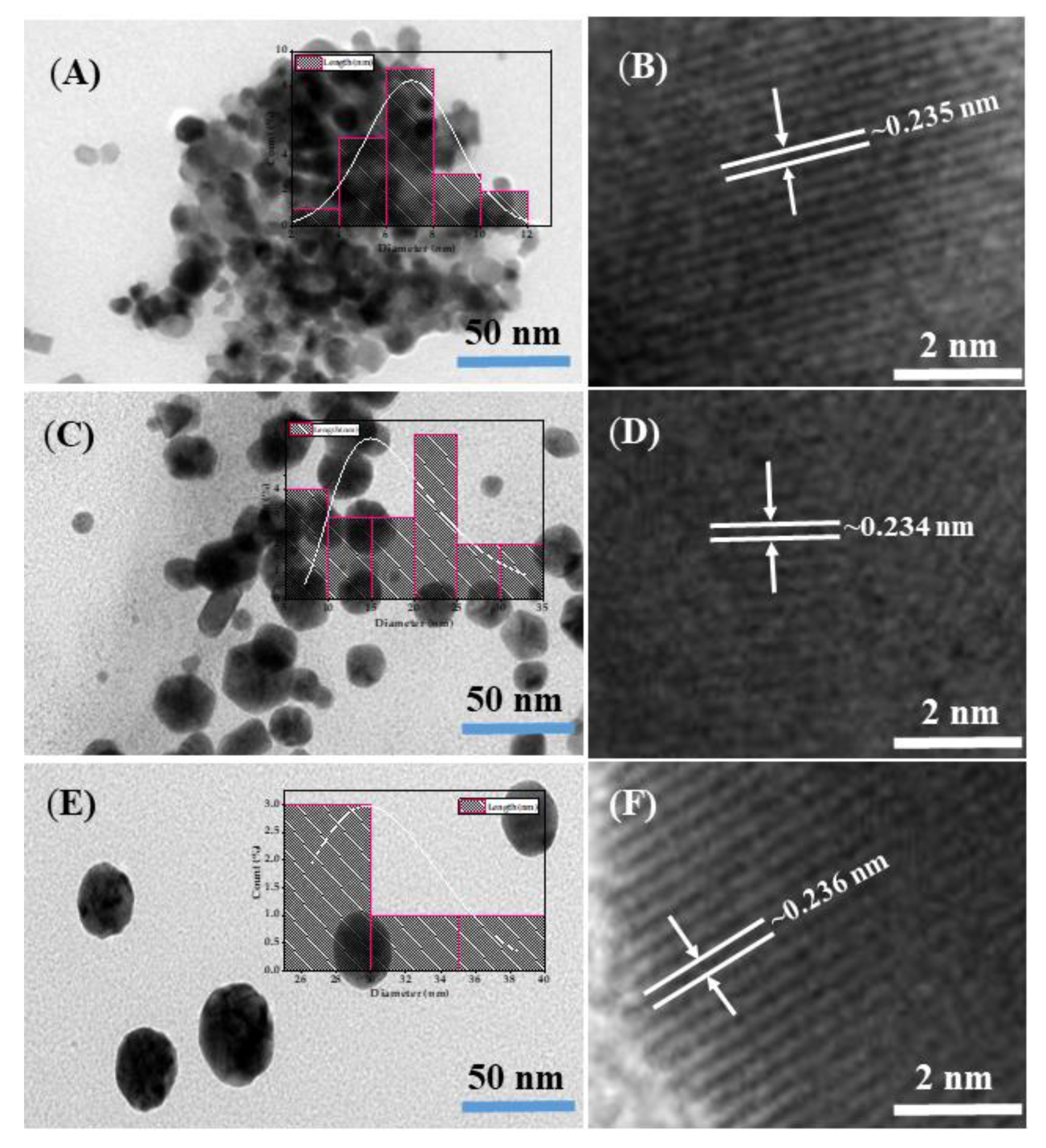
3.1. Morphological Characterization of GNPs

The morphology and crystallinity of the prepared GNPs were scrutinized via TEM equipped with high resolution (HR-TEM) and displayed as shown in Figure 1. In Figure 1A, low magnification shows that the entire surface is covered with spherical-shaped GNPs within the range of 10–15 nm in diameter. The inset of Figure 1A shows a histogram of the size distribution of GNPs-1. The lattice between the two fringes is 0.235 nm (Figure 1B) in HR-TEM, revealing the crystallinity of the prepared material. Figure 1C,D show the morphology of GNPs-2. The inset of Figure 1C shows a histogram of the 20–30 nm size distribution of GNPs-2; the GNPs are spherical in shape with smooth surfaces and free from agglomeration. The HR-TEM image of GNPs-2 (Figure 1D) was also checked, and shows that the lattice difference between the two fringes is 0.234 nm, well-defined, and analogous to previously published literature [59,60]. The data clearly show that prepared GNPs exhibit a lattice constant of face-centered cubic (FCC) crystals of GNPs that are consistent with published literature [59,60].
Figure 1E,F show the morphology of GNPs-3, as detailed in the Materials and Methods section. Due to the high concentration of salt solution (1.5 mM), it provides bigger-sized GNPs, with an individual nanoparticle size of 45 nm. The inset of Figure 1E illustrates a histogram of the size distribution of GNPs-3, which are spherical in shape, with a clean and smooth-surfaced morphology. The corresponding HR-TEM image shows that the lattice distance between the two fringes is 0.236 nm (Figure 1F), further confirming that the grown materials exhibit high crystallinity with good GNP shape and size [59,60,61].

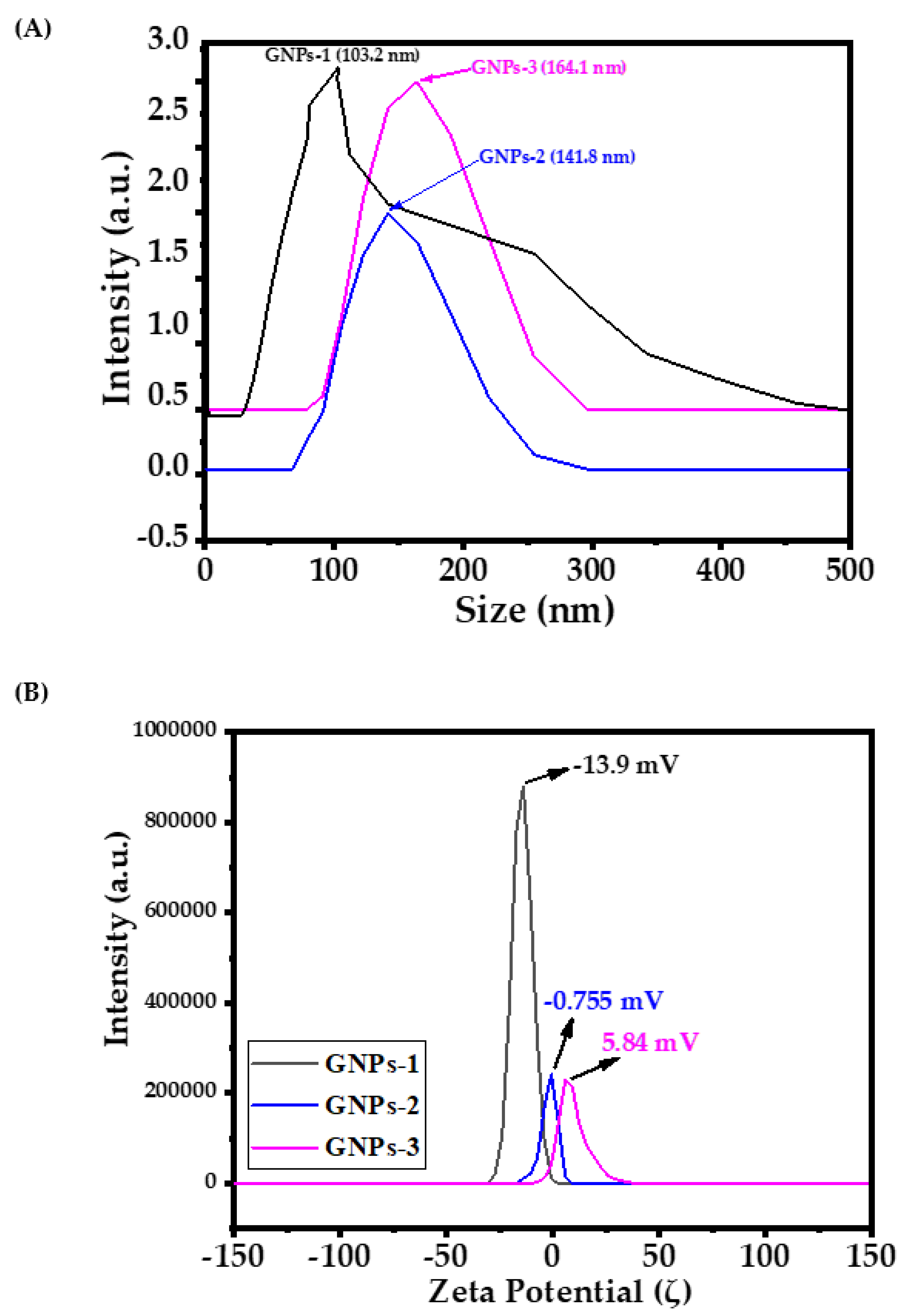
3.2. Hydrodynamic Size and Zeta Potential of GNPs

Aggregation in solutions appears to be an omnipresent phenomenon among all grown colloidal nanostructures. The achieved results showed that small NPs in an aqueous solution may possibly come closer together to form aggregation. The dynamic light scattering (DLS) of GNPs-1, GNPs-2, and GNPs-3 were analyzed, showing that the size of particulate matter was 103.2, 141.8, and 164.1 nm, respectively (Figure 2A). DLS was used to analyze the size of NPs in colloidal suspensions, where the particles have a tendency to colloid and form aggregation in an aqueous medium; DLS showed the primary and secondary sizes of the NPs. The sizes of GNPs increases with collisions and interactions with other molecules. The present finding corroborates the findings of previously published literature [61].
Zeta (ζ) potential is a physical property used understand the behavior of particles in suspension, macromolecule, or charges existing on material surfaces. It reduces the time required to produce trial formulations, and can be used to assist in predicting long-term stability. From the experimental perspective, the obtained magnitude of zeta potential provides an indication of the potential stability of the colloidal system [62,63]. For those particles, which have very small dimensions, a high zeta potential will indicate stability, i.e., the solution or dispersion will resist aggregation. At the same time, when the zeta potential is small, attractive forces may exceed repulsive forces, and the dispersion may break and flocculate. Hence, colloidal particles with high zeta potential (negative or positive) are electrically stable, whereas colloidal particles with low zeta potential tend to coagulate or flocculate in the solution. The general dividing line between stable and unstable suspensions is generally either +30 or −30 mV. In our case, the three different-sized gold nanoparticles express zeta potential with values of −13.9 (GNPs-1), −0.75 (GNPs-2), and 5.84 mV (GNPs-3), respectively (Figure 2B). It is evident from the data that small particles are stable in nature, but if the size of the particles increases the particles become coagulate or floccule, with sediment on the surface [64].

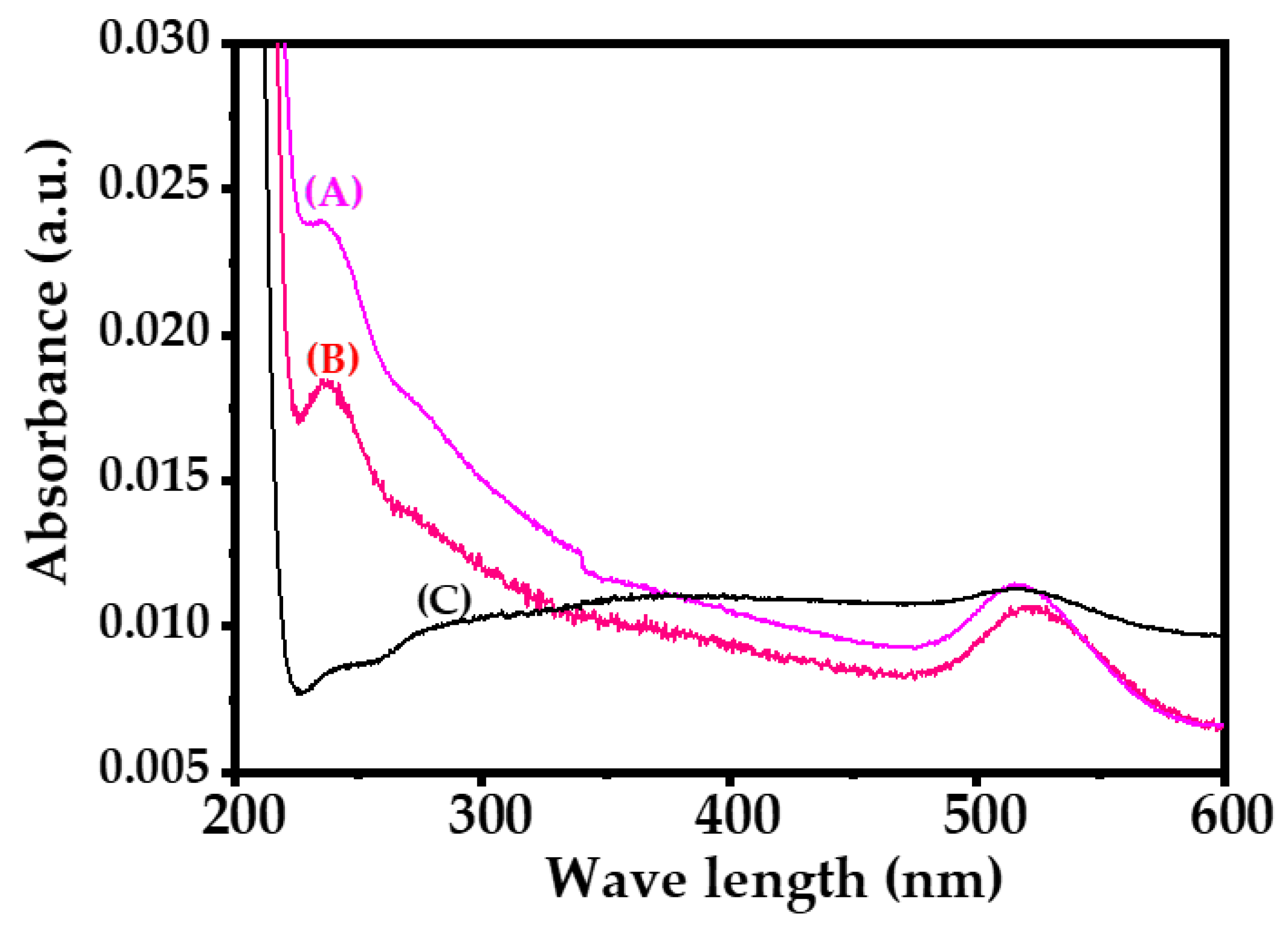
3.3. UV-Visible Spectroscopy

The optical property of GNPs was analyzed via UV-visible spectroscopy. A shallow peak and a centered peak were detected in the spectrum at 515, 522, and 516 nm (with band gaps of 2.40, 2.37, and 2.40 eV, respectively) (Figure 3), due the existence of colloidal gold ions. The band gaps express that the material is very efficient and optically active, exhibiting good chemical and optical characteristic [59,60,61].

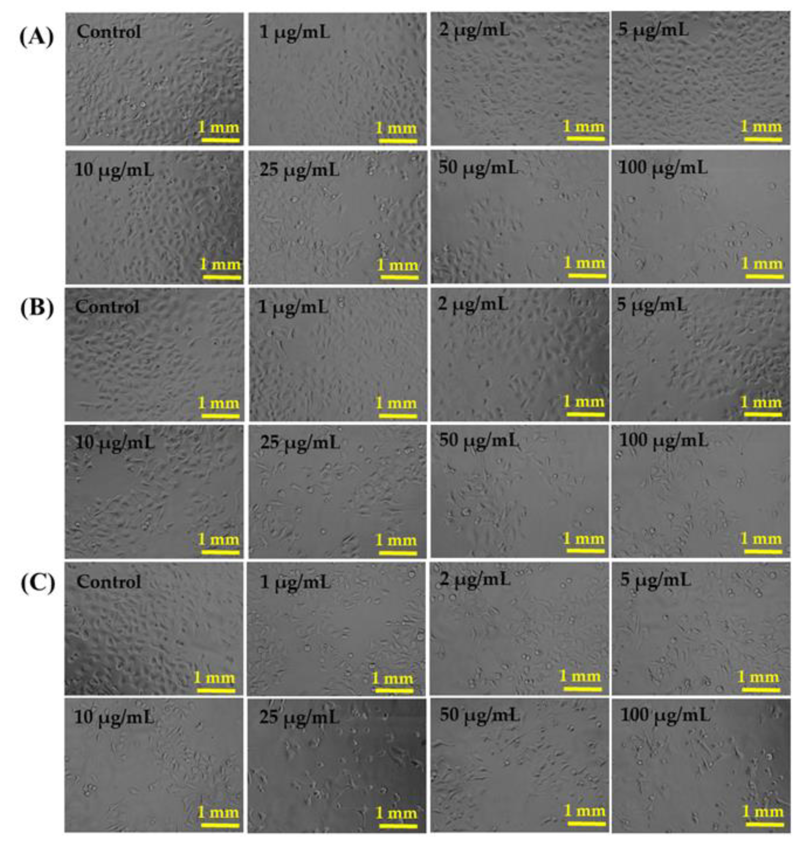
3.4. Morphological Changes of Cancer Cells (HepG2) Control and Treated with GNPs

The HepG2 cells were cultured and their structural details were observed via microscopy during a 24 h incubation period, with a number of selected concentrations (1 µg/mL, 2 µg/mL, 5 µg/mL, 10 µg/mL, 25 µg/mL, 50 µg/mL, and 100 µg/mL) and treated with samples of GNPs-1, GNPs-2, and GNPs-3 (Figure 4). The data received from the inverted microscopy for HepG2 cells (Figure 4) were used as control (Panel-A control, Panel-B control, and Panel-C control), whereas other images were received with interactions of a number of concentrations (1 µg/mL, 2 µg/mL, 5 µg/mL, 10 µg/mL, 25 µg/mL, 50 µg/mL and 100 µg/mL) of GNPs-1 (Panel-A), GNPs-2 (Panel-B), and GNPs-3 (Panel-C), respectively (Figure 4). As observed, the cells were initially nucleated, and when their confluences reached their maximum level (90–95%), the confluent cells were treated with various concentrations (1 µg/mL, 2 µg/mL, 5 µg/mL, 10 µg/mL, 25 µg/mL, 50 µg/mL, and 100 µg/mL) of GNPs and analyzed (Figure 4). The data reveal that at a low concentration (1–10 µg/mL) of GNPs, minimal change in cell morphology is observed, but when the concentration of GNPs increases to 25, 50, and 100 µg/mL, the growth of cells is significantly influenced by the GNPs. The obtained microscopic images in the HepG2 cells reveal that the cells were damaged by the GNPs (Figure 4).

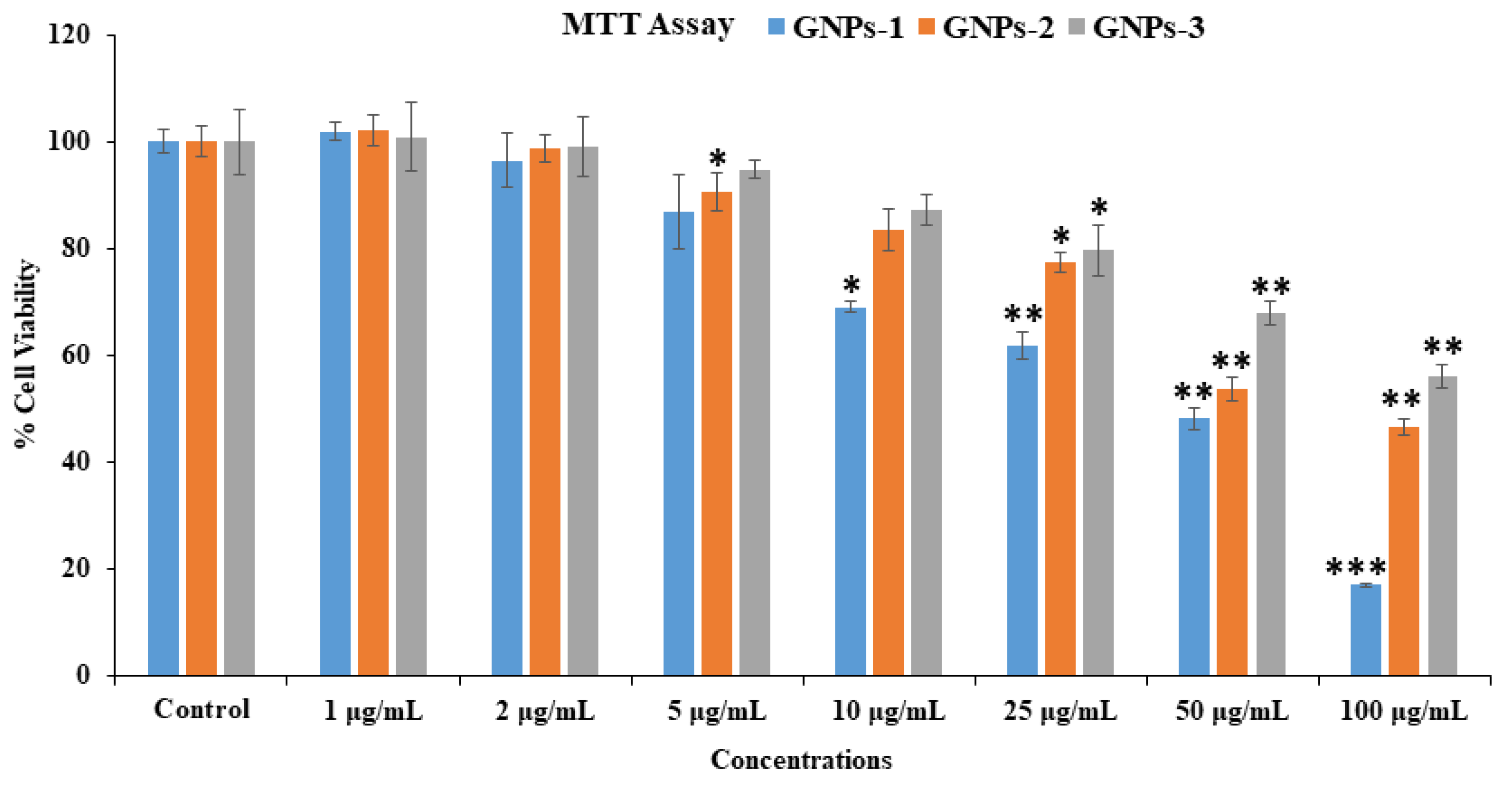
3.5. Cytotoxicity Assessment with MTT Assay in HepG2 Cells with GNPs

As mentioned in the Materials and Methods section, the confluent or grown cancer cells (HepG2) were used as a control, while a number of different concentrations (1–100 µg/mL) with GNPs were used as treated samples, exposed for 24 h incubation. Cytotoxicity was examined by a set and prescribed MTT assay method. The obtained data show that the viability of HepG2 cells is reduced with the interaction of GNPs and is dose- or concentration-reliant. In HepG2 cells, cell viability according to MTT assay was observed for GNPs-1 at 24 h as 101%, 96%, 86%, 69%, 61%, 48%, and as 17% (Figure 5) for concentrations of 1, 2, 5, 10, 25, 50, and 100 μg/mL, respectively (p < 0.05 for each concentration). Similar trends were found in samples of GNPs-2, once treated with HepG2 cells. The viability of HepG2 cells assessed by MTT assay for GNPs 2, at 24 h, was 102%, 98%, 90%, 83%, 77%, 53%, and 46% (Figure 5) for concentrations of 1, 2, 5, 10, 25, 50, and 100 μg/mL, respectively (p < 0.05 for each concentration).
The viability of HepG2 cells is significantly influenced by GNPs-3. The MTT assay result at 24 h was observed at 100%, 99%, 94%, 87%, 79%, 67%, and 55% (Figure 5) for concentrations of 1 μg/mL, 2 μg/mL, 5 μg/mL, 10 μg/mL, 25 μg/mL, 50 μg/mL, and 100 μg/mL, respectively (p < 0.05 for each concentration). The received cytotoxicity data of HepG2 cells show that initially the GNPs do not have much effect in reducing cancer cells, but once the concentration of GNPs upsurges, cytotoxicity was significantly influenced and diminished.

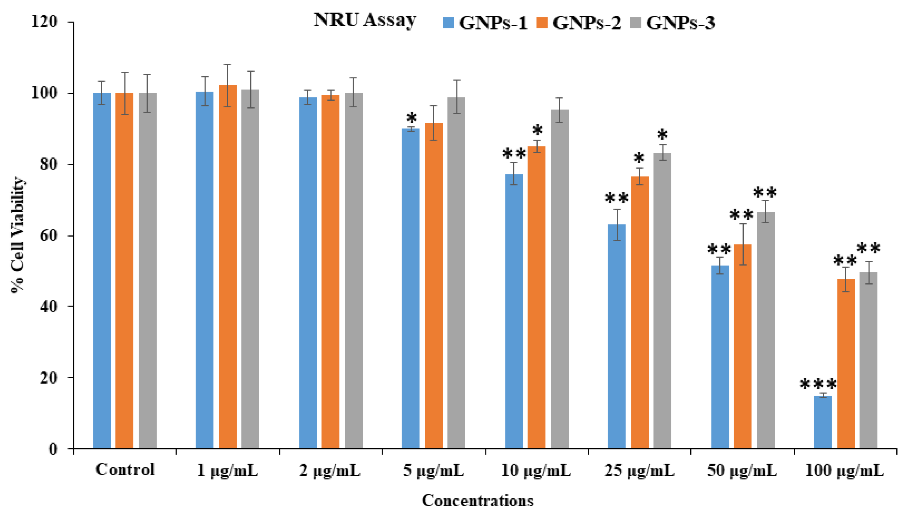
3.6. Cytotoxicity Assessment via NRU Assay in HepG2 Cells with GNPs

A cytotoxic study was also conducted in control and treated samples of GNPs via NRU assay, as detailed in Section 2.4.3. The NRU assay data are very analogous to the MTT assay data for HepG2 cancer cells and show a similar trend. Like the MTT assay, the NRU assay shows that at the initial concentration of GNPs, viability of cancer cells was not significantly affected, but when the concentration of GNPs rises, cell growth reduces. In the case of GNPs-1 exposed to HepG2 cells, cell viability by NRU assay at 24 h was 100%, 98%, 89%, 77%, 63%, 51%, and 15% (Figure 6) for concentrations of 1 μg/mL, 2 μg/mL, 5 μg/mL, 10 μg/mL, 25 μg/mL, 50 μg/mL, and 100 μg/mL, respectively (p < 0.05 for each concentration). In the case of GNPs-2 and HepG-2 cells, cell viability was recorded as 102%, 99%, 91%, 85%, 76%, 57%, and 47% (Figure 6) for concentrations of 1 μg/mL, 2 μg/mL, 5 μg/mL, 10 μg/mL, 25 μg/mL, 50 μg/mL, and 100 μg/mL, respectively (p < 0.05 for each concentration).
In the case of GNPs-3 and HepG-2 cells, cell viability was recorded as 100%, 100%, 98%, 95%, 83%, 66%, and 49% (Figure 6) for concentrations of 1 μg/mL, 2 μg/mL, 5 μg/mL, 10 μg/mL, 25 μg/mL, 50 μg/mL, and 100 μg/mL, respectively (p < 0.05 for each concentration). We also provide the comparative anticancer activity of gold NPs against liver cells by MTT (Table 1) and NRU (Table 2) assays.

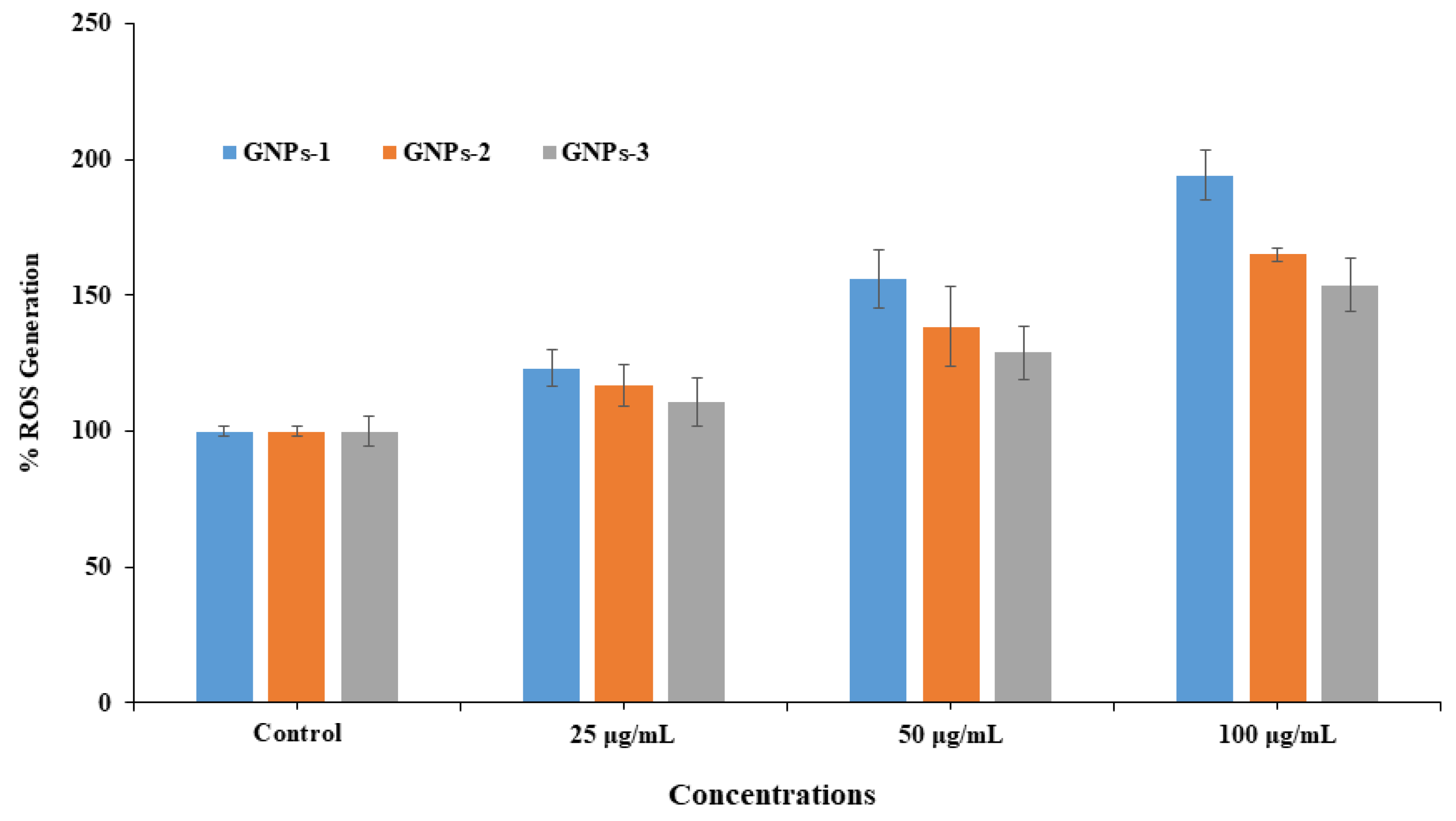
3.7. ROS Generation in HepG2 with and without Exposure GNPs

ROS generation was also examined in HepG2 cells, when exposed to GNPs at different concentrations (25, 50, and 100 μg/mL) for a 24 h incubation period (Figure 7). Compared to control, ROS levels increased in cancer cells, once exposed to GNPs-1, GNPs-2, and GNPs-3, as shown in Figure 7. At 25 μg/mL, ROS generation was increased to 123%, 156%, and 194% in HepG2 cells, respectively; at 50 μg/mL, ROS generation upsurged to 116%, 138%, and 164% in HepG2 cells, respectively. At a higher concentration (100 μg/mL), ROS generation increased to 110%, 128%, and 153% in HepG2 cells, respectively, as compared with controls (Figure 7).

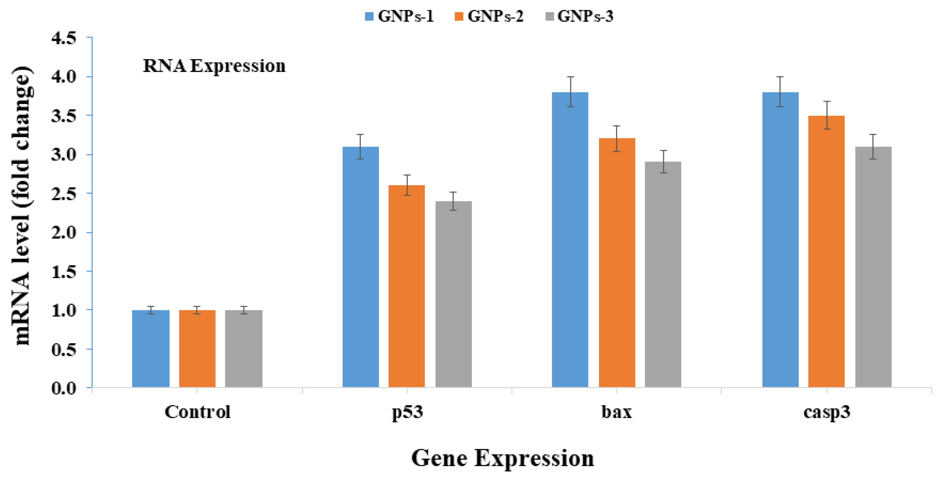
3.8. mRNA Expressions

The mRNA expressions with prepared NPs were analyzed with p53, Bax, and caspase-3 in HepG2 at a low concentration (25 µg/mL) of GNPs-1, GNPs-2, and GNPs-3 (Figure 8), with the control gene scrutinized via qPCR at a 24 h incubation period. The recovered data disclose that the p53, Bax, and caspase-3 with GNPs-1 results were 3.8, 3.1, and 3.8, respectively (Figure 8). The obtained data authenticate that GNPs-1 exhibits the possibility of interacting with cells and their marker gene (caspase 3) and causes an up-regulation in mRNA expression. Similar observations were found in p53 and Bax, when introduced with GNPs-2 at 24 h incubation times [7,8]. Other acquired data, related to p53, Bax, and caspase-3 in HepG2 cells, at a 25 µg/mL concentration of GNPs-2, disclose that the results were 3.2, 2.6, and 3.5, respectively (Figure 8). The larger-sized nanoparticles exhibit a special organization in their geometry. For instance, some are in polygonal, ellipsoidal, or circular shapes, and due to this, the surface area of the particles is reduced, as compared with the lower surface area of GNPs-1; this may cause a low up-regulation in gene expression.
In the case of GNPs-3 (Figure 8), after reacting with marker genes p53, Bax, and caspase-3 in HepG2 cells in a 24 h incubation period, the normalization fold change was 2.9, 2.4, and 3.1, respectively, at 25 µg/mL.
In all the cases of GNPs-1, GNPs-2 and GNPs-3 with the genes p53, Bax, and caspase-3, an apoptosis in the cells was created, leading to up-regulation in gene expression. All the data related to gene expression indicate that p53, Bax, and caspase-3 activity is directly related to the affected or apoptotic cells present in a population of grown cells. Based on the literature and obtained data, we conclude that the nanostructured materials act as an inhibitor on the growth/population of cancer cells [7,8]. The apoptosis in cell influences with various parameters, such as an increase in doses of nanostructures, higher incubation periods, and the sizes of the nanostructures, can be seen as an up-regulation in mRNA expression [7,8]. In the entire experiment of gene expression with GNPs-1, a higher expression of apoptotic genes was shown in cells, compared with the other nanostructures (GNPs-2 and GNPs-3), which may be due to their higher surface areas, low density, and spherical shape morphology, facilitating easy entry into the cells and easy reaction to their organelles [7,8].

3.9. Discussion

The different-sized nanoparticles GNPs-1, GNPs-2, and GNPs-3 were synthesized via the colloidal solution process, as described in the Materials and Methods section. Their special size facilitates application for various biological and biomedical purposes, such as drug delivery, DNA damage, cancer, and cytotoxicity, due to their enhanced surfaces and catalytic properties [7]. The processed GNPs-1, GNPs-2, and GNPs-3 were characterized with various instruments such as TEM and HR-TEM, which disclosed their particle size and crystalline properties. The morphology of processed structures were confirmed through TEM, which illustrated that the nanostructures possessed three different-sized NPs, spherical in shape, with nearly smooth surfaces and crystalline in nature. The average diameters of the GNPs were, respectively, 10–15 nm for GNPs-1, 20–30 nm for GNPs-2, and 45 nm for GNPs-3. HR-TEM of the prepared nanostructures showed lattice spacing of 0.235 nm, 0.234 nm, and 0.236 nm, respectively, between the two fringes, equal to the findings of previously published literature and confirming that the materials are highly crystalline in nature [65]. The hydrodynamic size and zeta potential results are in agreement with other data and consistent with TEM and HR-TEM. Very small and different-sized NPs have the ability to change their physiological and physicochemical character in optoelectronic applications, such as LEDs and solar cells.
A number of applications in optoelectronics are well established, but limited work is available in relation to anticancer applications with nanostructures, in terms of their diverse sizes and varied concentrations. The colloidal solution provides worthy results, which are very significant with respect to cancer cells that have underlying assumptions and restrictions. The diverse-sized nanoparticles GNPs-1, GNPs-2, and GNPs-3 have the ability to inhibit cancer cell growth [66]. It is hypothesized that the different sizes and shapes of NPs can possibly produce ROS generation in cell suspension, which is an important factor responsible for forming free radicals (FRs), which may be able to penetrate the outer walls of cells and enter into the inner walls of cell membranes. FRs also exhibit the possibility of reacting with internal organelles, creating an enzymatic change and resulting in disorganization of cells and cells contents.
ROS plays a significant role in cytotoxicity with NPs, suggesting that extreme generation reduces cellular antioxidant capacity [67]. Gold nanoparticles have the potential to regulate the growth/death of cell organelles, as well as biochemical and enzymatic changes of cancer cells [67]. This result shows that GNPs-1, GNPs-2, and GNPs-3 are potent materials for use in growth inhibition of cancer cells. These results indicate that varied concentrations and different sizes of nanostructures are able to easily enter into and react with cells, as is evident from the MTT, NRU, and ROS studies.
In most of the previous works, researchers have employed either a high concentration of nanostructures or complex materials for toxicity studies, which may be harmful for human exposure [68,69]. From the entirety of relevant experiments, it appears that low-concentration and small-size NPs (10 nm) would be increasingly helpful in controlling the growth of cancer cells without any adverse effects on the human body [68,69].

3.10. Techno-Economic Challenges

The study of cancer/oncology assists patients with cancer. The field of oncology is a major health sector that generates revenue in the healthcare economy. The new technologies and related advanced instruments provide care and treatment for patients, with impacts on hospitals, health insurers, doctors, and, most importantly, patients and their families. Focusing on these issues is important, as they are directly related to advancements in cancer treatment, economics strategies related to cancer care, and the effects on patients and healthcare systems.
Healthcare expenditures on technologies, instruments, drugs, clinical trials, etc., are high. Treatment approaches require strong evidence and cost-effectiveness. The core and fundamental objective of the experimental approach is to reduce the cost of treatment via technological means.
In this study, the roles of three different-sized nanoparticles are defined, involving synthesization via the colloidal solution process for use against cancer cells. Gold nanoparticles exhibit good biocompatibility and high reactivity with respect to cancer cells. Therefore, these materials may also be effective against other negative biological entities [70,71].
For the treatment of cancer cells in human beings, a number of physical approaches to complete eradication have been applied, e.g., chemotherapy, radiotherapy, and immunotherapy. Although these physical approaches eradicate cancer cells but they are very costly and the outcomes may not provide complete satisfaction because if any cell remains, it grows and forms further tumor cells in the body. At the same time, surgery is an expensive and complex procedure compared to other means of treatment, especially for low-income families; therefore, further work is required to establish a successful approach that can provide effective treatment at affordable prices for lower- and middle-class families.
The nanotechnology of metal-based drugs is an alternative approach for cancer studies, playing a huge role in the control and cure of different types of cancers [72]. Prepared gold nanoparticles, which are very small in size, are able to enter quickly and easily into cellular organelles, compared to the available drugs. These metal-based NPs employed against cancer can minimize the cost of organic and complex drugs and curtail the anxiety of surgery for patients from underprivileged and low-income families.

4. Conclusions

In summary, the synthesis of small-dimension GNPs was successfully performed via the colloidal chemical reduction process. The formed nanoparticles were well characterized in terms of their morphology and crystalline character, analyzed via TEM equipped with high resolution (HR-TEM) to validate high crystallinity and good structural morphology in the obtained products. TEM revealed that the diameter size of GNPs ranged from 10–15 nm for GNPs-1 to 20–30 nm for GNPs-2 to 45 nm for GNPs-3, with spherical shapes. The different-sized GNPs were applied against HepG2 cancer cells, with results that indicated that cytotoxicity in cancer cells is dose-dependent, from very low concentrations (1 μg/mL) to high concentrations (100 μg/mL). In all three different sizes of GNPs, minimal effect was observed initially from 1 μg/mL, 2 μg/mL, and 5 μg/mL concentrations, but as the concentrations were increased to 10 μg/mL, 25 μg/mL, and 50 μg/mL, and 100 μg/mL, respectively, the growth of cancer cells was significantly affected.
In this study, cell viability was significantly affected by small-sized NPs, as compared with the effect of other-sized NPs. This result may be due to the high surface area of small-sized NPs, as compared to other types; in addition, NPs that are very small in size exhibit low density, as compared with other NPs.
In the current study, we found that of the three different-sized nanoparticles GNPs-1, GNPs-2, and GNPs-3, small-sized nanoparticles (GNPs-1, 10–15 nm) is most effective in controlling cancer cells. The small size facilitates easy entry into cells and reaction with cell organelles.
NPs can possibly be used as anticancer nanodrugs for cancer treatment. This study indicates that different-sized gold nanoparticles induce cytotoxicity and apoptosis through different genes (i.e., the p53, Bax, and caspase pathways) in cancer cells, and mediate via ROS generation. At present, the annihilation of cancer cells is controlled by various surgical and therapeutic means, as well as by a number of organic drugs. These methods, including, chemotherapy, radiotherapy, immunotherapy, and radiosurgery, involve the use of complex drugs that are costly and fail to provide complete satisfaction. The aim of this study is to achieve a more fruitful option at a very low cost. Nanotechnology provides an excellent platform and a good alternative in forming a number of inorganic or colloidal nanoparticle-based drugs to diminish the growth of cancer cells much more effectively. Nano-based drugs are very cost-effective and affordable for economically-deprived persons. Due to their very small size, they exhibit high surfaces and sufficient volume ratio to allow for entry directly into cancer cells.
Based on the obtained results, gold nanoparticles show great efficiency and effectiveness in controlling the growth of cancer cells, compared to other organic or more complex molecules or drugs. This study demonstrates that the use of colloidal-based nanostructured materials can possibly reduce the costs of available drugs for the eradication of cancer cells, and that these small nanoparticles can reduce anxieties and medical costs for underprivileged patients.

Author Contributions

Conceptualization, A.A.A.-K.; methodology, A.A.A.-K.; software, R.W.; validation, R.W.; formal analysis, R.W.; investigation, R.W.; resources, A.A.A.-K.; data curation, R.W.; writing—original draft preparation, A.A.A.-K.; writing—review and editing, A.A.A.-K.; visualization, A.A.A.-K.; supervision, A.A.A.-K.; project administration, A.A.A.-K.; funding acquisition, A.A.A.-K. All authors have read and agreed to the published version of the manuscript.

Funding

Abdulaziz A. Alkhedhairy would like to thank Zoology Department, College of Science and Scientific Council, King Saud University, Riyadh, Saudi Arabia for support of this work over Sabbatical Leave for the academic year 2021–2022.

Institutional Review Board Statement

Not applicable.

Informed Consent Statement

Not applicable.

Data Availability Statement

Not applicable.

Acknowledgments

Abdulaziz A. Alkhedhairy would like to thank Zoology Department, College of Science and Scientific Council, King Saud University, Riyadh, Saudi Arabia for support of this work over Sabbatical Leave for the academic year 2021–2022.

Conflicts of Interest

The authors declare that they have no known competing or financial interests.

Abbreviations

ATCCAmerican Type Culture Collection
cDNAComplementary DNA
DMSODimethylsulfoxide
DLSDynamic light scattering
DMEMDulbecco’s modified eagle medium
DCF-DA2, 7-dichlorofluorescin diacetate
FBSFetal bovine serum
GNPsGold nanoparticles
GAPDHGlyceraldehyde 3-phosphate dehydrogenase
HR-TEMHigh-resolution transmission electron microscopy
HepG2Liver cancer cells
TEMTransmission electron microscopy
MTT3-(4,5-dimethylthiazol-2-yl)-2,5-diphenyltetrazolium bromide
NRUNeutral red uptake
qPCRQuantitative polymerase chain reaction
ROSReactive oxygen species
RTRoom temperature
SEMScanning electron microscope
NMsNanomaterials

References

  1. Nguyen, H.P.T.; Arafin, S.; Piao, J.; Cuong, T.V. Nanostructured Optoelectronics: Materials and Devices. J. Nanomater. 2016, 2016, 2051908. [Google Scholar] [CrossRef]
  2. Wang, Z.; Hu, T.; Liang, R.; Wei, M. Application of Zero-Dimensional Nanomaterials in Biosensing. Front. Chem. 2020, 8, 320. [Google Scholar] [CrossRef] [PubMed]
  3. Samykano, M. Progress in one-dimensional nanostructures. Mater. Charact. 2021, 179, 111373. [Google Scholar] [CrossRef]
  4. Li, X.; Wang, J. One-dimensional and two-dimensional synergized nanostructures for high-performing energy storage and conversion. InfoMat 2019, 2, 3–32. [Google Scholar] [CrossRef] [Green Version]
  5. Zeng, M.; Chen, M.; Huang, D.; Lei, S.; Zhang, X.; Wang, L.; Cheng, Z. Engineered two-dimensional nanomaterials: An emerging paradigm for water purification and monitoring. Mater. Horiz. 2021, 8, 758–802. [Google Scholar] [CrossRef]
  6. Song, G.; Gong, W.; Cong, S.; Zhao, Z.-G. Ultrathin Two-Dimensional Nanostructures: Surface Defects for Morphology-Driven Enhanced Semiconductor SERS. Angew. Chem. Int. Ed. 2020, 60, 5505–5511. [Google Scholar] [CrossRef]
  7. Wahab, R.; Khan, F.; Yang, Y.B.; Hwang, I.H.; Shin, H.-S.; Ahmad, J.; Dwivedi, S.; Khan, S.T.; Siddiqui, M.A.; Saquib, Q.; et al. Zinc oxide quantum dots: Multifunctional candidates for arresting C2C12 cancer cells and their role towards caspase 3 and 7 genes. RSC Adv. 2016, 6, 26111–26120. [Google Scholar] [CrossRef]
  8. Wahab, R.; Dwivedi, S.; Khan, F.; Mishra, Y.K.; Hwang, I.; Shin, H.-S.; Musarrat, J.; Al-Khedhairy, A.A. Statistical analysis of gold nanoparticle-induced oxidative stress and apoptosis in myoblast (C2C12) cells. Colloids Surf. B Biointerfaces 2014, 123, 664–672. [Google Scholar] [CrossRef] [PubMed]
  9. Khan, N.U.; Lin, J.; Younas, M.R.; Liu, X.; Shen, L. Synthesis of gold nanorods and their performance in the field of cancer cell imaging and photothermal therapy. Cancer Nanotechnol. 2021, 12, 20. [Google Scholar] [CrossRef]
  10. Feng, C.-J.; Guan, A.-Q.; Xie, Z. First-principle study of Ag-Au double wall nanotube and nanowire. Phys. Lett. A 2021, 407, 127468. [Google Scholar] [CrossRef]
  11. Ansari, S.G.; Ansari, Z.A.; Wahab, R.; Kim, Y.S.; Khang, G.; Shin, H.S. Glucose sensor based on nano-baskets of tin oxide templated in porous alumina by plasma enhanced CVD. Biosens. Bioelectron. 2008, 23, 1838–1842. [Google Scholar] [CrossRef] [PubMed]
  12. Kannan, K.; Radhika, D.; Sadasivuni, K.K.; Reddy, K.R.; Raghu, A.V. Nanostructured metal oxides and its hybrids for photocatalytic and biomedical applications. Adv. Colloid Interface Sci. 2020, 281, 102178. [Google Scholar] [CrossRef] [PubMed]
  13. Nakielski, P.; Pawłowska, S.; Rinoldi, C.; Ziai, Y.; De Sio, L.; Urbanek, O.; Zembrzycki, K.; Pruchniewski, M.; Lanzi, M.; Salatelli, E.; et al. Multifunctional Platform Based on Electrospun Nanofibers and Plasmonic Hydrogel: A Smart Nanostructured Pillow for Near-Infrared Light-Driven Biomedical Applications. ACS Appl. Mater. Interfaces 2020, 12, 54328–54342. [Google Scholar] [CrossRef]
  14. Bansal, S.A.; Kumar, V.; Karimi, J.; Singh, A.P.; Kumar, S. Role of gold nanoparticles in advanced biomedical applications. Nanoscale Adv. 2020, 2, 3764–3787. [Google Scholar] [CrossRef]
  15. Broadhead, E.J.; Tibbetts, K.M. Fabrication of Gold–Silicon Nanostructured Surfaces with Reactive Laser Ablation in Liquid. Langmuir 2020, 36, 10120–10129. [Google Scholar] [CrossRef] [PubMed]
  16. Danielson, E.; Sontakke, V.A.; Porkovich, A.J.; Wang, Z.; Kumar, P.; Ziadi, Z.; Yokobayashi, Y.; Sowwan, M. Graphene basedfield-effect transistor biosensors functionalized using gas-phase synthesized gold nanoparticles. Sens. Actuators B Chem. 2020, 320, 128432. [Google Scholar] [CrossRef]
  17. Teker, K.; AlKhaldi, A. Impact of gold nanoparticles on low-voltage operating GaN ultraviolet photodetector. Opt. Eng. 2020, 59, 127110. [Google Scholar] [CrossRef]
  18. Liu, X.; He, F.; Zhang, F.; Zhang, Z.; Huang, Z.; Liu, J. Dopamine and Melamine Binding to Gold Nanoparticles Dominates Their Aptamer-Based Label-Free Colorimetric Sensing. Anal. Chem. 2020, 92, 9370–9378. [Google Scholar] [CrossRef]
  19. Ding, J.; Lu, Z.; Wang, R.; Shen, G.; Xiao, L. Piezoelectric immunosensor with gold nanoparticles enhanced competitive immunoreaction technique for 2,4-dichlorophenoxyacetic acid quantification. Sens. Actuators B Chem. 2014, 193, 568–573. [Google Scholar] [CrossRef]
  20. Zhao, D.-W.; Yu, M.-Y.; Zheng, L.-L.; Li, M.; Dai, S.-J.; Chen, D.-C.; Lee, T.-C.; Yun, D.-Q. Enhanced Efficiency and Stability of Planar Perovskite Solar Cells Using a Dual Electron Transport Layer of Gold Nanoparticles Embedded in Anatase TiO2 Films. ACS Appl. Energy Mater. 2020, 3, 9568–9575. [Google Scholar] [CrossRef]
  21. Selopal, G.S.; Mohammadnezhad, M.; Besteiro, L.V.; Cavuslar, O.; Liu, J.; Zhang, H.; Pardo, F.N.; Liu, G.; Wang, M.; Durmusoglu, E.G.; et al. Synergistic Effect of Plasmonic Gold Nanoparticles Decorated Carbon Nanotubes in Quantum Dots/TiO2 for Optoelectronic Devices. Adv. Sci. 2020, 7, 2001864. [Google Scholar] [CrossRef] [PubMed]
  22. Lee, S.Y.; Kang, D.; Jeong, S.; Do, H.T.; Kim, J.H. Photocatalytic Degradation of Rhodamine B Dye by TiO2 and Gold Nanoparticles Supported on a Floating Porous Polydimethylsiloxane Sponge under Ultraviolet and Visible Light Irradiation. ACS Omega 2020, 5, 4233–4241. [Google Scholar] [CrossRef] [Green Version]
  23. Lee, K.X.; Shameli, K.; Yew, Y.P.; Teow, S.-Y.; Jahangirian, H.; Rafiee-Moghaddam, R.; Webster, T.J. Recent Developments in the Facile Bio-Synthesis of Gold Nanoparticles (AuNPs) and Their Biomedical Applications. Int. J. Nanomed. 2020, 15, 275–300. [Google Scholar] [CrossRef]
  24. He, M.Q.; Yu, Y.L.; Wang, J.H. Biomolecule-tailored assembly and morphology of gold nanoparticlesfor LSPR applications. Nano Today 2020, 35, 101005. [Google Scholar] [CrossRef]
  25. Ma, F.H.; Li, C.; Liu, Y.; Shi, L. Mimicking Molecular Chaperones to Regulate Protein Folding. Adv. Mater. 2020, 32, e1805945. [Google Scholar] [CrossRef]
  26. He, Z.; Yin, H.; Chang, C.-C.; Wang, G.; Liang, X. Interfacing DNA with Gold Nanoparticles for Heavy Metal Detection. Biosensors 2020, 10, 167. [Google Scholar] [CrossRef]
  27. Wu, Y.; Zhou, Y.; Huang, H.; Chen, X.; Leng, Y.; Lai, W.; Huang, X.; Xiong, Y. Engineered gold nanoparticles as multicolor labels for simultaneous multi-mycotoxin detection on the immunochromatographic test strip nanosensor. Sens. Actuators B Chem. 2020, 316, 128107. [Google Scholar] [CrossRef]
  28. Siddique, S.; Chow, J.C.L. Gold Nanoparticles for Drug Delivery and Cancer Therapy. Appl. Sci. 2020, 10, 3824. [Google Scholar] [CrossRef]
  29. Goddard, Z.R.; Marín, M.J.; Russell, D.A.; Searcey, M. Active targeting of gold nanoparticles as cancer therapeutics. Chem. Soc. Rev. 2020, 49, 8774–8789. [Google Scholar] [CrossRef]
  30. Lvarez, R.G.; Chen, L.; Nedilko, A.; Iglesias, A.S.; Rix, A.; Lederle, W.; Pathak, V.; Lammers, T.; Plessen, G.V.; Kostarelos, K.; et al. Optimizing the Geometry of Photoacoustically Active Gold Nanoparticles for Biomedical Imaging. ACS Photonics 2020, 7, 646–652. [Google Scholar]
  31. Li, H.; Pan, S.; Xia, P.; Chang, Y.; Fu, C.; Kong, W.; Yu, Z.; Wang, K.; Yang, X.; Qi, Z. Advances in the application of gold nanoparticles in bone tissue engineering. J. Biol. Eng. 2020, 14, 14–15. [Google Scholar] [CrossRef]
  32. Munawer, U.; Raghavendra, V.B.; Ningaraju, S.; Krishna, K.L.; Ghosh, A.R.; Melappa, G.; Pugazhendhi, A. Biofabrication of gold nanoparticles mediated by the endophytic Cladosporium species: Photodegradation, in vitro anticancer activity and in vivo antitumor studies. Int. J. Pharm. 2020, 588, 119729. [Google Scholar] [CrossRef] [PubMed]
  33. Wahab, R.; Yang, Y.B.; Umar, A.; Singh, S.; Hwang, I.H.; Shin, H.-S.; Kim, Y.-S. Platinum quantum dots and their cytotoxic effect towards myoblast cancer cells (C2C12). J. Biomed. Nanotechnol. 2012, 8, 424–431. [Google Scholar] [CrossRef]
  34. Hu, X.; Zhang, Y.; Ding, T.; Liu, J.; Zhao, H. Multifunctional Gold Nanoparticles: A Novel Nanomaterial for Various Medical Applications and Biological Activities. Front. Bioeng. Biotechnol. 2020, 8, 990. [Google Scholar] [CrossRef] [PubMed]
  35. Sun, H.; Jia, J.; Jiang, C.; Zhai, S. Gold Nanoparticle-Induced Cell Death and Potential Applications in Nanomedicine. Int. J. Mol. Sci. 2018, 19, 754. [Google Scholar] [CrossRef] [Green Version]
  36. Hoenerhoff, M.J.; Pandiri, A.R.; Lahousse, S.A.; Hong, H.H.; Ton, T.V.; Masinde, T.; Auerbach, S.S.; Gerrish, K.; Bushel, P.R.; Shockley, K.R.; et al. Global gene profiling of spontaneous hepatocellular carcinoma in B6C3F1 mice: Similarities in the molecular landscape with human liver cancer. Toxicol. Pathol. 2011, 39, 678–699. [Google Scholar] [CrossRef] [Green Version]
  37. Zhu, A.X. Systemic treatment of hepatocellular carcinoma: Dawn of a new era? Ann. Surg. Oncol. 2010, 17, 1247–1256. [Google Scholar] [CrossRef]
  38. Nault, J.C.; Rossi, J.Z. Genetics of hepatobiliary carcinogenesis. Semin. Liver Dis. 2011, 31, 173–187. [Google Scholar] [CrossRef] [PubMed]
  39. Siegel, A.B.; Zhu, A.X. Metabolic syndrome and hepatocellular carcinoma: Two growing epidemics with a potential link. Cancer 2009, 115, 5651–5661. [Google Scholar] [CrossRef] [Green Version]
  40. Alqahtani, A.; Khan, Z.; Alloghbi, A.; Ahmed, T.S.S.; Ashraf, M.; Hammouda, D.M. Hepatocellular Carcinoma: Molecular Mechanisms and Targeted Therapies. Medicina 2019, 55, 526. [Google Scholar] [CrossRef] [Green Version]
  41. Chakraborty, A.; Das, A.; Raha, S.; Barui, A. Size-dependent apoptotic activity of gold nanoparticles on osteosarcoma cells correlated with SERS signal. J. Photochem. Photobiol. B Biol. 2020, 203, 111778. [Google Scholar] [CrossRef] [PubMed]
  42. Yohan, D.; Cruje, C.; Lu, X.; Chithrani, D.B. Size-Dependent Gold Nanoparticle Interaction at Nano–Micro Interface Using Both Monolayer and Multilayer (Tissue-Like) Cell Models. Nano-Micro Lett. 2015, 8, 44–53. [Google Scholar] [CrossRef] [PubMed] [Green Version]
  43. Tomić, S.; Đokić, J.; Vasilijić, S.; Ogrinc, N.; Rudolf, R.; Pelicon, P.; Vučević, D.; Milosavljević, P.; Janković, S.; Anžel, I.; et al. Size-Dependent Effects of Gold Nanoparticles Uptake on Maturation and Antitumor Functions of Human Dendritic Cells In Vitro. PLoS ONE 2014, 9, e96584. [Google Scholar] [CrossRef] [Green Version]
  44. Xia, Q.; Huang, J.; Feng, Q.; Chen, X.; Liu, X.; Li, X.; Zhang, T.; Xiao, S.; Li, H.; Zhong, Z.; et al. Size- and cell type-dependent cellular uptake, cytotoxicity and in vivo distribution of gold nanoparticles. Int. J. Nanomed. 2019, 14, 6957–6970. [Google Scholar] [CrossRef] [Green Version]
  45. Vijayakumara, S.; Ganesan, S. Size-dependent in vitro cytotoxicity assay of gold nanoparticles. Toxicol. Environ. Chem. 2013, 95, 277–287. [Google Scholar] [CrossRef]
  46. Vodyashkin, A.A.; Rizk, M.G.H.; Kezimana, P.; Kirichuk, A.A.; Stanishevskiy, Y.M. Application of Gold Nanoparticle-Based Materials in Cancer Therapy and Diagnostics. ChemEngineering 2021, 5, 69. [Google Scholar] [CrossRef]
  47. Luo, D.; Wang, X.; Burda, C.; Basilion, J. Recent Development of Gold Nanoparticles as Contrast Agents for Cancer Diagnosis. Cancers 2021, 13, 1825. [Google Scholar] [CrossRef] [PubMed]
  48. Huang, K.; Ma, H.; Liu, J.; Huo, S.; Kumar, A.; Wei, T.; Zhang, X.; Jin, S.; Gan, Y.; Wang, P.C.; et al. Size-Dependent Localization and Penetration of Ultrasmall Gold Nanoparticles in Cancer Cells, Multicellular Spheroids, and Tumors In Vivo. ACS Nano 2012, 6, 4483–4493. [Google Scholar] [CrossRef] [Green Version]
  49. Yafout, M.; Ousaid, A.; Khayati, Y.; El Otmani, I.S. Gold nanoparticles as a drug delivery system for standard chemotherapeutics: A new lead for targeted pharmacological cancer treatments. Sci. Afr. 2021, 11, e00685. [Google Scholar] [CrossRef]
  50. Alhussan, A.; Bozdoğan, E.; Chithrani, D. Combining Gold Nanoparticles with Other Radiosensitizing Agents for Unlocking the Full Potential of Cancer Radiotherapy. Pharmaceutics 2021, 13, 442. [Google Scholar] [CrossRef]
  51. Chauhan, A.; Khan, T.; Omri, A. Design and Encapsulation of Immunomodulators onto Gold Nanoparticles in Cancer Immunotherapy. Int. J. Mol. Sci. 2021, 22, 8037. [Google Scholar] [CrossRef] [PubMed]
  52. Suchomel, P.; Kvitek, L.; Prucek, R.; Panacek, A.; Halder, A.; Vajda, S.; Zboril, R. Simple size-controlled synthesis of Au nanoparticles and their size-dependent catalytic activity. Sci. Rep. 2018, 8, 1–11. [Google Scholar] [CrossRef] [PubMed] [Green Version]
  53. Turkevich, J.; Stevenson, P.C.; Hillier, J. A study of the nucleation and growth proce -sses in the synthesis of colloidal gold, Discuss. Faraday Soc. 1951, 11, 55–75. [Google Scholar] [CrossRef]
  54. Siddiqui, M.A.; Singh, G.; Kashyap, M.P.; Khanna, V.K.; Yadav, S.; Chandra, D.; Pant, A.B. Influence of cytotoxic doses of 4-hydroxynonenal on selected neurotransmitter receptors in PC-12 cells. Toxicol. In Vitro 2008, 22, 1681–1688. [Google Scholar] [CrossRef]
  55. Mosmann, T. Rapid colorimetric assay for cellular growth and survival: Application to proliferation and cytotoxicity assays. J. Immunol. Methods 1983, 65, 55–63. [Google Scholar] [CrossRef]
  56. Borenfreund, E.; Puerner, J.A. Toxicity determined in vitro by morphological alterations and neutral red absorption. Toxicol. Lett. 1985, 24, 119–124. [Google Scholar] [CrossRef]
  57. Siddiqui, M.A.; Kashyap, M.P.; Kumar, V.; Al-Khedhairy, A.A.; Musarrat, J.; Pant, A.B. Protective potential of trans-resveratrol against 4-hydroxynonenal induced damage in PC12 cells. Toxicol. In Vitro 2010, 24, 1592–1598. [Google Scholar] [CrossRef]
  58. Wang, H.; Joseph, J.A. Quantifying cellular oxidative stress by dichlorofluorescein assay using microplate reader. Free Radic. Biol. Med. 1999, 27, 612–616. [Google Scholar] [CrossRef]
  59. Wang, Y.Q.; Liang, W.S.; Geng, C.Y. Coalescence Behavior of Gold Nano particles. Nanoscale Res. Lett. 2009, 4, 684–688. [Google Scholar] [CrossRef] [Green Version]
  60. Arunachalam, K.D.; Annamalai, S.K.; Hari, S. One-step green synthesis and characterization of leaf extract-mediated biocompatible silver and gold nanoparticles from Memecylon umbellatum. Int. J. Nanomed. 2013, 8, 1307–1315. [Google Scholar] [CrossRef] [Green Version]
  61. Balmes, O.; Malm, J.-O.; Pettersson, N.; Karlsson, G.; Bovin, J.-O. Imaging Atomic Structure in Metal Nanoparticles Using High-Resolution Cryo-TEM. Microsc. Microanal. 2005, 12, 145–150. [Google Scholar] [CrossRef] [PubMed]
  62. Smith, M.C.; Crist, R.M.; Clogston, J.D.; McNeil, S.E. Zeta potential: A case study of cationic, anionic, and neutral liposomes. Anal. Bioanal. Chem. 2017, 409, 5779–5787. [Google Scholar] [CrossRef]
  63. Zeta Potential—An Introduction in 30 Minutes. 2005. Available online: https://www.malvernpanalytical.com/ (accessed on 4 December 2021).
  64. Cui, W.; Li, J.; Zhang, Y.; Rong, H.; Lu, W. Long Jiang, Nanomedicine: Nanotechnology. Biol. Med. 2012, 8, 46–53. [Google Scholar]
  65. Wahab, R.; Kaushik, N.; Khan, F.; Kaushik, N.K.; Lee, S.-J.; Choi, E.H.; A Al-Khedhairy, A. Gold quantum dots impair the tumorigenic potential of glioma stem-like cells via β-catenin downregulation in vitro. Int. J. Nanomed. 2019, 14, 1131–1148. [Google Scholar] [CrossRef] [PubMed] [Green Version]
  66. Engstrom, A.M.; Faase, R.A.; Marquart, G.W.; Baio, J.E.; Mackiewicz, M.R.; Harper, S.L. Size-Dependent Interactions of Lipid-Coated Gold Nanoparticles: Developing a Better Mechanistic Understanding through Model Cell Membranes and In Vivo Toxicity. Int. J. Nanomed. 2020, 15, 4091–4104. [Google Scholar] [CrossRef]
  67. Misawa, M.; Takahashi, J. Generation of reactive oxygen species induced by gold nanoparticles under x-ray and UV Irradiations. Nanomed. Nanotechnol. Biol. Med. 2011, 7, 604–614. [Google Scholar] [CrossRef]
  68. Contini, C.; Hindley, J.W.; Macdonald, T.J.; Barritt, J.D.; Ces, O.; Quirke, N. Size depend ency of gold nanoparticles interacting with model membranes. Commun. Chem. 2020, 3, 130. [Google Scholar] [CrossRef] [PubMed]
  69. Ortega, D.A.M.; Tovar, G.N.; Castanon, G.M.; Gonzalez, C. Effect of gold nanoparticles (AuNPs) on isolated rat tracheal segments. Toxicol. Rep. 2021, 8, 1412–1418. [Google Scholar] [CrossRef]
  70. Ramsey, S.D.; Dusetzina, S.B. Weighing Costs and Benefits in the Economics of Cancer Care. J. Clin. Oncol. 2020, 38, 289–291. [Google Scholar] [CrossRef]
  71. Meropol, N.J.; Schulman, K.A. Cost of Cancer Care: Issues and Implications. J. Clin. Oncol. 2007, 25, 180–186. [Google Scholar] [CrossRef]
  72. Taylor, D.G. The political economics of cancer drug discovery and pricing. Drug Discov. Today 2020, 25, 2149–2160. [Google Scholar] [CrossRef] [PubMed]
Figure 1. TEM images displaying the size of the prepared nanoparticles and HR-TEM of their corresponding images: (A,B) show the TEM and HR-TEM of GNPs-1; (C,D) illustrate the TEM and HR-TEM GNPs-2; (E,F) show the TEM and HR-TEM of GNPs-3. Insets of Figure 1A,C,E, show histograms of size distribution for GNPs-1, GNPs-2, and GNPs-3, respectively.
Applsci 12 00901 g001
Figure 2. (A), dynamic light scattering (DLS) for particle size distribution; and (B), represent the zeta potential of GNPs-1, GNPs-2, and GNPs-3 respectively.
Applsci 12 00901 g002
Figure 3. The UV-visible spectroscopy of GNPs: (A), GNPs-1; (B), GNPs-2; and (C), GNPs-3.
Applsci 12 00901 g003
Figure 4. Morphological changes in HepG2 cells exposed to GNPs-1 (A), GNPs-2; (B), and GNPs-3 (C) for 24 h. Cell images were obtained via an inverted microscope at 20× magnification. Scale bar = 1 mm.
Applsci 12 00901 g004
Figure 5. Cytotoxicity assessments by MTT assay in HepG2 cells after the exposure of GNPs-1, GNPs-2, and GNPs-3 at different concentrations for 24 h. The values are mean ± SE of three independent experiments. * p < 0.05, ** p < 0.01, and *** p < 0.001 vs. control.
Applsci 12 00901 g005
Figure 6. Cytotoxicity assessment by NRU assay in HepG2 cells after exposure to GNPs-1, GNPs-2, and GNPs-3 at different concentrations for 24 h. The values are mean ± SE of three independent experiments. * p < 0.05, ** p < 0.01, *** p < 0.001 vs. control.
Applsci 12 00901 g006
Figure 7. Bar diagram showing percentage change in ROS generation in HepG2 cells exposed to 25–100 μg/mL of GNPs-1, GNP-2, and GNPs-3.
Applsci 12 00901 g007
Figure 8. Quantification of mRNA fold change and apoptotic genes (p53, Bax, and casp3) were evaluated. HepG2 cells were treated with 25 μg/mL of GNPs-1, GNPs-2, and GNPs-3 for 24 h. For internal control, glyceraldehyde 3-phosphate dehydrogenase (GAPDH) was used. The values are mean ± SE of three independent experiments.
Applsci 12 00901 g008
Table 1. Comparative anticancer activity of gold NPs against liver cells by MTT assay.
ConcentrationsGNPs-1 (% Cell Viability)GNPs-2 (% Cell Viability)GNPs-3 (% Cell Viability)
Control100.00 ± 2.23100.00 ± 2.93100.00 ± 6.13
1 μg/mL101.91 ± 1.82102.00 ± 2.89100.86 ± 6.44
2 μg/mL96.39 ± 5.0298.70 ± 2.4999.02 ± 5.49
5 μg/mL86.86 ± 6.9690.56 ± 3.6494.77 ± 1.60
10 μg/mL69.08 ± 1.0083.50 ± 3.9287.35 ± 2.88
25 μg/mL61.87 ± 2.6277.35 ± 1.8679.58 ± 4.65
50 μg/mL48.23 ± 2.0653.79 ± 2.2367.89 ± 2.21
100 μg/mL17.05 ± 0.3746.49 ± 1.5255.94 ± 2.17
Table 2. Comparative anticancer activity of gold NPs against liver cells by NRU assay.
ConcentrationsGNPs-1 (% Cell Viability)GNPs-2 (% Cell Viability)GNPs-3 (% Cell Viability)
Control100.00 ± 3.24100.00 ± 5.97100.00 ± 5.31
1 μg/mL100.48 ± 3.95102.16 ± 5.97100.97 ± 5.12
2 μg/mL98.84 ± 1.9399.35 ± 1.46100.19 ± 4.07
5 μg/mL89.95 ± 0.6991.57 ± 4.7798.95 ± 4.80
10 μg/mL77.34 ± 3.2085.02 ± 1.8095.31 ± 3.45
25 μg/mL63.03 ± 4.4676.49 ± 2.3283.26 ± 2.19
50 μg/mL51.46 ± 2.3557.54 ± 5.7466.60 ± 3.14
100 μg/mL15.10 ± 0.5247.66 ± 3.4649.54 ± 3.21
Publisher’s Note: MDPI stays neutral with regard to jurisdictional claims in published maps and institutional affiliations.

