Article

The Impact of Segmentation Method and Target Lesion Selection on Radiomic Analysis of 18F-FDG PET Images in Diffuse Large B-Cell Lymphoma

1
Medical Physics Unit, IEO European Institute of Oncology IRCCS, 20141 Milan, Italy
2
Department of Experimental Oncology, IEO European Institute of Oncology IRCCS, 20141 Milan, Italy
3
Onco-Hematology Division, IEO European Institute of Oncology IRCCS, 20141 Milan, Italy
4
Division of Nuclear Medicine, IEO European Institute of Oncology IRCCS, 20141 Milan, Italy
5
Department of Oncology and Hemato-Oncology, University of Milan, 20122 Milan, Italy
6
Department of Health Sciences, University of Milan, 20146 Milan, Italy
*
Author to whom correspondence should be addressed.
Appl. Sci. 2022, 12(19), 9678; https://doi.org/10.3390/app12199678
Submission received: 9 August 2022 / Revised: 16 September 2022 / Accepted: 23 September 2022 / Published: 27 September 2022
(This article belongs to the Special Issue Medical Image Processing and Analysis Methods for Cancer Applications)

Abstract

:

Featured Application

The present work contributes to the investigation on the robustness and reproducibility of radiomic studies, a fundamental step for bringing radiomics towards clinical implementation.

Abstract

Radiomic analysis of 18F[FDG] PET/CT images might identify predictive imaging biomarkers, however, the reproducibility of this quantitative approach might depend on the methodology adopted for image analysis. This retrospective study investigates the impact of PET segmentation method and the selection of different target lesions on the radiomic analysis of baseline 18F[FDG] PET/CT images in a population of newly diagnosed diffuse large B-cell lymphoma (DLBCL) patients. The whole tumor burden was segmented on PET images applying six methods: (1) 2.5 standardized uptake value (SUV) threshold; (2) 25% maximum SUV (SUVmax) threshold; (3) 42% SUVmax threshold; (4) 1.3∙liver uptake threshold; (5) intersection among 1, 2, 4; and (6) intersection among 1, 3, 4. For each method, total metabolic tumor volume (TMTV) and whole-body total lesion glycolysis (WTLG) were assessed, and their association with survival outcomes (progression-free survival PFS and overall survival OS) was investigated. Methods 1 and 2 provided stronger associations and were selected for the next steps. Radiomic analysis was then performed on two target lesions for each patient: the one with the highest SUV and the largest one. Fifty-three radiomic features were extracted, and radiomic scores to predict PFS and OS were obtained. Two proportional-hazard regression Cox models for PFS and OS were developed: (1) univariate radiomic models based on radiomic score; and (2) multivariable clinical–radiomic model including radiomic score and clinical/diagnostic parameters (IPI score, SUVmax, TMTV, WTLG, lesion volume). The models were created in the four scenarios obtained by varying the segmentation method and/or the target lesion; the models’ performances were compared (C-index). In all scenarios, the radiomic score was significantly associated with PFS and OS both at univariate and multivariable analysis (p < 0.001), in the latter case in association with the IPI score. When comparing the models’ performances in the four scenarios, the C-indexes agreed within the confidence interval. C-index ranges were 0.79–0.81 and 0.80–0.83 for PFS radiomic and clinical–radiomic models; 0.82–0.87 and 0.83–0.90 for OS radiomic and clinical–radiomic models. In conclusion, the selection of either between two PET segmentation methods and two target lesions for radiomic analysis did not significantly affect the performance of the prognostic models built on radiomic and clinical data of DLBCL patients. These results prompt further investigation of the proposed methodology on a validation dataset.

1. Introduction

Diffuse large B-cell lymphoma (DLBCL) is one of the most frequent and aggressive lymphomas [1]. Patient risk stratification is generally assessed through clinical parameters, grouped into the International Prognostic Index (IPI) score [2]. However, disease relapse occurs in approximately 20% of patients within 5 years after first-line therapy [3]. The early identification of patients at higher risk of relapse might allow the optimization of the treatment strategy, possibly improving the outcome after first-line therapy. Molecular imaging and genetic data are emerging as potential predictive biomarkers [4,5,6] to improve risk stratification and treatment optimization.
18F fluorodeoxyglucose positron emission tomography (18F [FDG] PET) imaging is a standard-of-care imaging procedure for staging DLBCL patients [7]. Quantitative analysis of 18F[FDG] PET images is conventionally performed by measuring FDG uptake (standardized uptake value [SUV] derivates) and metabolic volumes (metabolic tumor volume [MTV], total lesion glycolysis [TLG]). The role of these conventional PET parameters as prognostic biomarkers for DLBCL patients has been suggested in different clinical settings [8,9,10]. In addition, radiomics is an emerging approach [11] that allows the exploration of PET images by exponentially increasing the number of quantitative parameters analyzed. Promising results have been obtained also in the case of different lymphomas, including DLBCL, with a different purpose in relation to the different prognostic role of PET/CT for each lymphoma sub-type [12,13,14]. These preliminary studies support the hypothesis that PET radiomic imaging biomarkers might be reliable predictors in the early assessment of DLBCL patients’ treatment response, laying the basis for the present investigation [15].
Radiomic analysis should be robust, reliable, and reproducible [16]; nevertheless, the use of different techniques for image acquisition and analysis affects the comparison and reproducibility of the results obtained among different centers and studies. Noise in the image signal might be observed in the case of a heterogeneous dataset, thus affecting the identification of potential associations with survival outcomes. For this reason, when performing a radiomic analysis on PET images, it is crucial to test the stability of the results by applying different methodologies as an indicator of robustness and reproducibility.
This methodological study has been designed to investigate the impact of different PET segmentation methods on the extraction of radiomic features (RFs) in a population of DLBCL patients who performed baseline 18F[FDG] PET/CT prior to first-line therapy. The selection of two different target lesions for radiomics analysis and the subsequent impact on the overall analysis have also been explored.

2. Materials and Methods

2.1. Patient Selection and Data Collection

Clinical records of biopsy-proven DLBCL patients diagnosed between September 2008 and October 2019, referred to the Oncohematology Division of our Institute immediately after diagnosis for standard-of-care therapy, were retrospectively evaluated. Inclusion criteria were: (1) baseline 18F[FDG] PET/CT performed at our institute prior to any treatment for DLBCL; (2) patients suitable for standard-of-care first-line chemotherapy; (3) availability of all clinical, pathology, and imaging data; (4) PET images acquired with 3D acquisition modality and 256 × 256 matrix; (5) no lost follow-up. Patients were excluded in case of parameters impairing quantitative image analysis (e.g., wrong input of injection data, glycemia >200 mg/dL, para-vein injection). The study was approved by the institutional ethical committee and scientific review board (ID trial 2863).
Among 376 patients identified from clinical records, one hundred and twelve patients (n = 112) were considered for the analysis according to inclusion/exclusion criteria. Among them, 9 patients were classified as tumor stage I, 31 patients as stage II, 8 patients as stage III, and 64 patients as stage IV. For 9 patients the prognostic score was IPI = 0, for 33 patients IPI = 1, for 28 patients IPI = 2, for 29 patients IPI = 3, for 12 patients IPI = 4, and for 1 patient IPI = 5. All patients were treated with first-line R-CHOP/R-CHOP-like chemoimmunotherapy, according to the best standard-of-care clinical practice at our institution and to international guidelines. Population characteristics and survival data are reported in the Supplementary Materials together with PET acquisition parameters (Tables S1–S3).
18F-FDG PET/CT acquisitions were performed on a Discovery ST or a Discovery 600 PET-CT scanner (GE Healthcare, Waukesha, WI, USA), according to international procedural guidelines [17]. PET images acquired on the Discovery ST scanner were reconstructed with OSEM algorithm VUE point (2 iterations, 30 subsets, and 4.5 mm Gaussian post-filter), whereas those acquired on the Discovery 600 scanner were reconstructed with OSEM algorithm VUE point HD (2 iterations, 16 subsets, and 5 mm Gaussian post-filter).
Progression-free survival (PFS) was calculated from the date of the diagnosis to the date of progression or last follow-up, whichever occurred first. Overall survival (OS) was calculated from the date of the diagnosis to the date of death or last follow-up, whichever occurred first. Specifically, no deaths occurred as the first event, thus, they were not counted in the definition of PFS. Follow-up was updated in March 2021.

2.2. Identification of the Optimal Lesion Segmentation Method

Quantitative PET image analysis was performed with LifeX package, version 5.1 (Inserm, CEA, CNRS, Université Paris Sud, France) [18]. Different semi-automatic PET segmentation methods were applied according to standardized measurements of metabolic tumor burden in DLBCL [19]. First, an experienced nuclear medicine physician (LLT) drew a region of interest (ROI) totally encompassing each area of pathological uptake. Then, four different threshold methods were applied within each ROI: a fixed threshold equal to 2.5 standardized uptake value (SUV), referred to as Method-1; a threshold equal to 25% of the maximum SUV in the ROI (Method-2); a threshold equal to 42% of the maximum SUV in the ROI (Method-3); a threshold equal to 1.3 times the average liver SUV, assessed by drawing a 45 cm3 ROI in a not pathological liver region (Method-4). In addition, for each lesion, the intersection among the ROIs obtained with methods 1, 2, and 4, and among those obtained with methods 1, 3, and 4 were identified, defining Method-5 and Method-6, respectively. For each method, total metabolic tumor volume (TMTV) was calculated as the sum of all the lesions’ volumes. Accordingly, whole-body total lesion glycolysis (WTLG) was calculated as the sum of the total lesion glycolysis values obtained for each lesion. For each method, the association of TMTV and WTLG with PFS and OS was investigated using proportional-hazard Cox regression models both in univariate and bivariate analysis, in the latter case including the IPI score as an independent prognostic factor. The method yielding the highest association with PFS/OS was selected for the main analysis, whereas the second-ranking method was used for comparison and sensitivity analysis.

2.3. Radiomic Features Extraction

The radiomic analysis was performed on the lesion exhibiting the highest SUV value (SUVmax lesion) and on the largest lesion (VOLmax lesion). First, the 0–80 SUV interval was discretized into 256 bins resulting in a 0.31 SUV fixed bin size. Fifty-three radiomic features (RFs) were extracted from each lesion ROI, including 2 Shape features, 14 first-order features (HISTO category), and 37 second-order texture features. In particular, texture features were obtained from gray level co-occurrence matrix (GLCM) (calculated considering both 1 and 2 voxels offset, referred to as GLCM_1 and GLCM_2, respectively), gray level run length matrix (GLRM), neighboring gray level dependence matrix (NGLDM) and gray level zone length matrix (GLZLM). The full list of extracted RFs is reported in Table 1.
Clinical and PET imaging data possibly affecting the PET image texture and the RFs value were collected: glucose level, injected activity/body weight, time between injection and acquisition, scanner model, frame duration. Then, after RFs log transformation, the reproducibility of RFs was assessed by fitting an ANOVA model for each RF and each clinical/imaging parameter. False discovery rate (FDR) correction was applied to account for multiple comparisons. RFs with FDR-adjusted p-values < 0.05 were excluded from the analysis.

2.4. Radiomic and Clinical–Radiomic Predictive Models

The RFs resulting reproducible at ANOVA were used to build a radiomic score. More in detail, a Cox-elastic net model regression was applied to build a RS significantly associated with PFS and a RS significantly associated with OS, applying the R function “glmnet”. This function requires specific values of the regularization parameters, α and λ. λ values were chosen by means of a 10-fold cross-validation to avoid overfitting, and the best α and λ combination was chosen as the one maximizing the C-index. The final elastic net model was fitted using the optimal values of α and λ, providing—for each RF retained in the model – the corresponding coefficient β for the calculation of the radiomic score. Then, indicating with β1, β2, ..., βp the elastic net coefficients for each of the p RFs in the model, and xi1, xi2, …, xip the observed values of the features for the ith patient, the radiomic score was calculated for each patient as follows: radiomic score = β1xi1 + β2xi2 + …. + βpxip.
Univariate analysis was performed to assess the association between PFS/OS and the corresponding radiomic score. The radiomic scores were considered, first, as dichotomous variables according to the median value (log-rank test), and then as continuous variables using Cox univariate model (radiomic model). Subsequently, a clinical–radiomic multivariable model to predict PFS/OS was built, including as independent predictors the prognostic clinical variable IPI score, the conventional PET parameters (SUVmax, TMTV, WTLG, and the volume of the representative lesion), and the radiomic score. In particular, SUVmax, TMTV, and WTLG were included as potential predictors [8,9], whereas the volume of the representative lesion was included for adjusting a potential correlation between the radiomic score and the lesion volume. To create the clinical–radiomic model, Cox univariate models were preliminary performed, and only variables showing significant contribution in the univariate model (p < 0.05) were included in the multivariable proportional hazard Cox model. The performances of the radiomic and clinical–radiomic models were quantified and compared using Harrell’s C-index; 95% confidence intervals (CI) were obtained using the R package survcomp. Statistical analysis was performed with R software, version 4.1.0.

2.5. Main Analysis and Sensitivity Analysis

The main analysis was performed on the data obtained by applying the top-ranking segmentation method and selecting the “SUVmax lesion” for radiomic features calculation. The sensitivity analysis included three replications of the main analysis, performed on the data obtained when applying the remaining combinations of segmentation method (top or second ranking) and representative lesion for RFs extraction (“SUVmax lesion” or “VOLmax lesion”). Case 1 will be referred to the use of the second-ranking segmentation method and “SUVmax lesion”; Case 2 to the use of the top-ranking segmentation method and radiomic analysis on “VOLmax lesion”; Case 3 to the use of the second-ranking segmentation method and radiomic analysis on “VOLmax lesion”. The models produced by the main analysis and sensitivity analysis were compared using the C-index to assess the robustness of the main analysis model and exclude any eventual significance dependence of model performance on methodological choices.

3. Results

3.1. Identification of the Optimal Lesion Segmentation Method

A significant association was observed between TMTV and PFS at univariate analysis with segmentation methods 1, 2, 4, and 5. Method-1 and Method-2 yielded the lowest p-value (p = 0.01). In addition, Method-2 provided a lower p-value in comparison to Method-1 also for the association between TMTV and OS, even if without statistical significance. Accordingly, Method-2 was selected for the main analysis and Method-1 for the sensitivity analysis. The results on the association of TMTV and WTLG with PFS and OS for the different segmentation methods are reported in Table 2.

3.2. Main Analysis: Radiomic and Clinical–Radiomic Predictive Models

Among 53 RFs extracted from the “SUVmax lesion” applying Method-2, only one feature (GLCM_Correlation calculated with two voxels offset) was non-reproducible at ANOVA due to significant dependence on scanner type, and it was excluded from the subsequent analysis. The remaining 52 RFs were used as input for the elastic-net Cox regression model, to create the radiomic score. Figure 1 reports the most relevant RFs selected by the model, together with the corresponding average model coefficients. Only RFs with coefficients larger than 1.00 are represented.
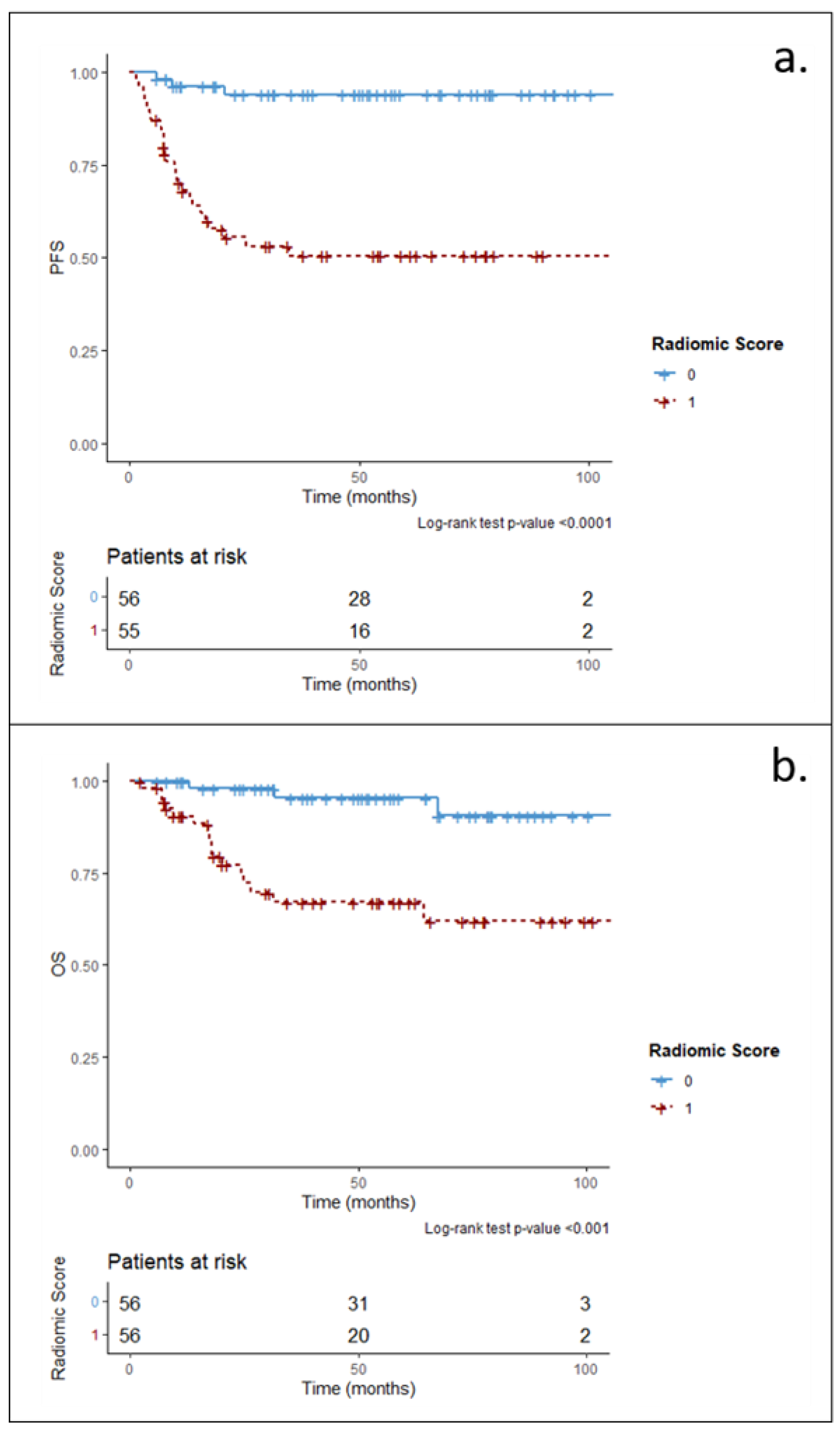
RFs significantly associated with PFS and OS were: NGLDM_Coarseness, GLZLM_LGZE, GLZLM_SZLGE, GLRLM_SRLGE, GLCM_1_Energy, GLRLM_LGRE, GLZLM_SZE, Sphericity, and HISTO_Uniformity. The radiomic scores were significantly associated with both PFS and OS in the univariate log-rank test, with p < 0.0001 and p < 0.001, respectively (Figure 2).
In addition, the radiomic scores were significantly associated with PFS and OS at Cox univariate and multivariate models (p < 0.001). In the multivariate model, the radiomic score retained significance also considering clinical (IPI score) and standard PET parameters (SUV derivates and metabolic volumes) as independent predictors (Table 3).

3.3. Sensitivity Analysis

The number of reproducible RFs at ANOVA was 51 in Case 1 (“SUVmax lesion” segmented with Method-1), 51 in Case 2 (“VOLmax lesion” segmented with Method-2), and 53 in Case 3 (“VOLmax lesion” segmented with Method-1). Most of the features included in the radiomics score in Case 1, 2, and 3 were in common with those obtained in the main analysis, as illustrated in Supplementary Figures S1–S3. Moreover, in all scenarios, the significant association of the radiomic score with PFS and OS was confirmed, both in long-rank univariate analysis (p < 0.001 in all cases, Supplementary Figures S4–S6 for Case 1, Case 2, and Case 3, respectively) and at Cox univariate and multivariable analysis (Supplementary Tables S4–S6). Table 4 illustrates the comparison of the models in terms of C-index and the corresponding 95% CI. The model accuracy of the main analysis was comparable to the accuracy obtained when applying different methodological approaches.

4. Discussion

This analysis explores the impact of different segmentation methods and target lesion selection on the performance of radiomic models in a cohort of DLBCL patients, whereas the clinical considerations of the obtained results are the object of another publication.
As a preliminary step, we took into account the fact that among different factors possibly affecting the robustness of radiomic studies, PET acquisition and reconstruction settings might alter the image texture and, consequently, the value and significance of the RFs [20]. To avoid the most relevant confounding factors and improve the robustness of our analysis, in this study, we included only images acquired at our Institute with comparable settings (3D modality, 256 × 256 matrix). This choice was performed since suitable data to test the generalizability of our findings to different datasets, after proper application of harmonization procedures [21], were not available at the time of this work. Such data might be collected for a future validation study. Even with the selection of images acquired only at our Institute, some imaging and clinical parameters potentially affecting image quality and texture unavoidably varied among patients (scanner model, frame duration, injected activity/body weight, time between injection and acquisition, glucose level). Such parameters were specifically investigated for their potential impact on radiomic features as the first step of our analysis. However, only one RF (GLCM_Correlation) was identified as not reproducible in the main analysis. This high RFs reproductivity is probably related to the standardization of the diagnostic procedure in our center, including standardized injection and acquisition protocols, PET/CT scanner manufactured by the same vendor, and similar reconstruction algorithms.
We then investigated the role of the segmentation method used to encompass the lesion since it is also crucial for radiomic analysis reproducibility. In our study, the application of a threshold equal to 25% of the SUVmax in each ROI (Method-2) was selected as the optimal segmentation method, as also already reported in the literature [22]. However, consensus on the optimal methodology has not been reached yet, with fixed SUV threshold methods appearing promising when searching for agreement with experienced observers [23,24]. Additional methodological studies designed to compare the performance of different methods are still ongoing [25]. In accordance, the second method we tested was a fixed threshold method (2.5 SUV, Method-1).
The selection of one representative lesion for the radiomic analysis might also introduce an element of arbitrariness in a radiomic study, especially when patients have multiple localizations, as generally happens in lymphoma. Previous studies investigated the potential role of whole-body SUVmax [26] or RFs extracted from the highest uptake lesions [14] as prognostic biomarkers. Hence, we selected the lesions exhibiting the highest uptake lesion (SUVmax lesion) as the target lesion for our main radiomic analysis. Nonetheless, other authors identified the largest lesion as potentially representative of clinical endpoints [12], therefore, we replicated our analysis by extracting RFs from the largest lesion as well (VOLmax lesion investigated in the sensitivity analysis). Further analysis might include the radiomic analysis of multiple lesions for each patient, attempting to capture the intra-patient heterogeneity and characterize the whole tumor burden.
The main analysis of this study highlighted the potential role of the RFs as prognostic biomarkers (Figure 2), and the sensitivity analysis confirmed that such a result was retained with the use of a different segmentation method or a different selection for target lesion (Supplementary Figure S4–S6). Indeed, in all scenarios, the prognostic role of RFs was confirmed with comparable model performance (Table 3, Supplementary Tables S4–S6, and Table 4) when switching among two segmentation methods and two different criteria for the selection of the lesion for RFs calculation. A partial explanation for these results can be found in the fact that, for many patients (91/112 patients with segmentation Method-1, and 98/112 patients with segmentation Method-2), the target lesion was at same time the one exhibiting both the highest uptake (SUVmax lesion) and the largest volume (VOLmax lesion). However, in spite of this, the observed reproducibility of model performance is an important result in support of the hypothesis that prognostic information is contained in PET images, it can be captured by radiomic analysis, and its assessment is robust since it is not strictly related to the use of a specific image analysis methodology. Robustness of radiomic analysis and the generalizability of its results are currently the most important issues to be overcome in order to make radiomics useful for clinical practice. Demonstrating that a radiomic finding is robust to methodology, as is the case of the present study, is an important step in indicating that the hypothesis deserves to be further investigated on independent populations. Such kinds of analysis are highly encouraged by researchers in the field but are still quite uncommon [27].
Of note, our results are in agreement with the previous literature. More in detail, in the main analysis, the RFs contributing mostly to the prediction of both PFS and OS were texture features, as evidenced in Figure 1. Notably, most features were in common between the radiomic score for PFS and OS, supporting the hypothesis that relevant and novel prognostic information can be derived by the RFs selected by the elastic net Cox model. The feature Coarseness calculated from the neighboring gray level dependence matrix (NGLDM) was among the top three features for both clinical endpoints, and is related to the rate of the spatial variation of radiopharmaceutical uptake, quantifying the prevalence of regions with sudden or low uptake variation. The features low gray-level zone emphasis (LGZE) and short-zone low gray-level emphasis (SZLGE) calculated from the gray level zone length matrix (GLZLM) were among the top two features for PFS and OS respectively, and in our database they exhibited similar behaviour (linear correlation R2 = 0.90). These RFs are both related to the presence and distribution of zones with low intensity, in particular small zones with homogeneous low uptake. The top feature in the radiomic score calculated for PFS was the feature short-run low gray-level emphasis (SRLGE) calculated from the gray level run length matrix (GLRLM), which, however, played a minor role as a predictor of OS. Similar to the previous GLZLM features, it is related to the distribution of short runs (consecutive voxels along a defined direction) with homogeneous low uptake. Previously, Parvez et al. [14] showed that GLZLM features (related to the distribution of long homogeneous zones) were predictive biomarkers associated with disease free survival, whereas a first-order feature, kurtosis, was associated with OS. Similarly, Aide et al. [28] found a significant association of GLZLM features extracted from the largest lesion with 2-year event-free survival. The same research group [29] showed that a first-order metric still related to heterogeneity, skewness-H, was the only independent predictor of PFS. Kun-Han-Lue et al. [30] found a significant association also using multivariate analysis of the feature RLN, extracted from GLRLM matrix, with both PFS and OS.
In our study, the radiomic score was the only imaging parameter retaining statistical significance in a multivariable analysis comprehending the clinical parameter (IPI score) and conventional PET parameters, as reported in Table 3. These results emphasize that the population included in this study was adequately representative of a real-world clinical scenario (the IPI score was a strong predictive parameter) and that radiomics has the potential of introducing an added value in the quantitative PET image analysis.
Among study limitations, we acknowledge the relatively small sample size and the lack of external validation as a fundamental step to confirm the accuracy of our model. Moreover, further studies are needed to assess if the inclusion of different scanners and different image reconstruction algorithms will affect the robustness of our analysis.
As already mentioned, the reproducibility of the radiomic score encourages further validation of these results, both on a clinically comparable population to confirm the generalizability of the predictive models, and on different populations to support the robustness of the methodology.

5. Conclusions

In this study, the performance of prognostic models built on radiomic and clinical data of DLBCL patients was not affected by the use of either among two PET segmentation methods (fixed threshold equal to 2.5 SUV and percentage threshold equal to 25% of the maximum SUV in the lesion) and two target lesions for radiomic analysis (highest uptake lesion and highest volume lesion)
The robustness of the results to variations in image analysis methodology supports the hypothesis that prognostic information is contained in baseline PET images of DLBCL patients, prompting further investigation on a validation dataset.

Supplementary Materials

The following supporting information can be downloaded at https://www.mdpi.com/article/10.3390/app12199678/s1, Table S1: Baseline characteristics of study population; Table S2: PET acquisition parameters of study population; Table S3: Summary of Progression-Free Survival (PFS) and Overall Survival (OS) times; Table S4: Sensitivity Analysis Case 1 (SUVmax lesion, segmentation Method-1). Univariate and multivariable Hazard Ratio (HR) with 95% Confidence Intervals (CI) for the association of continuous variables with Progression Free Survival (PFS) and Overall Survival (OS); Table S5: Sensitivity Analysis Case 2 (VOLmax lesion, segmentation Method-2). Univariate and multivariable Hazard Ratio (HR) with 95% Confidence Intervals (CI) for the association of continuous variables with Progression Free Survival (PFS) and Overall Survival (OS); Table S6: Sensitivity Analysis Case 3 (VOLmax lesion, segmentation Method-1). Univariate and multivariable Hazard Ratio (HR) with 95% Confidence Intervals (CI) for the association of continuous variables with Progression Free Survival (PFS) and Overall Survival (OS). Figure S1: Elastic Net Cox model average coefficients (among 500 repetitions of the model based on a bootstrap resampling) for prediction of a: PFS and b: OS according to RFs extracted from “SUVmax lesion” segmented with Method-1. Only coefficients larger than 1.00 are represented; Figure S2: Elastic Net Cox model average coefficients (among 500 repetitions of the model based on a bootstrap resampling) for prediction of a: PFS and b: OS according to RFs extracted from “VOLmax lesion” segmented with Method-2. Only coefficients larger than 1.00 are represented; Figure S3: Elastic Net Cox model average coefficients (among 500 repetitions of the model based on a bootstrap resampling) for prediction of a: PFS and b: OS according to RFs extracted from “VOLmax lesion” segmented with Method-1. Only coefficients larger than 1.00 are represented; Figure S4: Univariate association (Log-Rank rank test) between dichotomized RS and a: PFS; b: OS. RS were obtained from radiomic features extracted from “SUVmax lesion” segmented with Method-1; Figure S5: Univariate association (Log-Rank rank test) between dichotomized RS and a: PFS; b: OS. RS were obtained from radiomic features extracted from “VOLmax lesion” segmented with Method-2; Figure S6: Univariate association (Log-Rank rank test) between dichotomized RS and a: PFS; b: OS. RS were obtained from radiomic features extracted from “VOLmax lesion” segmented with Method-1.

Author Contributions

F.B. was involved in conceptualization, methodology, formal analysis, data curation, investigation, writing—original draft preparation, writing—review and editing, visualization, supervision; M.F. was involved in conceptualization, methodology, data curation, visualization, writing—review and editing; S.R. was involved in conceptualization, methodology, formal analysis, visualization, software, investigation, writing—review and editing; G.L.P. and F.C. (Federica Corso) were involved in methodology, formal analysis, visualization, software, writing—review and editing; S.M., L.S.A.F., T.R. and A.V. were involved in formal analysis and writing—review and editing; E.D. was involved in conceptualization, formal analysis, investigation, data curation, writing—review and editing; L.L.T. was involved in conceptualization, formal analysis, data curation, investigation, writing—review and editing; F.C. (Francesco Ceci) was involved in investigation, visualization, supervision, writing—review and editing. All authors have read and agreed to the published version of the manuscript.

Funding

This research received no external funding.

Institutional Review Board Statement

The study was approved by the institutional ethical committee and scientific review board of IEO European Institute of Oncology IRCCS (ID trial 2863).

Informed Consent Statement

Patient consent was waived by the institutional ethical committee and scientific review board of IEO European Institute of Oncology IRCCS.

Data Availability Statement

The datasets used and/or analyzed during the current study are available from the corresponding author upon reasonable request.

Acknowledgments

The work was partially supported by the Italian Ministry of Health with Ricerca Corrente and 5 × 1000 funds.

Conflicts of Interest

The authors declare no conflict of interest.

References

  1. Sant, M.; Allemani, C.; Tereanu, C.; De Angelis, R.; Capocaccia, R.; Visser, O.; Marcos-Gragera, R.; Maynadié, M.; Simonetti, A.; Lutz, J.-M.; et al. Incidence of hematologic malignancies in Europe by morphologic subtype: Results of the HAEMACARE project. Blood 2010, 116, 3724–3734. [Google Scholar] [CrossRef]
  2. International Non-Hodgkin's Lymphoma Prognostic Factors Project A Predictive Model for Aggressive Non-Hodgkin's Lymphoma. N. Engl. J. Med. 1993, 329, 987–994. [CrossRef] [PubMed]
  3. Harrysson, S.; Eloranta, S.; Ekberg, S.; Enblad, G.; Jerkeman, M.; Wahlin, B.E.; Andersson, P.-O.; Smedby, K.E. Incidence of relapsed/refractory diffuse large B-cell lymphoma (DLBCL) including CNS relapse in a population-based cohort of 4243 patients in Sweden. Blood Cancer J. 2021, 11, 9. [Google Scholar] [CrossRef] [PubMed]
  4. Gillies, R.J.; Kinahan, P.E.; Hricak, H. Radiomics: Images Are More than Pictures, They Are Data. Radiology 2016, 278, 563–577. [Google Scholar] [CrossRef] [PubMed]
  5. Harrington, F.; Greenslade, M.; Talaulikar, D.; Corboy, G. Genomic characterisation of diffuse large B-cell lymphoma. Pathology 2021, 53, 367–376. [Google Scholar] [CrossRef]
  6. Khan, S.; Naim, S.; Bilwani, R.; Salem, A.; Gorlin, D.; Muhammad, A.; Gul, M.; Imam, M.H.; Chaudhry, A. Radiogenomics and Its Role in Lymphoma. Curr. Hematol. Malign. Rep. 2020, 15, 211–224. [Google Scholar] [CrossRef]
  7. Barrington, S.F.; Mikhaeel, N.G.; Kostakoglu, L.; Meignan, M.; Hutchings, M.; Müeller, S.P.; Schwartz, L.H.; Zucca, E.; Fisher, R.I.; Trotman, J.; et al. Role of Imaging in the Staging and Response Assessment of Lymphoma: Consensus of the International Conference on Malignant Lymphomas Imaging Working Group. J. Clin. Oncol. 2014, 32, 3048–3058. [Google Scholar] [CrossRef]
  8. Kim, J.; Hong, J.; Kim, S.G.; Hwang, K.H.; Kim, M.; Ahn, H.K.; Sym, S.J.; Park, J.; Cho, E.K.; Shin, D.B.; et al. Prognostic Value of Metabolic Tumor Volume Estimated by 18 F-FDG Positron Emission Tomography/Computed Tomography in Patients with Diffuse Large B-Cell Lymphoma of Stage II or III Disease. Nucl. Med. Mol. Imaging 2014, 48, 187–195. [Google Scholar] [CrossRef]
  9. Xie, M.; Zhai, W.; Cheng, S.; Zhang, H.; Xie, Y.; He, W. Predictive value of F-18 FDG PET/CT quantization parameters for progression-free survival in patients with diffuse large B-cell lymphoma. Hematology 2015, 21, 99–105. [Google Scholar] [CrossRef]
  10. Cottereau, A.-S.; Lanic, H.; Mareschal, S.; Meignan, M.; Vera, P.; Tilly, H.; Jardin, F.; Becker, S. Molecular Profile and FDG-PET/CT Total Metabolic Tumor Volume Improve Risk Classification at Diagnosis for Patients with Diffuse Large B-Cell Lymphoma. Clin. Cancer Res. 2016, 22, 3801–3809. [Google Scholar] [CrossRef] [Green Version]
  11. Mayerhoefer, M.E.; Materka, A.; Langs, G.; Häggström, I.; Szczypiński, P.; Gibbs, P.; Cook, G. Introduction to Radiomics. J. Nucl. Med. 2020, 61, 488–495. [Google Scholar] [CrossRef] [PubMed]
  12. Ben Bouallègue, F.; Al Tabaa, Y.; Kafrouni, M.; Cartron, G.; Vauchot, F.; Mariano-Goulart, D. Association between textural and morphological tumor indices on baseline PET-CT and early metabolic response on interim PET-CT in bulky malignant lymphomas. Med. Phys. 2017, 44, 4608–4619. [Google Scholar] [CrossRef] [PubMed]
  13. Hanaoka, K.; Hosono, M.; Tatsumi, Y.; Ishii, K.; Im, S.-W.; Tsuchiya, N.; Sakaguchi, K.; Matsumura, I. Heterogeneity of intratumoral 111In-ibritumomab tiuxetan and 18F-FDG distribution in association with therapeutic response in radioimmunotherapy for B-cell non-Hodgkin’s lymphoma. EJNMMI Res. 2015, 5, 10. [Google Scholar] [CrossRef] [PubMed]
  14. Parvez, A.; Tau, N.; Hussey, D.; Maganti, M.; Metser, U. 18F-FDG PET/CT metabolic tumor parameters and radiomics features in aggressive non-Hodgkin’s lymphoma as predictors of treatment outcome and survival. Ann. Nucl. Med. 2018, 32, 410–416. [Google Scholar] [CrossRef]
  15. Jiang, H.; Li, A.; Ji, Z.; Tian, M.; Zhang, H. Role of Radiomics-Based Baseline PET/CT Imaging in Lymphoma: Diagnosis, Prognosis, and Response Assessment. Mol. Imaging Biol. 2022, 24, 537–549. [Google Scholar] [CrossRef] [PubMed]
  16. Ibrahim, A.; Primakov, S.; Beuque, M.; Woodruff, H.; Halilaj, I.; Wu, G.; Refaee, T.; Granzier, R.; Widaatalla, Y.; Hustinx, R.; et al. Radiomics for precision medicine: Current challenges, future prospects, and the proposal of a new framework. Methods 2020, 188, 20–29. [Google Scholar] [CrossRef]
  17. Boellaard, R.; Delgado-Bolton, R.; Oyen, W.J.G.; Giammarile, F.; Tatsch, K.; Eschner, W.; Verzijlbergen, F.J.; Barrington, S.F.; Pike, L.C.; Weber, W.A.; et al. FDG PET/CT: EANM procedure guidelines for tumour imaging: Version 2.0. Eur. J. Nucl. Med. Mol. Imaging 2015, 42, 328–354. [Google Scholar] [CrossRef]
  18. Nioche, C.; Orlhac, F.; Boughdad, S.; Reuzé, S.; Goya-Outi, J.; Robert, C.; Pellot-Barakat, C.; Soussan, M.; Frouin, F.; Buvat, I. LIFEx: A Freeware for Radiomic Feature Calculation in Multimodality Imaging to Accelerate Advances in the Characterization of Tumor Heterogeneity. Cancer Res. 2018, 78, 4786–4789. [Google Scholar] [CrossRef]
  19. Barrington, S.F.; Meignan, M.A. Time to Prepare for Risk Adaptation in Lymphoma by Standardizing Measurement of Metabolic Tumor Burden. J. Nucl. Med. 2019, 60, 1096–1102. [Google Scholar] [CrossRef]
  20. Zwanenburg, A. Radiomics in nuclear medicine: Robustness, reproducibility, standardization, and how to avoid data analysis traps and replication crisis. Eur. J. Pediatr. 2019, 46, 2638–2655. [Google Scholar] [CrossRef]
  21. Orlhac, F.; Boughdad, S.; Philippe, C.; Stalla-Bourdillon, H.; Nioche, C.; Champion, L.; Soussan, M.; Frouin, F.; Frouin, V.; Buvat, I. A Postreconstruction Harmonization Method for Multicenter Radiomic Studies in PET. J. Nucl. Med. 2018, 59, 1321–1328. [Google Scholar] [CrossRef] [PubMed]
  22. Ceriani, L.; Milan, L.; Johnson, P.W.M.; Martelli, M.; Presilla, S.; Giovanella, L.; Zucca, E. Baseline PET features to predict prognosis in primary mediastinal B cell lymphoma: A comparative analysis of different methods for measuring baseline metabolic tumour volume. Eur. J. Pediatr. 2019, 46, 1334–1344. [Google Scholar] [CrossRef] [PubMed]
  23. Barrington, S.F.; Zwezerijnen, B.G.; de Vet, H.C.; Heymans, M.W.; Mikhaeel, N.G.; Burggraaff, C.N.; Eertink, J.J.; Pike, L.C.; Hoekstra, O.S.; Zijlstra, J.M.; et al. Automated Segmentation of Baseline Metabolic Total Tumor Burden in Diffuse Large B-Cell Lymphoma: Which Method Is Most Successful? A Study on Behalf of the PETRA Consortium. J. Nucl. Med. 2020, 62, 332–337. [Google Scholar] [CrossRef] [PubMed]
  24. Burggraaff, C.N.; On behalf of the PETRA Consortium; Rahman, F.; Kaßner, I.; Pieplenbosch, S.; Barrington, S.F.; Jauw, Y.W.; Zwezerijnen, G.J.; Müller, S.; Hoekstra, O.S.; et al. Optimizing Workflows for Fast and Reliable Metabolic Tumor Volume Measurements in Diffuse Large B Cell Lymphoma. Mol. Imaging Biol. 2020, 22, 1102–1110. [Google Scholar] [CrossRef]
  25. Eude, F.; Toledano, M.; Vera, P.; Tilly, H.; Mihailescu, S.-D.; Becker, S. Reproducibility of Baseline Tumour Metabolic Volume Measurements in Diffuse Large B-Cell LymphomA: Is There a Superior Method? Metabolites 2021, 11, 72. [Google Scholar] [CrossRef]
  26. Chihara, D.; Oki, Y.; Onoda, H.; Taji, H.; Yamamoto, K.; Tamaki, T.; Morishima, Y. High maximum standard uptake value (SUVmax) on PET scan is associated with shorter survival in patients with diffuse large B cell lymphoma. Int. J. Hematol. 2011, 93, 502–508. [Google Scholar] [CrossRef]
  27. Park, J.E.; Kim, D.; Kim, H.S.; Park, S.Y.; Kim, J.Y.; Cho, S.J.; Shin, J.H.; Kim, J.H. Quality of science and reporting of radiomics in oncologic studies: Room for improvement according to radiomics quality score and TRIPOD statement. Eur. Radiol. 2019, 30, 523–536. [Google Scholar] [CrossRef]
  28. Aide, N.; Fruchart, C.; Nganoa, C.; Gac, A.-C.; Lasnon, C. Baseline 18F-FDG PET radiomic features as predictors of 2-year event-free survival in diffuse large B cell lymphomas treated with immunochemotherapy. Eur. Radiol. 2020, 30, 4623–4632. [Google Scholar] [CrossRef]
  29. Aide, N.; Talbot, M.; Fruchart, C.; Damaj, G.; Lasnon, C. Diagnostic and prognostic value of baseline FDG PET/CT skeletal textural features in diffuse large B cell lymphoma. Eur. J. Pediatr. 2017, 45, 699–711. [Google Scholar] [CrossRef]
  30. Lue, K.-H.; Wu, Y.-F.; Lin, H.-H.; Hsieh, T.-C.; Liu, S.-H.; Chan, S.-C.; Chen, Y.-H. Prognostic Value of Baseline Radiomic Features of 18F-FDG PET in Patients with Diffuse Large B-Cell Lymphoma. Diagnostics 2020, 11, 36. [Google Scholar] [CrossRef]
Figure 1. Elastic net Cox model average coefficients (among 500 repetitions of the model based on a bootstrap resampling) for prediction of (a): PFS and (b): OS according to RFs extracted from “SUVmax lesion” segmented with Method-2. Only coefficients larger than 1.00 are represented.
Applsci 12 09678 g001
Figure 2. Univariate association (log-rank rank test) between dichotomized RS and (a): PFS; (b): OS. RS were obtained from radiomic features extracted from “SUVmax lesion” segmented with Method-2.
Applsci 12 09678 g002
Table 1. Overview of the RFs analysed in the study. Mathematical definitions are available at: https://www.lifexsoft.org/images/phocagallery/documentation/ProtocolTexture/UserGuide/TextureUserGuide.pdf (version 5.nn), accessed on 1 February 2020.
ShapeFirst-Order (HISTO)Second-Order
SphericitySUVminGLCMHomogeneity (=Inverse Difference)
CompacitySUVmeanEnergy (=Angular Second Moment)
SUVstdContrast (=Variance)
SUVmax (*)Correlation
SUVQ1Entropy_log2 (=Joint Entropy)
SUVQ2Dissimilarity
SUVQ3GLRMShort-Run Emphasis (SRE)
SUVpeak (sphere 0.5 mL)Long-Run Emphasis (LRE)
SUVpeak (sphere 1 mL)Low Gray Level Run Emphasis (LGRE)
TLGHigh Gray Level Run Emphasis (HGRE)
SkewnessShort-Run Low Gray-level Emphasis (SRLGE)
KurtosisShort-Run High Gray-level Emphasis (SRHGE)
ExcessKurtosisLong-Run Low Gray-level Emphasis (LRLGE)
Entropy_log2Long-Run High Gray-level Emphasis (LRHGE)
Uniformity (=Energy)Gray-Level Non-Uniformity (GLNU)
Run Length Non-Uniformity (RLNU)
Run Percentage (RP)
NGLDMCoarseness
Contrast
Busyness
GLZLMShort-Zone Emphasis (SZE)
Long-Zone Emphasis (LZE)
Low Gray-level Zone Emphasis (LGZE)
High Gray-level Zone Emphasis (HGZE)
Short-Zone Low Gray-level Emphasis (SZLGE)
Short-Zone High Gray-level Emphasis (SZHGE)
Long-Zone Low Gray-level Emphasis (LZLGE)
Long-Zone High Gray-level Emphasis (LZHGE)
Gray-Level Non-Uniformity (GLNU)
Zone Length Non-Uniformity (ZLNU)
Zone Percentage (ZP)
(*) only in sensitivity analysis when considering the VOLmax lesion as representative for radiomic analysis.
Table 2. Univariate and bivariate Cox-regression models for the association of TMTV and WTLG with progression-free survival (PFS) and overall survival (OS), for the different segmentation methods.
p-Value
Method-1Method-2Method-3Method-4Method-5Method-6
PFSUnivariateTMTV0.010.010.060.040.050.08
WTLG0.390.490.650.470.570.67
BivariateTMTV
IPI score
0.11
0.13
0.13
0.08
0.27
0.05
0.25
0.08
0.24
0.08
0.30
0.05
WTLG
IPI score
0.92
0.03
0.98
0.02
0.90
0.02
0.98
0.02
0.94
0.02
0.88
0.02
OSUnivariateTMTV0.910.510.570.560.870.75
WTLG0.590.780.750.480.670.71
BivariateTMTV
IPI score
0.70
0.30
0.59
0.41
0.69
0.39
0.35
0.24
0.95
0.34
0.86
0.35
WTLG
IPI score
0.41
0.25
0.64
0.30
0.60
0.30
0.30
0.23
0.52
0.28
0.57
0.29
Table 3. Univariate and multivariable hazard ratio (HR) with 95% confidence intervals (CI) for the association of continuous variables with progression-free survival (PFS) and overall survival (OS).
VariablePFSOS
RadiomicClinical–RadiomicRadiomicClinical–Radiomic
HR (95% CI)p-ValueHR (95% CI)p-ValueHR (95% CI)p-ValueHR (95% CI)p-Value
SUVmax0.98
(0.94–1.01)
0.15--1.00
(0.96–1.04)
0.92--
TMTV *1.07
(1.01–1.13)
0.020.99
(0.93–1.05)
0.661.09
(1.0–1.16)
0.020.98
(0.92–1.05)
0.54
Vol(SUVmax lesion) *1.04
(0.98–1.10)
0.17--1.06
(0.99–1.12)
0.10--
WTLG **1.02
(0.96–1.07)
0.55--1.04
(0.99–1.10)
0.11--
IPI scoreLow1.00 (Ref)-1.00 (Ref)-1.00 (Ref)-1.00 (Ref)-
High2.60
(1.25–5.48)
0.012.68
(1.22–5.86)
0.0110.20
(2.97–35.01)
0.00028.12
(2.25–29.4)
0.001
Radiomic Score3.56
(2.29–5.54)
<0.00013.65
(2.26–5.89)
<0.00013.83
(2.37–6.20)
<0.00013.08
(1.94–4.88)
<0.0001
* 100 units increase (from mL to dL); ** 1000 units increase (from SUV*mL to SUV*L).
Table 4. C-index (95% CI) values for the radiomic and clinical–radiomic models obtained with the different methodological choices implemented in the main analysis and in the sensitivity analysis.
AnalysisSegmentation MethodRepresentative Lesion
for Radiomic Analysis
ModelC-Index (95% CI)
PFSOS
MainMethod-2SUVmax lesionRadiomic0.81 (0.75–0.88)0.84 (0.76–0.91)
Clinical–Radiomic0.83 (0.76–0.90)0.90 (0.82–0.98)
Sensitivity
Case 1
Method-1SUVmax lesionRadiomic0.79 (0.72–0.86)0.82 (0.72–0.92)
Clinical–Radiomic0.80 (0.72–0.87)0.87 (0.78–0.96)
Sensitivity
Case 2
Method-2VOLmax lesionRadiomic0.80 (0.72–0.87)0.87 (0.78–0.96)
Clinical–Radiomic0.82 (0.74–0.90)0.85 (0.76–0.94)
Sensitivity
Case 3
Method-1VOLmax lesionRadiomic0.79 (0.72–0.89)0.84 (0.73–0.94)
Clinical–Radiomic0.81 (0.73–0.89)0.83 (0.73–0.94)
Publisher’s Note: MDPI stays neutral with regard to jurisdictional claims in published maps and institutional affiliations.