Brief Report

A Brief Report on the Role of SPECT/TC in the Optimization of Radiotherapy Treatment with Radical Intent for Unresectable Stage III NSCLC

1
Radiotherapy Unit, Department of Oncology and Hematology, University Hospital of Modena, 41125 Modena, Italy
2
Department of Radiation Oncology, University ASST Spedali Civili of Brescia, 25123 Brescia, Italy
3
Nuclear Medicine, University of Brescia and Spedali Civili of Brescia, 25121 Brescia, Italy
4
Medical Physics Unit, University ASST Spedali Civili di Brescia, 25123 Brescia, Italy
5
Oncology, University ASST Spedali Civili of Brescia, 25123 Brescia, Italy
*
Author to whom correspondence should be addressed.
Appl. Sci. 2022, 12(18), 9351; https://doi.org/10.3390/app12189351
Submission received: 19 August 2022 / Revised: 13 September 2022 / Accepted: 15 September 2022 / Published: 18 September 2022
(This article belongs to the Special Issue Medical Imaging for Radiotherapy)

Abstract

:
Background: The standard treatment for locally advanced non-small cell lung cancer (LA-NSCLC) is represented by concomitant chemo-radiotherapy followed by consolidation with durvalumab that ensures a 5-year survival of 46%. However, the risk of radiotherapy-induced pneumonia (RIP) is almost 10–15%. Complete cardiological examination is also usually performed during the cardiopulmonary pre-treatment evaluation and pulmonary function testing is one of the most used tool to predict the risk of RIP development. Aim: The aim of this study is to investigate the impact of Tc-99 macroaggregated albumin (MAA) lung perfusion scan with single photon emission-computed tomography/computed tomography (SPECT/CT) in the preliminary assessment of lung functions and its potential role for the optimization of the radiotherapy treatment planning. Methods: Descriptive and statistical analysis were performed on eight patients affected by unresectable stage III LA-NSCLC treated with chemo-radiotherapy. Before starting radiotherapy, patients underwent lung perfusion SPECT/CT. The SPECT/CT images were firstly co-registered with the simulation CT scan ones, then a specific region of interest (ROI) of lung volumes was created to represent the areas with a perfusion of at least 20% 40%, 60% and 80% of maximum perfusion, respectively. Finally, optimization of the standard treatment plan was performed with the aim of preserving the better perfused lung volumes. The dosimetric correlations of both plans were made comparing pulmonary V20 and V5, mean pulmonary, esophagus and heart dose. Results: From the DVH comparative analysis of the two treatment plans (standard one versus SPECT optimized one) obtained for each patient, the data confirmed an equal coverage of the target volume while respecting all lungs, heart and esophagus dose constraints. At the same time, SPECT-optimized plans allowed to reduce the average dose to the better perfused lung volumes. Conclusions: Lung perfusion scintigraphy could be considered a preliminary assessment tool to explore lung functions and stratify the risk of RIP development. SPECT/TC may also be proposed as a dose painting tool to optimize radiotherapy treatment plans. Only prospective analysis will be enable us to confirm the real reduction of RIP risk in lung areas with an optimal perfusion.

1. Introduction

Definitive chemo-radiotherapy (CRT) followed by consolidation with durvalumab is the standard treatment for patients with unresectable locally advanced non-small cell lung cancer (LA-NSCLC) due to the findings showed in the Pacific trial [1] and in other “real world” studies [2]. Regarding toxicities, old data showed a radiation induced pneumonia (RIP) rate of 15–20% [3,4,5], while in the Pacific trial, the risk of RIP was 13.1% and 7.7% in the durvalumab and placebo arm, respectively. These findings confirmed that RIP still remains a critical issue when radical radiotherapy (RT) is performed. RIP may arise approximately 4–12 weeks after the end of RT, and it is clinically characterized by dyspnea, cough, pain and low-grade fever [6]. Radiologically, it presents distinctive changes on computed tomography (CT) scan, such as ground glass opacity, areas of pulmonary consolidation and consolidations with air bronchogram.
Lung perfusion evaluation with Tc-99 MAA SPECT/CT is a quantitative differential analysis of the lung functions able to provide additional information. Lung perfusion scintigraphy is often used by thoracic surgeons to test the residual respiratory function and to estimate the hemodynamic tolerance of the residual lung parenchyma after surgical resection [7].
Similarly, the lung perfusion scan with SPECT/CT could be used to estimate the risk of RIP in patients who are candidates for CRT with radical intent, without precluding the indication for treatment even in patients with comorbidities or respiratory symptoms [8].
However, the image co-registration of lung perfusion information with CT scan images may be useful during the RT treatment planning.
A prospective study showed that the functional dose–volume-normalized SPECT/CT-weighted parameters (functional MLD, V20 and V30) are more reliable to evaluate the risk of RIP than the commonly used standard dose–volume parameters based on simulation CT scan [9].
Lung perfusion images, using SPECT/CT, provide information about the different lung perfusions showing a concrete gradient. Different functional lung regions could be integrated into the global information available in the treatment planning process. Furthermore, this information may be used to optimize radiation treatment plans in terms of dose painting in order to preserve the best perfused lung regions without compromising target coverage and respecting dose constraints for organs at risk.
Several studies in the literature showed that 8–10% reduction in V20/V30 significantly reduces the risk of RIP using SPECT/CT during the planning workflow [10,11].
The exploratory nature of this descriptive analysis investigated the potential impact of the SPECT-CT scan on the RT treatment plan workflow for LA-NSCLC. The aim of this dosimetric study is to set the basis for a pilot and prospective clinical trial to evaluate the integration of SPECT data into treatment planning plans to more accurately estimate the risk of RIP.

2. Materials and Methods

All the eligible patients had a diagnosis of unresectable LA-NSCLC and were candidates for standard CRT followed or not by durvalumab as decided by a multidisciplinary meeting. The tumor was staged with total body CT scan and 18F-fluorodeoxyglucose (18F-FDG) positron emission tomography/computed tomography (PET/CT). In addition, all patients underwent a lung perfusion assessment with Tc-99 MAA SPECT-CT scan within 10 days before starting the treatment. A standard RT treatment plan was obtained using a four-dimensional simulation CT. All treatment plans were delivered using volumetric modulated arc therapy (VMAT) technique and daily image-guided radiation therapy (IGRT).
All treatment volumes were identified and contoured following the ESTRO ACROP guidelines [12]. The ICRU 83 [13] normalization and optimization guidelines were used as a reference for PTV coverage, with V95% > 95% and D105% < 5% so that a minimum dose of 95%, a maximum of 105% and an average of 95% of the prescribed dose were accepted. According to the QUANTEC [14], dose constraints for organs at risk were defined as follow: mean lung dose 20–23 Gy, mean dose to the esophagus < 34 Gy, volume of the lung receiving 20 Gy (V20) < 35% and volume of the lung receiving 5 Gy (V5) < 60%.
In addition, over the standard plan obtained for treatment, an in silico one was calculated for each patient introducing lung perfusion provided by SPECT CT scan. For this second treatment plan obtained after the SPECT optimization, deformable co-registration between CT simulation images and the SPECT/CT ones was performed using the VelocityTM software 4.0 (Varian Medical Systems, Crawley, UK). First performed a fusion between the simulation CT and the PET CT. Then, we automatically fused them with the corresponding PET perfusion images (Figure 1). The planning system used was Pinnacle. From the literature, the best perfused lung areas could be considered those with a perfusion range ranging from 20% of maximum perfusion to 80% of maximum perfusion [15].
Since the exploratory nature of this study, the regions of interest representing lung volumes with a perfusion of at least 20% 40%, 60% and 80% of the maximum perfusion value were created on the treatment plan (xx%IsoROI) (Figure 2).
Finally, a second plan optimization was developed giving priority to 80% isoROI with the purpose to preserve the most perfused lung areas and to maintain the same OARs doses obtained from the standard plan.
The dosimetric parameters that most frequently correlate with pulmonary toxicity, such as mean lung dose (Dm), lung V5, lung V20 and the PTV volume (cc), were analyzed. PTV coverage dose, mean heart and esophageal dose were also considered.
Volumes, ROI x%—PTV mean dose, V20 and V5 were extracted for each isoROI. The mean values of these parameters were used to compare the standard and the second RT treatment plan. The t-test was applied, and a p value < 0.05 was considered statistically significant.

3. Results

From July 2020 to November 2021, eight patients with unresectable LA-NSCLC and SPECT staging met the inclusion criteria and were selected for the analysis. All patients completed the planned chemo-radiotherapy course, represented by conventionally fractionated RT and platinum-based doublet chemotherapy. Maintenance immunotherapy with durvalumab was then prescribed to all the patients. Patient characteristics are summarized in Table 1. Median age was 66 years (range 56–75); no pre-existing pulmonary diseases were reported for all patients. Regarding histology, three patients had squamous cell carcinoma and five had adenocarcinoma. All patients completed the RT course receiving a total dose of 60 Gray (Gy), 2 Gy per fraction (2 Gy/fr), five fractions per week. Concomitant chemotherapy was proposed for six patients: four patients had carboplatin-paclitaxel, while two had cisplatin and pemetrexed and the last two patients received sequential chemo-radiotherapy with carboplatin plus paclitaxel. All patients were candidates to receive durvalumab as maintenance. Only one case of G3 RIP was reported (Table 1).
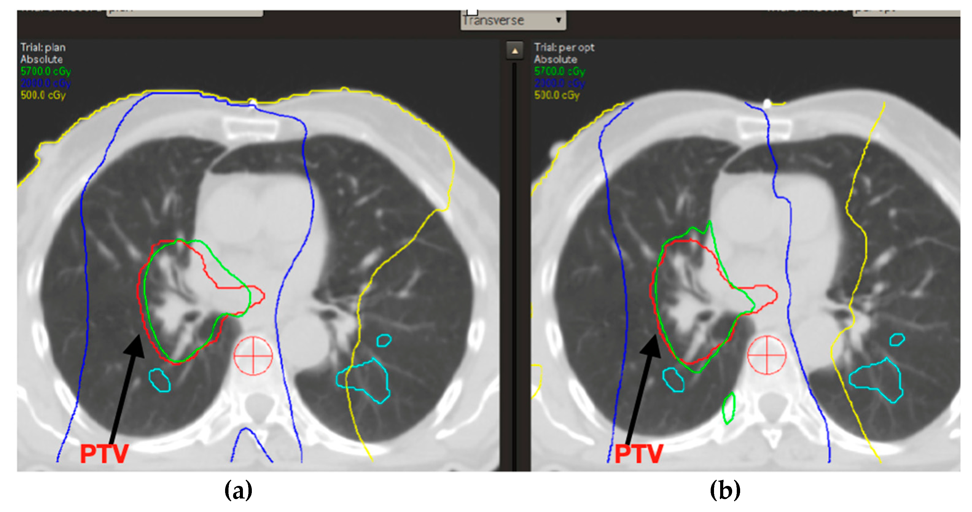
In terms of target coverage, a dosimetric comparison between the standard RT plan (plane 1) and the SPECT/CT-based one (plane 2) did not show relevant differences. No differences were also documented regarding the organs at risk identified. (Table 2) (Figure 3).
Mean dose, V5 and V20 for lung-PTV were substantially equivalent for standard RT plans and SPECT-based ones. No differences were observed for isoROI 20%, while a slight but significant improvement in mean lung dose was found in terms of isoROI 40%. Better results for all abovementioned dosimetric parameters were achieved for the isoROI 60% and 80% in the SPECT-optimized plan (Table 3).

4. Discussion

An example of application of radiological parameters to discriminate pathophysiological features is the DTI in the brain neoplasm, where different ADCmean values are related to different histologies [16,17]. In the same way, the pulmonary function test is well described as a useful tool to assess lung functionality and can potentially predict the risk of RIP in candidates to curative treatment for LA-NSCLC [8]. The SPECT-optimized plan was developed by giving higher priority to the isoROI 80% corresponding to very small lung volumes. This procedure allowed us to maintain the same dose volume histogram (DVH) between the standard and SPECT optimized plan in term of target coverage. At the same time, the SPECT-optimized RT plan allowed us to better satisfy the selected dose–volume parameters such as V5, V20 and MLD. This optimization also allowed us to obtain a clear advantage for isoROI 60%, while it was just marginally achieved for isoROI 40%. This suggests that the optimization effort profused on small lung volumes (isoROI 80%) could improve dose distribution even on larger lung volumes.
Pulmonary perfusion assessment could also be particularly relevant for RIP damage. Preserving lung parenchyma with optimal perfusion is likely to reduce the resulting vascular damage and the risk of pulmonary toxicity [18].
The main limits of this study are its retrospective nature, the limited number of cases and the lack of correlation between dosimetric data and clinical outcomes. Nonetheless, results of this pilot analysis are promising in terms of possibility to spare the better perfused lung volumes, with the chance to reduce the risk of RIP in functional lung. This preliminary hypothesis-generating experience warrants the evaluation of SPECT-guided radiotherapy in prospective and larger series to confirm the real clinical impact of integrating SPECT information into the treatment planning workflow to better avoid severe pulmonary toxicity.

Author Contributions

Conceptualization: P.B. and J.I.; methodology: A.B. and J.I.; software, C.T.; validation: S.L. and F.V.; formal analysis: G.V. and N.S.; investigation: J.I.; resources: G.F.; data curation: S.L.M. and A.G.; writing—original draft preparation: J.I. and N.S.; writing—review and editing: J.I. and P.B.; visualization: P.B. and A.B.; pervision: M.B.d.M.e.B.; project administration: P.B. All authors have read and agreed to the published version of the manuscript.

Funding

Authors declare that no funds, grants or other support were received during the preparation of this manuscript.

Institutional Review Board Statement

This is a retrospective study, based on anonymized data, no ethical approval and no informed consent regarding participation or publishing are required by the involved center.

Conflicts of Interest

Absence of any kind of any active or previous conflict of interest: no relevant financial or non-financial interests to disclose.

References

  1. Spigel, D.R.; Faivre-Finn, C.; Gray, J.E.; Vicente, D.; Planchard, D.; Paz-Ares, L.; Vansteenkiste, J.F.; Garassino, M.C.; Hui, R.; Quantin, X.; et al. Five-Year Survival Outcomes From the PACIFIC Trial: Durvalumab After Chemoradiotherapy in Stage III Non–Small-Cell Lung Cancer. J. Clin. Oncol. 2022, 40, 1301–1311. [Google Scholar] [CrossRef] [PubMed]
  2. Bruni, A.; Scotti, V.; Borghetti, P.; Vagge, S.; Cozzi, S.; D’Angelo, E.; Giaj Levra, N.; Fozza, A.; Taraborrelli, M.; Piperno, G.; et al. Corrigendum: A Real-World, Multicenter, Observational Retrospective Study of Durvalumab After Concomitant or Sequential Chemoradiation for Unresectable Stage III Non-Small Cell Lung Cancer. Front. Oncol. 2021, 11, 802949. [Google Scholar] [CrossRef]
  3. Chun, S.G.; Hu, C.; Choy, H.; Komaki, R.U.; Timmerman, R.D.; Schild, S.E.; Bogart, J.A.; Dobelbower, M.C.; Bosch, W.; Galvin, J.M.; et al. Impact of Intensity-Modulated Radiation Therapy Technique for Locally Advanced Non–Small-Cell Lung Cancer: A Secondary Analysis of the NRG Oncology RTOG 0617 Randomized Clinical Trial. J. Clin. Oncol. 2017, 35, 56–62. [Google Scholar] [CrossRef]
  4. Bradley, J.; Graham, M.V.; Winter, K.; Purdy, J.A.; Komaki, R.; Roa, W.H.; Ryu, J.K.; Bosch, W.; Emami, B. Toxicity and outcome results of RTOG 9311: A phase I–II dose-escalation study using three-dimensional conformal radiotherapy in patients with inoperable non–small-cell lung carcinoma. Int. J. Radiat. Oncol. Biol. Phys. 2005, 61, 318–328. [Google Scholar] [CrossRef]
  5. Cox, J.D. Are the Results of RTOG 0617 Mysterious? Int. J. Radiat. Oncol. Biol. Phys. 2012, 82, 1042–1044. [Google Scholar] [CrossRef]
  6. National Cancer Institute. Common Terminology Criteria for Adverse Events: CTCAE.
  7. Kristersson, S.; Lindell, S.-E.; Svanberg, L. Prediction of Pulmonary Function Loss Due to Pneumonectomy Using 133Xe-Radiospirometry. Chest 1972, 62, 694–698. [Google Scholar] [CrossRef]
  8. Evans, E.S.; Hahn, C.A.; Kocak, Z.; Zhou, S.-M.; Marks, L.B. The Role of Functional Imaging in the Diagnosis and Management of Late Normal Tissue Injury. Semin. Radiat. Oncol. 2007, 17, 72–80. [Google Scholar] [CrossRef]
  9. Farr, K.P.; Kallehauge, J.F.; Møller, D.S.; Khalil, A.A.; Kramer, S.; Bluhme, H.; Morsing, A.; Grau, C. Inclusion of functional information from perfusion SPECT improves predictive value of dose–volume parameters in lung toxicity outcome after radiotherapy for non-small cell lung cancer: A prospective study. Radiother. Oncol. 2015, 117, 9–16. [Google Scholar] [CrossRef] [PubMed]
  10. Graham, M.V.; A Purdy, J.; Emami, B.; Harms, W.; Bosch, W.; Lockett, M.A.; A Perez, C. Clinical dose–volume histogram analysis for pneumonitis after 3D treatment for non-small cell lung cancer (NSCLC). Int. J. Radiat. Oncol. Biol. Phys. 1999, 45, 323–329. [Google Scholar] [CrossRef]
  11. Tsujino, K.; Hirota, S.; Endo, M.; Obayashi, K.; Kotani, Y.; Satouchi, M.; Kado, T.; Takada, Y. Predictive value of dose-volume histogram parameters for predicting radiation pneumonitis after concurrent chemoradiation for lung cancer. Int. J. Radiat. Oncol. Biol. Phys. 2003, 55, 110–115. [Google Scholar] [CrossRef]
  12. Nestle, U.; De Ruysscher, D.; Ricardi, U.; Geets, X.; Belderbos, J.; Pöttgen, C.; Dziadiuszko, R.; Peeters, S.; Lievens, Y.; Hurkmans, C.; et al. ESTRO ACROP guidelines for target volume definition in the treatment of locally advanced non-small cell lung cancer. Radiother. Oncol. 2018, 127, 1–5. [Google Scholar] [CrossRef] [PubMed]
  13. Hodapp, N. Der ICRU-Report 83: Verordnung, Dokumentation und Kommunikation der fluenzmodulierten Photonenstrahlentherapie (IMRT) [The ICRU Report 83: Prescribing, recording and reporting photon-beam intensity-modulated radiation therapy (IMRT)]. Strahlenther. Onkol. 2012, 188, 97–100. [Google Scholar] [CrossRef] [PubMed]
  14. Bentzen, S.M.; Constine, L.S.; Deasy, J.O.; Eisbruch, A.; Jackson, A.; Marks, L.B.; Haken, R.K.T.; Yorke, E.D. Quantitative Analyses of Normal Tissue Effects in the Clinic (QUANTEC): An Introduction to the Scientific Issues. Int. J. Radiat. Oncol. Biol. Phys. 2010, 76, S3–S9. [Google Scholar] [CrossRef] [PubMed]
  15. Hoover, D.A.; Reid, R.H.; Wong, E.; Stitt, L.; Sabondjian, E.; Rodrigues, G.B.; Jaswal, J.K.; Yaremko, B.P. SPECT-based functional lung imaging for the prediction of radiation pneumonitis: A clinical and dosimetric correlation. J. Med. Imaging Radiat. Oncol. 2014, 58, 214–222. [Google Scholar] [CrossRef] [PubMed]
  16. Minh Duc, N. The Effects of Applying Apparent Diffusion Coefficient Parameters on the Differentiation between Fourth Ventricular Ependymoma and Diffuse Intrinsic Pontine Glioma. J. Child Sci. 2020, 10, e169–e174. [Google Scholar] [CrossRef]
  17. Thong, P.M.; Duc, N.M. The Role of Apparent Diffusion Coefficient in the Differentiation between Cerebellar Medulloblastoma and Brainstem Glioma. Neurol. Int. 2020, 12, 34–40. [Google Scholar] [CrossRef] [PubMed]
  18. Siva, S.; Devereux, T.; Ball, D.L.; MacManus, M.; Hardcastle, N.; Kron, T.; Bressel, M.; Foroudi, F.; Plumridge, N.; Steinfort, D.; et al. Ga-68 MAA Perfusion 4D-PET/CT Scanning Allows for Functional Lung Avoidance Using Conformal Radiation Therapy Planning. Technol. Cancer Res. Treat. 2016, 15, 114–121. [Google Scholar] [CrossRef] [PubMed]
Figure 1. Deformable co-registration between CT simulation images and the SPECT/CT.
Applsci 12 09351 g001
Figure 2. Red: PTV; green: isoROI 20%; magenta: isoROI 40%; yellow: isoROI 60%; light-blue: isoROI 80%.
Applsci 12 09351 g002
Figure 3. (a) Standard RT plan and (b) SPECT/CT-optimized RT plan.
Applsci 12 09351 g003
Table 1. Patient and treatment characteristics.
Variables Patients
Age66 (56–75)55
Diagnosissquamous cell carcinoma
adenocarcinoma
3
5
StageIIIA
IIIB
IIIC
1
5
2
BPCO o other respiratory diseases0
Charlson comorbidity index<78
>70
chemotherapyCarboplatin/paclitaxel trisettimanale
CDDP/Pemetrexed
6
2
RIPG3
G2
G1
G0
1
0
0
7
Table 2. Results of the DVH of plane 1 and plane 2.
Plane 1Plane 2p
D95 PTV5555ns
Mean dose of cumulative lung minus PTV1313ns
Mean dose of ipsilateral lung2323ns
Mean dose of contralateral lung99ns
V556%56%ns
V2023%23%ns
Mean dose of esophagus2727ns
Mean dose of heart1515ns
Table 3. Results of the DVH analysis of the isoROIs of plane 1 and plane 2.
IsoROI > 20% Max PerfusionPlane 1Plane 2p
Mean dose cumulative lung minus PTV1414ns
V202020ns
V55457ns
IsoROI > 40% max perfusionPlane 1Plane 2p
Mean dose cumulative lung minus PTV14130.042
V202019ns
V55656ns
IsoROI > 60% max perfusionPlane 1Plane 2p
Mean dose cumulative lung minus PTV14110.025
V202316ns (0.05)
V557540.023
Publisher’s Note: MDPI stays neutral with regard to jurisdictional claims in published maps and institutional affiliations.