Article

Nephroprotective and Antioxidant Effects of Flavonoid-Rich Extract of Thymelaea microphylla Coss. et Dur Aerial Part

1
Laboratory of Bioresources, Biotechnology, Ethnopharmacology and Health, Faculty of Sciences, Mohammed First University, B.P. 717, Oujda 60040, Morocco
2
Department of Basic Science, College of Medicine, Princess Nourah bint Abdulrahman University, P.O. Box 84428, Riyadh 11671, Saudi Arabia
3
Central Laboratory Service—CHU, Mohammed VI, Faculty of Medicine and Pharmacy, University Mohamed Premier, Oujda 60000, Morocco
*
Authors to whom correspondence should be addressed.
Appl. Sci. 2022, 12(18), 9272; https://doi.org/10.3390/app12189272
Submission received: 30 August 2022 / Revised: 9 September 2022 / Accepted: 12 September 2022 / Published: 15 September 2022
(This article belongs to the Special Issue Natural Products: Sources and Applications)

Abstract

:
Thymelaea microphylla Coss. et Dur (T. microphylla), a medicinal herb that grows in arid and desert pastures, has been traditionally utilized by Moroccans to treat many diseases, including kidney failure. This study aims to evaluate the nephroprotective effect against gentamicin-induced nephrotoxicity (GM), and thus the antioxidant activity of an aqueous extract rich in flavonoids from the aerial part of T. microphylla (APTM). The antioxidant activity of APTM was assessed using the 2-2-diphenyl-1-picrylhydrazyl (DPPH) scavenging test and the β-carotene bleaching assay. The nephroprotective effect of this extract was evaluated in two doses of 200 and 400 mg/kg in GM-exposed male rats. Acute toxicity of the APTM was tested out on Swiss albino mice using 2000 mg/kg as the dose limit. The findings showed that the aqueous extract of APTM is abundant in total polyphenols and flavonoids and has significant antioxidant properties against DPPH radicals and β-carotene oxidation. According to the acute toxicity research findings, the administered doses of the APTM extract do not cause toxicity and death. A significant increase in the serum concentrations of creatinine, urea, uric acid, sodium, chloride, calcium, gamma-glutamyl transpeptidase (gamma-GT), and alkaline phosphatase, as well as an increase in urinary volume, water consumption, and relative kidney weight, were all caused by the administration of GM to rats. In addition, a significant reduction in urinary concentrations of creatinine, uric acid, urea, and albumin, and thus the clearance of creatinine and weight gain were observed in rats injected with GM. Also, the administration of GM dramatically raised the malondialdehyde level in the kidneys. Likewise, rats that had been poisoned with GM had histological kidney abnormalities. However, the daily treatment of APTM aqueous extract to rats given GM injections dramatically improved the biochemical and histological parameters affected by GM administration in rats. Finally, APTM extract enhanced GM’s biochemical and histological indicators of nephrotoxicity, supporting its use as an ethnomedicinal.

1. Introduction

The renal system, characterized by a significant amount of blood supply from the cardiac output, carries out several vital tasks, including eliminating exogenous medications and toxins from the blood, controlling water fluid levels, and adjusting the body’s acid–base balance [1]. This vital organ’s high metabolic activities and transporter capacity make it more vulnerable to nephrotoxicity and increase its sensitivity to several types of failure [2]. Renal impairment caused by medication treatments is a regular occurrence in clinical care. The use of nephrotoxic drugs is responsible for about 20% of renal insufficiencies among hospitalized patients [3].
Aminoglycosides (AG) have long been among the most common causes of drug-induced nephrotoxicity [4]. Despite their adverse side effects, AG remains the main viable treatment option for bacteria resistant to other antibiotics [5]. This is mainly attributed to their chemical stability, immediate bactericidal activity, synergy with β-lactam antibiotics, limited resistance, and inexpensive cost [6]. Gentamicin (GM), the most nephrotoxic antibiotic in the AG class, externalizes its toxicity even at low doses [7,8]. Furthermore, in experimental studies, GM has been frequently utilized as a suitable substance to provoke animal nephrotoxicity [9]. Numerous researchers have shown that one of the fundamental mechanisms for the nephrotoxicity caused by this antibiotic is the oxidative stress induced by GM metabolism [7,10]. The formation of reactive oxygen species such as superoxide anion, hydrogen peroxide, and hydroxyl radicals is caused by renal cortical mitochondria due to the GM nephrotoxicity, which is also accompanied by an increase in lipid peroxidation. In this context, natural resources, such as medicinal plants, which are a source of natural antioxidants, can be employed as a treatment for this issue to lessen the nephrotoxicity brought on by drugs that promote oxidative stress. Among medicinal flora of Morocco, Thymelea microphylla Coss. et Dur. (T. microphylla), an endemic desert species [11], is a herbaceous plant growing in the Saharan region under harsh conditions [12]. This plant, classified under the genus Thymelaea, has smaller leaves and flowers, is well known for its vernacular name “Methnane” in Morocco, and is frequently used traditionally to treat various ailments [13]. Recently, our team conducted ethnobotanical fieldwork in a remote area of North Eastern Morocco and revealed that this plant species had been used to treat pyelonephritis, diuretics, renal colic, and kidney stones [14]. The scientific literature regarding the pharmacology of T. microphylla, indicated that this species has different properties such as antibacterial [12,15], antioxidant [11], and antidiabetic ones [12]. Also, the phytochemistry of this plant showed that the aerial part of T. microphylla is rich in phenolic compounds such as resorcinol, catechin, chlorogenic acid, syringic acid, sinapic acid, trans-3-hydroxycinnamic acid, isoquercitrin, ellagic acid, myricetin, kaempferol 3-O-rutinoside, trans-cinnamic acid, luteolin, and apigenin [16]. In addition, the pharmacological activities of flavonoids and phenolic substances of plant origin, including antioxidant and nephroprotective actions, are well established [17]. To our best knowledge, the aerial part of this Moroccan plant species has not been studied for its nephroprotective properties. In this respect, we conducted this research to see if the aqueous extract of the APTM could protect Wistar rats from nephrotoxicity caused by GM, and if it has antioxidant activity.

2. Results and Discussion

2.1. Total Phenolic and Flavonoids Contents

Quantification of total phenol and flavonoid components exhibited that APTM aqueous extract is rich in total polyphenols (2423.13 ± 23 µg gallic acid equivalent/mg) and flavonoids (317 ± 15 µg quercetin equivalent/mg). These results confirm previous research, which showed that T. microphylla extracts are rich in total polyphenols [11,18]. These high levels of total polyphenols in the extracts of T. microphylla are probably related to the climatic stress to which this plant is exposed in the desert of northeastern Morocco (high temperatures, high solar exposure, drought, and short growing season) [18]. Due to their interesting antioxidant capacity, these compounds have various pharmacological activities, including nephroprotective [17]. Phenolic compounds can operate as natural antioxidants and impact the synthesis or activity of other antioxidant molecules in the body [19].

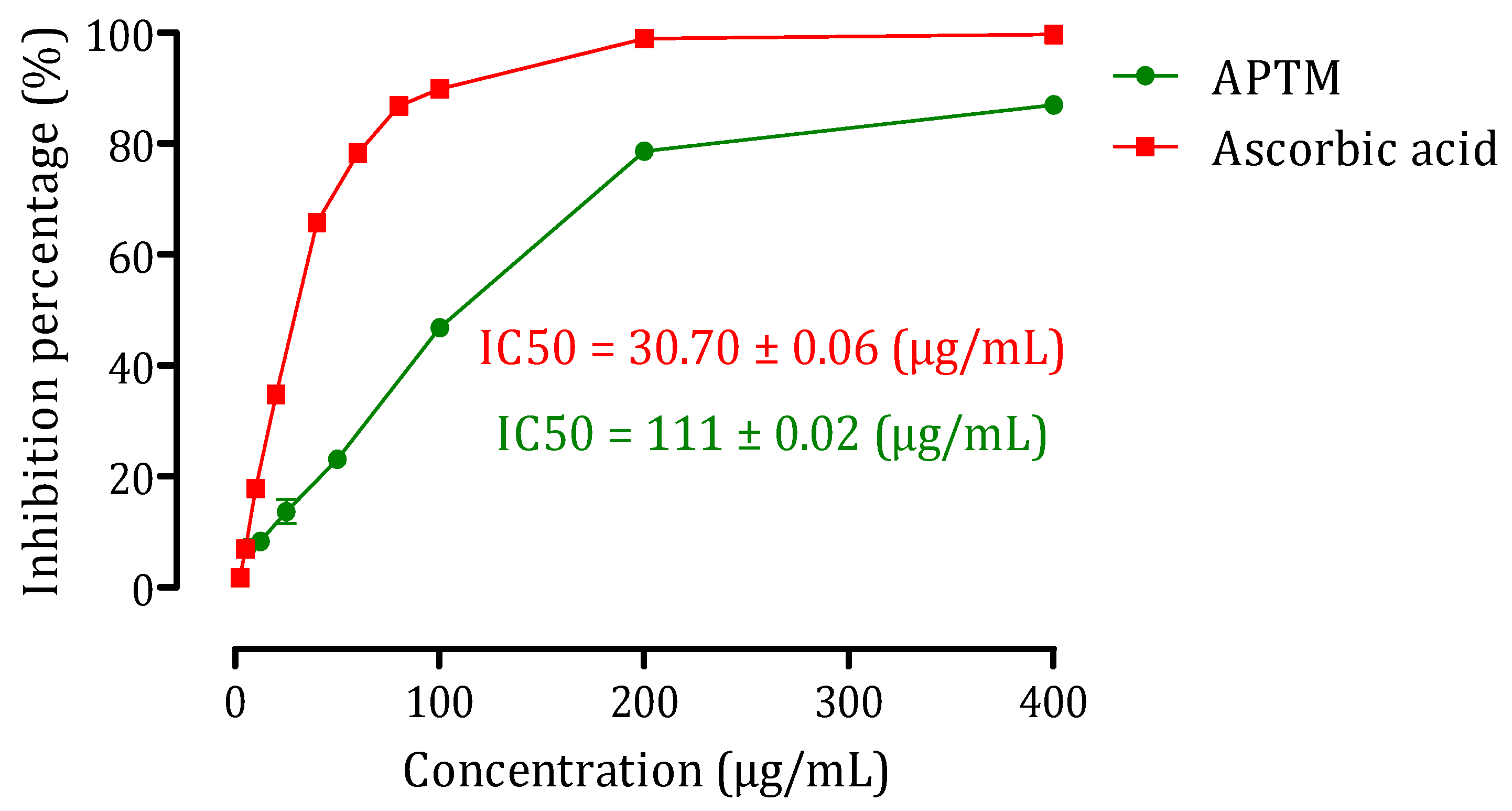
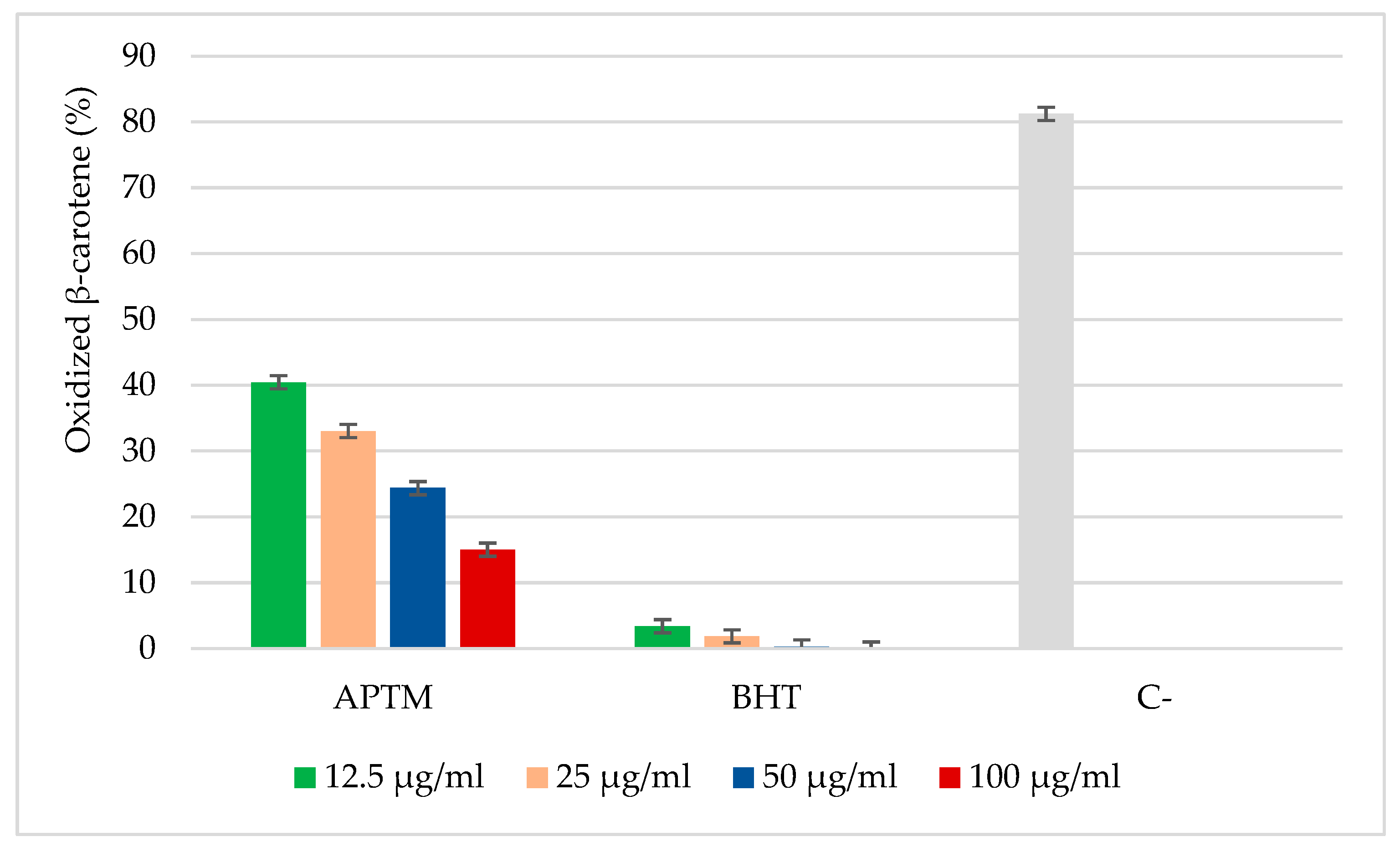
2.2. Antioxidant Activity of APTM Aqueous Extract

Antioxidants can prevent the human body from producing the harmful effects of free radicals by administering certain drugs, including aminoglycosides such as GM. They halt the course of specific acute and chronic disorders and the development of lipid peroxidation. Indeed, the aqueous extract of APTM may exhibit antioxidant properties against DPPH free radicals and lipid oxidation in biological systems.
The diversity and complexity of secondary metabolites present in plant extracts requires several techniques to assess the antioxidant effects and evaluate the efficacy of these phytochemicals. Most of these techniques rely on a reagent’s coloring or discoloration in the reaction mixture [12]. Usually, the methods of evaluation of antioxidant activity can be grouped into two classes: the tests used to evaluate the trapping power of free radicals, such as the DPPH test, and tests used to evaluate and estimate lipid peroxidation in biological systems while assessing the degree of inhibition of oxidation, such as the β-carotene/linoleic acid test. Our study used two antioxidant tests: the DPPH trapping test and the β-carotene bleaching test.
The results of the antioxidant effect of the aqueous extract APTM against free radicals DPPH were presented in Figure 1. Indeed, the aqueous extract of APTM has a dose-dependent inhibition effect on the free radical DPPH. Results showed a moderate ability to trap free radicals in APTM aqueous extract with an IC50 of 111 ± 0.02 µg/mL, which is even lower than that of ascorbic acid (IC50 = 30.70 ± 0.06 µg/mL). These findings demonstrated that APTM’s aqueous extract had moderate antioxidant activity against DPPH free radicals. The current antioxidative activity results from the aqueous APTM extract is consistent with those shown by Dahmna et al. (2015) in an extract of T. microphylla’s leaves and flowers from Draa Elhadja region in M’sila, Algeria [12].
Measurements of the inhibition of volatile organic molecules and the production of conjugated diene hydroperoxide from linoleic acid oxidation are used to determine the antioxidant activity in the β-carotene bleaching assay [20]. Figure 2 displays the outcomes of the aqueous APTM extract’s β-carotene bleaching test. The findings of this test reveal that our extract significantly inhibited β-carotene oxidation as a function of concentration. The percentages of β-carotene that are oxidized in the presence of APTM aqueous extract are 40.43%; 33.02%; 24.38% and 15.02%, respectively, for the concentrations of 12.5, 25, 50 et 100 µg/mL, and those of butylated hydroxytoluene were respectively 3.40%, 1.85%, 0.31% and 0.01% for the concentrations of 12.5, 25, 50 and 100 g/mL. The aqueous extract of APTM’s ability to prevent the oxidation of β-carotene may result from the extract’s high concentration of polyphenols and flavonoids. This may be due to the correlation between polyphenols or flavonoid content in a plant and its antioxidant effects [21].

2.3. Acute Oral Toxicity Test of APTM Aqueous Extract

The acute toxicity of APTM aqueous extract was investigated in mice (Table 1). In fact, at the dosage limit of 2000 mg/kg, administration of the aqueous extract of APTM causes neither mortality nor behavioral abnormalities in the mice. Additionally, the extract demonstrated a good and typical aspect of mice’s general behavior. Doses of 1/10 and 1/5 of the dose limit were used to test the nephroprotective impact of the aqueous extract of APTM.

2.4. Nephroprotective Effect of APTM

2.4.1. Effects of APTM on Urine Volume and Water Consumption

As shown in Figure 3, by comparing to animals in the control group (NCG), GM provoked a considerable rise in water consumption (p < 0.01) and urinary volume (p < 0.001). However, the treatment with the APTM extract at 200 and 400 mg/kg substantially recovered consumption of water (p < 0.05) and urine output (p < 0.05 and p < 0.01, respectively) in GM-intoxicated rats as compared to GGM (80 mg/kg). A possible explanation to this is GM accumulation in the renal tissue, which results in the loss of proximal tubular cells and the inability of Henle’s loop to reabsorb water, which results in dehydration [10,17]. A rise in osmolarity brought on by the depletion of the rats’ bodily fluid or an effort to replenish lost bodily fluid volume following GM treatment may also be to blame for the increase in water intake. As a result, osmoreceptor cells are stimulated, activating the thirst center in the hypothalamus and promoting increased water consumption [10].

2.4.2. Weight Gain and Relative Kidney Weight to Body Weight

Table 2 shows the impact of the APTM extract on body weight increase and relative kidney weight. When compared to the NCG, treatment with GM for 14 days caused a significant decrease in weight growth (p < 0.001) and hence a substantial increase in relative kidney weight (p < 0.001). However, as compared to GGM rats (80 mg/kg), treatment with APTM extract at dosages of 200 and 400 mg/kg in conjunction with GM boosted weight growth in a dose-dependent manner (p < 0.01) and lowered relative kidney weight (p < 0.001). These results support those of Erdem et al. (2000) and El-Zawahry and Abu El Kheir (2007), who showed that the administration of GM caused a decrease in body weight [22,23]. The same sources claim that GM decreases hunger and increases catabolism, both observed in GM-induced acute renal failure, which results in acidosis and anorexia. Because of this, oral nutritional intake declines, which may result in weight loss. Furthermore, the GM-induced rise in kidney weight is undoubtedly the consequence of oedema brought on by acute tubular necrosis brought on by the medication. This was confirmed histopathologically by the study of El-Kashef et al. (2016), in which electron microscope examination revealed mitochondrial swelling and cytoplasmic degeneration and accumulation of the myeloid body in the renal tissues of GM-exposed rats [24]. The observed improvement in body weight and relative kidney weight may be associated with the anti-inflammatory and antioxidant effects of APTM aqueous extract, which prevent the nephrotoxic effects of GM.

2.4.3. Effect of APTM on Serum and Urinary Creatinine, Uric Acid, and Urea Levels

In GM-induced acute nephrotoxicity, the activities of aqueous APTM extract on renal biomarkers were investigated (Table 3). In comparison to rats in the NCG, rats receiving GM (80 mg/kg) for 14 days had significantly higher serum creatinine (p < 0.001), uric acid (p < 0.05), and urea (p < 0.01). In addition, GM caused a substantial reduction in creatinine (p < 0.001), uric acid (p < 0.01), and urea (p < 0.01) levels in the urine. Changes in renal biochemical marker levels caused by GM suggest acute renal toxicity [25,26]. Since urea and creatinine are produced during purine metabolism, their elevated serum levels could indicate filtration rate alterations and be linked to kidney injury [27,28]. These outcomes are in line with those of other researchers, who found that giving GM to rats caused an increase in urea and creatinine concentrations in the serum with a lowering in those levels in the urine [29,30,31]. As shown in Table 3, before 3 h of the GM injection, daily administration of the aqueous extract of APTM (for 14 days of treatment) reversed the GM-induced alterations in kidney biomarkers to near-normal levels. When compared to GM-poisoned rats not treated with extract, the 200 and 400 mg/kg doses of the APTM extract substantially lowered serum creatinine (p < 0.01, p < 0.001), urea (p < 0.05), and significantly increased urine creatinine (p < 0.01, p < 0.001), uric acid (p < 0.05, p < 0.01), and urea (p < 0.05). Various plant extracts have been demonstrated to have similar anti-GM protective properties in other research [25,32,33]. According to several studies, GM can cause nephrotoxicity by increasing reactive oxygen species [34,35]. Natural antioxidants like polyphenols and flavonoids are known to capture free radicals in vivo and in vitro [17]. In addition, the results of our study indicate that the aqueous extract of APTM is rich in phenolic compounds, suggesting that our extract improved kidney function in GM-poisoned by removing free radicals brought on by GM in rats.

2.4.4. Effect of APTM on Creatinine Clearance

The effect of APTM on glomerular filtration was measured using creatinine clearance in all of the research animals, as shown in Figure 4. Indeed, treatment with GM during 14 days of treatment resulted in a very significant reduction in clearance of creatinine (p < 0.001) compared with the normal controlled group (NCG). A helpful indicator of the glomerular filtration rate is creatinine clearance. Reduced renal blood flow and glomerular filtration rate due to increased renal blood vessel constriction or injury to the glomerular capillary endothelium are indicated by a decline in creatinine clearance [30]. The treatment of GM-intoxicated rats with aqueous APTM extract at dosages of 200 and 400 mg/kg enhanced creatinine clearance in a dose-dependent manner (p < 0.05 and p < 0.01, respectively, for doses 200 and 400 mg/kg). This finding points to a significant rise in glomerular filtration rate, resulting in improved blood flow and kidney function. According to earlier investigations, reactive oxygen species may have a role in the development of GM nephrotoxicity. In addition, reactive oxygen species are also involved in proximal tubular necrosis and acute renal failure caused by GM [29]. For this reason, the suggested nephroprotective effect of our extract on creatinine clearance could be attributed to its antioxidant potential, as oxygen-reactive species are involved in the alteration of glomerular filtration rate [36].

2.4.5. Effect of APTM on Urinary Total Protein and Albumin Levels

In all test groups, the effect of the APTM extract on total urine protein and albumin levels was investigated, and the results are shown in Figure 5. When rats were given GM, urine concentrations of total proteins and albumin were significantly higher (p < 0.01 and p < 0.001, respectively) than in the NCG. These results are in agreement with those found by several researchers [8,17]. Increased urinary protein levels in GM-treated rats have been confirmed in other relevant studies [17,29]. However, compared to GGM (80 mg/kg), the APTM extract at dosages of 200 and 400 mg/kg significantly reduced the urine rate of total protein and albumin in GM-intoxicated rats. The bioactive components in the aqueous extract of APTM may be responsible for the increasing effect on total plasma proteins and albumin. This finding implies that kidney cells are generally protected by these natural substances.

2.4.6. Effect of APTM on Serum Gamma-GT and Alkaline Phosphatase (ALP) Level

The impact of APTM extract on the serum concentrations of gamma-GT and ALP in GM-intoxicated rats was tested for its hepatoprotective efficacy against GM hepatotoxicity, as shown in Figure 6. These results support the findings of Umbreen et al. (2017), who also reported an increase in serum ALP and gamma-GT after eight days of GM treatment [25]. GM-injection generates cell membrane damage and permeability and increases serum ALP, and gamma-GT levels, generally deemed sensitive markers of liver function [37]. Hepatocyte membranes have localized gamma-GT activity, and ALP, an indicator for the endoplasmic reticulum, is routinely used to examine the integrity of the plasma membrane. [8]. The high concentration of these enzymes in the serum highlighted that the GM’s oxidative stress had damaged the rat’s liver. The rat treated with GM had higher levels of these enzymes in its serum [8,38,39]. The findings of our study show that the pretreatments with APTM at doses of 200 and 400 mg/kg significantly reduced increases in blood gamma-GT and ALP in GM-poisoned rats. The study’s findings were in line with prior investigations, in which chemical or plant extracts improved the steady-state concentration of these enzymes and lessened hepatocyte damage [38,39].

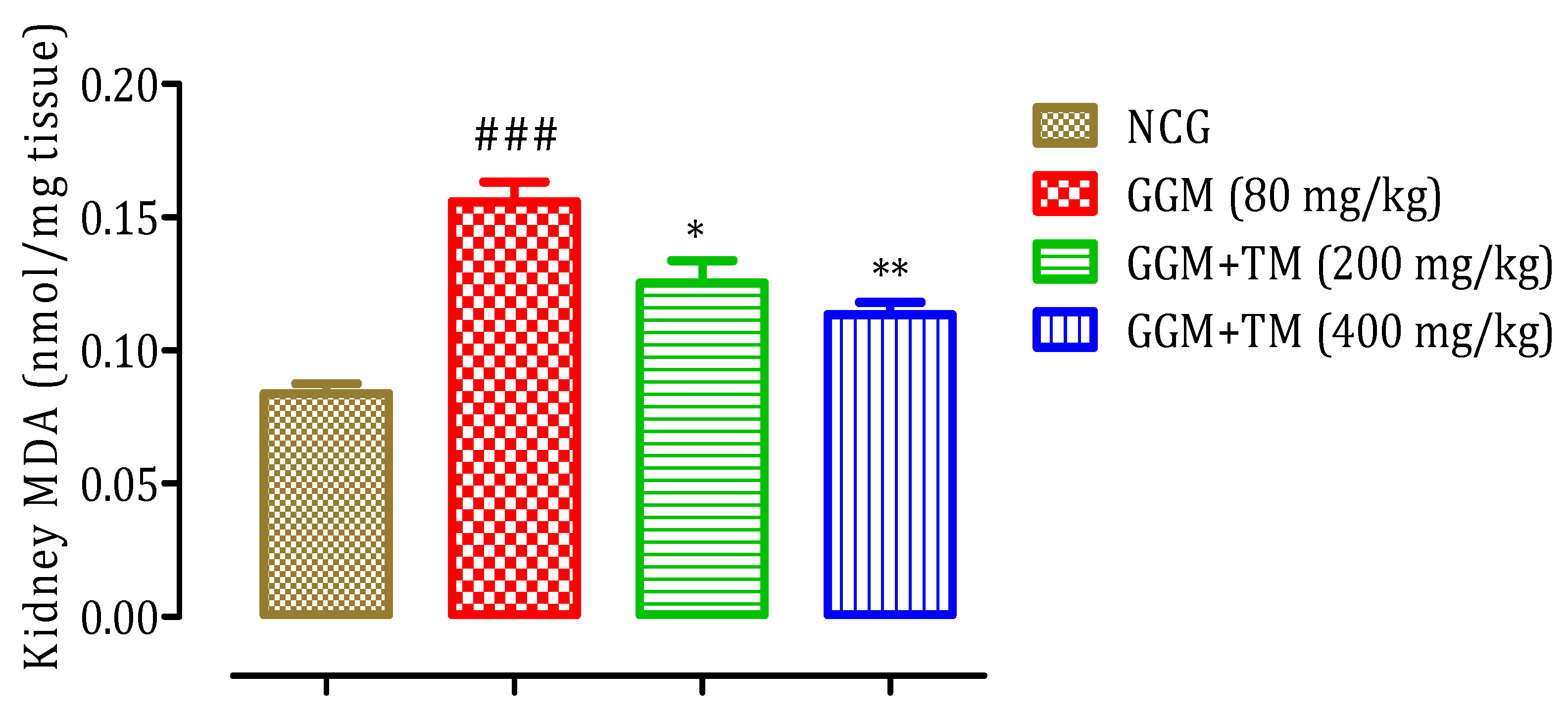
2.4.7. Effect of APTM on Renal Malondialdehyde (MDA) Level

Numerous investigations have identified lipid peroxidation in renal tissue as the damaging process of GM injection on renal function [40]. Increased malondialdehyde level in renal tissue suggests increased oxidative stress in GM-induced nephropathy [41]. The increased free radical generation has been linked to the degradation of several structural macromolecules, which can lead to cell lesions and tubular necrosis [40,42]. To this end, we assessed the impact of APTM aqueous extract on lipid peroxidation in GM-treated experimental animals. As shown in Figure 7, rats exposed to GM had a substantial increase (p < 0.001) in MDA rates compared to rats in the normal control group. The groups treated with aqueous APTM extract at two doses administered, with the GM-injection, had a marked decline (p < 0.05 and p < 0.01, respectively) in MDA concentration compared to rats in the GGM (80 mg/kg). These findings are consistent with prior studies, in which plant extract reduces lipid peroxidation to protect against GM-induced nephrotoxicity [25,43].

2.4.8. Effect of APTM on Serum Electrolytes Level

Table 4 summarizes the effects of aqueous APTM extract on serum electrolyte concentrations in GM-poisoned rats. Indeed, as compared to healthy rats (NCG), GM treatment substantially raised sodium (p < 0.01), chloride (p < 0.05), calcium (p < 0.01), and lowered potassium in a non-significant way (NCG). These outcomes align with some investigations where GM led to rat electrolyte imbalance [10,44]. This could be explained by how GM affects the basolateral membranes and the membranes that transport the epithelial cells’ brush borders, causing an electrolyte imbalance. Additionally, GM inhibits several membrane transporters from brush borders and basolateral membranes, regardless of the cellular lesion, which results in aberrant electrolysis [10]. Transport inhibition impacts tubular reabsorption and cell viability, leading to necrosis or apoptosis in some cases [45,46]. However, treatment of rats (intoxicated by GM) with aqueous APTM extract reduced sodium, chloride, and calcium levels in a non-significant way, while increasing potassium levels in a non-significant way. These findings agree with other researchers, who found that plant extract restored electrolyte concentration in rats poisoned by GM [30,44].

2.4.9. Effect of APTM on Histopathological Alterations in the Kidneys

Examination of histological sections separated from the normal control group (NCG) showed normal and intact tubules and glomeruli (Figure 8A). While analysis of histological sections from the negative control group (GGM) revealed loss of glomerular and tubular cells, dilation of renal tubules, deformation of Bowman space, and vascular congestion causing epithelial cell atrophy. These results are congruent with those obtained by several authors [7,47,48]. In contrast, the concomitant administration of GM and APTM aqueous extract at 200 mg/kg (Figure 8C) and 400 mg/kg (Figure 8D) in rats improved the histoarchitecture of the kidneys compared with those of healthy rats (NCG). In addition, this, improvement in kidney histology in GM-poisoned rats is comparable to that in normal control rats (Figure 8A). One potential mechanism to protect the kidneys from the oxidative stress caused by GM, according to several studies, is to reduce oxidative stress [7,8,10,49]. Polyphenols and flavonoids have been shown to reduce the nephrotoxicity of GM through increased antioxidant enzyme activity, decreased lipid peroxidation, trapping free radicals and improving kidney tissue architecture [17,43]. Our findings demonstrate that the APTM extract is rich in phenolic compounds and flavonoids, which are well known for their antioxidant capacity and have been generated and utilized by plants to defend themselves from pathogens [45,46]. According to these findings, our extract, based on these bioactive molecular activities, by scavenging free radicals produced by GM metabolism, protects the kidneys against GM nephrotoxicity.

3. Materials and Methods

3.1. Reagents

Gentamicin (GM) was provided by the pharmacy. Creatinine, uric acid, urea, alkaline phosphatase (ALP), gamma-glutamyl transpeptidase (gamma-GT), albumin, electrolytes kits, and thiobarbituric acid and trichloroacetic acid were acquired from the Sigma Aldrich Company (St. Louis, MO, USA). The 2,2-diphenyl-1-picrylhydrazyl (DPPH), β-carotene, butylated hydroxytoluene (BHT), tween-80, gallic acid, methanol, ascorbic acid, chloroform, aluminium chloride, Folin–Ciocalteu reagent, and quercetin were purchased from Sigma-Aldrich (Steinheim, Germany). All reagents and chemicals used are of analytical grade.

3.2. Plant Material

The Thymelaea microphylla Coss. et Dur aerial parts were obtained in April 2020 in a desert location between Tendrara and Figuig (Eastern Morocco) and identified by botanist Mohammed Fennane of Mohammed V University’s scientific institute. The sample was prepared and stored in the university’s herbarium number “HUMPOM715”.

3.3. APTM Extract Preparation

The aqueous extract was prepared according to the Moroccan population’s traditional technique, with slight modification [14]. Indeed, the aerial part of the plant was cut into small pieces. Then, a quantity of 150 g of the aerial part of this plant was mixed with 2 L of boiled distilled water. Then, the mixture was decocted for 10 min at a temperature of 75 °C. The decocted mixture was filtered and evaporated using a rotary vacuum evaporator with a temperature of 40 °C. After complete drying, the extract was stored at 20 °C for later use.

3.4. Estimation of Total Phenolic and Flavonoid Levels

The flavonoids estimation was determined following the protocol established by Kim et al. (2003) [50], with slight adjustments. A reaction mixture comprised 200 µL of aqueous extract of T. microphylla Coss. et Dur (1000 µg/mL), 1000 µL of purified water, and 50 µL of NaNO2 (5%). We added 120 L of AlCl3 (10%) to the reaction mixture after 6 min of homogenization, incubated for five min, and then added 400 µL of NaOH (1 M) and 230 µL of distilled water. Absorbance was read at 510 nm against a blank solution sample of all reagents without plant extract. The outcomes were given in µg quercetin equivalent/mg of extract.
The Folin–Ciocalteu approach was employed to determine the quantity of phenols in the APTM extract [51]. In summary, 200 µL of APTM extract (1000 µg of extract/mL of distilled water) were combined with 1000 µL of Folin–Ciocalteu reaction and 800 µL of Na2CO3 (75 g/L). The solution combination was vortexed and maintained for one hour at room temperature. A standard calibration curve was plotted using gallic acid (0–200 mg/mL). The absorbance was then measured against a blank (solution without extract) using a spectrophotometer at 765 nm. The values are given in µg equivalent acid gallic/mg of extract.

3.5. Evaluation of the Antioxidant Properties

3.5.1. DPPH Free Radical-Scavenging Activity Test

The capacity of the extract to trap the free radical DPPH of dark violet color, which transforms into a yellowish color (after reduction), is the basis of this test. Spectrophotometry is used to measure discoloration. The DPPH radical-scavenging test was carried out, as reported in [52]. Indeed, increasing doses of aqueous APTM extract (6, 12, 25, 50, 100, 200, and 400 µg/mL) were created in test tubes, then 0.1 mL from every tube was collected and combined with 2.5 mL of ethanolic DPPH solution (4%). The solutions were then kept in the dark for 30 min. The absorbance of the combinations was measured immediately against a blank (ethanol) after incubation, using spectrophotometry at 517 nm. Ascorbic acid was employed as an ordinary point. Every one of the tests was carried out in triplicate. The APTM extract trapping activity was determined using the following formula:
D P P H   t r a p p i n g   a c t i v i t y   % = A 0 A 1 A 0 * 100
The absorbance of the control reaction is A0, while the absorbance of all extract samples and standards is A1.

3.5.2. β-Carotene Bleaching test

The antioxidant properties of the APTM extract were measured using the β-carotene bleaching procedure described by Sun and Ho [53]. Indeed, 2 mg of β-carotene was blended with 200 mg of tween 80 and 20 mg of linoleic acid, and the whole solution was added to 10 mL of chloroform. The chloroform was removed from the mixture by rotavapor at 40 °C, and 100 mL of purified water was introduced and vigorously mixed by the vortex. After that, 0.2 mL of β-carotene solution was added to the simple solution (12.5, 25, 50 and 100 µg/mL), and the tubes were incubated at 50° C in a bain-marie for 2 h with continuous agitation. The absorbance was measured at 470 nm at 0 and 2 h of incubation. BHT (12.5, 25, 50 and 100 µg/mL) was used as a reference. The steps were repeated three times to reduce the risk of error. The equation below was used to compute the proportion of β-carotene oxidation:
% o x i d i z e d   β c a r o t e n e = A i t o A f   2 h A i t o * 100
Ai: optical density in to, and Af: optical density after incubation (2 h).

3.6. Acute Oral Toxicity Study

Acute toxicity was assessed, referring to OECD recommendations (425) [54]. Swiss albino mice (20–30 g) were utilized in the toxicity assessment of APTM extract. In fact, 36 mice were divided into six groups to assess acute oral toxicity. Each of the six mice (n = 6; ♂/♀ = 1) was given a single oral dosage of 50, 300, 500, 700, and 2000 mg/kg body weight of APTM extract, whereas the control group was given purified water (10 mL/kg).
After oral administration of the extract, the animals were monitored for signs of toxicity and/or mortality for the first 4 h and then for the 14 days of treatment.

3.7. Nephroprotective Study

3.7.1. Experimental Design

The male rats were arbitrarily divided into four groups after a two-week adaptation period, with each group consisting of six rats [7]. In the normal control group (NCG), animals received only 10 mL/kg of distilled water. The rats of the group GMG (80 mg/kg) were treated with distilled water (10 mL/kg) and were injected intraperitoneally with the GM (80 mg/kg; b.w.). The animals in the group GGM+TM (200 mg/kg) received an aqueous extract of APTM orally at 200 mg/kg, and the rats of the group GGM+TM (400 mg/kg) received 400 mg/kg of the aqueous extract of APTM orally. Subsequently, after 3 h, these two last groups were injected with GM (80 mg/kg) via the intraperitoneal route. Following the last injection, all of the rats in the study were kept in separate metabolic cages for 24 h to collect urine simple. The urine samples were centrifuged with a centrifugal force of 704× g.

3.7.2. Sample Collection

All rats were anaesthetized with light ethyl ether and euthanized 14 h after the last dose of intraperitoneal GM injection. The blood samples were taken from the aorta in the abdomen and placed in dry blood collection tubes; then, they were centrifuged for 10 min at 3000 rpm at about 4 °C to separate the serum. The serum was removed and kept chilled at 20 °C for additional examination. To calculate the amount of malondialdehyde (MDA), the liver was also weighed and stored for the production of a liver homogenate (10% w/v) in sodium phosphate buffer (pH 7.0).

3.7.3. Biochemical Analysis

Several biochemical parameters related to renal markers were evaluated in this study. Calcium, sodium, potassium, chloride, urea, creatinine, alkaline phosphatase (ALP), albumin, uric acid, and gamma-GT were measured with the COBAS INTEGRA® 400-Plus analyzer (Roche Diagnostics, Meylan, France).

3.7.4. Creatinine Clearance (CCL)

Based on serum and urine creatinine concentrations, the creatinine clearance was calculated using the formula below to determine the glomerular filtration rate [7].
C C L   m L m i n = U r i n e   c r e a t i n i n e   m g / m L * U r i n e   f l o w m L / m i n S e r u m   c r e a t i n i n e   m g / m L
This formula was used to determine the urine output: urine flow (mL/min) = value of urine volume (24 h)/1440 (60 min × 24 h = 1440)

3.7.5. Relative Kidney Weight (RKW)

The following equation was used to determine the relative kidney weight [10]:
R K W   % = A b s o l u t e   k i d n e y   w e i g h t   g * 100 B o d y   w e i g h t   o f   t h e   r a t   g

3.7.6. Kidney Lipid Peroxidation

The level of renal lipid peroxidation was evaluated using the experimental technique reported by Bueg and Aust [55]. In this test, the amount of TBARS produced is quantified. The kidney homogenate was prepared, and 0.5 mL of it was combined with 0.5 mL of TCA (30% w:v). After that, this solution was centrifuged at 4 °C using a 959× g centrifugal force. Then, 1 mL of supernatant and 1 mL of TBA (0.67% w:v) were mixed, implanted in hot water (100 C for ten min.), and covered with ice. The opacity of the combination was evaluated using a spectrophotometer calibrated to 535 nm. The following molar extinction coefficient was used to get the finding in moles (MDA quantity)/g (tissue). 1.56 × 10⁵ M−1 cm−1.

3.7.7. Histopathological Analysis

The rats’ left kidneys were removed for histopathological examination. The paraffin wax-encased 10% buffered formalin is used to preserve kidney tissues. They were then processed, divided into pieces measuring 3 to 4 µm, and stained with eosin and hematoxylin. Following this, histological pictures at ×40 magnification were obtained after the renal histology sections had been examined under an optical microscope.

3.8. Statistical Analysis

The data were indicated as means ± SEM. The ANOVA test followed by Tukey’s post hoc test was used in the statistical analysis of the data using Graph Pad Prism 5, San Diego, CA, USA. The result is considered significant if p < 0.05.

4. Conclusions

The current findings showed that the aqueous extract of APTM is rich in total polyphenols and flavonoids. The antioxidant effect produced by this extract was moderate against the free radical DPPH and oxidation of β-carotene in a dose-dependent manner. Thus, the acute toxicity study of APTM in mice indicated that this extract is devoid of toxicity and mortality at the administered doses. This is consistent with the daily use of this plant by the Moroccan population. Regarding the nephroprotective effect of the APTM extract, the biochemical and histological results showed that this extract improved the parameters modified during GM-induced nephrotoxicity. These findings offer preclinical, experimental support for the widespread use of this plant to treat kidney issues by indicating potential renal protection.

Author Contributions

Conceptualization, N.B.; methodology, N.B. and S.O.; software, H.O., and S.E.A.; validation, M.E., N.B. and M.C.; formal analysis, N.B., M.E. and J.F.; data curation, N.B.; writing—original draft preparation, N.B.; supervision, M.E.; review and editing, M.A.W.C. and A.A. All authors have read and agreed to the published version of the manuscript.

Funding

This research was supported by Princess Nourah bint Abdulrahman University Researchers Supporting Project number (PNURSP2022R147), Princess Nourah bint Abdulrahman University, Riyadh, Saudi Arabia.

Institutional Review Board Statement

The study was conducted according to the guidelines of the Declaration of Helsinki, and approved by the Institutional Review Board of the Faculty of Sciences, Oujda, Morocco (01/20-LBBEH-04 and 09/01/2020).

Data Availability Statement

All the data supporting the findings of this study are included in this article.

Conflicts of Interest

The authors declare no conflict of interest.

References

  1. Jose, S.P.; Asha, S.; IM, K.; Ratheesh, M.; Santhosh, S.; Sandya, S.; Girish Kumar, B.; Pramod, C. Nephro-protective effect of a novel formulation of unopened coconut inflorescence sap powder on gentamicin induced renal damage by modulating oxidative stress and inflammatory markers. Biomed. Pharmacother. 2017, 85, 128–135. [Google Scholar] [CrossRef] [PubMed]
  2. Martins, E. Nephrotoxicity and Nephroprotective Potential of African Medicinal Plants. In Toxicological Survey of African Medicinal Plants; Elsevier: Amsterdam, The Netherlands, 2014; pp. 357–393. ISBN 9780128000182. [Google Scholar]
  3. Schortgen, F. Néphrotoxicité et médicaments. Reanimation 2005, 14, 436–441. [Google Scholar] [CrossRef]
  4. Mingeot-Leclercq, M.-P.; Tulkens, P.M. Aminoglycoside nephrotoxicity. Curr. Drug Targets Infect. Disord. 2004, 4, 153–162. [Google Scholar] [CrossRef] [PubMed]
  5. Nagai, J.; Takano, M. Molecular aspects of renal handling of aminoglycosides and strategies for preventing the nephrotoxicity. Drug Metab. Pharmacokinet. 2004, 19, 159–170. [Google Scholar] [CrossRef]
  6. Edson, R.; Terrell, C.L. The Aminoglycosides. Mayo Clin. Proc. 1999, 74, 519–528. [Google Scholar] [CrossRef]
  7. Bencheikh, N.; Bouhrim, M.; Kharchoufa, L.; Al Kamaly, O.M.; Mechchate, H.; Es-safi, I.; Dahmani, A.; Ouahhoud, S.; El Assri, S.; Eto, B.; et al. The Nephroprotective Effect of Zizyphus lotus L. (Desf.) Fruits in a Gentamicin-Induced Acute Kidney Injury Model in Rats: A Biochemical and Histopathological Investigation. Molecules 2021, 26, 4806. [Google Scholar] [CrossRef]
  8. Bouhrim, M.; Bencheikh, N.; Imtara, H.; Daoudi, N.E.; Mechchate, H.; Ouassou, H.; Kharchoufa, L.; Elachouri, M.; Mekhfi, H.; Ziyyat, A.; et al. Protective Effect of Opuntia dillenii (Ker Gawl.) Haw. Seed Oil on Gentamicin-Induced Nephrotoxicity: A Biochemical and Histological Analysis. Sci. World J. 2021, 2021, 7. [Google Scholar] [CrossRef]
  9. Lopez-Novoa, J.M.; Quiros, Y.; Vicente, L.; Morales, A.I.; Lopez-Hernandez, F.J. New insights into the mechanism of aminoglycoside nephrotoxicity: An integrative point of view. Kidney Int. 2011, 79, 33–45. [Google Scholar] [CrossRef]
  10. Govindappa, P.K.; Gautam, V.; Tripathi, S.M.; Sahni, Y.P.; Raghavendra, H.L.S. Effect of Withania somnifera on gentamicin induced renal lesions in rats. Rev. Bras. Farmacogn. 2019, 29, 234–240. [Google Scholar] [CrossRef]
  11. Belyagoubi-Benhammou, N.; Belyagoubi, L.; Bekkara, F.A. Phenolic contents and antioxidant activities in vitro of some selected Algerian plants. J. Med. Plants Res. 2014, 8, 1198–1207. [Google Scholar]
  12. Dahamna, S.; Dehimi, K.; Merghem, M.; Djarmouni, M.; Bouamra, D.; Harzallah, D.; Khennouf, S. Antioxidant, Antibacterial and Hypoglycemic Activity of Extracts from Thymelaea microphylla Coss. et Dur. Int. J. Phytocosmetics Nat. Ingredients 2015, 2, 15. [Google Scholar] [CrossRef] [Green Version]
  13. Fakchich, J.; Elachouri, M. An overview on ethnobotanico-pharmacological studies carried out in Morocco, from 1991 to 2015: Systematic review (part 1). J. Ethnopharmacol. 2021, 267, 113–200. [Google Scholar] [CrossRef] [PubMed]
  14. Bencheikh, N.; Elbouzidi, A.; Kharchoufa, L.; Ouassou, H.; Merrouni, I.A.; Mechchate, H.; Es-Safi, I.; Hano, C.; Addi, M.; Bouhrim, M.; et al. Inventory of medicinal plants used traditionally to manage kidney diseases in north-eastern Morocco: Ethnobotanical fieldwork and pharmacological evidence. Plants 2021, 10, 1966. [Google Scholar] [CrossRef] [PubMed]
  15. Noman, L.; Zellagui, A.; Hallis, Y.; Yaglioglu, A.S.; Demirtas, I.; Gherraf, N.; Rhouati, S. Antioxidant and antimicrobial activities of an endemic desert species Thymelea microphylla Coss. et Dur. Der Pharm. Lett. 2015, 7, 118–121. [Google Scholar]
  16. Allam, H.; Bennaceur, M.; Ksouri, R.; Sahki, R.; Marouf, A.; Benamar, H. Identification of phenolic compounds and assessment of the antioxidant and antibacterial properties of thymelaea microphylla coss. Et dur. from Western Algerian Sahara (Ain-Sefra Province). Jordan J. Pharm. Sci. 2020, 13, 363–377. [Google Scholar]
  17. Zrouri, H.; Elbouzidi, A.; Bouhrim, M.; Bencheikh, N.; Kharchoufa, L.; Ouahhoud, S.; Ouassou, H.; El Assri, S.; Choukri, M. Phytochemical analysis, antioxidant activity, and nephroprotective effect of the Raphanus sativus aqueous extract. Mediterr. J. Chem. 2021, 11, 84. [Google Scholar] [CrossRef]
  18. Benhammou, N.; Bekkara, F.A.; Coustard, J. Antioxidant Activity of Methanolic and Water Extracts From Marrubium deserti (de Noë) and Thymelaea microphylla from Algerian sahara. Adv. Food Sci. 2009, 31, 194–201. [Google Scholar]
  19. Bencheikh, N.; Bouhrim, M.; Merrouni, I.A.; Boutahiri, S.; Legssyer, A.; Elachouri, M. Antihyperlipidemic and Antioxidant Activities of Flavonoid-Rich Extract of Ziziphus lotus (L.) Lam. Fruits. Appl. Sci. 2021, 11, 7788. [Google Scholar] [CrossRef]
  20. Obame, L.C.; Edou, P.; Bassolé, I.H.N.; Koudou, J.; Agnaniet, H.; Eba, F.; Traore, A.S. Chemical composition, antioxidant and antimicrobial properties of the essential oil of Dacryodes edulis (G. Don) H.J. Lam from Gabon. Adv. J. Microbiol. Res. 2012, 2012, 148. [Google Scholar]
  21. Khoulati, A.; Ouahhoud, S.; Mamri, S.; Alaoui, K.; Lahmass, I. Saffron extract stimulates growth, improves the antioxidant components of Solanum lycopersicum L., and has an antifungal effect Annals of Agricultural Sciences Saffron extract stimulates growth, improves the antioxidant components of Solanum lycopersic. Ann. Agric. Sci. 2019, 64, 138–150. [Google Scholar] [CrossRef]
  22. El-Zawahry, B.H.; Abu El Kheir, E.M. The Protective Effect of Curcumin against Gentamicin-Induced Renal Dysfunction and Oxidative Stress in Male Albino Rats. Egypt. J. Hosp. Med. 2007, 29, 546–556. [Google Scholar] [CrossRef]
  23. Erdem, A.; Gündoǧan, N.Ü.; Usubütün, A.; Kilinç, K.; Erdem, Ş.R.; Kara, A.; Bozkurt, A. The protective effect of taurine against gentamicin-induced acute tubular necrosis in rats. Nephrol. Dial. Transplant. 2000, 15, 1175–1182. [Google Scholar] [CrossRef] [PubMed]
  24. El-Kashef, D.H.; El-Kenawi, A.E.; Rahim, M.A.; Suddek, G.M.; Salem, H.A. Agmatine improves renal function in gentamicin-induced nephrotoxicity in rats. Can. J. Physiol. Pharmacol. 2015, 94, 278–286. [Google Scholar] [CrossRef] [PubMed]
  25. Rashid, U.; Khan, M.R. Fagonia olivieri prevented hepatorenal injuries induced with gentamicin in rat. Biomed. Pharmacother. 2017, 88, 469–479. [Google Scholar] [CrossRef]
  26. Salih, N.A. Effect of nettle (Urtica dioica) extract on gentamicin induced nephrotoxicity in male rabbits. Asian Pac. J. Trop. Biomed. 2015, 5, 756–760. [Google Scholar] [CrossRef] [Green Version]
  27. El Gamal, A.A.; Alsaid, M.S.; Raish, M.; Al-Sohaibani, M.; Al-Massarani, S.M.; Ahmad, A.; Hefnawy, M.; Al-Yahya, M.; Basoudan, O.A.; Rafatullah, S. Beetroot (Beta vulgaris L.) extract ameliorates gentamicin-induced nephrotoxicity associated oxidative stress, inflammation, and apoptosis in rodent model. Mediators Inflamm. 2014, 2014, 12. [Google Scholar] [CrossRef]
  28. Salem, N.A.; Salem, E.A. Renoprotective effect of grape seed extract against oxidative stress induced by gentamicin and hypercholesterolemia in rats. Ren. Fail. 2011, 33, 824–832. [Google Scholar] [CrossRef]
  29. Mestry, S.N.; Gawali, N.B.; Pai, S.A.; Gursahani, M.S.; Dhodi, J.B.; Munshi, R.; Juvekar, A.R. Punica granatum improves renal function in gentamicin-induced nephropathy in rats via attenuation of oxidative stress. J. Ayurveda Integr. Med. 2018, 11, 1–9. [Google Scholar] [CrossRef]
  30. Nafiu, A.O.; Akomolafe, R.O.; Alabi, Q.K.; Idowu, C.O.; Odujoko, O.O. Effect of fatty acids from ethanol extract of Moringa oleifera seeds on kidney function impairment and oxidative stress induced by gentamicin in rats. Biomed. Pharmacother. 2019, 117, 109154. [Google Scholar] [CrossRef]
  31. Ogundipe, D.J.; Akomolafe, R.O.; Sanusi, A.A.; Imafidon, C.E.; Olukiran, O.S.; Oladele, A.A. Effects of two weeks administration of Ocimum gratissimum leaf on feeding pattern and markers of renal function in rats treated with gentamicin. Egypt. J. Basic Appl. Sci. 2016, 3, 219–231. [Google Scholar]
  32. Kuatsienu, L.E.; Ansah, C.; Adinortey, M.B. Toxicological evaluation and protective effect of ethanolic leaf extract of Launaea taraxacifolia on gentamicin induced rat kidney injury. Asian Pac. J. Trop. Biomed. 2017, 7, 640–646. [Google Scholar] [CrossRef]
  33. Raju, S.; Kavimani, S.; Uma Maheshwara Rao, V.; Sreeramulu Reddy, K.; Vasanth Kumar, G. Floral extract of Tecoma stans: A potent inhibitor of gentamicin-induced nephrotoxicity in vivo. Asian Pac. J. Trop. Med. 2011, 4, 680–685. [Google Scholar] [CrossRef]
  34. Perazella, M.A. Pharmacology behind common drug nephrotoxicities. Clin. J. Am. Soc. Nephrol. 2018, 13, 1897–1908. [Google Scholar] [CrossRef] [PubMed]
  35. Randjelović, P.; Veljković, S.; Stojiljković, N.; Sokolović, D.; Ilić, I. Gentamicin nephrotoxicity in animals: Current knowledge and future perspectives. EXCLI J. 2017, 16, 388–399. [Google Scholar] [PubMed]
  36. Abdel-Rahmeen, T.I.; Ahmed Ali, A.-G.; Gamal Abdallah, M. Protective Effect of Quercetin against Gentamicin-Induced Nephrotoxicity in Rats. Biol. Pharm. Bull. 2009, 32, 61–67. [Google Scholar] [CrossRef]
  37. Khan, M.R.; Badar, I.; Siddiquah, A. Prevention of hepatorenal toxicity with Sonchus asper in gentamicin treated rats. BMC Complement. Altern. Med. 2011, 11, 3–11. [Google Scholar] [CrossRef] [Green Version]
  38. Aboubakr, M.; Abdelazem, A.M. Hepatoprotective effect of aqueous extract of cardamom against gentamicin induced hepatic damage in rats. Int. J. Basic Appl. Sci. 2015, 5, 1. [Google Scholar] [CrossRef]
  39. Nale, L.P.; More, P.R.; More, B.K.; Ghumare, B.C.; Shendre, S.B.; Mote, C.S. Protective Effect of Carica papaya L. Seed Extract in Gentamicin Induced Hepatotoxicity and Nephrotoxicity in Rats. Int. J. Pharma Bio Sci. 2012, 3, 508–515. [Google Scholar]
  40. Parlakpinar, H.; Tasdemir, S.; Polat, A.; Bay-Karabulut, A.; Vardi, N.; Ucar, M.; Acet, A. Protective role of caffeic acid phenethyl ester (cape) on gentamicin-induced acute renal toxicity in rats. Toxicology 2005, 207, 169–177. [Google Scholar] [CrossRef]
  41. Boroushaki, M.T.; Asadpour, E.; Sadeghnia, H.R.; Dolati, K. Effect of pomegranate seed oil against gentamicin-induced nephrotoxicity in rat. J. Food Sci. Technol. 2014, 51, 3510–3514. [Google Scholar] [CrossRef]
  42. Baliga, R.; Ueda, N.; Walker, P.D.; Shah, S.V. Oxidant mechanisms in toxic acute renal failure. Drug Metab. Rev. 1999, 31, 971–997. [Google Scholar] [CrossRef] [PubMed]
  43. Dungca, N.T.P. Protective effect of the methanolic leaf extract of Eclipta alba (L.) Hassk. (Asteraceae) against gentamicin-induced nephrotoxicity in Sprague Dawley rats. J. Ethnopharmacol. 2016, 184, 18–21. [Google Scholar] [CrossRef]
  44. Chatterjee, P.; Mukherjee, A.; Nandy, S. Protective effects of the aqueous leaf extract of Aloe barbadensis on gentamicin and cisplatin-induced nephrotoxic rats. Asian Pac. J. Trop. Biomed. 2012, 2, S1754–S1763. [Google Scholar] [CrossRef]
  45. Raghavan, V.; Weisz, O.A. Discerning the role of mechanosensors in regulating proximal tubule function. Am. J. Physiol. Ren. Physiol. 2021, 15261, 1–5. [Google Scholar] [CrossRef] [PubMed]
  46. Rai, D.; Bhatia, G.; Sen, T.; Palit, G. Anti-stress Effects of Ginkgo biloba and Panax ginseng: A Comparative Study. J. Pharmacol. Sci. 2003, 464, 458–464. [Google Scholar] [CrossRef]
  47. Helal, M.G.; Zaki, M.M.A.F.; Said, E. Nephroprotective effect of saxagliptin against gentamicin-induced nephrotoxicity, emphasis on anti-oxidant, anti-inflammatory and anti-apoptic effects. Life Sci. 2018, 208, 64–71. [Google Scholar] [CrossRef]
  48. Kojom, J.J.W.; Nguemfo, E.L.; Djouatsa, Y.N.N.; Bogning, C.Z.; Azebaze, A.G.B.; Llorent-Martínez, E.J.; Córdova, M.L.F.D.; Dongmo, A.B. Phytochemical, antihypertensive and nephroprotective study of aqueous extract of the stems and roots of Selaginella vogelii Mett (Selaginellaceae) in rats. S. Afr. J. Bot. 2019, 127, 256–264. [Google Scholar] [CrossRef]
  49. Abdelrahman, R.S.; Abdelmageed, M.E. Renoprotective effect of celecoxib against gentamicin-induced nephrotoxicity through suppressing NFκB and caspase-3 signaling pathways in rats. Chem. Biol. Interact. 2020, 315, 1–4. [Google Scholar] [CrossRef] [PubMed]
  50. Kim, D.O.; Chun, O.K.; Kim, Y.J.; Moon, H.Y.; Lee, C.Y. Quantification of Polyphenolics and Their Antioxidant Capacity in Fresh Plums. J. Agric. Food Chem. 2003, 51, 6509–6515. [Google Scholar] [CrossRef]
  51. Folin, O.; Ciocalteau, V. Tyrosine and Tryptophane in Proteins. J. Biol. Chem. 1927, 73, 627–648. [Google Scholar] [CrossRef]
  52. Braca, A.; De Tommasi, N.; Di Bari, L.; Pizza, C.; Politi, M.; Morelli, I. Antioxidant principles from Bauhinia tarapotensis. J. Nat. Prod. 2001, 64, 892–895. [Google Scholar] [CrossRef] [PubMed]
  53. Sun, T.; Ho, C.T. Antioxidant activities of buckwheat extracts. Food Chem. 2005, 90, 743–749. [Google Scholar] [CrossRef]
  54. OECD. Guideline 425: Acute Oral Toxicity–Up-and-Down-Procedure (UDP); OECD: Paris, France, 2008. [Google Scholar]
  55. Buege, J.A.; Aust, S.D. Microsomal lipid peroxidation. Methods Enzymol. 1978, 52, 302–310. [Google Scholar] [PubMed]
Figure 1. DPPH free radical-scavenging properties of APTM aqueous extract and ascorbic acid.
Applsci 12 09272 g001
Figure 2. Effect of aqueous extract of APTM against β-carotene oxidation, C-: negative control; BHT: butylated hydroxytoluene.
Applsci 12 09272 g002
Figure 3. Effect of APTM on water supply (a) and urine output (b) in GM-poisoned rats. The data is provided as mean ± SEM, (n = 6). ### p < 0.001, ## p < 0.01, comparing to NCG. ** p < 0.01, * p < 0.05 related to GGM (80 mg/kg).
Applsci 12 09272 g003
Figure 4. Effect of APTM on creatinine clearance in GM-intoxicated rats. Data are expressed as means ± SEM, (n = 6). ### p < 0.001 related to NCG. * p < 0.05, ** p < 0.01 compared to GGM (80 mg/kg, b.w).
Applsci 12 09272 g004
Figure 5. Effect of APTM on total urinary protein (a) and albumin (b) in GM-poisoned rats. Data are given as means ± SEM, (n = 6). ### p < 0.001, ## p < 0.01 related to NCG. * p < 0.05, ** p < 0.01 compared to GGM (80 mg/kg, b.w). Ns: not significant.
Applsci 12 09272 g005
Figure 6. Effect of aqueous APTM extract on serum gamma-GT (b) and ALP (a) concentrations in GM-poisoned rats. ## p < 0.01 related to NCG. * p < 0.05, ** p < 0.01 compared to GGM. Ns: not significant compared to GGM. Ns: not significant.
Applsci 12 09272 g006
Figure 7. Effect of aqueous APTM extract on kidney MDA concentrations in GM-poisoned rats. The results are posted as mean ± SEM, (n = 6). ### p < 0.001 related to NCG. * p < 0.05, ** p < 0.01 compared to GGM (80 mg/kg).
Applsci 12 09272 g007
Figure 8. The effect of APTM aqueous extract on renal histology in rats poisoned with GM. Histological sections of renal tissue from different study groups were stained with hematoxylin and eosin and examined under an optical microscope at X40 magnification. (A): normal control group, (B): GMG (80 mg/kg), (C): GGM+TM (200 mg/kg), (D): GGM+TM (400 mg/kg), (G): Glomerulus, (DT): Distal convoluted tubule, (BS): Bowman space, (PT): Proximal convoluted tubule.
Applsci 12 09272 g008
Table 1. Signs of acute toxicity of the aqueous extract of APTM.
Route of AdministrationDoses (mg/kg Body Weight)Toxicity Symptoms
OralControl Group (Distilled Water)None
APTM (50 mg/kg)None
APTM (300 mg/kg)None
APTM (500 mg/kg)None
APTM (700 mg/kg)None
APTM (2000 mg/kg)None
Table 2. The impact of APTM aqueous extract on growth indices in GM-exposed rats.
GroupsWeight Gain (g)Relative Kidney
to Body Weight (g)
NCG20.3 ± 2.730.25 ± 0.02
GGM (80 mg/kg)7.15 ± 0.69 ###0.45 ± 0.05 ###
GGM+TM (200 mg/kg)11.07 ± 2.33 Ns0.30 ± 0.02 ***
GGM+TM (400 mg/kg)14.15 ± 2.27 **0.25 ± 0.03 ***
The data are shown as mean ± SEM; (n = 6): ### p < 0.001 with relation to NCG. *** p < 0.001, ** p < 0.01 related to GGM (80 mg/kg). Ns: not significant.
Table 3. Toxicity biomarkers in the kidneys of control and experimental rats.
In SerumIn Urine
GroupsCreatinine (mg/L)Uric Acid (mg/L)Urea (g/L)Creatinine (mg/L)Uric Acid (mg/L)Urea (g/L)
NCG3.75 ± 0.9622.2 ± 1.920.268 ± 0.061551.76 ± 48.4076.7 ± 12.1822.79 ± 2.46
GGM (80 mg/kg)10.05 ± 1.73 ###29.50 ± 3.70 #0.454 ± 0.068 ##206.71 ± 15.13 ###49.76 ± 4.11 ##10.81 ± 3.66 ##
GGM+TM (200 mg/kg)7.00 ± 0.81 **27.20 ± 5.17 Ns0.336 ± 0.058 *383.96 ± 41.19 **66.33 ± 5.50 *20.07 ± 3.90 *
GGM+TM (400 mg/kg)4.66 ± 1.13 ***22.60 ± 1.95 *0.314 ± 0.065 *469.35 ± 56.27 ***70.17 ± 2.47 **22.21 ± 2.59 *
The data are shown as mean ± SEM; (n = 6): ### p < 0.001, ## p < 0.01, # p < 0.05 with relation to NCG. *** p < 0.001, ** p < 0.01, * p < 0.05 related to GGM (80 mg/kg). Ns: not significant.
Table 4. Effect of APTM extract on serum electrolyte amounts in GM-exposed rats.
GroupsSodium
(mmol/L)
Potassium (mmol/L)Chloride (mmol/L)Calcium
(mg/L)
NCG117.00 ± 20.703.45 ± 1.2077.50 ± 18.9470.55 ± 19.67
GGM (80 mg/kg)154.20 ± 12.00 ##2.60 ± 0.78 Ns102.25 ± 3.59 #104.27 ± 4.18 ##
GGM+TM (200 mg/kg)145.50 ± 4.65 Ns2.84 ± 0.53 Ns99.25 ± 5.85 Ns97.06 ± 6.94 Ns
GGM+TM (400 mg/kg)143.50 ± 11.93 Ns3.11 ± 0.29 Ns94.60 ± 7.60 Ns94.17 ± 10.96 Ns
The findings are displayed as mean ± SEM, (n = 6). # p < 0.05, ## p < 0.01 comparing to NCG. Ns: not significant related to GGM (80 mg/kg).
Publisher’s Note: MDPI stays neutral with regard to jurisdictional claims in published maps and institutional affiliations.

