Article

The Suppressive Activity of Water Mimosa Extract on Human Gastric Cancer Cells

1
Institute of Applied Science and Technology, Van Lang University, Ho Chi Minh 700000, Vietnam
2
NTT Hi-Tech Institute, Nguyen Tat Thanh University, Ho Chi Minh 700000, Vietnam
3
Institute of Applied Technology, Thu Dau Mot University, Thu Dau Mot 75100, Vietnam
*
Authors to whom correspondence should be addressed.
Appl. Sci. 2022, 12(13), 6817; https://doi.org/10.3390/app12136817
Submission received: 14 June 2022 / Revised: 30 June 2022 / Accepted: 2 July 2022 / Published: 5 July 2022
(This article belongs to the Special Issue Natural Products: Sources and Applications)

Abstract

:
Epidemiological studies have evidenced that natural dietary products can prevent or manage gastric cancer. Neptunia oleracea, an aquatic vegetable and edible plant, has been reported to have anti-cancer properties. In this study, N. oleracea extract’s suppression of gastric cancer cells was investigated on an in vitro experimental model. We found that ethyl acetate (EtOAc) extract inhibited cell proliferation at IC50 value of 172 µg/mL. Moreover, the treatment of EtOAc extract at a concentration of 50 µg/mL for 24 h caused suppression of cancer cell migration. Notably, a real-time PCR assay revealed that EtOAc extract induced the process of apoptosis via upregulating the mRNA expression level of caspase-8, Bax, caspase-9, and caspase-3 in cancer cells. In conclusion, N. oleracea had potential anti-cancer activity against gastric cancer cells, suggesting its role in the prevention and management of gastric cancer.

1. Introduction

Gastric cancer has a high rate of incidence and mortality worldwide, causing a severe public health problem [1]. This type of cancer can develop from damages caused by stomach diseases, such as a Helicobacter pylori infection, Epstein–Barr virus, gastric polyps, and chronic gastritis. In addition, other factors, such as gender, age, smoking, and especially familial and personal diet, also increase the risk of gastric cancer development [2]. Currently, therapies for the treatment of gastric cancer mainly include chemotherapy, radiation therapy, and gastrectomy [3]. A healthy diet contributes to the prevention and management of gastric cancer [4].
It was evidenced that a healthy diet with fruits, vegetables, edible mushrooms, and cereals can reduce the risk of cancer development [2]. Among them, Neptunia oleracea, an aquatic vegetable and edible plant belonging to the family of Fabaceae, has been studied for its anti-cancer property. For the first time, Nakamura and colleagues successfully isolated pheophorbide A and B from N. oleracea leaves. These compounds were reported to suppress the Epstein–Barr-virus-induced tumor promoter’s activation of human B lymphoblastoid tumor cells at IC50 values of 3.3 and 4.5 µM, respectively. The anti-tumor activity of these compounds was comparable with that of curcumin (3.1 µM) [5]. Recently, the methanol extract of N. oleracea was shown to downregulate the expression of Bcl-2, c-Myc, and pERK in human leukemia. Meanwhile, it enhanced the expression of a cleaved PARP molecule and induced an apoptotic cell population [6]. These studies indicated that N. oleracea possesses a potential inhibition on hematological malignancies. Although the potential anti-cancer activity of N. oleracea was evidenced, its effects were reported only on leukemia. Therefore, the inhibitory activity of N. oleracea on a wide range of other cancer cell lines should be further evaluated. Notably, Das and colleagues mentioned that young shoots of N. oleracea can be used to treat gastritis problems [7]. As the result, a question is raised whether N. oleracea is able to protect against the initiation and development of gastric cancer. Up to now, there are no reports on the anti-cancer activity of N. oleracea on gastric cancer cells. Accordingly, this study investigated the inhibitory effect as well as the active mechanism of N. oleracea against gastric cancer cell proliferation via an in vitro experimental model.

2. Materials and Methods

2.1. Materials

Neptunia oleracea was purchased from Greengrocer in Ho Chi Minh. BGC-823 cells were donated by Chinese Academy of Sciences (Shanghai, China). Reagents for real-time PCR were purchased from Qiagen (Hilden, Germany). Primers for qPCR were obtained from Intergrated DNA Technologies, Iowa, USA. All other reagents were obtained from Sigma-Aldrich (St. Louis, MO, USA).

2.2. Extraction

The Neptunia oleracea was dried at 60 °C, and the extraction procedure was performed following Lee and colleagues with some modification [8]. For crude extract production, the powder of the dried N. oleracea (100 g) was soaked in ethanol at a ratio of 1:8 (w/v) for 24 h at room temperature. The supernatant was collected and concentrated using a rotary evaporator at 40 °C. The crude extract was then suspended in distilled water (1:1, w/v) and subsequently partitioned with different polar organic solvents (1:1, v/v) to achieve n-hexane, dichloromethane (CH2Cl2), ethyl acetate (EtOAc), and n-butanol fractions. The extracts were dried to lower the moisture content to less than 13% and subsequently stored in the fridge until use.

2.3. Total Phenolic Content

The Folin–Ciocalteau method was used to determine the total phenolic content (TPC) [9]. Briefly, the extract solution was prepared by adding 10 mg of the N. oleracea extract into 10 mL of ethanol to achieve a dose of 1 mg/mL. About 100 µL of the extract solution (1 mg/mL) was well-mixed with 500 µL of the 10% Folin–Ciocalteau solution in a test tube. This mixture was held for 5 min at a room temperature. Subsequently, 400 µL of Na2CO3 7.5% was added into the mixture, and the tube was well-shaken for 1 min. The mixture was maintained in the dark for 60 min. Finally, the absorption was measured using a microplate reader at 750 nm, and the TPC was calculated using the calibration curve of gallic acid. The experiment was repeated three separate times.

2.4. MTT Assay

The cytotoxicity of the extract was examined by MTT assay [10]. Briefly, BGC-823 cells were seeded in triplicates (2 × 10⁵ cells/mL) in a 96-well plate. After 24 h of incubation, the cells were treated either with the extracts (200 µg/mL) or a range concentration of 10–500 µg/mL for 24 h. The culture medium was removed before adding MTT solution (0.5 mg/mL) to each well. The cell culture plate was maintained in a CO2 incubator at 37 °C until the color appeared. Subsequently, the supernatant was carefully removed, and the formed formazan salt was solubilized by adding 100 µL of DMSO. The plate was well-shaken to complete the solubilization of the formazan crystal before the absorbance was read at 540 nm under a microplate reader. The optical density (OD) of the culture medium and DMSO-treated cells were considered as the blank and control, respectively. The percentage of cell viability was expressed as a ratio of OD treatment to OD control. The fifty-percent inhibitory concentration of the extract (IC50 value) was calculated using Excel.

2.5. Morphological Identification Assay

BGC-823 cells were seeded in triplicates (2 × 10⁵ cells/mL) in a 96-well plate and subsequently incubated with either 100 µg/mL or 200 µg/mL of EtOAc extract for 24 h. The supernatant was then replaced by PBS, and the cell morphology was visualized under an inverted microscope at a magnification of 10× (Oxion, Euromex, The Netherlands).

2.6. Cell Migration Assay

The cell migration was examined following the protocol of Kwak and Ju [11]. Briefly, BGC-823 cells were seeded 2 × 10⁴ cells per well (96-well plates) and maintained in CO2 incubator at 37 °C until the density reached 90% confluence. Next, a straight gap was scratched on the confluent cell monolayer using a yellow pipette tip. The floating cells were removed, and the fresh medium containing the EtOAc extract (50 µg/mL) was replaced. The group without treatment was considered blank. The gap was captured at 0 h and after 24 h via an inverted microscope (Oxion, Euromex, The Netherlands). Moreover, the relative gap area between 24 h and 0 h was measured by ImageJ (NIH, Bethesda, MD, USA) and expressed as the following formula:
The relative gap area (%) = (Gap area of 24 h/Gap area of 0 h) × 100%

2.7. Real-Time PCR

The BGC-823 cells were exposed using 50 µg/mL of EtOAc extract for 24 h. The total RNA from the treated cells was extracted, and the cDNA was subsequently synthesized. qPCR was run under conditions following those of Motadi and colleagues [12]. The sequences of the primers were designed following Genecards and the NCBI gene database. Primer for caspase-3 (F: TCGCTTTGTGCCATGCTGAA and R: ACTCAAATTCTGTTGCCACC); for caspase-8 (F: AATGGAACACACTTGGATGC and R: GCTCTACTGTGCAGTCATCG); for caspase-9 (F: TTGAGGACCTTCGACCAGCT and R: CAACGTACCAGGAGCCACTC); for Bax (F: CTGACGGCAACTTCAACTGG and R: CCAATGTCCAGCCCATGATG); for MMP-2 (F: TGGCACCCATTTACACCTAC and R: GATCTCAGGAGTGACAGGG); for MMP-9 (F: GCAACGTGAACATCTTCGAC and R: TCCTCAAAGACCGAGTCCAG); for GAPDH (F: GGGCTCTCCAGAACATCATC and R: GGTCCACCACTGACACGTTG). The expression level of the genes was calculated using the following formula [13]:
∆Cq = Cq (Tar) − Cq (Ref)
∆∆Cq = ∆Cq (Exp) − ∆Cq (Con)
2−∆∆Cq
where Cq = quantification cycle; Tar = Target gene; Ref = Reference gene (GAPDH); Exp = Experimental; Con = Control.

2.8. Statistical Analysis

Statistical analysis was carried out by using ANOVA test of SPSS Statistics 23. The significant differences among groups were evaluated by Tukey’s multiple range tests (p < 0.05).

3. Results

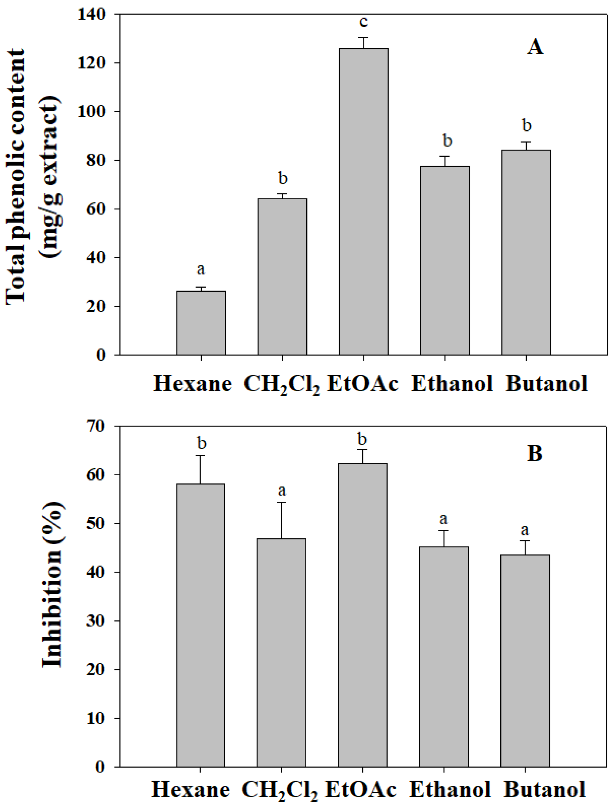
3.1. The TPC and Cell Proliferation Inhibition of N. oleracea Extracts

The TPC of N. oleracea extracts including hexane, CH2Cl2, EtOAc, ethanol, and butanol was shown in Figure 1A. The highest total phenolic content was found from EtOAc extract (126 ± 4 mg/g), followed by butanol (84 ± 3 mg/g), ethanol (77 ± 4 mg/g), CH2Cl2 (64 ± 2 mg/g), and hexane (26 ± 1 mg/g). The extraction solvent of EtOAc exhibited a higher solubility of the phenolic compounds, where significant differences among these solvents were indicated by different letters (p < 0.05). Moreover, the suppression of these extracts on the growth of human gastric cancer cells (BGC-823) was also shown in Figure 1B. The results showed that the hexane and EtOAc extracts exhibited high inhibition and cytotoxicity on cell growth at 200 µg/mL. The ratio of inhibition was up to 62% for EtOAc extract, followed by 58% for hexane extract, 47% for CH2Cl2 extract, 45% for ethanol extract, and 44% for butanol extract. Accordingly, the EtOAc extract with high total phenolic content and an anti-proliferation effect was chosen for further investigation.

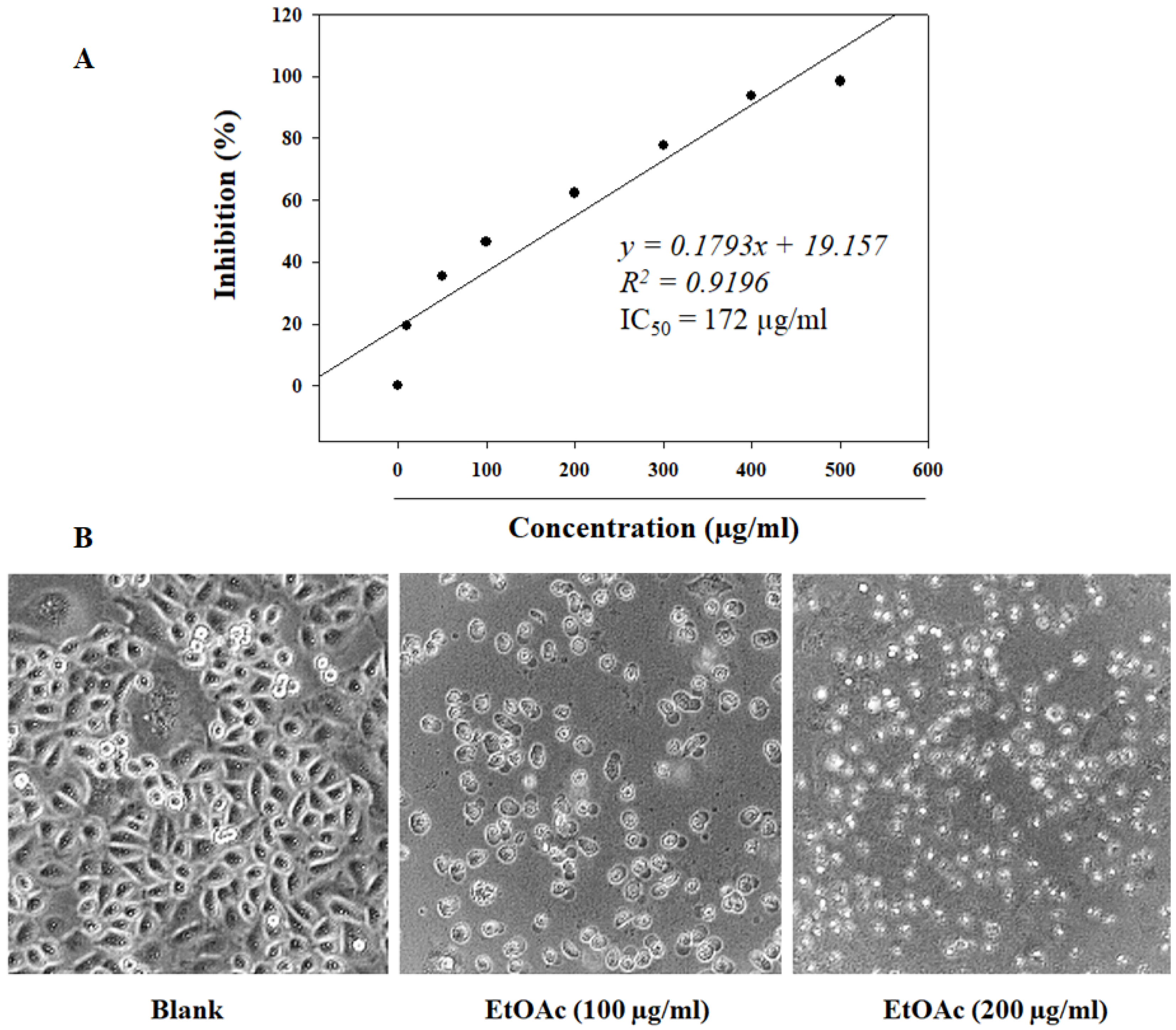
3.2. The Proliferation Inhibition of EtOAc Extract on BGC-823 Cells

The MTT assay showed that the treatment of the EtOAc extract significantly inhibited cell proliferation in a range concentration of 25–500 µg/mL (Figure 2A). The inhibitory effect of the EtOAc extract was determined at IC50 value of 172 ± 8 µg/mL that was inferred from the equation of regression line of y = 0.1793x + 19.157 (Uncertainty slope = 0.02; Uncertainty intercept = 5.68). On the other hand, microscopy assay showed that the EtOAc-treated cells changed their morphology as exposed by 100 and 200 µg/mL extract (Figure 2B). The cells reduced their cell size, formed a round shape, and had irregular surfaces compared with the blank cells. The assay indicated that EtOAc treatment caused proliferation inhibition and induced injury and death in gastric cancer cells.

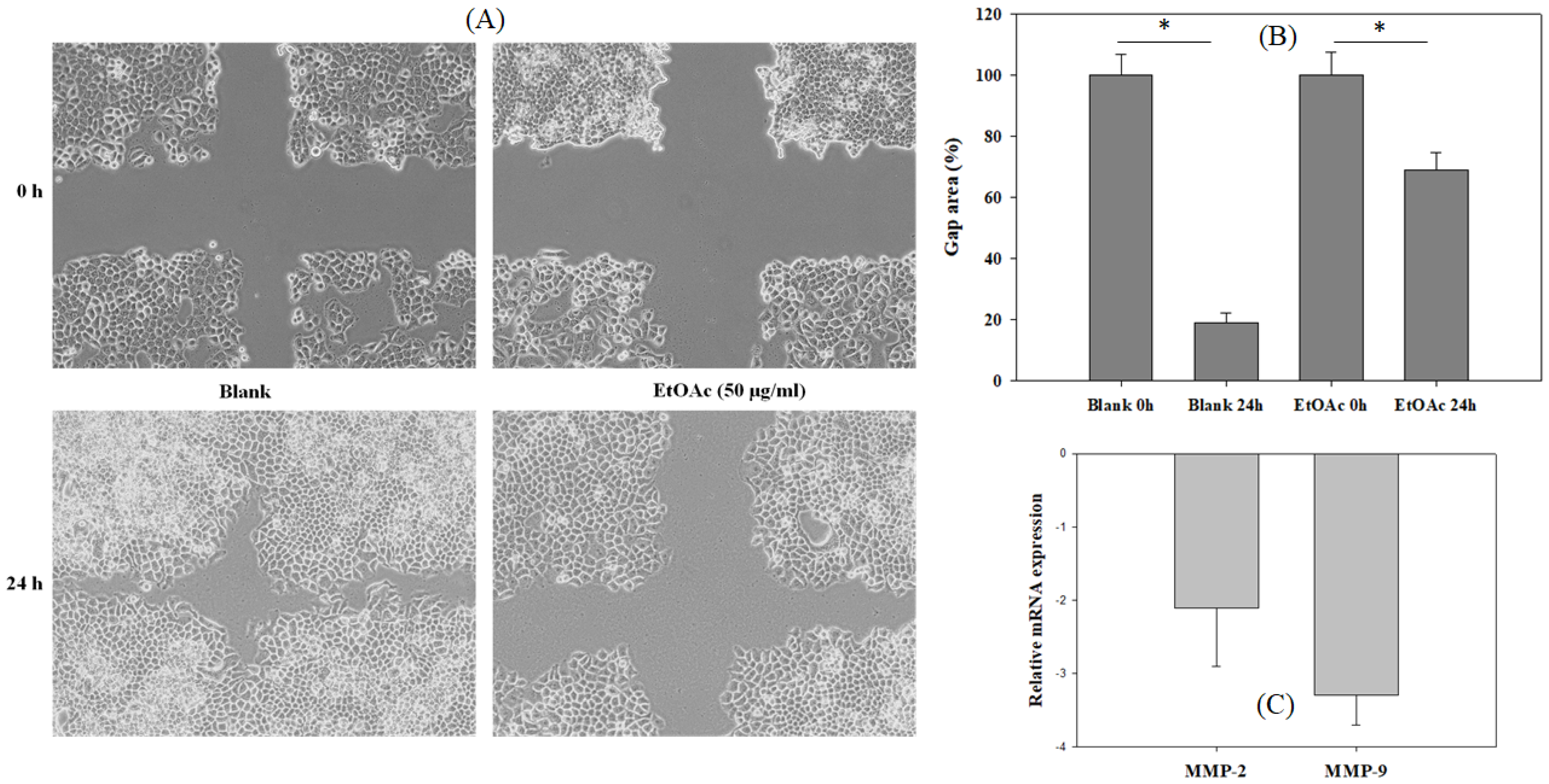
3.3. The Inhibition of EtOAc Extract on Cancer Cell Migration

In order to investigate the effect of EtOAc extract on cancer cell migration, a wound healing assay was performed on monolayer cell culture seeded on 6-well plates. The monolayer of BGC-823 cells was scratched before treatment with or without EtOAc extract (50 µg/mL) for 24 h. The result showed that the EtOAc treatment significantly suppressed the spreading of the cells to the wound area compared with that of the untreated cells (Figure 3A,B). Moreover, the mRNA expression level of matrix metalloproteinase (MMP)-2 and 9 from the cells was also downregulated in the presence of EtOAc extract. The suppressive activity of EtOAc extract on MMP-9 was stronger than that of MMP-2. These results indicated that EtOAc extract may inhibit the spread of gastric cancer cells.

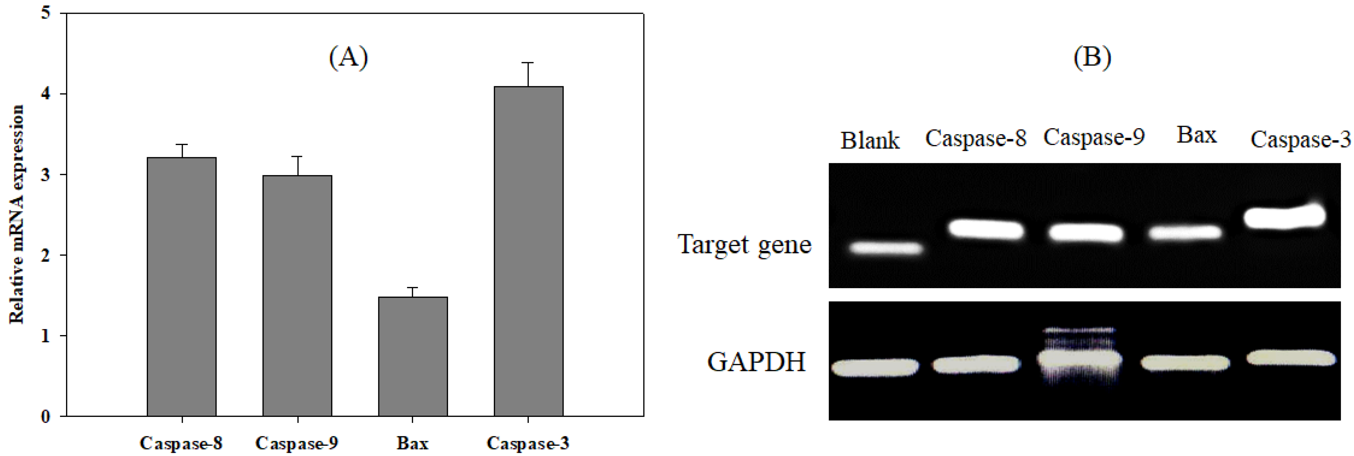
3.4. Effect of EtOAc Extract on Apoptotic Signaling

In order to clarify the inhibition of the EtOAc extract on theproliferation of gastric cancer cells, the expression level of the genes regulating the process of apoptosis was investigated. The cells were treated by EtOAC extract (50 µg/mL) for 24 h, and the expression of genes was quantified by real-time PCR. It was found that the EtOAC treatment significantly upregulated the gene expression of apoptotic signaling molecules, including caspase-3, -8, -9, and Bax (Figure 4). The gene expression levels of caspase-3, -8, -9, and Bax in the treated cells were 4.1-fold, 3.2-fold, 3.0-fold, and 1.5-fold higher than that of the untreated cells, respectively. These findings suggested that EtOAC extract may inhibit cell growth via the initiation of apoptosis in cancer cells.

4. Discussion

Cancer cells were charactered due to the inactivated apoptosis, the uncontrolled growth, and metastasis [14]. Therefore, suppression of growth, inhibition of metastasis, and induction of apoptosis in cancer cells were considered as potential strategies for the prevention and treatment of cancers [15]. In this sense, various herbs have been screened to be effective in the inhibition of the proliferation of cancer cells [15,16]. In this study, the MTT assay revealed that the EtOAc extract containing high total phenolic content exhibited potential inhibition of the proliferation of human gastric cancer cells. Numerous studies have suggested that polyphenols such as flavonoids, phenolic acids, lignans, and stilbenes have the capacity to inhibit tumor generation and thus could be used for the control of cancer [17,18]. Therefore, the potential inhibitory activity of EtOAc extract on cell proliferation may be due to its phenolic content. In particular, the inhibitory effect of the EtOAc extract of N. oleracea on the proliferation of gastric cancer cells was observed to be higher than that of the aqueous extracts of ziziphora, ginger, saffron, aloe vera [19], dandelion (Taraxacum spp.) [20], and Curcuma manga [21]. Hence, N. oleracea was also considered as a potential agent for targeting gastric cancer cell growth. In addition, anti-cancer therapy not only targets cell proliferation but also focuses on suppression of cell migration and invasion [22]. Notably, MMP-2 and MMP-9 were evidenced to play a crucial role in degrading extracellular matrices, thus facilitating the invasion and metastasis of cancer cells [23]. The inhibition of MMP-2 and MMP-9 may contribute to the suppression of cancer cell migration, bringing about the prevention of metastasis [24,25]. In this study, the EtOAc extract of N. oleracea was shown to inhibit MMP-2 and MMP-9 and suppress the cell migration at a low concentration of treatment (50 µg/mL). As a result, N. oleracea has potential in suppression of the proliferation at the primary sites as well as the prevention of migration of gastric cancer cells to new sites.
It was observed that the EtOAc extract of N. oleracea exhibited a cytotoxic effect toward gastric cancer cells, thus inhibiting cell growth and migration. However, this result should clearly indicate whether its cytotoxic effect was due to necrosis or apoptosis. Certainly, a natural product is considered as a potential anti-cancer agent when its cytotoxic effect is due to the promotion of apoptosis in the cancer cells [26,27]. Apoptosis is a caspase-mediated programmed cell death, leading to chromosome condensation, nuclear fragmentation, and membrane blebbing [28]. First, the binding of the Fas ligand, TNF-α, or TRAIL to the corresponding death receptors triggers the activation of caspase-8, which then activates caspase-3. Moreover, the caspase-8 activation also results in conformational changes in Bax, which causes the release of cytochrome C from mitochondria. Subsequently, the combination of cytochrome C and Apaf-1 activated caspase-9 and caspase-3 [28]. Finally, the activation of caspase-3 induces the process of apoptosis [29]. As a result, the induction of apoptosis due to the upregulation of caspase-3, -8, -9, and Bax activation in cancer cells can trigger the process of cell death. In this study, the treatment of the EtOAc extract of N. oleracea caused a significant increase in the gene expression levels of caspase-3, -8, -9, and Bax in gastric cancer cells. Likewise, various plant extracts, such as Brucea javanica, Camellia sinensis, Cinnamomum kanehirai Hayata, Corni Fructus, Cucurbita ficifolia, and Cyperus rotundus L. have been reported to induce apoptosis via the upregulation of caspase-3, -8, -9, and Bax in cancer cells [30]. This result suggested that N. oleracea extract can promote the process of apoptosis in gastric cancer cells, thus resulting in the cell’s death.
Recently, the role of phenolic compounds from vegetables, fruits, and beverages in the management of cancers has received much attention. Phenolic compounds have been evidenced to inhibit the initiation and development of cancer cells in in vitro and in vivo experiment models. The mechanism of action was found due to causing cell cycle arrest, suppressing cell survival and invasion, and inducing apoptosis [31,32]. Notably, N. oleracea have been revealed to contain a wide range of phenolic compounds, including quercetin, myricetin, kaempferol, and their derivatives, as well as phenolic acid [8,33]. These phenolic compounds from N. oleracea may be responsible for the suppression of the growth and induction of apoptosis in gastric cancer cells.

5. Conclusions

This study determined the anti-cancer effect of N. oleracea extract against human gastric cancer cells. Its anti-cancer activity was found due to suppressing proliferation, suppressing migration, and promoting apoptosis in BGC-823 cells. However, the mechanism of action of N. oleracea extract on the suppression of cell migration could be further evaluated in the next study. As a result, N. oleracea may be suggested as a potential dietary vegetable for the prevention and management of gastric cancer.

Author Contributions

Conceptualization, T.S.V.; methodology, D.-H.N.; validation, D.-H.N.; formal analysis, T.Q.N.; investigation, T.Q.N. and H.N.M.N.; resources, T.S.V.; data curation, T.Q.N.; writing—original draft preparation, T.Q.N.; writing—review and editing, D.-H.N. and P.-H.P.; visualization, T.Q.N.; supervision, T.S.V.; project administration, T.S.V.; funding acquisition, T.S.V. All authors have read and agreed to the published version of the manuscript.

Funding

This research received no external funding.

Institutional Review Board Statement

Not applicable.

Informed Consent Statement

Not applicable.

Data Availability Statement

Not applicable.

Acknowledgments

This study was funded by Van Lang University and supported by Nguyen Tat Thanh University.

Conflicts of Interest

The authors declare no conflict of interest.

References

  1. Yan, S.; Li, B.; Bai, Z.Z.; Wu, J.Q.; Xie, D.W.; Ma, Y.C.; Ma, X.X.; Zhao, J.H.; Guo, X.J. Clinical epidemiology of gastric cancer in Hehuang valley of China: A 10-year epidemiological study of gastric cancer. World J. Gastroenterol. 2014, 20, 10486–10494. [Google Scholar] [CrossRef]
  2. Mao, Q.Q.; Xu, X.Y.; Shang, A.; Gan, R.Y.; Wu, D.T.; Atanasov, A.G.; Li, H.B. Phytochemicals for the prevention and treatment of gastric cancer: Effects and mechanisms. Int. J. Mol. Sci. 2020, 21, 570. [Google Scholar] [CrossRef] [PubMed] [Green Version]
  3. Izuishi, K.; Mori, H. Recent strategies for treating stage IV gastric cancer: Roles of palliative gastrectomy, chemotherapy, and radiotherapy. J. Gastrointestin Liver Dis. 2016, 25, 87–94. [Google Scholar] [CrossRef] [PubMed] [Green Version]
  4. Bertuccio, P.; Rosato, V.; Andreano, A.; Ferraroni, M.; Decarli, A.; Edefonti, V.; La Vecchia, C. Dietary patterns and gastric cancer risk: A systematic review and meta-analysis. Ann. Oncol. 2013, 24, 1450–1458. [Google Scholar] [CrossRef] [PubMed]
  5. Nakamura, Y.; Murakami, A.; Koshimizu, K.; Ohigashi, H. Identification of pheophorbide a and its related compounds as possible anti-tumor promoters in the leaves of Neptunia oleracea. Biosci. Biotechnol. Biochem. 1996, 60, 1028–1030. [Google Scholar] [CrossRef]
  6. Bhumireddy, A.; Nellore, K.; Alapati, K.S. Anticancer activity of Neptunia oleracea methanolic extracts. Nat. Prod. Res. 2020, 36, 1053–1057. [Google Scholar] [CrossRef]
  7. Das, H.B.; Majumdar, K.; Datta, B.; Ray, D. Ethnobotanical uses of some plants by Tripuri and Reang tribes of Tripura. Nat. Prod. Radiance 2009, 8, 172–180. [Google Scholar]
  8. Lee, S.Y.; Mediani, A.; Ismail, I.S.; Maulidiani; Abas, F. Antioxidants and α-glucosidase inhibitors from Neptunia oleracea fractions using 1H NMR-based metabolomics approach and UHPLC-MS/MS analysis. BMC Complement. Ther. Med. 2019, 19, 7. [Google Scholar] [CrossRef]
  9. Le, P.; Ngo, D.; Vo, T. Optimization of extraction conditions for achieving high content and antioxidant activities of the total phenolic compounds of Phu Quoc sim fruit (Rhodomyrtus tomentosa (aiton) hasak.). J. Med. Mater. 2018, 23, 157–166. [Google Scholar]
  10. Mazloum-Ardakani, M.; Barazesh, B.; Moshtaghioun, S.M.; Sheikhha, M.H. Designing and optimization of an electrochemical substitute for the MTT (3-(4,5-Dimethylthiazol-2-yl)-2,5-diphenyltetrazolium bromide) cell viability assay. Sci. Rep. 2019, 9, 14966. [Google Scholar]
  11. Kwak, Y.; Ju, J. Inhibitory activities of Perilla frutescens britton leaf extract against the growth, migration, and adhesion of human cancer cells. Nutr. Res. Pract. 2015, 9, 11–16. [Google Scholar] [CrossRef] [PubMed] [Green Version]
  12. Motadi, L.R.; Choene, M.S.; Mthembu, N.N. Anticancer properties of Tulbaghia violacea regulate the expression of p53-dependent mechanisms in cancer cell lines. Sci. Rep. 2020, 10, 12924. [Google Scholar] [CrossRef] [PubMed]
  13. Livak, K.J.; Schmittgen, T.D. Analysis of relative gene expression data using real-time quantitative PCR and the 2(-Delta Delta C(T)) Method. Methods 2001, 25, 402–408. [Google Scholar] [CrossRef] [PubMed]
  14. Sarkar, S.; Horn, G.; Moulton, K.; Oza, A.; Byler, S.; Kokolus, S.; Longacre, M. Cancer development, progression, and therapy: An epigenetic overview. Int. J. Mol. Sci. 2013, 14, 21087–21113. [Google Scholar] [CrossRef]
  15. Tian, X.; Li, Y.; Shen, Y.; Li, Q.; Wang, Q.; Feng, L. Apoptosis and inhibition of proliferation of cancer cells induced by cordycepin. Oncol. Lett. 2015, 10, 595–599. [Google Scholar] [CrossRef] [Green Version]
  16. Dutta, S.; Mahalanobish, S.; Saha, S.; Ghosh, S.; Sil, P.C. Natural products: An upcoming therapeutic approach to cancer. Food Chem. Toxicol. 2019, 128, 240–255. [Google Scholar] [CrossRef]
  17. Sharma, A.; Kaur, M.; Katnoria, J.K.; Nagpal, A.K. Polyphenols in food: Cancer prevention and apoptosis induction. Curr. Med. Chem. 2018, 25, 4740–4757. [Google Scholar] [CrossRef]
  18. Zhou, Y.; Zheng, J.; Li, Y.; Xu, D.P.; Li, S.; Chen, Y.M.; Li, H.B. Natural polyphenols for prevention and treatment of cancer. Nutrients 2016, 8, 515. [Google Scholar] [CrossRef]
  19. Ghazanfari, T.; Yaraee, R.; Shams, J.; Rahmati, B.; Radjabian, T.; Hakimzadeh, H. Cytotoxic effect of four herbal medicines on gastric cancer (AGS) cell line. Food Agr. Immunol. 2013, 24, 1–7. [Google Scholar] [CrossRef]
  20. Han, H.; Chen, G.Z.; Zhou, S.K.; Xu, R.R.; Wu, C.L. In vitro anti-tumor activity in SGC-7901 human gastric cancer cells treated with dandelion extract. Trop. J. Pharm. Res. 2018, 17, 65–70. [Google Scholar] [CrossRef] [Green Version]
  21. Liu, Y.; Nair, M.G. Labdane diterpenes in Curcuma mangga rhizomes inhibit lipid peroxidation, cyclooxygenase enzymes and human tumour cell proliferation. Food Chem. 2011, 124, 527–532. [Google Scholar] [CrossRef]
  22. Guan, X. Cancer metastases: Challenges and opportunities. Acta Pharm. Sin. B 2015, 5, 402–418. [Google Scholar] [CrossRef] [PubMed] [Green Version]
  23. Li, H.; Qiu, Z.; Li, F.; Wang, C. The relationship between MMP-2 and MMP-9 expression levels with breast cancer incidence and prognosis. Oncol. Lett. 2017, 14, 5865–5870. [Google Scholar] [CrossRef] [PubMed] [Green Version]
  24. Palmer, T.D.; Ashby, W.J.; Lewis, J.D.; Zijlstra, A. Targeting tumor cell motility to prevent metastasis. Adv. Drug Deliv. Rev. 2011, 63, 568–581. [Google Scholar] [CrossRef] [PubMed] [Green Version]
  25. Lyu, Y.; Xiao, Q.; Yin, L.; Yang, L.; He, W. Potent delivery of an MMP inhibitor to the tumor microenvironment with thermosensitive liposomes for the suppression of metastasis and angiogenesis. Sig. Transduct. Target Ther. 2019, 4, 1–9. [Google Scholar] [CrossRef] [PubMed]
  26. Wang, Y.; Zhong, J.; Bai, J.; Tong, R.; An, F.; Jiao, P.; He, L.; Zeng, D.; Long, E.; Yan, J.; et al. The application of natural products in cancer therapy by targeting apoptosis pathways. Curr. Drug Metab. 2018, 19, 739–749. [Google Scholar] [CrossRef] [PubMed]
  27. Safarzadeh, E.; Sandoghchian Shotorbani, S.; Baradaran, B. Herbal medicine as inducers of apoptosis in cancer treatment. Adv. Pharm. Bull. 2014, 4, 421–427. [Google Scholar]
  28. Chen, Q.; Kang, J.; Fu, C. The independence of and associations among apoptosis, autophagy, and necrosis. Signal Transduct. Target. Ther. 2018, 3, 1–11. [Google Scholar] [CrossRef] [Green Version]
  29. Porter, A.G.; Jänicke, R.U. Emerging roles of caspase-3 in apoptosis. Cell Death Differ. 1999, 6, 99–104. [Google Scholar] [CrossRef]
  30. Rajabi, S.; Maresca, M.; Yumashev, A.V.; Choopani, R.; Hajimehdipoor, H. The most competent plant-derived natural products for targeting apoptosis in cancer therapy. Biomolecules 2021, 11, 534. [Google Scholar] [CrossRef]
  31. Wahle, K.W.; Brown, I.; Rotondo, D.; Heys, S.D. Plant phenolics in the prevention and treatment of cancer. Adv. Exp. Med. Biol. 2010, 698, 36–51. [Google Scholar] [PubMed]
  32. Matulja, D.; Vranješević, F.; Kolympadi Markovic, M.; Pavelić, S.K.; Marković, D. Anticancer Activities of Marine-Derived Phenolic Compounds and Their Derivatives. Molecules 2022, 27, 1449. [Google Scholar] [CrossRef] [PubMed]
  33. Devi, O.S.; Singh, T.N.; Singh, L.J.; Singh, T.P. Biological Importance and Phytochemistry of Neptunia oleracea Lour: A Mini Review. Asian J. Chem. 2021, 33, 2276–2280. [Google Scholar] [CrossRef]
Figure 1. The TPC (A) and the suppression of N. oleracea extracts on cell proliferation (B). (A) The PC was examined using Folin–Ciocalteau assay. (B) BGC-823 cells were exposed with 200 µg/mL of different extracts, and the inhibition of cell proliferation was assessed by MTT assay. a–c showed significant difference among groups (p < 0.05).
Applsci 12 06817 g001
Figure 2. The dose-dependent inhibition of EtOAc extract on cell proliferation (A) and cell morphological change (B). (A) The cells were treated with a concentration range of EtOAc extract for 24 h, and the 50% inhibitory concentration (IC50) value was indicated by a linear regression equation. (B) The cells were treated with EtOAc extract for 24 h, and cell morphology was then visualized under an inverted microscope (10× magnification).
Applsci 12 06817 g002
Figure 3. The suppression of EtOAc extract on cell migration. BGC-823 cells were seeded on 6-well plates before a wound line was made by a yellow pipette tip. (A) The images of gap were captured by a microscope (10× magnification). (B) The relative gap area between 24 h and 0 h in each group was measure by ImageJ. (C) The expression of MMP-2 and MMP-9 was assessed by qPCR. The differences compared between groups were considered significant at * p < 0.05.
Applsci 12 06817 g003
Figure 4. EtOAc extract induced apoptosis in BGC-823 cells. The cells were exposed by EtOAc extract, and total RNA was then collected. The expression levels of caspase-3, -8, -9, and Bax were identified using real-time PCR (A) and gel electrophoresis (B). GAPDH was used as an internal control.
Applsci 12 06817 g004
Publisher’s Note: MDPI stays neutral with regard to jurisdictional claims in published maps and institutional affiliations.

