Abstract
It has been shown that the cultivation of plants under glass coated with nano-sized upconversion luminophores led to an increase in plant productivity and the acceleration of plant adaptation to ultraviolet radiation. In the present work, we examined the effect of upconversion nanopowders with the nominal composition Sr0.955Yb0.020Er0.025F2.045 on plant (Solanum lycopersicum) photochemistry. The composition, structure and size of nanoparticles were tested using X-ray pattern diffraction, scanning electron microscopy, and dynamic light scattering. Nanoparticles are capable of converting infrared radiation into red and green photons. Glasses coated with upconversion luminophores increase the intensity of photosynthetically active radiation and absorb the ultraviolet and far-red radiation. The chlorophyll a fluorescence method showed that plants growing under photoconversion and those growing under common film demonstrate different ability to utilize excitation energy via photosynthesis. It was shown that under ultraviolet and high light conditions, the efficiency of the photochemical reactions, the non-photochemical fluorescence quenching, and the electron transport remained relatively stable in plants growing under photoconversion film in contrast to plants growing under common film. Thus, cultivation of Solanum lycopersicum under photoconversion glasses led to the acceleration in plant growth due to greater efficiency of plant photochemistry under stress conditions.
1. Introduction
Sustainable agriculture has an important role to play in the continuous provision of food to the population. Consequently, for farmers, the problem of increasing the productivity of agricultural crops is always in the forefront of their minds. Everyone knows that for good harvest production, it is necessary to provide the most favorable conditions: lighting, watering, nutrition, and temperature mode. The intensification of agriculture implies a transition to the year-round cultivation of plants, which is effectively realized in modern greenhouses. The design features of greenhouses allow for optimized growth conditions and increased crop yields while reducing costs [1,2,3]. The protective environment of greenhouse allows the growth of thermophilic plant species ubiquitously, as well as the planting of seeds and harvest of crops several times a year. The most important feature of greenhouses is their maximum transmittance of sunlight to the plants. However, even this property is not always sufficient to achieve the best result, due to sunlight deficit in temperate regions. Currently, artificial lighting is used to solve the problem of the lack of sunlight, but this leads to an increase in energy consumption [4,5,6]. Photoconversion coatings of greenhouse glass are an example of smart and efficient production technologies, which reduce costs and increase yields. Normally, agricultural photoconversion covers transform the light, which is least absorbed by plants, into photosynthetically active radiation (PAR). It is assumed that the potential economic effect of using photoconversion coatings can be achieved by reducing the cost of electricity, and of the purchase and maintenance of equipment for artificial lighting, as well as increasing the productivity of crops.
Light quantity and quality affect photosynthesis efficiency, plant growth and metabolism [7,8,9], and farmers should ensure that plants have optimal lighting. PAR is a light with wavelengths 400–700 nm. This waveband at sunny weather can contain approximately 25% of the sunlight photons, which leaves a lot of potential for the application of photoconversion technologies in the greenhouse. In the 19th century, the research of T. W. Engelmann and K. A. Timiryazev demonstrated that red and blue photons are most able to maximize the productivity of plant photosynthesis, and this was later confirmed by McCree [10]. Despite all the advantages of red illumination for photosynthesis, blue, green, yellow, and far-red lights additionally intensify the photosynthesis, optimize plant biochemistry, and improve crop quality [11,12,13,14,15,16,17,18].
A wide range of devices are being developed that are made with luminescent materials (metal complexes, semiconductors, nanoparticles, fluorescent proteins, organic dyes), including some for agriculture [19,20,21,22]. The main limitations for the agricultural application of photoconversion devices is the inability to manufacture ideal covers, which have a light transmittance and harvesting performance in given parts of the solar spectrum, high quantum yield and good stability when exposed to rain, high intensity light and extreme temperatures.. Unfortunately, almost all photoconversion coatings produced so far have critical problems. The organic fluorophores incorporated in coatings rapidly photobleach, with a decrease in the quantum yield of photoconversion. The relatively stable rare-earth elements-based fluorophores have a low yield of luminescence [23,24,25,26,27]. The illumination of nanoparticles with plasmon or exciton emissions (cadmium selenide, zinc sulfide, etc.) [28] leads to the generation of reactive oxygen species, which cause damage to phosphors. Moreover, this type of particles is hardly integrated in polymers. An important goal of modern research is the creation of photoconversion coatings, which will include durable phosphors with optimal photoabsorbing and emitting properties [22,29]. Previously, stable photoconversion materials, such as nanoluminophores that are not damaged by reactive oxygen species due to high vapor-proofing, were obtained [30,31,32]. Moreover, the coverings are characterized by a very high efficiency of photoconversion of ultraviolet radiation into red and blue light. Therefore, photoconversion technologies have great potential to enhance plant growing without additional energy consumption.
Most luminescent materials convert high energy photons into light quanta having lower energy, but a few contrarily phosphoresce with higher energy photons (so-called upconversion luminescence). Upconversion luminophores, suggested in the last century [33,34], may be applied in agriculture, but only in the case of conversing infrared light into visible light. This requirement is best met by phosphors based on pair of rare-earth cations. Upconversion pair cations should contain a sensitizer with a wide absorption cross-section (for example, Yb3+) and an activator (Er3+, Tm3+, or Ho3+) [33,35,36,37] incorporated in the matrix to provide a high upconversion yield. Strontium or barium fluoride is preferred as the matrix, as this is proven to provide a high upconversion yield [38,39,40].
Previously, it has been shown that upconversion coatings with nano-sized upconversion luminophores increase plant productivity and accelerate their adaptation to ultraviolet radiation [41,42]. In this work, we studied the effect of glasses coated with an upconversion luminescent film on the photochemistry of agricultural plants. For this purpose, we prepared Sr0.955Yb0.020Er0.025F2.045 nanoparticles capable of performing upconversion. The present research is aimed at the design of versatile photoconversion coatings for use in greenhouses.
2. Materials and Methods
2.1. Synthesis of Nanoluminophore
In the study, nanopowders with a nominal composition Sr0.955Yb0.020Er0.025F2.045 were used. Nanoparticles have been produced by co-precipitation from nitrate solutions, as described in [43]. The solution containing 0.08 M nitrate was dropwise mixed with 0.16 M ammonium fluoride. Note that NH4F was taken with 7% excess. The resulting mixture was incubated for two hours with stirring. After incubation, the suspension was allowed to settle and the obtained precipitate was washed with ammonium fluoride until the nitrate ions were completely washed out. The washed sediment was then centrifuged at 10,000 rpm for 5 min. Centrifugation was followed by two-stage drying of the precipitate: first in air at 45 °C, then in a platinum crucible at 600 °C for 1 h. The heating rate was 10 degrees per min.
2.2. Production of Photoconversion Film
To manufacture the photoconversion film (PCF) a solution of nanoparticles in acetone and a liquid component of a fluoroplate polymer were used. Fluoroplast-32L (St. Petersburg Kraski, Russia) was applied to the preparation of the polymer varnish. A 7% solution of nanoluminophore was added to a fluoroplate polymer in a ratio of 1:100 and mixed. Then the resulting mixture was loaded into the spray gun, and the PCF containing luminescent nanoparticles was sprayed on to the glass surface.
2.3. Characterization of Nanoluminophore
X-ray pattern diffraction (XRD) was performed on a BRUKER D8 ADVANCE diffractometer (Billerica, MA, USA). Calculation of the unit cell parameters was performed using POWDER 2.0 software (Moscow State University, Moscow, Russia). Particle shape and size were established from a scanning electron microscopy (SEM) image. For this aim, a Carl Zeiss NVision 40 microscope (Zeiss AG, Oberkochen, Germany) connected with Oxford Instruments XMAX (80 mm2) set-up (Oxford Instruments plc, Abingdon, UK) (using ImageJ software) was used. Hydrodynamic diameter distribution of nanoparticles was determined using Zetasizer Ultra (Malvern Panalytical, Malvern, UK). Registration of particle luminescence was completed using fiber-optic spectrometer USB2000 (OceanOptics, Orlando, FL, USA). The sample was placed inside the integrating sphere Cintra 4040 (GBC Scientific, Braeside, Victoria, Australia) and irradiated by a 50 mW IR light diode (λ = 975 nm).
2.4. Growth Conditions and Morphometric Measurements
The tomato variety (Solanum lycopersicum) “Balcony Miracle” was the one used experimentally in this work. Plants were grown with a 16 h photoperiod at 25–26°C. Intensities of PAR and overall light (350–800 nm) were ≈ 70 μmol photons s−1 m−2 and about 140 μmol photons s−1 m−2. Plants were grown to the seventh leaf stage. Both experimental and control plants were then covered by glasses with either a common fluoroplastic polymer coating or containing photoconversion nanoparticles. The UVA component (λ = 370 nm, photon flux density (PFD) ≤ 10 μmol photons s−1 m−2) was added to the illumination spectrum. PG200N Spectral PAR Meter (UPRtek, Zhunan, Miaoli, Taiwan) was used to estimate light flux density. The relative chlorophyll concentration in leaves was evaluated using portative chlorophyll meter CL-01 (Hansatech, Norfolk, UK). The number of leaves was determined manually. The length of the stem was measured with a graduated ruler to the nearest millimeter. For the definition of the leaf area, the GreenImage software was applied.
2.5. Chlorophyll a Fluorescence Kinetics Measurement
The kinetics of chlorophyll fluorescence (ChlF) were measured by a DUAL-PAM-100 fluorometer (Waltz, Eichenring, Effeltrich, Germany). Experiments were performed using non-cut leaves at room temperature. Measurements were preceded by 1 h dark incubation of the plants at 25–26 °C to provide complete relaxation of all photoinduced processes. To measure maximum quantum yield of photosystem II photochemistry (Fv/Fm) and ChlF parameters, the leaves were illuminated with a saturating 500 ms flash (λ = 625 nm, 12,000 μmol photons s−1 m−2). Adaptation to actinic illumination (λ = 625 nm, 250 μmol photons s−1 m−2) lasted 10 min. ChlF registered at various intensities was studied after 30 min of light adaptation for the leaves. The calculation of the ChlF parameters was performed using DUAL-PAM Software [44] according to the equations:
where F0 is the initial fluorescence value, Fm is the maximum fluorescence value, Fv is the photoinduced changes of chlorophyll a fluorescence yield, Y(II) is the effective quantum yield of PSII, Y(NPQ) is the quantum yield of light-induced non-photochemical quenching, Y(NO) is the quantum yield of non-regulated heat dissipation and fluorescence emission, F is a fluorescence yield measured briefly before application of a saturating pulse, Fm′ is light-induced maximum level of chlorophyll fluorescence in light-adapted samples, ETR is the rate of linear electron transport through photosystem II, PAR(II) is the PAR absorbed by PS II (quanta/(PS II·s)).
Fv/Fm = (Fm − F0)/Fm
Y(II) = (Fm′ − F)/Fm′
Y(NPQ) = F/Fm′ − F/Fm
Y(NO) = F/Fm
ETR(II) = PAR(II)·Y(II)/(Fv/Fm)
2.6. Statistics
To determine statistically significant differences between plant groups, one-way analysis of variance (ANOVA) followed by post hoc comparisons by Tukey’s test and Student’s t test for independent means were performed. The normality (Shapiro–Wilk test) requirements were checked. The difference was considered significant if p ≤ 0.05.
3. Results
To understand which particles we are dealing with, we tested their composition, structure and size. XRD for the 600 °C-treated nanoparticles are shown in Figure 1. The data confirms that the chosen technique of synthesis of nanopowders leads to the formation of fluorite structure particles (JCPDS #06-0262, a = 5.800 Å). The monophase and purity of the obtained samples are also confirmed.
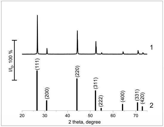
Figure 1.
XRD for the Sr0.955Yb0.020Er0.025F2.045 nanoparticles (1) and JCPDS #06-0262 for the strontium fluoride (2).
The scanning electron microscopy image for Sr0.955Yb0.020Er0.025F2.045 samples shown in Figure 2A made it possible to determine the shape and diameter of the particles. Two fractions of spherical particle were identified: primary particles were approximately 75 nm, and agglomerates with a 300 nm diameter. The dynamic light scattering method confirmed the formation of both primary particles and their agglomerates (Figure 2B). The energy-dispersive X-ray analysis clarified the elemental composition of the particles: Sr0.948Yb0.024Er0.028F2.052.
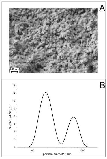
Figure 2.
Scanning electron microscopy image for Sr0.955Yb0.020Er0.025F2.045 (A) and particle number as a function of their diameter (B).
Figure 3A presents the spectra of the semiconductor laser-induced photoluminescence of nanoparticles measured in visible range. Under 976 nm excitation (curve 2), the nanoparticles emit spectra with maximum wavelengths of 660 nm, 545 nm and 525 nm (curve 1). Very weak blue (410 nm) and down-shifted (1520 nm) luminescence are not shown. It must be emphasized that the amplitudes of both the excitation light and the luminescence are given in arbitrary units and should not be used to calculate the luminescence efficiency. Difference spectrum (“light spectrum under photoconversion film” minus “light spectrum under common film”) allowed the visualization of changes in the spectral composition of growth light passed through photoconversion glasses, are shown in Figure 3B. It was shown that the photoconversion glasses we used were capable of increasing PAR intensity and decreasing intensity of UV and far-red. Thus, the luminescent nanoparticles are capable of optimizing the illumination spectrum (increasing PAR intensity and changing the ratio of red and far-red light) and can be used in photoconversion materials for agricultural applications.
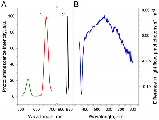
Figure 3.
(A) Photoluminescence (1) of the nanoparticles in acetone in PAR range, which was excited by a semiconductor laser (2). (B) Difference spectrum (“light spectrum under photoconversion film” minus “light spectrum under common film”), showing changes in the spectrum of growth light. In figure (A), green and red colors indicate the luminescence in the green and red regions of the spectrum, respectively.
Since our study is aimed at optimizing the conditions for growing agricultural plants with a shortage of natural light, we imitated a cloudy day in temperate regions using UV and incandescent lamps. UVA and PAR intensities were ≤ 10 μmol photons s−1 m−2 and ≈ 70 μmol photons s−1 m−2. The overall light intensity (350–800 nm) was greater than 140 μmol of photons s−1 m−2.
The results of morphometric studies are presented in Table 1. It was shown that photoconversion film has a moderate, but statistically significant, effect on the leaf formation. Under control films, the appearance of a new leaf was observed every sixth day, whereas under photoconversion films, new leaves appeared every fourth day. At the same time, the number of leaves in the control samples increased by 55% and by 85% in the experimental conditions.

Table 1.
Effect of an upconversion luminescent film on the growth of tomatoes. Presented data are the result of averaging from four (leaf area) to eleven (all other parameters) measurements.
In comparison to other parameters, the area of leaves showed the greatest increase. Before the experiment, the average leaf area was 23 cm2; after 25 days, control samples grew by 290% under common film and by 435% under photoconversion film. Thus, the acceleration of leaf growth under photoconversion films was 50%. Note that PCF contributes to acceleration in the length of the stem, while it practically does not affect the length of the internodes. It is interesting to mark that the chlorophyll concentration in the tomatoes leaf remained relatively unchanged under the common film, while PCF increased the amount of chlorophyll in the leaf. Thus, it is shown that plants grow and develop faster under the films containing photoconversion nanoparticles.
To understand the cause of the acceleration in tomato growth under the photoconversion films, we examined the impact of photoconversion films on the plant photochemistry. For this aim, we analyzed the development of changes in the maximum quantum yield of plant photochemistry (which provides information about the general state of the photosynthetic apparatus). This parameter reflects the utilization of excitation energy (Figure 4).
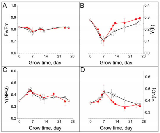
Figure 4.
Development of changes in maximum (A) and effective (B) quantum yield of photosystem II photochemistry, quantum yield of light-induced non-photochemical fluorescence quenching (C) and quantum yield of nonregulated heat dissipation and fluorescence emission (D) under common (red, closed squares) and photoconversion (black, opened circles) films. The data are the means of at least three measurements, with the standard deviation of the mean.
The level of photochemical activity recorded in the plants did not decrease during the experiment, which may indicate similar and stable overall states of the photosynthetic apparatus in both groups of plants (Figure 4A). Illumination of plants led to formation of exited states of pigments of photosynthetic antenna. Part of this energy is used for photosynthesis. The proportion of excited energy routed to photosynthesis is reflected in the level of effective Y(II) (quantum yield of photosystem II photochemistry). Under the unfavorable conditions, the plant can change the proportion of energy directed to photosynthesis and activate mechanisms aimed at the quenching of excitation energy via heat dissipation: so-called processes of regulated and non-regulated heat dissipation. Activity of these processes correlate well with levels of non-photochemical quenching of fluorescence (NPQ), and non-regulated heat dissipation and fluorescence emission (NO), respectively. The installation of the coating over the plants and the appearance of ultraviolet light in the growth spectrum induce the changes in ChlF parameters. These changes had similar trends in both control and experimental groups. The Y(II) decreases within the first 6–7 days from approximately 0.3 to 0.1 (Figure 4B), which indicates some suppression of photosynthesis in the plants. The reduction of Y(II) is accompanied by an increase in Y(NPQ) (Figure 4C) and Y(NO) (Figure 4D) in both control and experiment plants. The amplitude of Y(II) reduction was equal to the sums of growth of Y(NPQ) and Y(NO). However, the changes described above were reversible: the photochemistry parameters gradually reached initial values. The rate of relaxation of Y(NO) and Y(II) values in plants growing under PCF was relatively fast (initial levels were observed within 3–5 days and did not change later). In control plants, Y(II) and Y(NO) relaxed slowly (achievement of initial levels were observed in only three weeks). Note that the kinetics of Y(NPQ) in both control and experimental plants were practically identical. The above-mentioned data reflect a reversible decrease in efficiency of photosynthesis at the first week of the experiment.
The different capacities of the photosynthetic electron-transport chain, and the activity of enzymes during the dark stage of photosynthesis, show that electron transport and, concurrently the redistribution of excitation energy, changes in plants growing under different conditions, as a result of changes in light intensity. We studied the dependence of Y(II), Y(NPQ) and ETR (the rate of linear electron transport through photosystem II) on light intensity (Figure 5).
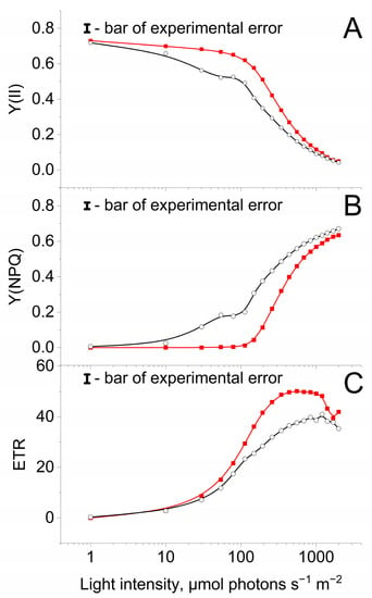
Figure 5.
Dependence of Y(II) (A), Y(NPQ) (B) and ETR (C) on light intensity in plants growing under common (red, closed squares) and photoconversion (black, opened circles) films. Measurements were done at room temperature. The data are the means of at least three measurements, with the standard deviation of the mean.
The Y(II) and Y(NPQ) in leaves of plants growing under PCF were relatively stable at weak light (up to 100 μmol photons s−1 m−2) (Figure 5A,B). When Y(II) was relatively high (about 0.72–0.73), the Y(NPQ) value close to zero. Further magnification of light intensity resulted in a simultaneous increase in Y(NPQ) and decrease in Y(NO), which achieved maximum and minimum, respectively, at 1000–1500 μmol photons s−1 m−2. In control plants, Y(II) and Y(NPQ) rapidly changed even at low light, however under high light (more than 1000 μmol photons s−1 m−2) they were equal to levels observed in experiment plants. This may indicate the different ability of plants growing under PCF and common film at utilizing excitation energy via photosynthesis. In addition, an increase in light intensity led to a gradual growth of the ETR (Figure 5C). ETR was higher in plants growing under PCF under all light intensities, in comparison to plants grown under common film. At 1200 μmol photons s−1 m−2, a decrease in ETR was observed, which may be induced by photoinhibition.
4. Discussion
It is known that the quality and quantity of light affects the growth and development of plants, determining the efficiency of photosynthetic processes, as well as the accumulation of secondary metabolites. Plants are capable of adapting to a wide variety of light conditions; however, improving the quality of lighting always increases the productivity of agricultural plants. Plants are believed to utilize between a third and a half of solar radiation. The remaining photons belong to the ultraviolet, infrared and other light ranges, which are generally unavailable to plants for photosynthesis. This makes the development of photoconversion technologies relevant for the temperate and cold regions of the planet, where usable light is at a premium. Based on the investigations of Engelmann [45], Timiryazev [46] and McCree [10], we know that additional red light can more efficiently activate photosynthesis in comparison to other light of similar intensity. This principle was applied in this study. For the intensification of plant development, we used a photoconversion coating containing Sr0.955Yb0.020Er0.025F2.045 nanoluminophores. The coating absorbs IR light and emits visible light (at bandwidths of 660 nm, 545 nm and 525 nm; Figure 3A). These luminescence bands are usually observed under the illumination of the phosphors containing the Er3+/Yb3+ ion pair [39,40,41,42,47,48,49,50]. Of course, the phosphor we used is much inferior to conventional phosphors in terms of light conversion efficiency (about 6% vs. more than 20%) [30,31,32,51,52,53,54,55,56,57,58,59,60,61]. However, in comparison with many other upconversion luminophores, this phosphor has a fairly high efficiency of photoconversion. The fact that similar particles can convert ultraviolet radiation into PAR increases their usefulness in greenhouses, as discussed in [39]. The difference spectrum shown in Figure 3B confirmed that photoconversion glasses moderately increase PAR intensity and absorb UVA and far-red. Obviously, this changes the red light:far-red light ratio. The difference spectrum is a broadband radiation with two pronounced peaks at 561 nm and 650 nm, which can be correlated with the erbium luminescence bands. The observed broadband radiation was studied earlier for nanoparticles of various chemical compositions [62,63,64,65,66,67]. The mechanism of the appearance of such luminescence is the subject of open discussion.
As can be seen in Table 1, plants grow and develop quicker under the PCF in comparison with common film, which is consistent with the previous data [41,42]. The appearance of leaves, and the increase in their area, are accelerated by 50%. PCF accelerates the growth of the stem, without affecting the length of the internodes, which is indicative of accelerated growth rather than avoidance of shadows. Moreover, photoconversion glasses stimulate the accumulation of chlorophyll in the tissues. According to the data published in [68], the obtained values indicate a two-fold increase. A similar improvement in plant growth was previously obtained using a down-shifting red luminophore [32]. As can be seen, luminophore substitution does not significantly reduce the efficiency of photoconversion coatings. It is important to note that only a negligible part of the light absorbed by the coating can be transformed into luminescence in the PAR region. Possible reasons include low absorption of ultraviolet and/ or infrared radiation and high yield of heat dissipation. However, several papers (including the present study) have clearly demonstrated that the application of photoconversion films increases crop yields [30,31,32,51,52,53,55,56,57,58,59,60,61,69,70,71,72,73,74,75,76]. There are several possible reasons for the activation of plant development under films. Firstly, the functioning of phytochromes, which are very sensitive to the change in the proportion of far-red light: the phytochrome system can regulate plant growth and increase resistance to adverse environmental conditions [55,77,78]. Moreover, a decrease in far-red availability may induce a reduction in chlorophyll content [79] that correlates well with our data (Table 1). Secondly, the positive effects of photoluminescent particles as protectants against UV: UVA radiation can damage the photosynthetic apparatus due to destruction of the pigments and pigment-protein complexes [80,81]. Previously, it was shown that, under insufficient illumination, photoconversion coatings change the content of phytohormones and thus affect plant growth [82]. Importantly, the described coatings luminesced in the red region. Furthermore, photoconversion films can affect soil microflora as well as plants. It was shown that PCF-induced changes in light spectrum stimulated reproduction of native soil microflora, which enhances the effect of transformed light on plant growth [61,83]. Changes in the partitioning of absorbed light energy and the inhibition of plant grow (Figure 3) are most likely induced by the appearance of UVA; although, the addition of UVA can play a positive role in plant development [81].
Later, plants acclimatize to the action of a stress factor, which is reflected in the restoration of the activity of the photosynthetic apparatus. Photoconversion films speed up this process, making it easier for the plant to withstand stress. The different rates of the restoration of the initial activity of the photosynthetic apparatus may be due to the slow establishment of a balance between electron transport and NADPH utilization in control plants, as well as different degrees of damage to the water-oxidizing complex of photosystem II in control and experimental plants [84]. It is not elucidated that minor alterations in the light spectrum (in light deficiency conditions) (Figure 3B) can increase in the rate of the adaptation processes of plants. Figure 5 demonstrates that after 30 min of adaptation to various actinic light conditions, Y(II) and Y(NPQ) in plants growing under photoconversion film remained relatively stable with increasing AL intensity to comparably high light intensity (in contrast to plants growing under common film). It can be presumed that plants growing under photoconversion films develop resistance to the inhibitory effects of light. It was shown that plants, which adapted to weak illumination, demonstrated the limited ability to acclimate, and experienced photoinhibition after exposition to high light [85].
Thus, it can be suggested that films containing upconversion luminophores can contribute to the acquisition by plants of properties that allow the plant to more effectively adapt to the ultraviolet and high light.
5. Conclusions
The photosynthetic apparatus of plants is very sensitive to the action of such abiotic environmental factors as ambient temperature, humidity, lighting, etc. Light quantity and quality are very important for the growth and development of plants. Even slight changes in the ratio of spectral components can be critical, and this fact opens up great opportunities for the use of photoconversion coatings in agriculture. This study shows that coatings that can convert far-red and ultraviolet light into visible photons can improve the efficiency of solar energy conversion in the photosynthetic apparatus of plants.
Author Contributions
Conceptualization, D.V.Y. and S.V.G.; methodology, D.V.Y., S.V.K. and S.V.G.; formal analysis, D.V.Y. and D.E.B.; investigation, D.E.B., D.V.Y., A.V.S. (Alexander V. Simakin), M.O.P., R.V.P., J.A.E., S.V.K., A.A.V., M.V.V., A.P.G., V.P.K., M.K., E.K., M.V.D., V.A.K., N.F.B., A.V.S. (Alexey V. Sibirev), A.G.A. and A.A.A.; writing—original draft preparation, D.V.Y.; writing—review and editing, D.V.Y., S.V.K. and S.V.G.; funding acquisition, S.V.G. All authors have read and agreed to the published version of the manuscript.
Funding
This research was funded by a grant from the Ministry of Science and Higher Education of the Russian Federation for large scientific projects in priority areas of scientific and technological development (subsidy identifier: 075-15-2020-774).
Institutional Review Board Statement
Not applicable.
Informed Consent Statement
Not applicable.
Data Availability Statement
Not applicable.
Acknowledgments
The authors are grateful to the Center for Collective Use of the GPI RAS for the equipment provided.
Conflicts of Interest
The authors declare no conflict of interest.
References
- Cook, R.; Calvin, L. Greenhouse Tomatoes Change the Dynamics of the North American Fresh Tomato Industry; US Department of Agriculture, Economic Research Service: Washington, DC, USA, 2005; Available online: https://www.ers.usda.gov/webdocs/publications/45465/15309_err2_1_.pdf?v=4201.2 (accessed on 17 July 2022).
- Ramankutty, N.; Evan, A.T.; Monfreda, C.; Foley, J.A. Farming the planet: 1. Geographic distribution of global agricultural lands in the year 2000. Glob. Biogeochem. Cycles 2008, 22. [Google Scholar] [CrossRef]
- Vos, R.; Bellu, L.; Stamoulis, K.; Haight, B. The Future of Food and Agriculture–Trends and Challenges; Food and Agriculture Organisation of the United Nations: Rome, Italy, 2017. [Google Scholar]
- Bot, G.; Van De Braak, N.; Challa, H.; Hemming, S.; Rieswijk, T.; Van Straten, G.; Verlodt, I. The solar greenhouse: State of the art in energy saving and sustainable energy supply. Acta Hortic. 2005, 691, 501. [Google Scholar] [CrossRef]
- Barbosa, G.L.; Gadelha, F.D.A.; Kublik, N.; Proctor, A.; Reichelm, L.; Weissinger, E.; Wohlleb, G.M.; Halden, R.U. Comparison of land, water, and energy requirements of lettuce grown using hydroponic vs. conventional agricultural methods. Int. J. Environ. Res. Public Health 2015, 12, 6879–6891. [Google Scholar] [CrossRef] [PubMed]
- Nadal, A.; Llorach-Massana, P.; Cuerva, E.; López-Capel, E.; Montero, J.I.; Josa, A.; Rieradevall, J.; Royapoor, M. Building-integrated rooftop greenhouses: An energy and environmental assessment in the Mediterranean context. Appl. Energy 2017, 187, 338–351. [Google Scholar] [CrossRef]
- Demotes-Mainard, S.; Péron, T.; Corot, A.; Bertheloot, J.; Le Gourrierec, J.; Pelleschi-Travier, S.; Crespel, L.; Morel, P.; Huché-Thélier, L.; Boumaza, R. Plant responses to red and far-red lights, applications in horticulture. Environ. Exp. Bot. 2016, 121, 4–21. [Google Scholar] [CrossRef]
- Huché-Thélier, L.; Crespel, L.; Le Gourrierec, J.; Morel, P.; Sakr, S.; Leduc, N. Light signaling and plant responses to blue and UV radiations—Perspectives for applications in horticulture. Environ. Exp. Bot. 2016, 121, 22–38. [Google Scholar] [CrossRef]
- Sukhova, E.; Sukhov, V. Electrical Signals, Plant Tolerance to Actions of Stressors, and Programmed Cell Death: Is Interaction Possible? Plants 2021, 10, 1704. [Google Scholar] [CrossRef] [PubMed]
- McCree, K.J. The action spectrum, absorptance and quantum yield of photosynthesis in crop plants. Agric. Meteorol. 1971, 9, 191–216. [Google Scholar] [CrossRef]
- Trouwborst, G.; Hogewoning, S.W.; van Kooten, O.; Harbinson, J.; van Ieperen, W. Plasticity of photosynthesis after the ‘red light syndrome’ in cucumber. Environ. Exp. Bot. 2016, 121, 75–82. [Google Scholar] [CrossRef]
- Hogewoning, S.W.; Trouwborst, G.; Maljaars, H.; Poorter, H.; van Ieperen, W.; Harbinson, J. Blue light dose–responses of leaf photosynthesis, morphology, and chemical composition of Cucumis sativus grown under different combinations of red and blue light. J. Exp. Bot. 2010, 61, 3107–3117. [Google Scholar] [CrossRef] [PubMed]
- Van Ieperen, W.; Savvides, A.; Fanourakis, D. Red and blue light effects during growth on hydraulic and stomatal conductance in leaves of young cucumber plants. In Proceedings of the VII International Symposium on Light in Horticultural Systems 956, Wageningen, The Netherlands, 15–18 October 2012; pp. 223–230. [Google Scholar]
- Nanya, K.; Ishigami, Y.; Hikosaka, S.; Goto, E. Effects of blue and red light on stem elongation and flowering of tomato seedlings. In Proceedings of the VII International Symposium on Light in Horticultural Systems 956, Wageningen, The Netherlands, 15–18 October 2012; pp. 261–266. [Google Scholar]
- Savvides, A.; Fanourakis, D.; van Ieperen, W. Co-ordination of hydraulic and stomatal conductances across light qualities in cucumber leaves. J. Exp. Bot. 2012, 63, 1135–1143. [Google Scholar] [CrossRef] [PubMed]
- Sun, J.; Nishio, J.N.; Vogelmann, T.C. Green light drives CO2 fixation deep within leaves. Plant Cell Physiol. 1998, 39, 1020–1026. [Google Scholar] [CrossRef]
- Terashima, I.; Fujita, T.; Inoue, T.; Chow, W.S.; Oguchi, R. Green light drives leaf photosynthesis more efficiently than red light in strong white light: Revisiting the enigmatic question of why leaves are green. Plant Cell Physiol. 2009, 50, 684–697. [Google Scholar] [CrossRef] [PubMed]
- Paradiso, R.; Meinen, E.; Snel, J.F.; De Visser, P.; Van Ieperen, W.; Hogewoning, S.W.; Marcelis, L.F. Spectral dependence of photosynthesis and light absorptance in single leaves and canopy in rose. Sci. Hortic. 2011, 127, 548–554. [Google Scholar] [CrossRef]
- Kitai, A. Luminescent Materials and Applications; John Wiley & Sons: Hoboken, NJ, USA, 2008; Volume 25. [Google Scholar]
- Mahata, M.; Hofsäss, H.; Vetter, U.; Thirumalai, J. Luminescence—An Outlook on the Phenomena and Their Applications; InTech: Rijeka, Croatia, 2016; pp. 109–131. [Google Scholar]
- Liu, N.; Chen, X.; Sun, X.; Sun, X.; Shi, J. Persistent luminescence nanoparticles for cancer theranostics application. J. Nanobiotechnol. 2021, 19, 1–24. [Google Scholar] [CrossRef] [PubMed]
- Fang, M.-J.; Tsao, C.-W.; Hsu, Y.-J. Semiconductor nanoheterostructures for photoconversion applications. J. Phys. D Appl. Phys. 2020, 53, 143001. [Google Scholar] [CrossRef]
- Schelokov, R. Polysvetanes and polysvetane effect. Her. Russ. Acad. Sci. 1986, 10, 50–55. [Google Scholar]
- Palkina, K.; Kuz’mina, N.; Strashnova, S.; Zaitsev, B.; Koval’chukova, O.; Nikitin, S.; Goncharov, O.; Schelokov, R. Synthesis and structure of the 2-Amino-3-Hydroxypyridine complexes with trivalent praseodymium, neodymium, samarium, and europium nitrates: Crystal structure of Tris (2-Amino-3-Hydroxypyridine) trinitratosamarium (III) monohydrate. Russ. J. Inorg. Chem. 2000, 45, 515–520. [Google Scholar]
- Pogreb, R.; Finkelshtein, B.; Shmukler, Y.; Musina, A.; Popov, O.; Stanevsky, O.; Yitzchaik, S.; Gladkikh, A.; Shulzinger, A.; Streltsov, V. Low-density polyethylene films doped with europium (III) complex: Their properties and applications. Polym. Adv. Technol. 2004, 15, 414–418. [Google Scholar] [CrossRef]
- Hemming, S.; Van Os, E.; Hemming, J.; Dieleman, J. The effect of new developed fluorescent greenhouse films on the growth of Fragaria x ananassa ‘Elsanta’. Eur. J. Hortic. Sci. 2006, 71, 145–154. [Google Scholar]
- Ziessel, R.; Diring, S.; Kadjane, P.; Charbonnière, L.; Retailleau, P.; Philouze, C. Highly efficient blue photoexcitation of europium in a bimetallic Pt–Eu complex. Chem.–Asian J. 2007, 2, 975–982. [Google Scholar] [CrossRef]
- Fitzmorris, B.C.; Pu, Y.-C.; Cooper, J.K.; Lin, Y.-F.; Hsu, Y.-J.; Li, Y.; Zhang, J.Z. Optical Properties and Exciton Dynamics of Alloyed Core/Shell/Shell Cd1–x Zn x Se/ZnSe/ZnS Quantum Dots. ACS Appl. Mater. Interfaces 2013, 5, 2893–2900. [Google Scholar] [CrossRef] [PubMed]
- Chen, Y.-C.; Liu, T.-C.; Hsu, Y.-J. ZnSe·0.5 N2H4 Hybrid nanostructures: A promising alternative photocatalyst for solar conversion. ACS Appl. Mater. Interfaces 2015, 7, 1616–1623. [Google Scholar] [CrossRef] [PubMed]
- Simakin, A.V.; Ivanyuk, V.V.; Dorokhov, A.S.; Gudkov, S.V. Photoconversion fluoropolymer films for the cultivation of agricultural plants under conditions of insufficient insolation. Appl. Sci. 2020, 10, 8025. [Google Scholar] [CrossRef]
- Gudkov, S.V.; Simakin, A.V.; Bunkin, N.F.; Shafeev, G.A.; Astashev, M.E.; Glinushkin, A.P.; Grinberg, M.A.; Vodeneev, V.A. Development and application of photoconversion fluoropolymer films for greenhouses located at high or polar latitudes. J. Photochem. Photobiol. B Biol. 2020, 213, 112056. [Google Scholar] [CrossRef] [PubMed]
- Ivanyuk, V.V.; Shkirin, A.V.; Belosludtsev, K.N.; Dubinin, M.V.; Kozlov, V.A.; Bunkin, N.F.; Dorokhov, A.S.; Gudkov, S.V. Influence of Fluoropolymer Film Modified With Nanoscale Photoluminophor on Growth and Development of Plants. Front. Phys. 2020, 8, 616040. [Google Scholar] [CrossRef]
- Ovsyankin, V.; Feofilov, P. Mechanism of summation of electronic excitations in activated crystals. Sov. J. Exp. Theor. Phys. Lett. 1966, 3, 322. [Google Scholar]
- Auzel, F. Quantum counter by energy transfer from Yb3+ to Tm3+ in a mixed tungstate and a germanate glass. CR Acad. Sci. 1966, 263, 819–821. [Google Scholar]
- Dieke, G.H.; Crosswhite, H. The spectra of the doubly and triply ionized rare earths. Appl. Opt. 1963, 2, 675–686. [Google Scholar] [CrossRef]
- Bloembergen, N. Solid state infrared quantum counters. Phys. Rev. Lett. 1959, 2, 84. [Google Scholar] [CrossRef]
- Auzel, F. Upconversion and anti-stokes processes with f and d ions in solids. Chem. Rev. 2004, 104, 139–174. [Google Scholar] [CrossRef] [PubMed]
- Ritter, B.; Haida, P.; Krahl, T.; Scholz, G.; Kemnitz, E. Core–shell metal fluoride nanoparticles via fluorolytic sol–gel synthesis–a fast and efficient construction kit. J. Mater. Chem. C 2017, 5, 5444–5450. [Google Scholar] [CrossRef]
- Reig, D.S.; Grauel, B.; Konyushkin, V.; Nakladov, A.; Fedorov, P.; Busko, D.; Howard, I.; Richards, B.; Resch-Genger, U.; Kuznetsov, S.; et al. Upconversion properties of SrF2: Yb3+, Er3+ single crystals. J. Mater. Chem. C 2020, 8, 4093–4101. [Google Scholar] [CrossRef]
- Madirov, E.I.; Konyushkin, V.A.; Nakladov, A.N.; Fedorov, P.P.; Bergfeldt, T.; Busko, D.; Howard, I.A.; Richards, B.S.; Kuznetsov, S.V.; Turshatov, A. An up-conversion luminophore with high quantum yield and brightness based on BaF2: Yb3+, Er3+ single crystals. J. Mater. Chem. C 2021, 9, 3493–3503. [Google Scholar] [CrossRef]
- Burmistrov, D.E.; Yanykin, D.V.; Simakin, A.V.; Paskhin, M.O.; Ivanyuk, V.V.; Kuznetsov, S.V.; Ermakova, J.A.; Alexandrov, A.A.; Gudkov, S.V. Cultivation of Solanum lycopersicum under Glass Coated with Nanosized Upconversion Luminophore. Appl. Sci. 2021, 11, 10726. [Google Scholar] [CrossRef]
- Yanykin, D.V.; Burmistrov, D.E.; Simakin, A.V.; Ermakova, J.A.; Gudkov, S.V. Effect of Up-Converting Luminescent Nanoparticles with Increased Quantum Yield Incorporated into the Fluoropolymer Matrix on Solanum lycopersicum Growth. Agronomy 2022, 12, 108. [Google Scholar] [CrossRef]
- Ermakova, Y.A.; Pominova, D.V.; Voronov, V.V.; Yapryntsev, A.D.; Ivanov, V.K.; Tabachkova, N.Y.; Fedorov, P.P.; Kuznetsov, S.V. Synthesis of SrF2:Yb:Er ceramics precursor powder by co-precipitation from aqueous solution with different fluorinating media: NaF, KF and NH4F. Dalton Trans. 2022, 51, 5448–5456. [Google Scholar] [CrossRef]
- Walz, H. Dual-PAM-100 Measuring System for Simultaneous Assessment of P700 and Chlorophyll Fluorescence, Instrument Description and Instructions for Users 2.151/07.06 2; Heinz Walz GmbH: Pfullingen, Germany, 2006. [Google Scholar]
- Engelmann, T.W. Untersuchungen über die quantitativen beziehungen zwischen absorption des lichtes und assimilation in pflanzenzellen. Bot. Zeit. 1884, 44, 43–52. [Google Scholar]
- Timiriazev, K.A.S.A. The Life of the Plant; Longmans, Green & Co.: London, UK; New York, NY, USA, 1912. [Google Scholar]
- Labbe, C.; Doualan, J.; Camy, P.; Moncorgé, R.; Thuau, M. The 2.8 μm laser properties of Er3+ doped CaF2 crystals. Opt. Commun. 2002, 209, 193–199. [Google Scholar] [CrossRef]
- Druon, F.; Ricaud, S.; Papadopoulos, D.N.; Pellegrina, A.; Camy, P.; Doualan, J.L.; Moncorgé, R.; Courjaud, A.; Mottay, E.; Georges, P. On Yb:CaF2 and Yb:SrF2: Review of spectroscopic and thermal properties and their impact on femtosecond and high power laser performance. Opt. Mater. Express 2011, 1, 489–502. [Google Scholar] [CrossRef]
- Ma, W.; Su, L.; Xu, X.; Wang, J.; Jiang, D.; Zheng, L.; Fan, X.; Li, C.; Liu, J.; Xu, J. Effect of erbium concentration on spectroscopic properties and 2.79 μm laser performance of Er:CaF2 crystals. Opt. Mater. Express 2016, 6, 409–415. [Google Scholar] [CrossRef]
- Xu, J.; Su, L.; Li, H.; Zhang, D.; Wen, L.; Lin, H.; Zhao, G. High quantum fluorescence yield of Er3+ at 1.5μm in an Yb3+, Ce3+-codoped CaF2 crystal. Opt. Mater. 2007, 29, 932–935. [Google Scholar] [CrossRef]
- Parrish, C.H.; Hebert, D.; Jackson, A.; Ramasamy, K.; McDaniel, H.; Giacomelli, G.A.; Bergren, M.R. Optimizing spectral quality with quantum dots to enhance crop yield in controlled environments. Commun. Biol. 2021, 4, 1–9. [Google Scholar] [CrossRef] [PubMed]
- Zhang, Z.; Zhao, Z.; Lu, Y.; Wang, D.; Wang, C.; Li, J. One-Step Synthesis of Eu3+-Modified Cellulose Acetate Film and Light Conversion Mechanism. Polymers 2021, 13, 113. [Google Scholar] [CrossRef] [PubMed]
- Wu, W.; Zhang, Z.; Dong, R.; Xie, G.; Zhou, J.; Wu, K.; Zhang, H.; Cai, Q.; Lei, B. Characterization and properties of a Sr2Si5N8: Eu2+-based light-conversion agricultural film. J. Rare Earths 2020, 38, 539–545. [Google Scholar] [CrossRef]
- Novoplansky, A.; Sachs, T.; Cohen, D.; Bar, R.; Bodenheimer, J.; Reisfeld, R. Increasing plant productivity by changing the solar spectrum. Sol. Energy Mater. 1990, 21, 17–23. [Google Scholar] [CrossRef]
- Khramov, R.N.; Kreslavski, V.D.; Svidchenko, E.A.; Surin, N.M.; Kosobryukhov, A.A. Influence of photoluminophore-modified agro textile spunbond on growth and photosynthesis of cabbage and lettuce plants. Opt. Express 2019, 27, 31967–31977. [Google Scholar] [CrossRef]
- Ke-li, Z.; Liang-jie, Y.; Mei-yun, X.; You-zu, Y.; Ju-tang, S. The application of lights-conversed polyethylene film for agriculture. Wuhan Univ. J. Nat. Sci. 2002, 7, 365. [Google Scholar] [CrossRef]
- Yoon, H.I.; Kim, J.H.; Park, K.S.; Namgoong, J.W.; Hwang, T.G.; Kim, J.P.; Son, J.E. Quantitative methods for evaluating the conversion performance of spectrum conversion films and testing plant responses under simulated solar conditions. Hortic. Environ. Biotechnol. 2020, 61, 999–1009. [Google Scholar] [CrossRef]
- HI, Y.; JH, K.; WH, K.; Son, J. Subtle changes in solar radiation under a green-to-red conversion film affect the photosynthetic performance and chlorophyll fluorescence of sweet pepper. Photosynthetica 2020, 58, 1107–1115. [Google Scholar] [CrossRef]
- Schettini, E.; De Salvador, F.; Scarascia-Mugnozza, G.; Vox, G. Radiometric properties of photoselective and photoluminescent greenhouse plastic films and their effects on peach and cherry tree growth. J. Hortic. Sci. Biotechnol. 2011, 86, 79–83. [Google Scholar] [CrossRef]
- Nishimura, Y.; Wada, E.; Fukumoto, Y.; Aruga, H.; Shimoi, Y. The effect of spectrum conversion covering film on cucumber in soilless culture. In Proceedings of the VII International Symposium on Light in Horticultural Systems 956, Wageningen, The Netherlands, 15–18 October 2012; pp. 481–487. [Google Scholar]
- Minich, A.; Minich, I.; Shaitarova, O.; Permyakova, N.; Zelenchukova, N.; Ivanitskiy, A.; Filatov, D.; Ivlev, G. Vital activity of Lactuca sativa and soil microorganisms under fluorescent films. TSPU Bull. 2011, 8, 78–84. Available online: https://vestnik.tspu.edu.ru/files/vestnik/PDF/articles/minich_a._s._78_84_8_110_2011.pdf (accessed on 20 July 2022).
- Strek, W.; Marciniak, L.; Bednarkiewicz, A.; Lukowiak, A.; Wiglusz, R.; Hreniak, D. White emission of lithium ytterbium tetraphosphate nanocrystals. Opt. Express 2011, 19, 14083–14092. [Google Scholar] [CrossRef] [PubMed]
- Marciniak, L.; Strek, W.; Hreniak, D.; Guyot, Y. Temperature of broadband anti-Stokes white emission in LiYbP4O12: Er nanocrystals. Appl. Phys. Lett. 2014, 105, 173113. [Google Scholar] [CrossRef]
- Ryabochkina, P.A.; Khrushchalina, S.A.; Kyashkin, V.M.; Vanetsev, A.S.; Gaitko, O.M.; Tabachkova, N.Y. Features of the interaction of near-infrared laser radiation with Yb-doped dielectric nanoparticles. JETP Lett. 2016, 103, 743–751. [Google Scholar] [CrossRef]
- Khrushchalina, S.A.; Ryabochkina, P.A.; Kyashkin, V.M.; Vanetsev, A.S.; Gaitko, O.M.; Tabachkova, N.Y. Broadband white radiation in Yb3+- and Er3+-doped nanocrystalline powders of yttrium orthophosphates irradiated by 972-nm laser radiation. JETP Lett. 2016, 103, 302–308. [Google Scholar] [CrossRef]
- Marciniak, L.; Tomala, R.; Stefanski, M.; Hreniak, D.; Strek, W. Laser induced broad band anti-Stokes white emission from LiYbF4 nanocrystals. J. Rare Earths 2016, 34, 227–234. [Google Scholar] [CrossRef]
- Vervald, A.M.; Lachko, A.V.; Kudryavtsev, O.S.; Shenderova, O.A.; Kuznetsov, S.V.; Vlasov, I.I.; Dolenko, T.A. Surface Photoluminescence of Oxidized Nanodiamonds: Influence of Environment pH. J. Phys. Chem. C 2021, 125, 18247–18258. [Google Scholar] [CrossRef]
- Kalaji, H.M.; Dąbrowski, P.; Cetner, M.D.; Samborska, I.A.; Łukasik, I.; Brestic, M.; Zivcak, M.; Tomasz, H.; Mojski, J.; Kociel, H. A comparison between different chlorophyll content meters under nutrient deficiency conditions. J. Plant Nutr. 2017, 40, 1024–1034. [Google Scholar] [CrossRef]
- Sánchez-Lanuza, M.B.; Menéndez-Velázquez, A.; Peñas-Sanjuan, A.; Navas-Martos, F.J.; Lillo-Bravo, I.; Delgado-Sánchez, J.M. Advanced Photonic Thin Films for Solar Irradiation Tuneability Oriented to Greenhouse Applications. Materials 2021, 14, 2357. [Google Scholar] [CrossRef]
- Hamada, K.; Shimasaki, K.; Ogata, T.; Nishimura, Y.; Nakamura, K.; OYAMA-EGAWA, H.; Yoshida, K. Effects of spectral composition conversion film and plant growth regulators on proliferation of Cymbidium protocorm like body (PLB) cultured in vitro. Environ. Control. Biol. 2010, 48, 127–132. [Google Scholar] [CrossRef][Green Version]
- Liu, X.; Chang, T.; Guo, S.; Xu, Z.; Li, J. Effect of different light quality of LED on growth and photosynthetic character in cherry tomato seedling. In Proceedings of the VI International Symposium on Light in Horticulture 907, Tsukuba, Japan, 15–19 November 2009; pp. 325–330. [Google Scholar]
- Edser, C. Auto applications drive commercialization of nanocomposites. Plast. Addit. Compd. 2002, 4, 30–33. [Google Scholar] [CrossRef]
- Rodríguez, R.; Bañón, S.; Franco, J.; Fernández, J.; Salmerón, A.; Espí, E.; González, A. Strawberry and cucumber cultivation under fluorescent photoselective plastic films cover. In Proceedings of the VI International Symposium on Protected Cultivation in Mild Winter Climate: Product and Process Innovation 614, Ragusa-Sicilia, Italy, 5–8 March 2002; pp. 407–413. [Google Scholar]
- De Salvador, F.; Scarascia Mugnozza, G.; Vox, G.; Schettini, E.; Mastrorilli, M.; Bou Jaoudé, M. Innovative photoselective and photoluminescent plastic films for protected cultivation. Acta Hortic. 2008, 801, 115–122. [Google Scholar] [CrossRef]
- Hidaka, K.; Yoshida, K.; Shimasaki, K.; Murakami, K.; Yasutake, D.; Kitano, M. Spectrum conversion film for regulation of plant growth. J. Fac. Agric. Kyushu Univ. 2008, 53, 549–552. [Google Scholar] [CrossRef]
- Yoon, H.I.; Kang, J.H.; Kim, D.; Son, J.E. Seedling Quality and Photosynthetic Characteristic of Vegetables Grown Under a Spectrum Conversion Film. J. Bio-Environ. Control 2021, 30, 110–117. [Google Scholar] [CrossRef]
- Kreslavski, V.D.; Los, D.A.; Schmitt, F.-J.; Zharmukhamedov, S.K.; Kuznetsov, V.V.; Allakhverdiev, S.I. The impact of the phytochromes on photosynthetic processes. Biochim. Biophys. Acta (BBA)-Bioenerg. 2018, 1859, 400–408. [Google Scholar] [CrossRef]
- Cao, K.; Yu, J.; Xu, D.; Ai, K.; Bao, E.; Zou, Z. Exposure to lower red to far-red light ratios improve tomato tolerance to salt stress. BMC Plant Biol. 2018, 18, 1–12. [Google Scholar] [CrossRef]
- Smith, H.; Whitelam, G.C. The shade avoidance syndrome: Multiple responses mediated by multiple phytochromes. Plant Cell Environ. 1997, 20, 840–844. [Google Scholar] [CrossRef]
- Salama, H.M.H.; Al Watban, A.A.; Al-Fughom, A.T. Effect of ultraviolet radiation on chlorophyll, carotenoid, protein and proline contents of some annual desert plants. Saudi J. Biol. Sci. 2011, 18, 79–86. [Google Scholar] [CrossRef]
- Chen, Y.; Li, T.; Yang, Q.; Zhang, Y.; Zou, J.; Bian, Z.; Wen, X. UVA radiation is beneficial for yield and quality of indoor cultivated lettuce. Front. Plant Sci. 2019, 10, 1563. [Google Scholar] [CrossRef]
- Minich, A.; Minich, I.; Zelen’chukova, N.; Karnachuk, R.; Golovatskaya, I.; Efimova, M.; Raida, V. The role of low intensity red luminescent radiation in the control of Arabidopsis thaliana morphogenesis and hormonal balance. Russ. J. Plant Physiol. 2006, 53, 762–767. [Google Scholar] [CrossRef]
- Marulanda, A.; Barea, J.-M.; Azcón, R. Stimulation of plant growth and drought tolerance by native microorganisms (AM fungi and bacteria) from dry environments: Mechanisms related to bacterial effectiveness. J. Plant Growth Regul. 2009, 28, 115–124. [Google Scholar] [CrossRef]
- Jursinic, P. Effects of hydroxylamine and silicomolybdate on the decay in delayed light emission in the 6–100 μs range after a single 10 ns flash in pea thylakoids. Photosynth. Res. 1982, 3, 161–177. [Google Scholar] [CrossRef] [PubMed]
- Tognetti, R.; Minotta, G.; Pinzauti, S.; Michelozzi, M.; Borghetti, M. Acclimation to changing light conditions of long-term shade-grown beech (Fagus sylvatica L.) seedlings of different geographic origins. Trees 1998, 12, 326–333. [Google Scholar] [CrossRef]
Publisher’s Note: MDPI stays neutral with regard to jurisdictional claims in published maps and institutional affiliations. |
© 2022 by the authors. Licensee MDPI, Basel, Switzerland. This article is an open access article distributed under the terms and conditions of the Creative Commons Attribution (CC BY) license (https://creativecommons.org/licenses/by/4.0/).