Article

Three-Dimensional Evaluation of the Root Apex of Permanent Maxillary Premolars: A Multicentric Study

1
Faculty of Dentistry, University of Salerno, 84084 Salerno, Italy
2
Faculty of Dentistry, University of Alexandria, Alexandria 5424041, Egypt
*
Author to whom correspondence should be addressed.
Appl. Sci. 2022, 12(12), 6159; https://doi.org/10.3390/app12126159
Submission received: 16 May 2022 / Revised: 15 June 2022 / Accepted: 16 June 2022 / Published: 17 June 2022
(This article belongs to the Section Applied Dentistry and Oral Sciences)

Abstract

:
Aim: Modern endodontics has advanced radically in the recent years, and recently, it has been focused on the concept of mini-invasive treatments. This study aimed to evaluate the sections of the root apices at 1 mm from the radiographic apex using a high-resolution CBCT. Materials and Methods: The current study was performed in three different dental centers. One hundred maxillary permanent premolars (50 first and 50 s premolars) were analyzed using measurement software of the CBCT radiographic pictures. The mesio-distal (M-D) and bucco–palatal (B-P) sections were measured at 1 mm from the radiographic apex of each root. The section of 0.3 mm or less, 1 mm from the anatomical apex of the upper premolars, was decided as the limit value. All values were statistically analyzed. Results: The mean value in the 304 sections analyzed was 1.03 (±0.37). Only 19 sections (6.3%) had measurements less than or equal to 0.3 mm. All these 19 sections were from first premolars (p = 0.002). Conclusions: Based on the data obtained, the authors recommend performing conservative shaping when endodontically treating first maxillary premolars, especially when a pre-operative CBCT to evaluate the actual apical dimensions cannot be acquired.

1. Introduction

The root canal system is defined as the space enclosed within the root dentin; it is invisible and variable. The physiological description of this system implicates the dental pulp. When the latter is in pathological conditions, it contains the products of tooth decay, bacteria, toxins, and filling material. Henceforth, the endodontic treatment’s main purpose is to resolve pain and lesions resulting from pulp or periapical pathologies by eliminating intra-root and extra-root infections [1,2].
Consequently, when selecting the material used for root canal obturation, the material of choice must comply with the biocompatibility criteria, and therefore should be harmless and preclude toxic tissue reactions or systemic complications [3].
Modern endodontics has evolved in recent years, and recently it has been focused on the concept of minimally invasive treatments. Explicitly, the conservative approach begins from the conservative access cavity, minimally invasive shaping, 3D cleaning, and finally, three-dimensional obturation [4].
In general, the root canal mechanical preparation is a vital phase on which the quality of the entire endodontic treatment depends. This phase removes all vital and necrotic pulp tissue, bacteria, and toxins from the main canal.
Moreover, the proper shaping of the canal must respect the original anatomy without any alteration; above all, the shaping must not modify the foramen. Likewise, it should avoid causing any excessive dentin removal to evade the hazard of microcracks or stripping.
Equally important, fracturing the rotating files during the shaping phase should be evaded by all means [5,6].
In conclusion, it is crucial to respect a series of general, mechanical, and biological criteria to assess mechanical preparation. Such standards turn out to be particularly difficult, especially in the presence of complex anatomies [7].
For decades, the traditional two-dimensional radiography was used to examine the endodontic space prior to the treatment. Unfortunately, it does not permit us to thoroughly analyze the actual existing anatomical features of the tooth under treatment.
With the spread and wide use of 3D radiological technologies, such as cone beam computed tomography (CBCT), the anatomical characteristics can be illustrated three-dimensionally without altering the actual geometry. Furthermore, in some cases where lower dose conventional radiography does not provide adequate or sufficient diagnostic information and additional information would potentially change or enhance the treatment plan, small FOV CBCT may be considered [8].
The current work aimed to evaluate the anatomy of sections of the apices of maxillary premolars 1 mm from the radiographic apex utilizing high-resolution cone beam computed tomography. These data were collected by measuring the examined tooth’s mesio-distal and bucco-palatal length.

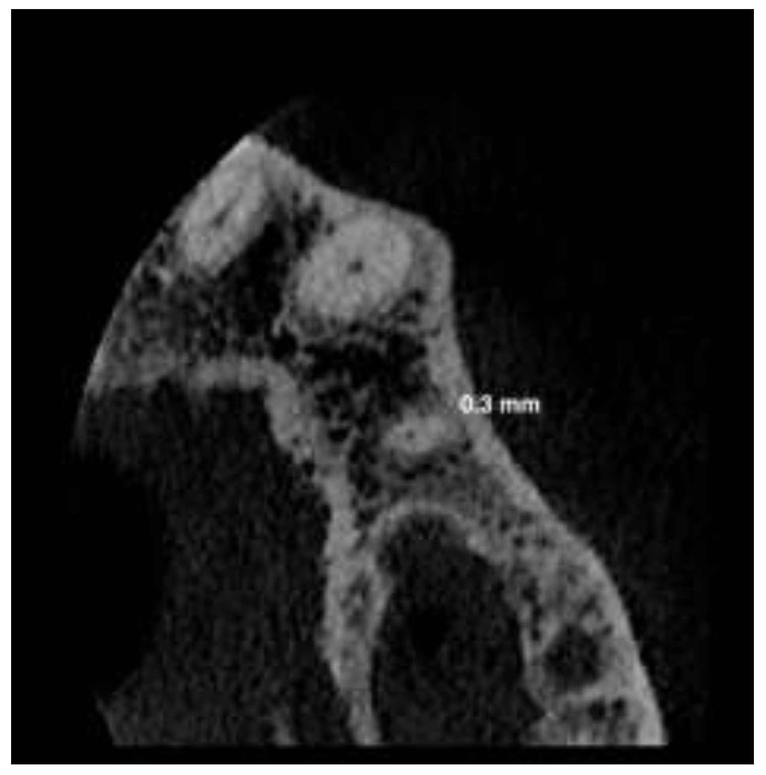
2. Materials and Methods

The current study was performed in three different dental centers. Via a three-dimensional CBCT radiographic examination, 100 permanent maxillary premolars (50 first and 50 s premolars) were analyzed.
All the CBCTs analyzed in the current work were independent from our research for diagnostic purposes for other dental problems non-related to our work.
The inclusion criteria were no sex differences, age between 20–60, premolars without any periapical lesions, external or internal resorption, non-treated endodontically, and mature apex. The exclusion criteria were all permanent premolars associated with periapical lesions, endodontically treated teeth, or immature permanent premolars.
In addition, CBCT devices with the highest resolution (75 microns) were selected (Carestream 8100 3D, Carestream Health Inc., Verona Street 150, 14,608 Rochester (NY), USA).
The selected teeth were analyzed using measurement software, CS 3D Imaging Software. The measurement was taken in an axial orientation 1 mm from the radiographic apex of each root, and the mesio-distal (M-D) and bucco-palatal (B-P) sections were measured.
Two trained operators ran two different measures. A third trained operator conducted the processes if disagreement was found between the two operators.
The section of 0.3 mm or less, 1 mm from the radiographic apex of the upper premolars, was chosen as the critical value (Figure 1).

Statistical Analysis

Frequencies, percentages, means, and standard deviations were calculated for the data. The weighted Cohen’s kappa (κ) test was used to assess the interobserver agreement. The range of variation of the weighted κ statistic is between 0 for no agreement and 1 for perfect agreement with five intermediate levels: slight agreement (0.01–0.20), fair agreement (0.21–0.40), moderate agreement (0.41–0.60), substantial agreement (0.61–0.80), and almost perfect agreement (0.81–0.99) [9].
A chi-square test was used to assess possible associations between measures ≤0.3 mm and the first or second premolar, between measures ≤0.3 mm and M-D or B-P, and between measures ≤0.3 mm and roots. An independent samples t-test was used to compare the means and standard deviations between teeth, roots, and sections.
A standard statistical software package (SPSS, version 22.0; IBM SPSS, Armonk, NY, USA) was used. The significance level was set at p < 0.05.

3. Results

The mean value in the 304 sections analyzed was 1.03 mm (±0.37). Only 19 sections (6.3%) had measurements less than or equal to 0.3 mm. All these 19 sections were from first premolars (p = 0.002).
Mean of the sections of second premolars was 1.12 mm (±0.3).
Mean of the sections of buccal roots of the first premolars was 0.98 mm (±0.38).
Mean of the sections of palatal roots of the first premolars was 1 mm (±0.39).
The results are presented in Table 1.
Interobserver agreement was almost perfect (κ = 0.91; p < 0.001).

4. Discussion

The minimally invasive endodontics concept is spreading in all the phases of the root canal treatment, starting from the access cavity to shaping, 3D cleaning, and ultimately to three-dimensional obturation [10].
In general, the root canal mechanical preparation is a crucial phase on which the quality of the entire endodontic treatment depends. All vital and necrotic pulp tissue, bacteria, and toxins are removed from the main canal during this phase. The root canal dentin is also shaped to create a space to accommodate for the cleaning and filling phases also to eliminate bacteria deep in the dentinal tubules.
Significantly, this step can be critical when treating necrotic teeth or in the case of endodontic retreatments.
The proper shaping of the canal must respect the original anatomy without altering it and evade excessive dentin removal to avoid the risk of microcracks or stripping.
Root canal shaping procedures and rotary instrumentation can potentially induce cracks. Accordingly, once the tooth is in function and rehabilitated, it can lead to complete fractures.
According to various authors, several factors regarding nickel–titanium tools can influence microcrack formation, such as heat treatments, design, cross-sectional shape, and kinematics [11,12].
Advances in Ni-Ti instruments and their kinematics have made it possible to model root canals with single-file systems activated with rotational or reciprocating motion [13,14].
Moreover, the shaping must maintain the foramen in its original anatomy without creating any modifications.
Sousa-Neto et al., in a review of the literature, analyzed three-dimensionally the biomechanical preparation of root canals using micro-computed tomography as regards to the final diameter; the studies examined by the authors showed that the final diameter of the instruments used for the biomechanical preparation varied from a diameter of 25/100 mm to a diameter of 40/100 mm, with a diameter of 30/100 mm being the most used in these canals. The mesial canals of the mandibular molars were the most used specimen among the studies examined, and the diameter of these canals at 1 mm of the apical foramen ranged between 0.28–0.40 mm in the buccal direction and 0.21–0.28 mm in the mesio-distal direction [13].
An equally important aspect to avoid during the mechanical preparation is fracturing the rotating files. Separating a file inside the canals can complicate the treatment and alter the prognosis, as retrieving it can be unachievable. Hence, a fracture of an endodontic instrument can also lead to treatment failure.
Nowadays, with the aid of powerful irrigant activation systems, it is feasible to perform conservative shaping and obtain outstanding results in cleaning the walls and the success rates. According to Iandolo et al., satisfactory outcomes are obtained using the intracanal heating of sodium hypochlorite associated with ultrasonic activation when performed after conservative preparations using rotary files 20.05 [15,16].
After performing the diagnosis steps, access cavity, shaping, and 3D cleaning during the root canal treatment, we reach the final phase, which is the obturation phase. Recent techniques and technologies in endodontics have removed the urgency of excessively enlarging the root canals by using modern sealers such as the biosealers combined with a simple obturation technique such as the single cone or hot modified technique [17,18].
This conservative approach is explained by the lack of need to use heat pluggers and other devices deep inside the root canal. In this way, by doing a conservative shaping and a powerful irrigant phase, it is possible to execute the endodontic treatment conservatively and obtain positive results.
Carrying out such minimally invasive preparations leads to a series of advantages; for instance, faster and more reproducible shaping even by less experienced operators. [18,19,20].
Moreover, it reduces the risk of altering the original anatomy of the canals or the apical foramen and lowers the risk of creating stripping in delicate anatomies [21,22].
Equally important, conservative approaches yield more significant dentin conservation and finally, by using small-sized rotating files of the new generation, the risk of instrument fracture is almost nil in the martensitic phase [23,24,25].
The analysis of our study showed that in the first maxillary premolars, even if at a low percentage, it is possible to find root sections 1 mm from the radiographic apex that are very small and less than or equal to 0.30 mm.
The use of rotating files 25/100 mm, 04 in taper, 30/100 mm, 04 in taper, 25/100 mm, 06 in taper, or files with diameters and tapers greater than these mentioned in root canals that have such reduced diameters can destroy the apical structure.
A rotating file 25/100 mm, 06 in taper at 1 mm from its tip measuring 31/100 mm used to shape delicate anatomy with a section at 1 mm from the radiographic apex of 0.30 mm would inevitably cause irreversible damage in the last apical millimeter.
In summary, when treating teeth with fragile apical diameters (such as maxillary first premolars), the application of minimally invasive endodontics, utilizing recent powerful diagnostic tools such as CBCT and revolutionary cleaning and shaping methods, can lead to more predictable and applicable treatment that guarantees the preservation of valuable tooth structure without altering the quality of the treatment [4,16,20].

5. Conclusions

Regarding the analyzed data, the authors recommend, in the case of endodontic treatments of maxillary premolars, performing a pre-operative high-resolution 3D CBCT to know in advance the measurements of the last apical millimeter of the roots. Consequently, selecting an adequate shaping protocol is advisable to avoid causing iatrogenic errors.
Moreover, suppose a high-resolution pre-operative 3D CBCT is unavailable. In that case, the authors suggest, in order to avoid risks caused by excessive shaping such as perforations, microcracks, file separation, alteration of apical anatomy, and similar risks, to use a conservative shaping combined with enhanced cleaning.

Author Contributions

Conceptualization, A.I. and D.A.; methodology, A.I.; software, M.P.; validation, G.S. and D.A.; formal analysis, M.P. and G.S.; investigation, S.M.; resources, D.A.; data curation, A.I.; writing—original draft preparation, D.A.; writing—review and editing, A.I.; visualization, S.M.; supervision, S.M. All authors have read and agreed to the published version of the manuscript.

Funding

This research received no external funding. The study was conducted in accordance with the Declaration of Helsinki.

Informed Consent Statement

Informed consent was obtained from all subjects involved in the study. Written informed consent has been obtained from the patients to publish this paper.

Conflicts of Interest

The authors declare no conflict of interest.

References

  1. Neuhaus, K.W.; Liebi, M.; Stauffacher, S.; Eick, S.; Lussi, A. Antibacterial efficacy of a new sonic irrigation device for root canal disinfection. J. Endod. 2016, 42, 1799–1803. [Google Scholar] [CrossRef] [PubMed]
  2. Amato, M.; Pantaleo, G.; Abdellatif, D.; Blasi, A.; Gagliani, M.; Iandolo, A. An in vitro evaluation of the degree of pulp tissue dissolution through different root canal irrigation protocols. J. Conserv. Dent. 2018, 21, 175–179. [Google Scholar] [PubMed]
  3. Friedman, S. Prognosis of initial endodontic therapy. Endod. Topics 2002, 2, 59–88. [Google Scholar] [CrossRef] [Green Version]
  4. Yared, G.; Ramli, G.A. Ex vivo ability of a noninstrumentation technique to disinfect oval-shaped canals. J. Conserv. Dent. 2020, 23, 10–14. [Google Scholar] [CrossRef]
  5. Zanza, A.; Seracchiani, M.; Di Nardo, D.; Reda, R.; Gambarini, G.; Testarelli, L. A Paradigm Shift for Torsional Stiffness of Nickel-Titanium Rotary Instruments: A Finite Element Analysis. J. Endod. 2011, 47, 1149–1156. [Google Scholar] [CrossRef] [PubMed]
  6. Ferreira, F.; Adeodato, C.; Barbosa, I.; Aboud, L.; Scelza, P.; Zaccaro Scelza, M. Movement kinematics and cyclic fatigue of NiTi rotary instruments: A systematic review. Int. Endod. J. 2017, 50, 143–152. [Google Scholar] [CrossRef]
  7. Iandolo, A.; Amato, A.; Martina, S.; Latif, D.A.; Pantaleo, G. Management of severe curvatures in root canal treatment with the new generation of rotating files using a safe and predictable protocol. Open Dent. J. 2020, 14, 421–425. [Google Scholar] [CrossRef]
  8. Patel, S.; Brown, J.; Semper, M.; Abella, F.; Mannocci, F. European Society of Endodontology position statement: Use of cone beam computed tomography in Endodontics: European Society of Endodontology (ESE) developed by. Int. Endod. J. 2019, 52, 1675–1678. [Google Scholar] [CrossRef] [Green Version]
  9. Viera, A.J.; Garrett, J.M. Understanding interobserver agreement: The kappa statistic. Fam. Med. 2005, 37, 360–363. [Google Scholar]
  10. Bürklein, S.; Schäfer, E. Minimally invasive endodontics. Quintessence Int. 2015, 46, 119–124. [Google Scholar]
  11. Liu, R.; Hou, B.X.; Wesselink, P.R.; Wu, M.-K.; Shemesh, H. The incidence of root microcracks caused by 3 different single-file systems versus the ProTaper system. J. Endod. 2013, 39, 1054–1056. [Google Scholar] [CrossRef] [PubMed]
  12. Barreto, M.S.; Moraes, R.D.A.; da Rosa, R.A.; Moreira, C.; Só, M.V.R.; Bier, C.A.S. Vertical root fractures and dentin defects: Effects of root canal preparation, filling, and mechanical cycling. J. Endod. 2012, 38, 1135–1139. [Google Scholar] [CrossRef] [PubMed]
  13. Sousa-Neto, M.D.; Silva-Sousa, Y.C.; Mazzi-Chaves, J.F.; Carvalho, K.K.T.; Barbosa, A.F.S.; Versiani, M.A.; Jacobs, R.; Leoni, G.B. Root canal preparation using micro-computed tomography analysis: A literature review. Braz. Oral Res. 2018, 32, e66. [Google Scholar] [CrossRef] [PubMed] [Green Version]
  14. Webber, J.; Machtou, P.; Pertot, W.; Kuttler, S.; Ruddle, C.; West, J. The Wave One single file reciprocating system. Roots 2011, 1, 28–33. [Google Scholar]
  15. Iandolo, A.; Abdellatif, D.; Pantaleo, G.; Sammartino, P.; Amato, A. Conservative shaping combined with three-dimensional cleaning can be a powerful tool: Case series. J. Conserv. Dent. 2020, 23, 648–652. [Google Scholar] [CrossRef]
  16. Iandolo, A.; Abdellatif, D.; Amato, M.; Pantaleo, G.; Blasi, A.; Franco, V.; Neelakantan, P. Dentinal tubule penetration and root canal cleanliness following ultrasonic activation of intracanal-heated sodium hypochlorite. Aust. Endod. J. 2020, 46, 204–209. [Google Scholar] [CrossRef]
  17. Abdellatif, D.; Amato, A.; Calapaj, M.; Pisano, M.; Iandolo, A. A novel modified obturation technique using biosealers: An ex vivo study. J. Conserv. Dent. 2021, 24, 369–373. [Google Scholar]
  18. Alves Silva, E.C.; Tanomaru-Filho, M.; da Silva, G.F.; Delfino, M.M.; Cerri, P.S.; Guerreiro-Tanomaru, J.M. Biocompatibility and Bioactive Potential of New Calcium Silicate-based Endodontic Sealers: Bio-C Sealer and Sealer Plus BC. J. Endod. 2020, 46, 1470–1477. [Google Scholar] [CrossRef]
  19. Gündoğar, M.; Uslu, G.; Özyürek, T.; Plotino, G. Comparison of the cyclic fatigue resistance of VDW.ROTATE, TruNatomy, 2Shape, and HyFlex CM nickel-titanium rotary files at body temperature. Restor. Dent. Endod. 2020, 22, 45. [Google Scholar] [CrossRef]
  20. Ruiz-Sánchez, C.; Faus-Matoses, V.; Alegre-Domingo, T.; Faus-Matoses, I.; Faus-Llácer, V.J. An in vitro cyclic fatigue resistance comparison of conventional and new generation nickel-titanium rotary files. J. Clin. Exp. Dent. 2018, 1, 10. [Google Scholar] [CrossRef]
  21. Martina, S.; Pisano, M.; Amato, A.; Abdellatif, D.; Iandolo, A. Modern rotary files in minimally invasive endodontics: A case report. Front. Biosci. 2021, 13, 299–304. [Google Scholar]
  22. Singh, S.; Mirdha, N.; Shilpa, P.H.; Tiwari, R.V.C.; Abdul, M.S.M.; Sainudeen, S. Shaping Ability of 2Shape and WaveOne Gold Files Using Cone-Beam Computed Tomography. J. Int. Soc. Prev. Community Dent. 2019, 9, 245–249. [Google Scholar] [CrossRef] [PubMed]
  23. Siang Lin, G.S.; Singbal, K.P.; Abdul Ghani, N.R.N. A Comparative evaluation of the shaping ability, canal straightening, and preparation time of five different NiTi rotary files in simulated canals. J. Conserv. Dent. 2021, 24, 67–71. [Google Scholar] [PubMed]
  24. Kuzekanani, M.; Sadeghi, F.; Hatami, N.; Rad, M.; Darijani, M.; Walsh, L.J. Comparison of Canal Transportation, Separation Rate, and Preparation Time between One Shape and Neoniti (Neolix): An In Vitro CBCT Study. Int. J. Dent. 2021, 7, 6457071. [Google Scholar] [CrossRef]
  25. Devi, T.P.; Kaur, A.; Priyadarshini, S.; Deepak, B.S.; Banerjee, S.; Sanjeeta, N. Microscopic Assessment of Dentinal Defects Induced by ProTaper Universal, ProTaper Gold, and Hyflex Electric Discharge Machining Rotary File Systems-An in vitro Study. Contemp. Clin. Dent. 2021, 12, 230–234. [Google Scholar] [CrossRef]
Figure 1. Representative image of a buccal root of first maxillary premolar showing the measurement of the diameter in a mesio-distal section. The diameter was 0.3 mm at 1 mm away from the radiographic apex.
Applsci 12 06159 g001
Table 1. Frequency of measurements, means, and standard deviations of each measurement. Independent two sample t-tests were performed to compare measurements between teeth (1st premolars vs. 2nd premolars), roots of monorradicular premolars (buccal vs. palatal), and sections (mesio-distal vs. bucco-palatal). Statistically significant differences are in bold type.
NMean ± SDp
Teeth1st Premolars501 ± 0.390.008
2nd Premolars501.12 ± 0.3
RootsBuccal Roots1040.98 ± 0.390.09
Palatal Roots1041 ± 0.39
SectionsM-D Sections1521 ± 0.350.14
B-P Sections1521.1 ± 0.39
Publisher’s Note: MDPI stays neutral with regard to jurisdictional claims in published maps and institutional affiliations.

