Article

Sasa borealis Ethanol Extract Protects PC12 Neuronal Cells against Oxidative Stress

Department of Herbal Crop Research, National Institute of Horticultural and Herbal Science (NIHHS), Rural Development Administration (RDA), Eumsung 27709, Korea
*
Author to whom correspondence should be addressed.
These authors contributed equally to this work.
Appl. Sci. 2022, 12(12), 6155; https://doi.org/10.3390/app12126155
Submission received: 19 April 2022 / Revised: 12 June 2022 / Accepted: 15 June 2022 / Published: 17 June 2022

Abstract

:
The overproduction of reactive oxygen species (ROS) can cause oxidative stress to biomolecules such as nucleic acids, proteins, and lipids, leading to neurodegenerative disorders. Sasa borealis (SB) has antioxidant, anti-inflammatory, antidiabetic, and anti-obesity effects. We evaluated the neuroprotective activity of SB on hydrogen peroxide (H2O2)-induced oxidative stress. We investigated the antioxidant and neuroprotective effects of SB water extract (SBW) and SB ethanol extract (SBE) by measuring the radical scavenging activities and intracellular ROS production. SBE, which had a high level of isoorientin, had higher antioxidative activities than SBW in 2,2′-azino-bis-(3-ethylbenzothiazolin-6-sulfonic acid) diammonium salt (ABTS+) and 2,2-diphenyl-1-picrylhydrazyl (DPPH) assays. It also reduced ROS generation in pheochromocytoma 12 (PC12) cells more significantly than SBW. It increased the translation of heme oxygenase-1 (HO-1), superoxide dismutase 2 (SOD2), catalase (CAT), and glutathione peroxidase (GPx) with a corresponding increase in the translation of NF-E2-related factor-2 (Nrf-2). In conclusion, SBE with high levels of phenolic compounds such as isoorientin shows promise for preventing neurodegenerative diseases.

1. Introduction

Free radicals generated by various factors are unstable and have high oxidative power, so if they cannot be removed from the body, oxidative stress is caused. An overproduction of reactive oxygen species (ROS) causes oxygen-induced damage, and many chronic diseases related to it include cancer, arteriosclerosis, diabetes, and cardiovascular disease. In particular, it is known that nerve cells are very vulnerable to oxidative stress due to their relatively high level of fatty acids [1,2]. An excessive production of ROS increases the rate of DNA, RNA, enzyme, protein, and lipid oxidation in the nerve cell, resulting in the dysfunction of the nerve cell and the disruption of cell membrane fluidity, leading to necrosis and apoptosis [1]. It has been reported that oxidative stress causes degenerative cranial nerve diseases such as stroke, Alzheimer’s disease (AD), and Parkinson’s disease (PD) [3]. In vivo, enzymatic antioxidant defense systems to minimize the toxicity consists mostly of Cu/Zn superoxide dismutase (SOD1), Mn superoxide dismutase (SOD2), catalase (CAT), and glutathione peroxidase (GPx). Therefore, the activation of an antioxidant response by inducing antioxidant enzymes could become a therapeutic target for neurodegenerative diseases [4].
Plant phenolic compounds, which are secondary metabolites of plants, are aromatic substances. Since they exhibit antioxidant activity, they can protect nerve cells from the oxidative stress induced by free radicals and are reported to contribute to the prevention of neurodegenerative diseases [5]. Sasa is a genus of the bamboo family, and a perennial wild grass distributed widely in Asian countries including South Korea, China, and Japan. Bamboo has been used in traditional medicine for its anti-inflammatory, antipyretic, and diuretic effects for centuries in South Korea and other Asian countries [6]. S. borealis (SB) has antidiabetic [7], hypoglycemic, hypolipidemic [8], anti-obesity [9], and antioxidant [10] effects. In addition, the use of bamboo for the treatment of hypertension, arteriosclerosis, cardiovascular disease, and cancer has been reported [11]. The diverse biological and pharmacological properties of bamboo leaf extract are attributed to the presence of flavones, glycosides, phenolic acids, coumarone lactones, anthraquinones, and amino acids [12].
S. borealis leaf extract has higher total phenolic and flavonoid contents compared to other bamboo species; the major phenolic compounds are protocatechuic acid, p-hydroxy benzoic acid, caffeic acid, syringic acid, p-coumaric acid, leuteolin-6-glucoside, and tricine-7-glucoside [13]. In addition, S. borealis contains friedelin and glutiol (triterpenoids), isoorientin, tricin 7-O-β-D-glucopyranoside (flavonoids), and luteolin 6-C-α-L-arabinopyranoside [14]. Isoorientin (3′,4′,5,7-tetrahydroxy-6-C-glucopyranosyl flavone) and a common C-glycosyl flavone, luteolin-6-C glucoside, have been isolated from several plant taxa, such as Patrinia villosa Juss, Crataegus monogyna, Crataegus pentagyna, Gentiana, Rumex, Swertia, and Vitex [15,16,17]. Isoorientin has antioxidant, anti-inflammatory, antitumor, and hepatoprotective effects [18,19,20].
Isoorientin is a flavonoid-like compound that exhibits various pharmacological activities such as antioxidant and anti-inflammatory properties. In Kim’s study [21], isoorientin reduced ROS production in Aβ25-35-induced BV2 cells, thereby blocking apoptosis and reportedly exhibiting anti-inflammatory action. According to Zhang [22] et al., flavonoid compounds protect cells from damage caused by oxidative stress, and isoorientin exhibits neuropathic anti-nociceptive activity.
Previous studies have demonstrated that isoorientin has a cytoprotective effect by inducing Nrf2, increasing detoxification molecules such as HO-1 and NQO-1, and activating the Sirt1/Sirt6 pathway. Nuclear transcription factor-E2-related factor 2-antioxidant responsive element (Nrf2-ARE) is one of the important antioxidant pathways in neuronal cells. When Nrf2 is exposed to external stimuli and oxidative stress, it translocates into the nucleus and binds to the ARE to induce the expression of other detoxifying enzymes and heme oxygenase-1 (HO-1), an important component, thereby eliminating reactive oxygen species and reducing toxicity. It has the properties of cell protection, cell damage and apoptosis inhibition, and antioxidant action [20,23,24].
Although the antioxidant, antibacterial, and anticancer effects of SB are known, the relationship between the component responsible for the neuroprotective effect of SB or the specific mechanism of action and the pharmacological effects have not yet been clearly elucidated. Therefore, in this study, an active ingredient of SB extract was identified and the protective effect of SB extracts against oxidative stress on rat pheochromocytoma cells (PC12) was investigated.

2. Materials and Methods

2.1. Sample Preparation

SB was harvested from Eumsung, Chungcheongbuk-do, South Korea, in 2019. The plant material was taxonomically identified by Dr. Jeong Hoon Lee and the voucher specimen (MPS006519) was stored at the Korea Medicinal Resources Herbarium, Eumseong, South Korea. Stem and leaf (100 g) were ground and sieved (aperture 1.40 mm, wire 0.71 mm). Next, the aerial part was refluxed with water (SB water extract, SBW) and 70% ethanol (SB ethanol extract, SBE) at a solvent-to-sample ratio of 10:1 (v/v) for 24 h three times at RT. The extracts were filtered and concentrated under vacuum. Afterwards, the extracts were lyophilized and kept at −80 °C. SB extracts were resuspended in DMSO (100 mg/mL) for cell-based assays, and methanol (50 mg/mL) for high-performance liquid chromatography (HPLC) analysis, respectively. DMSO does not have –OH, it has high solubility and cell penetrability, so it is widely used as an extracting solvent for dissolving in biochemistry and cell biology [25]. DMSO at a concentration of 0.1% was used as a solvent for cell-based assays, and this does not affect cell viability of PC12 and bioactivity of SB extracts. However, methanol (MeOH) can be used as a standard solvent to extract phenolic compounds [26]. Methanol at a concentration of 5% was used as a solvent for high-performance liquid chromatography (HPLC) analysis.

2.2. Antioxidant Activity Assay

2.2.1. ABTS+ Radical Scavenging Assay

ABTS radical scavenging activity was measured as described previously with slight modifications [27]. ABTS+ solution was prepared by mixing ABTS (7.4 mM) and potassium persulfate (2.6 mM) and allowing them to react for 24 h at RT. Next, a 40 µL sample was reacted with 160 µL ABTS+ solution, stored in dark conditions, and the mixture was reacted for half-hour at RT. Absorbance at 735 nm was gauged using a plate absorbance reader (BioTek, Winooski, VT, USA). Concentrations were estimated using an ascorbic acid (AA) standard curve and are expressed as mg of AA equivalent (AAE) per gram of dry sample [28].

2.2.2. DPPH Radical Scavenging Assay

DPPH solution was produced by dissolving DPPH (0.2 mM) into 99% ethanol. Next, sample (40 µL) was blended with DPPH solution (160 µL) and reacted for half-hour at RT. Optical density of mixture at 520 nm was gauged using a microplate absorbance reader (BioTek, Winooski, VT, USA). Concentrations were calculated using an AA standard curve and are expressed as mg of AAE per gram dry of sample [28].

2.3. Cell Culture

Pheochromocytoma cells (PC12) from rats were obtained from the ATCC (Manassas, VA, USA). PC12 cells were cultured in DMEM supplemented with 10% FBS and 1% penicillin and streptomycin (Gibco, Grand Island, NY, USA) in a humidified atmosphere containing 5% CO2 at 37 °C. Adherent PC12 cells were passaged when they were 70–80% confluent in well plates by using trypsin-EDTA solution to detach cells from the plates (every 2 or 3 days).

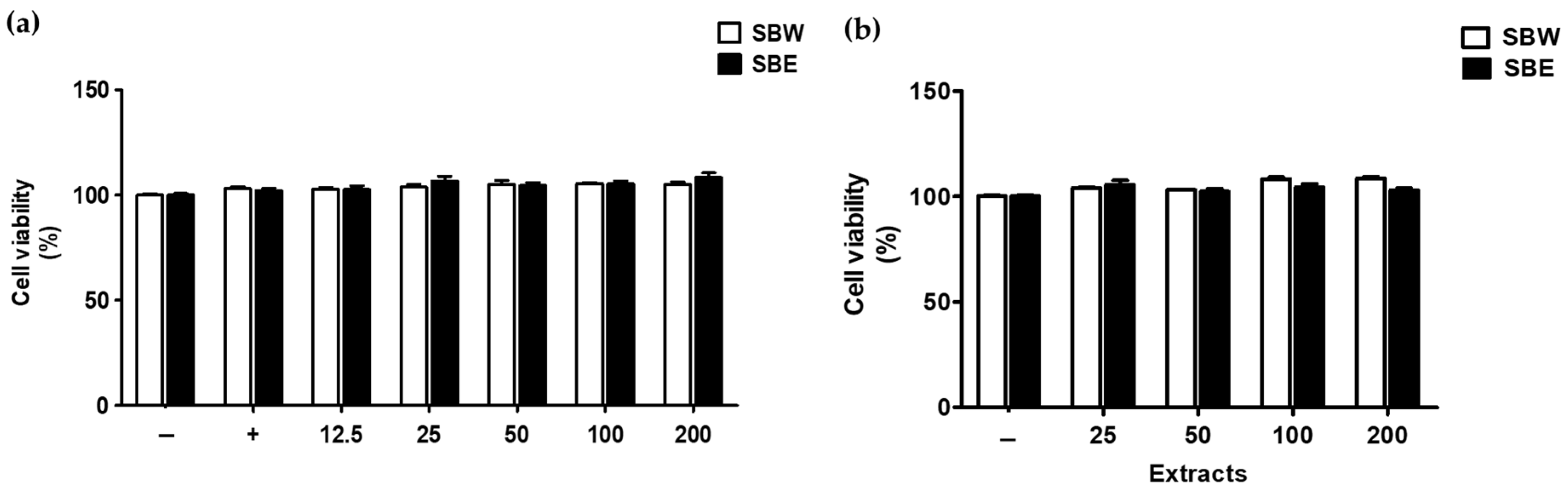
2.4. Cell Viability Assay

The cytotoxicity of SB extracts to PC12 cells was determined by MTS assay. PC12 cells were seeded at 1 × 10⁵ per mL in a plate (96-well) for 24 h. Thereafter, the cells were treated with SB extracts at different concentrations (12.5–200 microgram per mL) and cultured at 37 °C for 24 h. Next, MTS solution (100 μL) was added to each well and cultured for 1 h at 37 °C. Solvent-treated groups (DMSO 0.1%) were regarded as controls, and cell viability was measured as a fold change between control and treated groups. Optical density (490 nm) was gauged using a microplate absorbance reader. Assays were performed in triplicate.

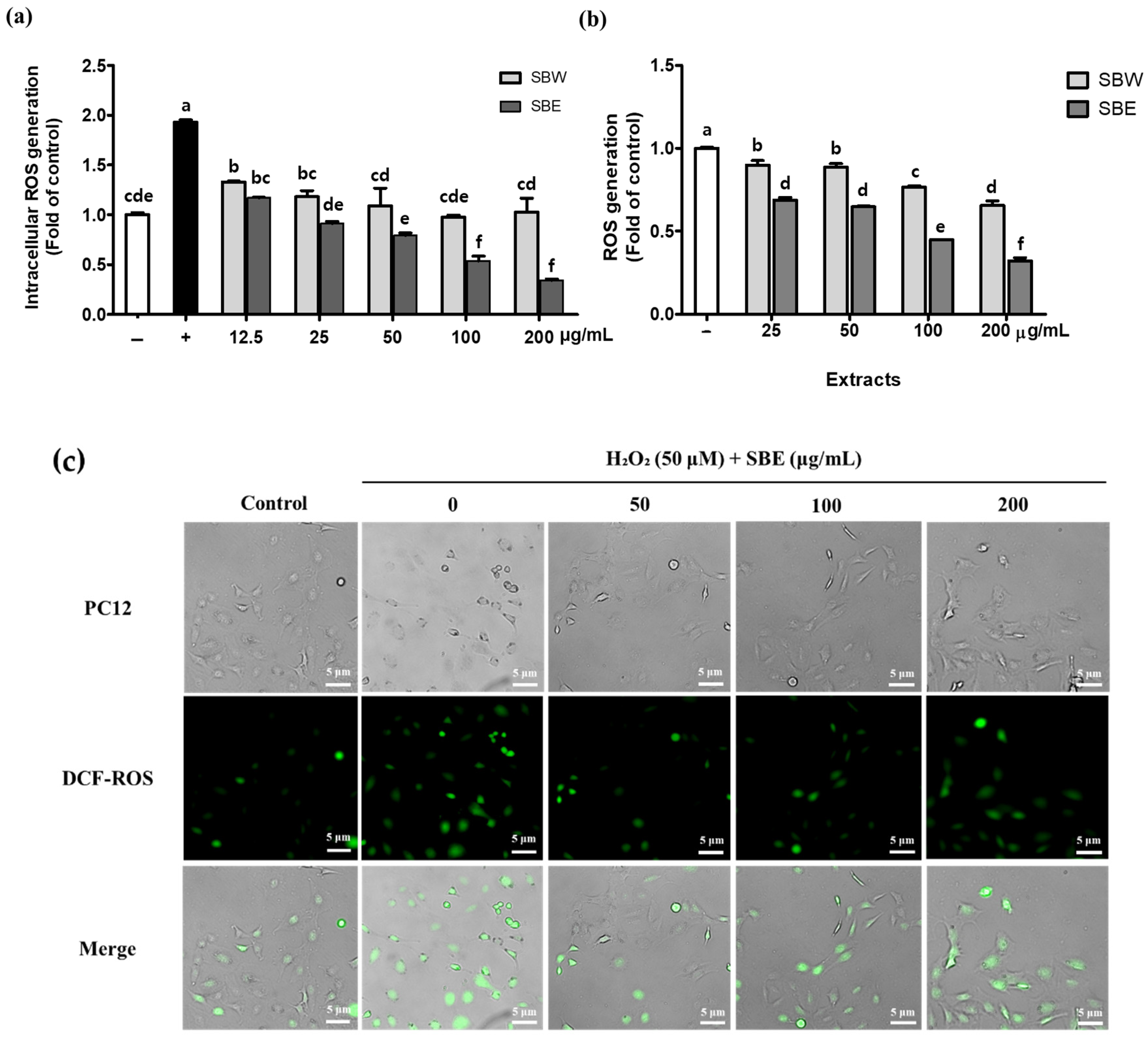
2.5. Measurement of Intracellular ROS

Intracellular ROS levels were measured using a 2′,7′-dichlorohydrofluorescein diacetate (DCF-DA; Sigma-Aldrich, St. Louis, MO, USA) assay. PC12 cells (1 × 10⁵ per mL) were seeded in plates (96-well) and cultured for 24 h. Thereafter, the cells were treated with SB extracts at different concentrations (12.5–200 μg/mL) and incubated at 37 °C for 24 h. The cells were exposed to 50 μM hydrogen peroxide H2O2 in serum-free medium (SFM) for 30 min and then to 20 μM DCF-DA in SFM for 30 min. After treatment, the cells were rinsed, and 100 µL DPBS was added to each well. Solvent-treated groups (DMSO 0.1%) were regarded as controls, and cellular ROS production was measured as a fold change between control and treated groups. Fluorescence was detected using a plate reader at 485/530 nm (excitation/emission). Fluorescence micrographs were acquired using a confocal microscope.

2.6. High-Performance Liquid Chromatography Analysis of Phenolic Acids and Flavonoids

Phenolic acids (gallic acid, homogentisic acid, protocatechuic acid, chlorogenic acid, vanillic acid, ferulic acid, veratric acid, ellagic acid, rosmarinic acid, and cinnamic acid) and flavonoids (quercetin, luteolin, naringenin, apigenin, kaempferol, isoorientin, and tricin) were used as standards. Extracts and standards were dissolved in methanol and filtered through a 0.45 μm PVDF membrane. Phenolic compounds in SB extracts were analyzed by high-performance liquid chromatography (HPLC) using a UV-VIS detector (HPLC: 1200 series, Agilent Technologies; column: XB-C18, 100 × 4.6 mm, 2.6 μm, Phenomenex). The mobile phase was composed of water with 0.1% formic acid (solvent A) and acetonitrile with 0.1% formic acid (solvent B). The gradient program for the mobile phase was 0–3 min (7–7% B), 3–10 min (7–12% B), 10–42 min (12–22% B), 42–50 min (22–50% B), 50–60 min (50–100% B), 60–68 min (100–100% B), and 68–75 min (100–7% B). Detection wavelength, injection volume, and flow rate were set to 280 nm, 10 μL, and 1.0 mL/min, respectively. The standard solutions were analyzed at concentrations of 0.03125, 0.0625, 0.125, 0.25, and 0.5 mg per mL. A standard curve was generated to quantify the amount of caffeic acid and isoorientin. In the linear regression equation (y = ax + b), x means the concentration of caffeic acid and isoorientin (mg per mL), and y is the peak area.

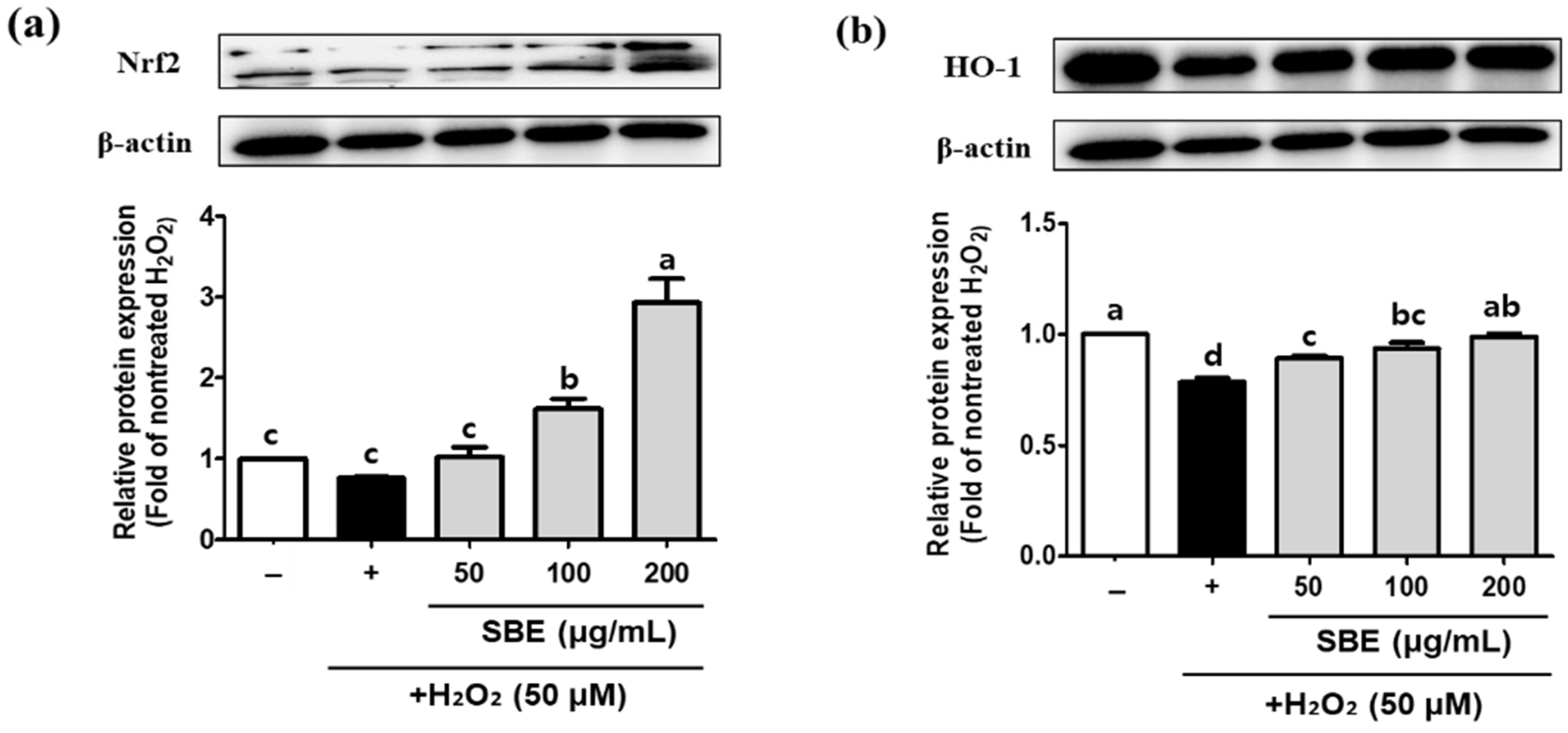
2.7. Western Blotting

PC12 cells were exposed to SB extracts (50–200 μg per mL) for 24 h, and then H2O2 (50 μM) for half-hour. Then the cells were scraped off and harvested. After centrifugation at 15,000 rpm, the cells were lysed with RIPA buffer (GenDEPOT, Katy, TX, USA) containing a phosphatase and protease inhibitor mixture (GenDEPOT, Katy, TX, USA). The amounts of protein in PC12 cells were quantified using a bicinchoninic acid assay kit. Equal quantities of protein (20 μg) were stacked on 10% SDS-PAGE gels, which were operated for 4 h at 80 V. The protein samples were moved to a PVDF membrane. The membrane was rinsed several times with TBST and blocked with 2% BSA for 1 h. Next, the membranes were treated with the primary antibodies for 4 h at RT. Nrf2 (dilution 1:1000), HO-1 (dilution 1:1000), SOD2 (dilution 1:1000), CAT (dilution 1:1000), GPx (dilution 1:500), and β-actin (dilution 1:1000) were detected using mouse monoclonal antibody (Santa Cruz, Dallas, TX, USA). The membranes were rinsed several times with TBST and incubated with the goat anti-mouse secondary IgG (dilution 1:2000; Santa Cruz, Dallas, TX, USA) for 1 h. Target proteins were visualized and detected using ECL solution with the Bio-Rad ChemiDoc System. Protein normalization was performed using ImageJ program.

2.8. Statistics

All data were processed using Microsoft Excel (2016) and the results are exhibited as means ± standard deviations. To decide the significance levels set at p < 0.05, p < 0.01, and p < 0.001, Duncan’s test and one-way ANOVA were carried out using R software (version 3.6.3). Correlation analysis was conducted using GraphPad Prism Software.

3. Results

3.1. SB Extracts Show an Antioxidant Effect by Scavenging Free Radicals

The yields of S. borealis water extract (SBW, 14.2%) and S. borealis ethanol extract (SBE, 16.9%) were measured as the dry weight of the plant material (Table 1). The antioxidative activities were evaluated by radical scavenging assays (DPPH and ABTS) and expressed as mg of AAE per gram of dry sample (Table 1). Ascorbic acid was used as the positive control. SBE (60.91 ± 0.13 mg AAE/g extract) exhibited higher DPPH radical scavenging activity than SBW (49.63 ± 1.02 mg AAE/g extract). The ABTS+ radical-scavenging activity was higher for SBE extract (165.14 ± 5.08 mg AAE/g extract) than SBW extract (144.84 ± 2.82 mg AAE/g extract); therefore, SBE has greater antioxidant activity than SBW.

3.2. SB Extracts Decrease H2O2-Induced ROS Production in PC12 Neuronal Cells

As shown in Figure 1, SB extracts (SBW and SBE) at up to 200 μg/mL did not affect cell viability compared to H2O2-treated PC12 cells. At 200 μg/mL SBW and SBE, cell viability remained >100%. Therefore, further experiments were carried out using SB extracts at ≤200 μg/mL.
To verify the effect of the SB extracts on H2O2-induced oxidative stress, the intracellular ROS generation was measured based on DCF-DA staining [29]. The intensity of the fluorescent signal from the liberated DCF in the H2O2-treated cells was significantly increased by 1.9-fold relative to the untreated control (Figure 2a). Treatment with SBW and SBE (12.5 to 200 μg/mL) decreased the H2O2-induced cellular ROS generation in a concentration-dependent manner. The inhibitory activity of SBE on ROS production was higher than that of SBW. SBE at 12.5, 25, 50, 100, and 200 μg/mL reduced the ROS level approximately 0.61-, 0.47-, 0.41-, 0.28-, and 0.18-fold, respectively; SBW decreased the ROS level by 0.69-, 0.61-, 0.56-, 0.51-, and 0.53-fold, respectively (Figure 2a). Additionally, SBE reduced the ROS generation in non-treated cells in a concentration-dependent manner. (Figure 2b). Next, we visually investigated the fluorescence changes in the PC12 cells treated with H2O2 (50 μM) and SBE (50, 100 and 200 μg/mL). As shown in Figure 2c, SBE (50 to 200 μg/mL) decreased H2O2-induced cellular ROS generation in a concentration-dependent manner.

3.3. Isoorientin and Caffeic Acid of SB Extracts Were Identified by HPLC Analysis

To identify the components responsible for the neuroprotective activity of the SB extracts, we used HPLC. A major component of SBW and SBE was isoorientin (Figure 3, peak 10). As shown in Table 2, the amounts of isoorientin (6.75 ± 0.17 mg/g extract, d.b.) and caffeic acid (0.73 ± 0.01 mg/g extract, d.b.) in SBE were higher than in SBW (2.70 ± 0.03 and 0.72 ± 0.00 mg/g extract, d.b., respectively). Therefore, isoorientin is linked to the neuroprotective activity of the SB extracts.
Next, we examined the correlations among antioxidant activity, isoorientin, caffeic acid content, and the intracellular ROS generation (Table 3). DPPH and ABTS+ radical-scavenging activities were positively correlated with isoorientin (0.995 and 0.959, respectively) and caffeic acid (0.889 and 0.902, respectively) contents, whereas antioxidative activities (DPPH and ABTS+) were inversely correlated with cellular ROS generation (−0.932 and −0.887, respectively). The isoorientin content was positively correlated with ROS generation in the PC12 cells (−0.925). Therefore, a major constituent of SB extracts, isoorientin, is associated with their antioxidant and neuroprotective activities.

3.4. SBE Increases the Protein Levels of Nrf2, HO-1, SOD2, CAT, and GPx
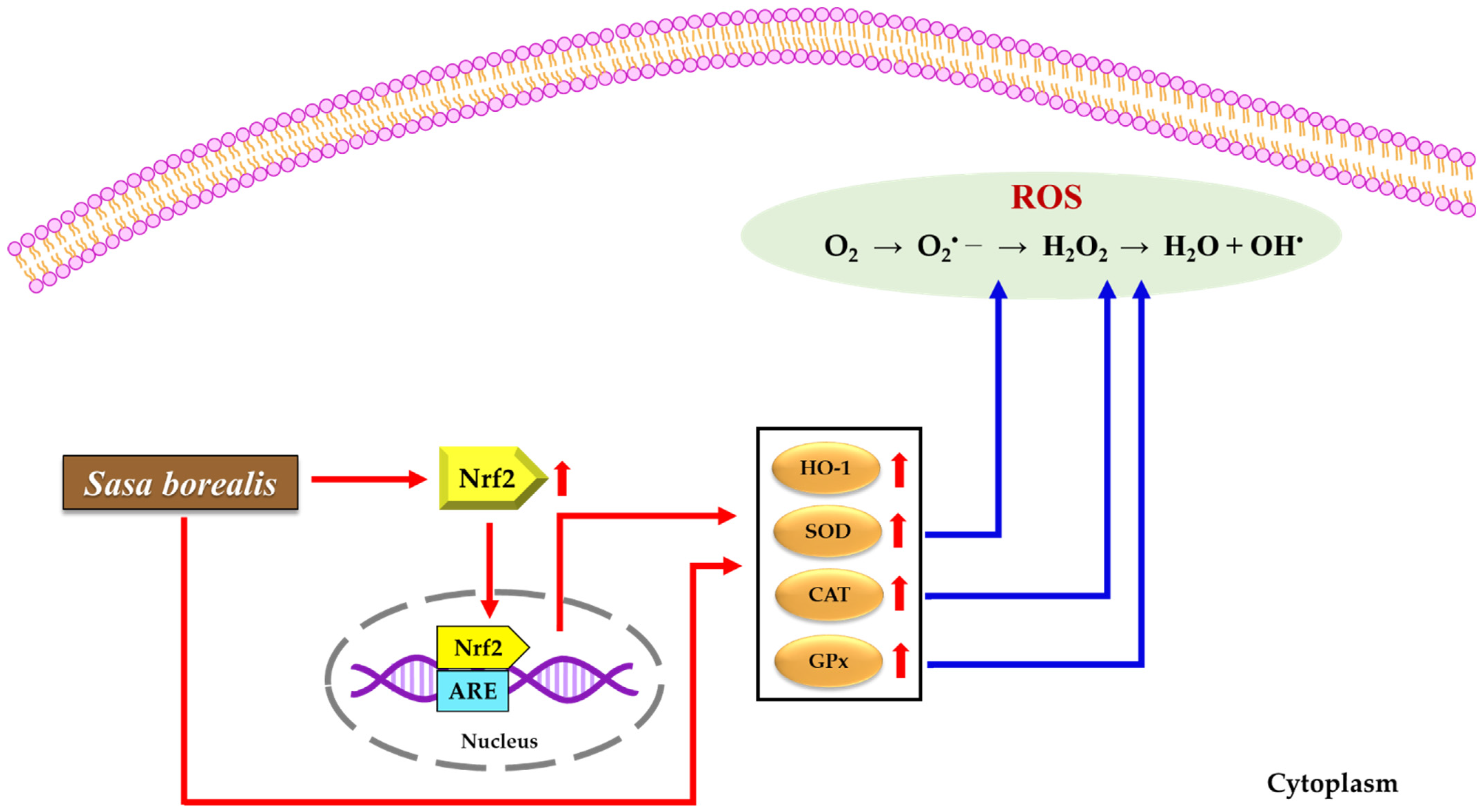
H2O2 has been generally used to cause ROS release. In order to counteract intracellular ROS, the antioxidant enzyme defense system including SOD2, CAT, and GPx is activated [28]. To determine whether PC12 cells were protected from oxidative stress by SBE, we induced oxidative stress via treatment with H2O2 in PC12 cells, and then added SBE to PC12 cells.
To investigate the effect of SBE on the protein levels of an antioxidant-related factor (Nrf2) and enzymes (HO-1, SOD, CAT, and GPx), we performed western blotting. Nrf2 is a key transcription factor that regulates antioxidant-related gene expression. The activation of Nrf2 leads to an induction of antioxidant enzymes, such as HO-1, SOD, CAT, and GPx [28]. As shown in Figure 4A, SBE (50, 100, and 200 μg/mL) increased the levels of Nrf2 (1.35-, 2.13-, and 3.87-fold, respectively, relative to H2O2-treated cells) in a concentration-dependent manner. The level of HO-1 in cells treated with SBE (50, 100, and 200 μg/mL) increased in a concentration-dependent manner (1.14-, 1.19-, and 1.26-fold, respectively, relative to the H2O2-treated cells). SBE (50, 100, 200 μg/mL) increased the protein levels of SOD2 (0.74-, 0.79-, and 1.42-fold, respectively, relative to H2O2-treated cells) and CAT (1.28-, 1.57-, and 1.68-fold, respectively, relative to H2O2-treated cells). The level of GPx in cells treated with SBE (50, 100, 200 μg/mL) was increased 1.20-, 1.19-, and 1.44-fold, respectively, relative to the H2O2-treated cells.
To confirm whether SBE has an impact on the protein levels of an antioxidant-related factor (Nrf2) and enzymes (HO-1, SOD, CAT, and GPx) in normal conditions, PC12 cells without a H2O2 stimulus were assessed using western blotting. As shown in Figure 4B, SBE (50, 100, and 200 μg/mL) raised the expression levels of Nrf2 and HO-1. SBE (50, 100, and 200 μg/mL) caused concentration-dependent increases in the levels of Nrf2 (1.20-, 1.24-, and 1.39-fold, respectively, relative to the non-treated cells). The level of HO-1 in cells treated with SBE (50, 100, and 200 μg/mL) increased in a concentration-dependent manner (2.61-, 2.97-, and 3.53-fold, respectively, relative to the non-treated cells). SBE (50, 100, 200 μg/mL) increased the protein levels of SOD2 (1.15-, 1.62-, and 1.35-fold, respectively, relative to the non-treated cells) and CAT (1.58-, 1.75-, and 1.98-fold, respectively, relative to the non-treated cells). The level of GPx in the cells treated with SBE (50, 100, 200 μg/mL) was increased 1.27-, 1.10-, and 1.41-fold, respectively, relative to the non-treated cells. Therefore, SBE protected the PC12 cells from H2O2-induced oxidative stress by inducing the expression of antioxidant-related proteins.

4. Discussion

Most neurodegenerative diseases are caused by the death of brain nerve cells due to oxidative stress [29]. An antioxidant can donate an electron or a hydrogen atom, transfer unpaired electrons, and can chelate metals [30]. Since DPPH and ABTS assays can be applied to both water-soluble and fat-soluble antioxidant measurements, they are widely used to measure the antioxidant activity of foods [31]. Park et al. [32] showed that 70% ethanol extract showed a significantly higher electron donating ability than water extraction, and our analysis showed that SBE had higher radical scavenging activity than SBW (Table 1). Thus, SBE can scavenge free radicals by delivering hydrogen (DPPH) or single electrons (ABTS assay). Cellular antioxidants quench free radicals by transferring a hydrogen or electron, thereby inducing the synthesis of detoxifying and antioxidant proteins in phase II [33].
Substances such as friedelin and glutinol (triterpene-based compounds) and isoorientin (flavonoid-based compounds) have been reported as component studies for SB [33]. Isoorientin, a family of rooibos flavonoids, has antioxidant activity and has been reported to protect cells from 6OHDA-induced neurotoxicity in PC12 cells [34]. In addition, it protects cells from oxidative damage caused by t-BOOH in HepG2 cells, and has effects on DPPH free radical scavenging activity, iron (III) reducing activity, linoleic acid peroxidation, and MDA formation [35]. Isoorientin reportedly has anti-inflammatory activity and the ability to diminish mitochondrial ROS production [36,37]. Isoorientin and orientin, rooibos flavonoids, enhance mitochondrial function by regulating mitochondrial respiration, leading to a reduced production of ROS in C2C12 skeletal muscle cells [38]. In addition, isoorientin attenuates the oxidative damage induced by oleic acid in buffalo rat liver 3A (BRL-3A) cells [39], and reduces ROS generation in UV-stimulated human dermal fibroblasts [36].
In this study, the isoorientin content of SBE was higher than that of SBW. This is consistent with our finding that the antioxidant and neuroprotective activities of SBE are superior to those of SBW. Therefore, the antioxidant and neuroprotective effects of SBE are likely mediated by isoorientin. However, the bioactive activities of various phytochemicals present in trace amounts cannot be excluded. Further studies on unidentified phenolic acids and flavonoids in SB extracts that mediate its antioxidant effect in vitro and in vivo are warranted.
SBE with a high radical scavenging activity reduced the intracellular ROS accumulation induced by H2O2 (Figure 2). This implies that the neuroprotective effect of SBE is mediated by quenching or neutralizing free radicals. The expression of genes encoding antioxidant proteins is induced by molecular mechanisms (Figure 5). Antioxidant response elements (ARE), Nrf2, and Keap1 regulate the expression of genes encoding SOD2, CAT, and GPx [40]. Under normal conditions, Nrf2 is bound to Keap1 in the cytoplasm. Upon exposure to oxidative stress, Nrf2 is released from Keap1, is translocated into the nucleus, and activates phase II antioxidant enzymes, such as heme oxygenase (HO-1) [41,42]. HO-1 catalyzes the conversion of heme to CO, free iron, and biliverdin, which is converted into bilirubin. Bilirubin has potent antioxidant and cytoprotective activities [43]. Thus, an elevated expression of Nrf2 and HO-1 can protect cells from oxidative damage. SOD catalyzes the conversion of superoxide anion (O2) into a molecule of H2O2. In peroxisomes, CAT converts H2O2 to water (H2O) [44]. Glutathione peroxidase (GPx) contains selenium and catalyzes the degradation of H2O2 to H2O. These enzymes act collectively in the metabolic pathway of free radicals [45,46]. We found that SBE increased the expression levels of Nrf2 and HO-1 in PC12 cells. In addition, SBE markedly increased the expression levels of SOD2, CAT, and GPx in PC12 cells, suggesting that cellular antioxidant activity is an indirect effect of antioxidant factors.
We showed that SBE with high levels of phenolic compounds such as isoorientin had neuroprotective effects against H2O2-induced oxidative stress via activation of the Nrf2 signaling. The molecular mechanisms need to be verified in vivo, which would facilitate the development of a natural medicine protecting neurons from oxidative stress, which is known as an essential factor of neurodegenerative diseases.

5. Conclusions

SBE had neuroprotective effects against H2O2-induced oxidative stress. It showed a higher radical scavenging activity and a more significant inhibitory effect on ROS generation in H2O2-treated PC12 cells. These effects were likely mediated by phenolic compounds such as isoorientin. In addition, SBE activated Nrf2 signaling and induced the synthesis of antioxidant enzymes, such as HO-1, SOD2, CAT, and GPx. Taken together, this study demonstrated that SBE has the potential for protecting neurons from oxidative stress, which is known as an essential factor of neurodegenerative diseases.

Author Contributions

Conceptualization, H.D.K.; methodology, M.H.K., Y.-J.J., Y.M.H. and G.Y.J.; data curation, M.H.K., Y.M.H., G.Y.J., G.-S.K. and S.J.C.; resources, D.H.K. and J.H.L.; writing—original draft preparation, M.H.K. and Y.-J.J. writing—review and editing, H.D.K.; funding acquisition, M.H.K. and H.D.K. All authors have read and agreed to the published version of the manuscript.

Funding

This work was carried out with the support of “Cooperative Research Program for Agriculture Science and Technology Development (Project No. PJ01415802)” and the RDA Fellowship Program of National Institute of Horticultural and Herbal Science, Rural Development Administration, Republic of Korea.

Institutional Review Board Statement

Not applicable.

Informed Consent Statement

Not applicable.

Data Availability Statement

Not applicable.

Conflicts of Interest

The authors declare no conflict of interest.

References

  1. Uttara, B.; Singh, A.V.; Zamboni, P.; Mahajan, R. Oxidative stress and neurodegenerative diseases: A review of upstream and downstream antioxidant therapeutic options. Curr. Neuropharmacol. 2009, 7, 65–74. [Google Scholar] [CrossRef] [PubMed] [Green Version]
  2. Limón-Pacheco, J.; Gonsebatt, M.E. The role of antioxidants and antioxidant-related enzymes in protective responses to environmentally induced oxidative stress. Mutat. Res. Genet. Toxicol. Environ. Mutagenes. 2009, 674, 137–147. [Google Scholar] [CrossRef] [PubMed]
  3. Bhatia, V.; Sharma, S. Role of mitochondrial dysfunction, oxidative stress and autophagy in progression of Alzheimer’s disease. J. Neurol. Sci. 2021, 421, 117253. [Google Scholar] [CrossRef]
  4. Lin, M.T.; Beal, M.F. Mitochondrial dysfunction and oxidative stress in neurodegenerative diseases. Nature 2006, 443, 787–795. [Google Scholar] [CrossRef] [PubMed]
  5. Lu, B.; Wu, X.; Shi, J.; Dong, Y.; Zhang, Y. Toxicology and safety of antioxidant of bamboo leaves. Part 2: Developmental toxicity test in rats with antioxidant of bamboo leaves. Food Chem. Toxicol. 2006, 44, 1739–1743. [Google Scholar] [CrossRef] [PubMed]
  6. Lu, B.; Wu, X.; Tie, X.; Zhang, Y.; Zhang, Y. Toxicology and safety of anti-oxidant of bamboo leaves. Part 1: Acute and subchronic toxicity studies on anti-oxidant of bamboo leaves. Food Chem. Toxicol. 2005, 43, 783–792. [Google Scholar] [CrossRef]
  7. Ko, B.-S.; Jun, D.-W.; Jang, J.-S.; Kim, J.-H.; Park, S.-M. Effect of Sasa borealis and white lotus roots and leaves on insulin action and secretion in vitro. Korean J. Food Sci. Technol. 2006, 38, 114–120. [Google Scholar]
  8. Choi, Y.-J.; Lim, H.-S.; Choi, J.-S.; Shin, S.-Y.; Bae, J.-Y.; Kang, S.-W.; Kang, I.-J.; Kang, Y.-H. Blockade of chronic high glucose–induced endothelial apoptosis by Sasa borealis bamboo extract. Exp. Biol. Med. 2008, 233, 580–591. [Google Scholar] [CrossRef]
  9. Kim, E.-Y.; Jung, E.-Y.; Lim, H.-S.; Heo, Y.-R. The effects of the Sasa borealis leaves extract on plasma adiponectin, resistin, C-reactive protein and homocysteine levels in high fat diet-induced obese C57/BL6J mice. J. Nutr. Health 2007, 40, 303–311. [Google Scholar]
  10. Park, H.-S.; Lim, J.H.; Kim, H.J.; Choi, H.J.; Lee, I.-S. Antioxidant flavone glycosides from the leaves of Sasa borealis. Arch. Pharmacal. Res. 2007, 30, 161–166. [Google Scholar] [CrossRef]
  11. Sangeetha, R.; Diea, Y.; Chaitra, C.; Malvi, P.; Shinomol, G. The amazing bamboo: A review on its medicinal and pharmacological potential. Indian J. Nutr. 2015, 2, 1–7. [Google Scholar]
  12. Sun, A.; Ren, G.; Deng, C.; Zhang, J.; Luo, X.; Wu, X.; Mani, S.; Dou, W.; Wang, Z. C-glycosyl flavonoid orientin improves chemically induced inflammatory bowel disease in mice. J. Funct. Foods 2016, 21, 418–430. [Google Scholar] [CrossRef]
  13. Park, E.-J.; Jhon, D.-Y. Effects of bamboo shoot consumption on lipid profiles and bowel function in healthy young women. Nutrition 2009, 25, 723–728. [Google Scholar] [CrossRef] [PubMed]
  14. Molina-Cortes, A.; Sanchez-Motta, T.; Tobar-Tosse, F.; Quimbaya, M. Spectrophotometric estimation of total phenolic content and antioxidant capacity of molasses and vinasses generated from the sugarcane industry. Waste Biomass Valorization 2020, 11, 3453–3463. [Google Scholar] [CrossRef] [Green Version]
  15. Bello, O.M.; Ogbesejana, A.B.; Adetunji, C.O.; Oguntoye, S.O. Flavonoids isolated from Vitex grandifolia, an underutilized vegetable, exert monoamine A & B inhibitory and anti-inflammatory effects and their structure-activity relationship. Turk. J. Pharm. Sci. 2019, 16, 437. [Google Scholar]
  16. Fan, X.; Lv, H.; Wang, L.; Deng, X.; Ci, X. Isoorientin ameliorates APAP-induced hepatotoxicity via activation Nrf2 antioxidative pathway: The involvement of AMPK/Akt/GSK3β. Front. Pharmacol. 2018, 9, 1334. [Google Scholar] [CrossRef]
  17. Wu, Q.-Y.; Wong, Z.C.-F.; Wang, C.; Fung, A.H.-Y.; Wong, E.O.-Y.; Chan, G.K.-L.; Dong, T.T.-X.; Chen, Y.; Tsim, K.W.-K. Isoorientin derived from Gentiana veitchiorum Hemsl. flowers inhibits melanogenesis by down-regulating MITF-induced tyrosinase expression. Phytomedicine 2019, 57, 129–136. [Google Scholar] [CrossRef]
  18. Yuan, L.; Wang, J.; Xiao, H.; Xiao, C.; Wang, Y.; Liu, X. Isoorientin induces apoptosis through mitochondrial dysfunction and inhibition of PI3K/Akt signaling pathway in HepG2 cancer cells. Toxicol. Appl. Pharmacol. 2012, 265, 83–92. [Google Scholar] [CrossRef]
  19. Lee, W.; Ku, S.-K.; Bae, J.-S. Vascular barrier protective effects of orientin and isoorientin in LPS-induced inflammation in vitro and in vivo. Vasc. Pharmacol. 2014, 62, 3–14. [Google Scholar] [CrossRef]
  20. Magesh, S.; Chen, Y.; Hu, L. Small molecule modulators of K eap1-N rf2-ARE pathway as potential preventive and therapeutic agents. Med. Res. Rev. 2012, 32, 687–726. [Google Scholar] [CrossRef] [Green Version]
  21. Kim, B.; Lee, K.Y.; Park, B. Isoorientin Inhibits Amyloid β25–35-Induced Neuronal Inflammation in BV2 Cells by Blocking the NF-κB Signaling Pathway. Molecules 2021, 26, 7056. [Google Scholar] [CrossRef] [PubMed]
  22. Zhang, G.; Liu, N.; Zhu, C.; Ma, L.; Yang, J.; Du, J.; Zhang, W.; Sun, T.; Niu, J.; Yu, J. Antinociceptive effect of isoorientin against neuropathic pain induced by the chronic constriction injury of the sciatic nerve in mice. Int. Immunopharmacol. 2019, 75, 105753. [Google Scholar] [CrossRef] [PubMed]
  23. Ma, Q. Role of nrf2 in oxidative stress and toxicity. Annu. Rev. Pharmacol. Toxicol. 2013, 53, 401–426. [Google Scholar] [CrossRef] [PubMed] [Green Version]
  24. Velichkova, M.; Hasson, T. Keap1 regulates the oxidation-sensitive shuttling of Nrf2 into and out of the nucleus via a Crm1-dependent nuclear export mechanism. Mol. Cell. Biol. 2005, 25, 4501–4513. [Google Scholar] [CrossRef] [Green Version]
  25. Cai, S.-Q.; Zhang, K.-F.; Cai, X.-H. Recent Advances in Dimethyl Sulfoxide (DMSO) Used as a Multipurpose Reactant. Curr. Org. Chem. 2022, 26, 91–121. [Google Scholar] [CrossRef]
  26. Truong, D.-H.; Nguyen, D.H.; Ta, N.T.A.; Bui, A.V.; Do, T.H.; Nguyen, H.C. Evaluation of the Use of Different Solvents for Phytochemical Constituents, Antioxidants, and In Vitro Anti-Inflammatory Activities of Severinia buxifolia. J. Food Qual. 2019, 2019, 8178294. [Google Scholar] [CrossRef] [Green Version]
  27. Van den Berg, R.; Haenen, G.R.; van den Berg, H.; Bast, A. Applicability of an improved Trolox equivalent antioxidant capacity (TEAC) assay for evaluation of antioxidant capacity measurements of mixtures. Food Chem. 1999, 66, 511–517. [Google Scholar] [CrossRef]
  28. Bitalebi, S.; Nikoo, M.; Rahmanifarah, K.; Noori, F.; Ahmadi Gavlighi, H. Effect of apple peel extract as natural antioxidant on lipid and protein oxidation of rainbow trout (Oncorhynchus mykiss) mince. Int. Aquat. Res. 2019, 11, 135–146. [Google Scholar] [CrossRef] [Green Version]
  29. Kelly, E.; Vyas, P.; Weber, J.T. Biochemical properties and neuroprotective effects of compounds in various species of berries. Molecules 2017, 23, 26. [Google Scholar] [CrossRef] [Green Version]
  30. Tsiepe, T.J.; Kabanda, M.M.; Serobatse, K. Antioxidant properties of kanakugiol revealed through the hydrogen atom transfer, electron transfer and M2+ (M2+ = Cu (II) or Co (II) ion) coordination ability mechanisms. A DFT study in vacuo and in solution. Food Biophys. 2015, 10, 342–359. [Google Scholar] [CrossRef]
  31. Wang, Y.; Yu, G.; Zang, X.; Ye, F. Optimization, antioxidant activity and bile salts adsorption capacity of the aqueous enzymatic extract from rice bran. Czech J. Food Sci. 2018, 36, 338–348. [Google Scholar]
  32. Park, Y.-O.; Lim, H.-S. Antioxidant activities of bamboo (Sasa borealis) leaf extract according to extraction solvent. J. Korean Soc. Food Sci. Nutr. 2009, 38, 1640–1648. [Google Scholar] [CrossRef]
  33. Furger, C. Live Cell Assays for the Assessment of Antioxidant Activities of Plant Extracts. Antioxidants 2021, 10, 944. [Google Scholar] [CrossRef] [PubMed]
  34. Ma, L.; Zhang, B.; Liu, J.; Qiao, C.; Liu, Y.; Li, S.; Lv, H. Isoorientin exerts a protective effect against 6-OHDA-induced neurotoxicity by activating the AMPK/AKT/Nrf2 signalling pathway. Food Funct. 2020, 11, 10774–10785. [Google Scholar] [CrossRef]
  35. Yuan, L.; Ren, X.; Wu, Y.; Wang, J.; Xiao, H.; Liu, X. Isoorientin protects BRL-3A rat liver cell against hydrogen peroxide-induced apoptosis by inhibiting mitochondrial dysfunction, inactivating MAPKs, activating Akt and scavenging ROS and NO. Biomed. Aging Pathol. 2013, 3, 153–159. [Google Scholar] [CrossRef]
  36. Zheng, H.; Zhang, M.; Luo, H.; Li, H. Isoorientin alleviates UVB-induced skin injury by regulating mitochondrial ROS and cellular autophagy. Biochem. Biophys. Res. Commun. 2019, 514, 1133–1139. [Google Scholar] [CrossRef]
  37. Anilkumar, K.; Reddy, G.V.; Azad, R.; Yarla, N.S.; Dharmapuri, G.; Srivastava, A.; Kamal, M.A.; Pallu, R. Evaluation of anti-inflammatory properties of isoorientin isolated from tubers of Pueraria tuberosa. Oxidative Med. Cell. Longev. 2017, 2017, 5498054. [Google Scholar] [CrossRef] [Green Version]
  38. Mthembu, S.X.; Muller, C.J.; Dludla, P.V.; Madoroba, E.; Kappo, A.P.; Mazibuko-Mbeje, S.E. Rooibos Flavonoids, Aspalathin, Isoorientin, and Orientin Ameliorate Antimycin A-Induced Mitochondrial Dysfunction by Improving Mitochondrial Bioenergetics in Cultured Skeletal Muscle Cells. Molecules 2021, 26, 6289. [Google Scholar] [CrossRef]
  39. Luo, T. Protective effect of isoorientin on oleic acid-induced oxidative damage and steatosis in rat liver cells. Front. Pharmacol. 2022, 13, 818159. [Google Scholar] [CrossRef]
  40. Dinkova-Kostova, A.T.; Talalay, P. Direct and indirect antioxidant properties of inducers of cytoprotective proteins. Mol. Nutr. Food Res. 2008, 52, S128–S138. [Google Scholar] [CrossRef]
  41. Bajpai, V.K.; Alam, M.B.; Quan, K.T.; Kwon, K.-R.; Ju, M.-K.; Choi, H.-J.; Lee, J.S.; Yoon, J.-I.; Majumder, R.; Rather, I.A. Antioxidant efficacy and the upregulation of Nrf2-mediated HO-1 expression by (+)-lariciresinol, a lignan isolated from Rubia philippinensis, through the activation of p38. Sci. Rep. 2017, 7, 46035. [Google Scholar] [CrossRef] [PubMed] [Green Version]
  42. Lakhan, S.E.; Kirchgessner, A.; Hofer, M. Inflammatory mechanisms in ischemic stroke: Therapeutic approaches. J. Transl. Med. 2009, 7, 97. [Google Scholar] [CrossRef] [PubMed] [Green Version]
  43. Jun, Y.-J.; Lee, M.; Shin, T.; Yoon, N.; Kim, J.-H.; Kim, H.-R. Eckol enhances heme oxygenase-1 expression through activation of Nrf2/JNK pathway in HepG2 cells. Molecules 2014, 19, 15638–15652. [Google Scholar] [CrossRef] [PubMed] [Green Version]
  44. Ighodaro, O.; Akinloye, O. First line defence antioxidants-superoxide dismutase (SOD), catalase (CAT) and glutathione peroxidase (GPX): Their fundamental role in the entire antioxidant defence grid. Alex. J. Med. 2018, 54, 287–293. [Google Scholar] [CrossRef] [Green Version]
  45. Wu, J.Q.; Kosten, T.R.; Zhang, X.Y. Free radicals, antioxidant defense systems, and schizophrenia. Prog. Neuro-Psychopharmacol. Biol. Psychiatry 2013, 46, 200–206. [Google Scholar] [CrossRef]
  46. Battin, E.E.; Brumaghim, J.L. Antioxidant activity of sulfur and selenium: A review of reactive oxygen species scavenging, glutathione peroxidase, and metal-binding antioxidant mechanisms. Cell Biochem. Biophys. 2009, 55, 1–23. [Google Scholar] [CrossRef]
Figure 1. (a) SB extracts (12.5 to 200 μg/mL) and H2O2 (50 μM) did not show toxicity in PC12 cells. (b) SB extracts (25 to 200 μg/mL) did not show toxicity in PC12 cells. SBW, Sasa borealis water extract; SBE, Sasa borealis ethanolic extract. Data are means ± SDs (n = 3).
Applsci 12 06155 g001
Figure 2. (a) SBW and SBE (12.5 to 200 μg/mL) reduces H2O2-induced ROS generation in PC12 cells. (b) SB extracts (25 to 200 μg/mL) reduced ROS generation in non-treated cells in a concentration-dependent manner. Negative control (-) means untreated group. SBW, Sasa borealis water extract; SBE, Sasa borealis ethanol extract. Data are expressed as mean ± SD (n = 3). Lowercase letters represent statistical differences at p < 0.05. (c) Cells were cultured with SBE (50–200 μg/mL) and H2O2 (50 μM) and stained with DCF-DA. H2O2-induced ROS production in PC12 cells was visualized by fluorescence microscopy. Scale bar, 5 μm.
Applsci 12 06155 g002
Figure 3. HPLC of phenolic compounds in SB extracts. (a) The peaks represent the following phenolics (0.5 mg/mL): 1, gallic acid; 2, homogentisic acid; 3, protocatechuic acid; 4, chlorogenic acid; 5, vanillic acid; 6, caffeic acid; 7, p-coumaric acid; 8, ferulic acid; 9, veratric acid; 10, isoorientin; 11, ellagic acid; 12, naringin; 13, rosmarinic acid; 14, cinnamic acid; 15, quercetin; 16, luteolin; 17, naringenin; 18, apigenin; 19, kampferol; 20, tricin. (b) Sasa borealis water extract (SBW), (c) Sasa borealis ethanol extract (SBE).
Applsci 12 06155 g003
Figure 4. (A) SBE (50, 100, and 200 μg/mL) increased the levels of antioxidant-related proteins in H2O2-treated PC12 cells. (ae) Nrf2, HO-1, SOD2, CAT, and GPx levels determined by Western blotting with densitometry. (B) SBE (50, 100 and 200 μg/mL) increased the levels of antioxidant-related proteins in non-treated PC12 cells. (ae) Nrf2, HO-1, SOD2, CAT, and GPx levels determined by Western blotting with densitometry. Values are presented as mean ± SD from three experimental replicates. Significance was statistically defined with small letters at p < 0.05. Protein band quantification was conducted using Image J software. All protein values were divided by the relative β-actin values in their respective lanes.
Applsci 12 06155 g004aApplsci 12 06155 g004b
Figure 5. Probable mechanism of the neuroprotective effect of Sasa borealis on H2O2-treated PC12 cells. ARE, antioxidant response element; CAT, catalase; GPx, glutathione peroxidase; HO-1, hemeoxygenase-1; H2O2; hydrogen peroxide; Nrf2, nuclear factor erythroid 2-related factor 2; ROS, reactive oxygen species; SOD, superoxide dismutase.
Applsci 12 06155 g005
Table 1. Antioxidant activity based on DPPH and ABTS+ assays of S. borealis extracts.
ExtractDPPH 1
(mg AAE 5/g, d.b.)
ABTS+ 2
(mg AAE 5/g, d.b.)
Yields (%)
SBW 360.91 ± 0.13 a165.14 ± 5.08 a14.2
SBE 449.63 ± 1.02 b 144.84 ± 2.82 b 16.9
1 DPPH: 2,2-Diphenyl-1-picrylhydrazyl. 2 ABTS: 2,2′-azino-bis-(3-ethylbenzothiazolin-6-sulfonic acid) diammonium salt. 3 SBW, S. borealis water extract. 4 SBE, S. borealis ethanol extract. 5 AAE, ascorbic acid equivalent. Data are means ± SDs (n = 3). Different letters (a, b) indicate statistical significance among mean values (p < 0.05).
Table 2. Phenolic acid (caffeic acid) and flavonoid (isoorientin) contents of SB extracts by HPLC.
No.Phenolic AcidsContent
(mg/g Extract, Dried Basis)
SBW 1SBE 2
6Caffeic acid0.72 ± 0.00 b0.73 ± 0.01 a
10Isoorientin2.70 ± 0.03 b6.75 ± 0.17 a
1 SBW, Sasa borealis water extract. 2 SBE, Sasa borealis ethanol extract. Values are means ± SDs (n = 3). Different letters (a,b) mean statistical significance among mean values at p < 0.05 in Duncan’s tests.
Table 3. Correlations among antioxidant activities, isoorientin content, caffeic acid content, and ROS generation in PC12 cells.
FactorsDPPH 1ABTS+ 1IsoorientinCaffeic AcidROS 2
DPPH 11.000
ABTS+ 10.933 **1.000
Isoorientin0.995 ***0.959 **1.000
Caffeic acid0.889 *0.902 *0.893 *1.000
ROS 2−0.932 **−0.887 *−0.925 **−0.7681.000
1 DPPH and ABTS+ radical scavenging activities were indicated as mg ascorbic acid (AA) equivalent per g. 2 ROS, reactive oxygen species generation in PC12 cells. Statistical significance was decided by Pearson’s correlation coefficient; * p < 0.05, ** p < 0.01, *** p < 0.001.
Publisher’s Note: MDPI stays neutral with regard to jurisdictional claims in published maps and institutional affiliations.

