Article

Core Microbiota Promotes the Development of Dental Caries

State Key Laboratory of Oral Diseases, National Clinical Research Center for Oral Diseases, Department of Cariology and Endodontics West China School of Stomatology, Sichuan University, Chengdu 610041, China
*
Authors to whom correspondence should be addressed.
These authors contributed equally to this work.
Appl. Sci. 2021, 11(8), 3638; https://doi.org/10.3390/app11083638
Submission received: 21 March 2021 / Revised: 13 April 2021 / Accepted: 14 April 2021 / Published: 18 April 2021
(This article belongs to the Special Issue Oral Microbial Communities and Oral Health)

Abstract

:
A previous longitudinal study about using microbiome as a caries indicator has successfully predicted early childhood caries (ECC) in healthy individuals, but there is no evidence to verify the composition of core microbiota and its pathogenicity in vitro and in vivo. Biofilm acidogenicity, S. mutans count, and biofilm composition were estimated by pH evaluation, colony-forming unit, and quantitative PCR, respectively. Extracellular polysaccharide production and enamel demineralization were observed by confocal laser scanning microscopy (CLSM) and transverse microradiography (TMR), respectively. A rat caries model was established for dental caries formation in vivo, and caries lesions were quantified by Keyes Scoring. We put forward that microbiota including Veillonella parvula, Fusobacterium nucleatum, Prevotella denticola, and Leptotrichia wadei served as the predictors for ECC may be the core microbiota in ECC. This study found that the core microbiota of ECC produced limited acid, but promoted growth and acidogenic ability of S. mutans. Besides, core microbiota could help to promote the development of biofilms. Moreover, the core microbiota enhanced the enamel demineralization in vitro and increased cariogenic potential in vivo. These results proved that core microbiota could promote the development of dental caries and plays an important role in the development of ECC.

Graphical Abstract

1. Introduction

Early childhood caries (ECC) is one of the most prevalent infectious diseases affecting around half of children worldwide [1,2]. ECC has been poorly controlled in many countries and has become a serious public-health problem, especially among socially vulnerable groups [3]. A better understanding of the etiology and mechanism can help with the prevention, diagnosis, treatment, and public intervention of this disease and further reduce the socioeconomic burden.
Dental caries including ECC has been considered as a multifactorial disease that is affected by the host genetic status, microorganisms, diet, and time. The etiology of dental caries is chemical-driven to microorganism-driven due to the rapid development of oral microbiology related studies. The dysbiosis of the dental plaque microbiota community could initiate this disease with the presence of fermentable carbohydrates mainly from diet. The affected plaque becomes more acidogenic and acid tolerant, which causes the decrease of local pH. Once the pH is lower than 5.5, the balance between demineralization and remineralization of dental hard tissue breaks down [4]. The continuous demineralization of dental hard tissue could eventually cause the irreversible dental carious lesions.
Preventive methods and treatments focused on traditional cariogenic bacteria like S. mutans and lactobacillus have shown some effects in clinical studies [5]. However, dental plaque, in which more than 700 different bacterial species have been identified, is one of the most complex microbiota communities co-existing with the human body, and the dysbiosis of dental plaque can cause diseases just like the gut microbiota [6]. It is increasingly recognized that it is not a specific pathogen, but the interactions among different bacterial species that leads to the physiological functions and pathogenic characteristics of dental plaque [7,8,9]. In recent years, several studies have demonstrated the existence of core microbiome in dental caries [10,11]. The existence of a “core microbiome” was first proposed by Turnbaugh et al. [12] and referred to the organisms, genes, or functions shared by all or most individuals such as the oral cavity, nasal cavity, skin, and intestinal tract.
A previous longitudinal study about using microbiome as a dental caries indicator has successfully predicted ECC in healthy individuals, and provided evidence that some microbial populations in plaque and saliva changed acutely along with ECC onset, which may be the core microbiota of ECC [13]. This microbiota prediction model can diagnose ECC from a healthy population with 70% accuracy and predict future ECC onset with 81% accuracy. However, the researchers did not conduct studies to verify the composition of core microbiota and its pathogenicity in vivo and in vitro. Therefore, to further explore the cariogenic ability of core microbiota and its impact on previously known cariogenic S. mutans, our study established the multiple-species biofilm model with four represented bacteria from the highest predictors of ECC [13] including Prevotella, Leptotrichia, Veillonella, and Fusobacterium, cultured with or without S. mutans and compared the acid production ability, biofilm structure, EPS synthesis, demineralization ability, and in vivo cariogenic potential on animal model.

2. Materials and Methods

2.1. Bacterial Strains and Culture Conditions

The bacteria used in this study were Streptococcus mutans UA159, Fusobacterium nucleatum ATCC 25586, Veillonella parvula DSM 2008, Prevotella denticola 33-5, and Leptotrichia wadei 33-10. Streptococcus mutans, Fusobacterium nucleatum, and Veillonella parvula were commercially obtained from the American Type Culture Collection (ATCC) and the Deutsche Sammlung von Mikroorganismen und Zellkulturen (DSMZ). Prevotella denticola and Leptotrichia wadei were from the National Clinical Research Center for Oral Diseases. S. mutans and L. wadei were routinely grown at 37 °C in brain heart infusion broth (BHI; Difco, Sparks, MD, USA) [14,15], F. nucleatum and P. denticola were grown in BHI supplemented with 7.7 µM hemin (Sigma, St. Louis, MO, USA) and 1.2 µM Vitamin K1 (Sigma; St. Louis, MO, USA) [16], and V. parvula was grown in BHI containing 0.6% sodium lactate [17]. All bacterial strains were grown under anaerobic conditions (80% N2, 10% H2, 10% CO2) at 37 °C [18].

2.2. Biofilm Development

In order to determine the role of the core microbiota, we established three experimental biofilm groups: “Core”, “S. mutans”, and “Core + S. mutans”. The S. mutans group contained only S. mutans. The Core group contained V. parvula, F. nucleatum, P. denticola, and L. wadei, and the Core + S. mutans group contained all bacteria in the Core group and S. mutans.
We first adjusted all bacterial suspensions to a 2 × 10⁸ colony-forming unit (CFU)/mL. Then, the suspension of four bacteria from the core microbiota with equal volume was mixed to generate the Core group suspension for the follow-up experiment. For the Core group, a 100 µL suspension was added onto a saliva-coated glass coverslip in a 24-well cell culture plate with 1900 µL of SHI media [19]; for the Core + S. mutans group, a 100 µL suspension of S. mutans together with a 100 µL suspension of core microbiota were added to the system with 1800 µL of SHI media; for the S. mutans group, a 100 µL suspension of S. mutans and 1900 µL of SHI media were added. By doing so, the amount of S. mutans equaled the total amount of core microorganisms at the beginning of the biofilm formation.
The biofilms were incubated anaerobically for 24 h, 48 h, or 72 h and 1 mL of medium in each well was renewed every 24 h. The biofilm discs were harvested at the end of the incubation time and washed out by dip-washing with phosphate-buffered saline (PBS) three times [20].

2.3. Biofilm Acidogenicity and S. Mutans Counting

For each group, pH in culture medium, used as an indicator of biofilm acidogenicity, was measured each time when the medium was changed. The medium was collected and transferred to polystyrene tubes for pH evaluation with an Orion Dual Star pH/ISE electrode (Thermo Scientific, Waltham, MA, USA) [18]. For S. mutans counting, the biofilms were transferred to microcentrifuge tubes containing 1 mL PBS. Serial dilutions were performed in PBS and the CFU/disc of S. mutans was determined by plating in triplicate on MSB plates as described in previous studies [21]. Three specimens were tested for each group.

2.4. DNA Isolation and Quantitative Analysis of Biofilm Composition

The bacterial composition of the Core + S. mutans group and Core group was further quantified via species-specific real-time quantitative polymerase chain reaction (qPCR) by the method described before [22]. According to the manufacturer’s instructions, the TIANamp Bacterial DNA Kit (TIANGEN, Beijing, China) was used to isolate and purify the total DNA of the biofilm. We used enzymatic lysis buffer (20 mM Tris-HCl, pH 8.0; 2 mM sodium EDTA, and 1.2% Triton X-100) containing 30 mg·mL-1 lysozyme to lyse the bacteria at 37 °C for 1 h. Three independent replicates from each parameter were analyzed in triplicate using a NanoDrop ND-1000 (Thermo Scientific) and stored at −20 °C before use. For qPCR, 20 μL mixture containing 10 μL of SYBR® Premix Ex Taq (Takara, Wan Chai, Hong Kong), 1.5 μL of template, and 250 nM (each) of the forward and reverse primer were placed in each well. Primer sequences are given in Table 1. Real-time PCR was performed as follows: 95 °C for 3 min, followed by 40 cycles of 95 °C for 10 s, and 56 °C for 30 s. The mean CT value was converted into the copy number for the calculation of the percentage of each strain in the biofilm. Melting curve analysis was performed on all primer sets to ensure a single peak, which indicates primer specificity.

2.5. Scanning Electron Microscopy (SEM)

For SEM analysis, the 72 h biofilms were carefully fixed with 2.5% glutaraldehyde solution for 12 h at 4 °C, then dehydrated in a series of ethanol (30, 50, 70, 80, 85, 90, 95, and 100% ethanol) and sputter-coated with gold. Specimens were examined at 10,000× magnification [25]. Three specimens were tested for each group and each sample was taken with three images.

2.6. Confocal Laser Scanning Microscopy (CLSM)

The bacteria and extracellular polysaccharides (EPS) of 72 h biofilms were labeled with SYTO 9 (Molecular Probes, Invitrogen Corp., Carlsbad, CA, USA) and Alexa Fluor 647-labeled dextran conjugate (Molecular Probes), respectively, as previously described [26]. Biofilm images were captured using a confocal laser scanning microscope (Olympus FV1000, Tokyo, Japan). The image collection gates were set to 495–515 nm for SYTO 9 and 655–690 nm for Alexa Fluor 647. Each biofilm was scanned at five selected positions, and then the confocal image series was generated by optical sectioning of each of these positions. Three-dimensional reconstruction of the biofilms and the quantification of EPS/bacteria biomass were performed with IMARIS 7.0.0 (Bitplane, Zurich, Switzerland). The EPS/bacteria ratio was calculated with ImageJ software [26,27]. Three specimens were tested for each group.

2.7. Enamel Demineralization Assessment

This experimental study was carried out on the extracted bovine teeth and measured as described previously [28,29] with some modification. Briefly, the baseline surface microhardness (SMH) was measured and bovine specimens in the range from 350 KHN to 550 KHN were collected for the demineralization investigations.
Enamel blocks (4 mm × 6 mm × 2 mm) obtained from bovine incisors were embedded in ethoxyline resin. All samples were cut again and polished by hand plane, using water-cooled silicon carbide disks (800–1000 grade paper) on both sides in parallel with a thickness range of approximately 150 nm. The enamel surface outside a 1.5 × 2.5 mm area was covered with nail polish to create the treatment window. Before the demineralization of the bovine enamel block sample, three observation points were randomly made in the above-mentioned area using a microhardness tester (Vickers indenter, 50 gf, 10 s) [30].
The blocks were randomly divided into three groups and disinfected by ethylene oxide before biofilm formation. After 72 h of biofilm incubation, the enamel blocks were collected for transverse microradiography (TMR) imaging.
The tooth slices were put into the exposure box and placed in the TMR cabinet 30 cm directly below the Cu-Kα radiation. The sample collection current was 20 mA, the voltage was 20 kV, and the collection time was 30 min [31]. Then, the image acquisition software TMR 2012 (Amsterdam, The Netherlands) was used to acquire the image of the sample [32]. The images were analyzed by using image analysis software (ImageJ; version 1.42q, Wayne Rasband, NIH, USA) [33] and customized image processing software to calculate the lesion depth (LD, µm) and the integrated mineral loss (vol%·µm, Z). The lesion depth was calculated using a threshold at 90% of the mineral content of sound enamel. The integrated mineral loss was calculated by integrating the difference between the mineral content (Vol.%) in sound and demineralized enamel over the depth of the lesion [34]. Three specimens were tested for each group.

2.8. Rat Caries Model and Caries Scoring

Rat models of dental caries were established according to previous studies [35]. The experiment was performed on 20 specific pathogen-free female Wister rats. Starting from the age of 20 days, all animals were fed with a cariogenic diet (Keyes 2000) and sterilized water containing 5% w/v sucrose ad libitum. For the first three treatment days, the water was supplemented with chloramphenicol, ampicillin, and carbenicillin [1 g/kg]). Then, the animals were screened for S. mutans by an oral swab streaked on MSB agar. Furthermore, PCR was performed to detect the four bacteria from the core group.
At the age of 24 days, the rats were randomized into four groups infected for seven consecutive days with core microbiota and S. mutans (Group A); S. mutans (Group B), core microbiota (Group C), and PBS solution (Group D) (approximately 109 CFU/mL, 0.2 mL/rat). After three weeks, the animals were euthanized by cervical dislocation for Keyes Scoring [36]. Briefly, the teeth of each rat were stained with 0.4% ammonium salt solution for the caries red. Then, the teeth were rinsed, dried, hemisectioned, and finally observed by a stereo microscope. The depth and size of the red ammonium salt solution spread into the teeth represent the severity and impact area of the caries. Five specimens were tested for each group (n = 5).

2.9. Statistical Analysis

Data were analyzed using the SPSS software v.18.0 (International Business Machines Corp., Armonk, NY, USA). The Shapiro–Wilk (W test) was used for the normality test. Independent sample Student’s t test was employed for the comparison of S. mutans counts in the biofilm of Core + S. mutans groups and S. mutans groups if normally distributed. One-way ANOVA was used for pH value, demineralization depth, mineral loss, and Keyes score analysis. Student–Newman–Keuls (SNK) test was used to identify the specific differences among groups. The significance level was set at 0.05.

3. Results

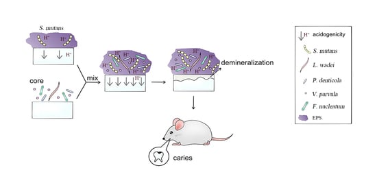
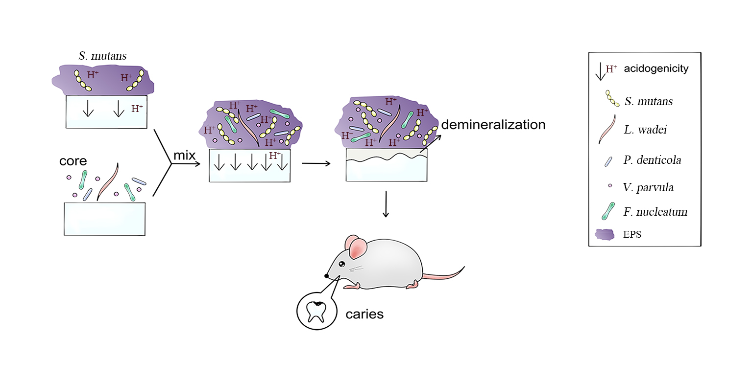
3.1. S. mutans Was Promoted by Core Microbiota and the Core + S. Mutans Biofilm Showed Increased Acidogenic Ability

To have a better consistency of the ECC plaque composition, we developed the core biofilm with an even amount of the selected four genus as well as the Core + S. mutans biofilm with 50% of the core microbiota and 50% of the Streptococcus mutans. qPCR was used for the analysis of biofilm composition. In the Core group, V. parvula was the most dominated species within the biofilm (69.85%, 69.87%, and 66.99%), followed by L. wadei (14.24%, 15.59%, and 13.77%), P. denticola (11.31%, 12.38%, and 17.36%), and F. nucleatum (2.44%, 1.53%, and 1.22%) (Figure 1a). In the Core + S. mutans biofilm, S. mutans counted for 51.16%, 54.63%, and 63.86% at 24 h, 48 h, and 72 h separately, followed by V. parvula (28.53%, 29.59%, and 19.96%); L. wadei (16.27%, 9.51%, and 4.75%); P. denticola (2.74%, 4.87%, and 7.45%), and F. nucleatum (1.30%, 1.40%, and 2.51%) (Figure 1b). In order to detect the influence of the core microbiota on the growth of S. mutans, we measured the count of S. mutans in the S. mutans group and in the Core + S. mutans group. S. mutans was promoted by the core microbiota and demonstrated a 3.41, 5.17, and 14.63 times increase in CFU counting at 24 h, 48 h, and 72 h compared to single S. mutans biofilm (p < 0.05) (Figure 1c,d). The pH of the culture medium measured during the experiments was taken as an indirect indicator of the biofilm acidogenicity. We found that the pH of all three groups was under 5, which can serve as the biological initiation of the enamel demineralization. The Core + S. mutans group showed the lowest pH at all time points (p > 0.05), followed by the S. mutans group and the Core group (p < 0.05) (Figure 1e).

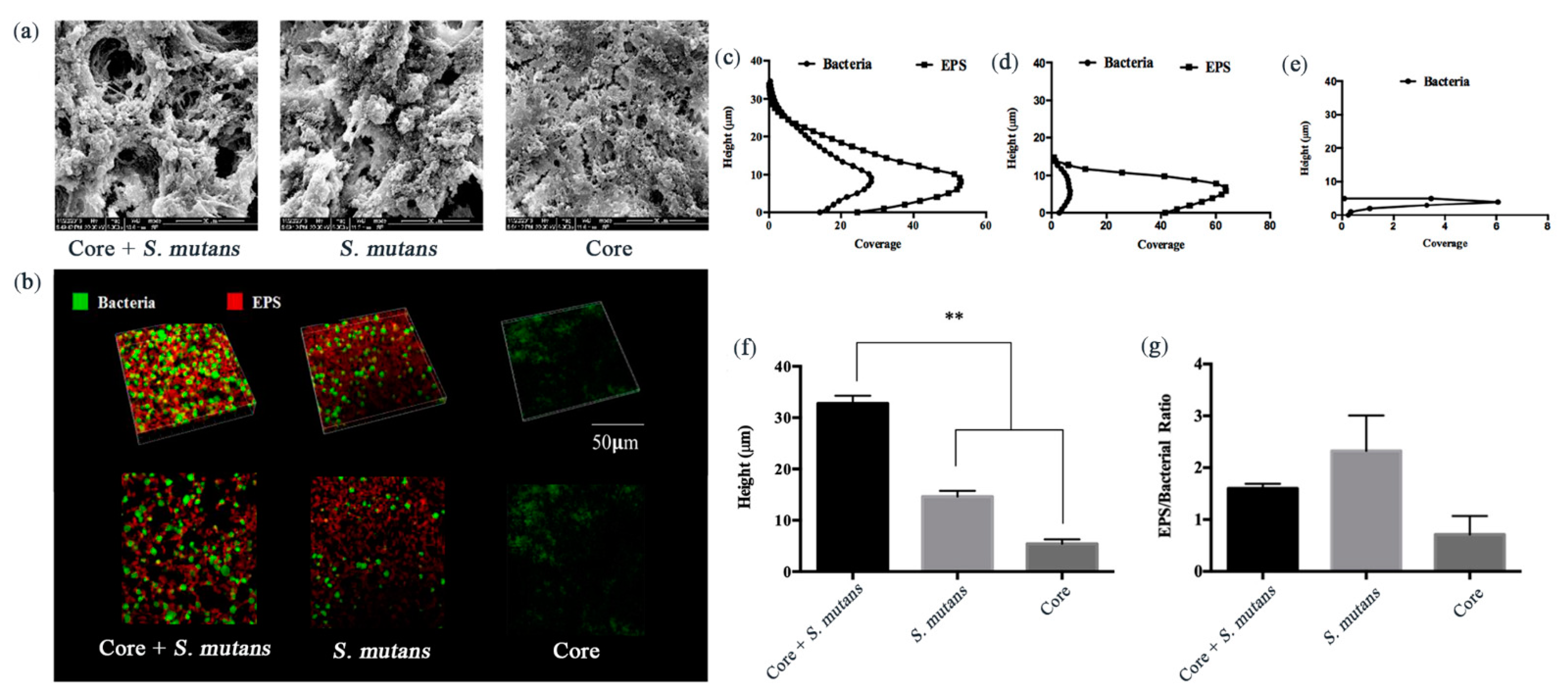
3.2. Core + S. Mutans Biofilm Showed a Net-Like Structure and Increased the Depth of the Biofilms

To have an integrated understanding of the biofilm, the SEM and CLSM were enrolled for observation of the biofilm formation, EPS synthesis, and biofilm structure in situ. The 72-h-old biofilms exhibited different morphologies under the SEM; the Core + S. mutans group biofilms with the existence of both EPS and bacillus represented a thick and net-like structural; the S. mutans group biofilms with rich EPS looked like honeycombs; the Core groups, however, without EPS, could only be detected with loose and thin biofilms (Figure 2a). Furthermore, consistent with the observation from SEM, the Core + S. mutans group biofilm images taken by CLSM showed a net-like structure that might improve the stability of the biofilm in complex environments (Figure 2b). The depth of the biofilms was significantly increased in the Core + S. mutans biofilm (p < 0.05) and EPS was not produced or rarely produced in the core biofilm (Figure 2c–g). The depth of the biofilm and EPS showed a significant increase in the Core + S. mutans group.

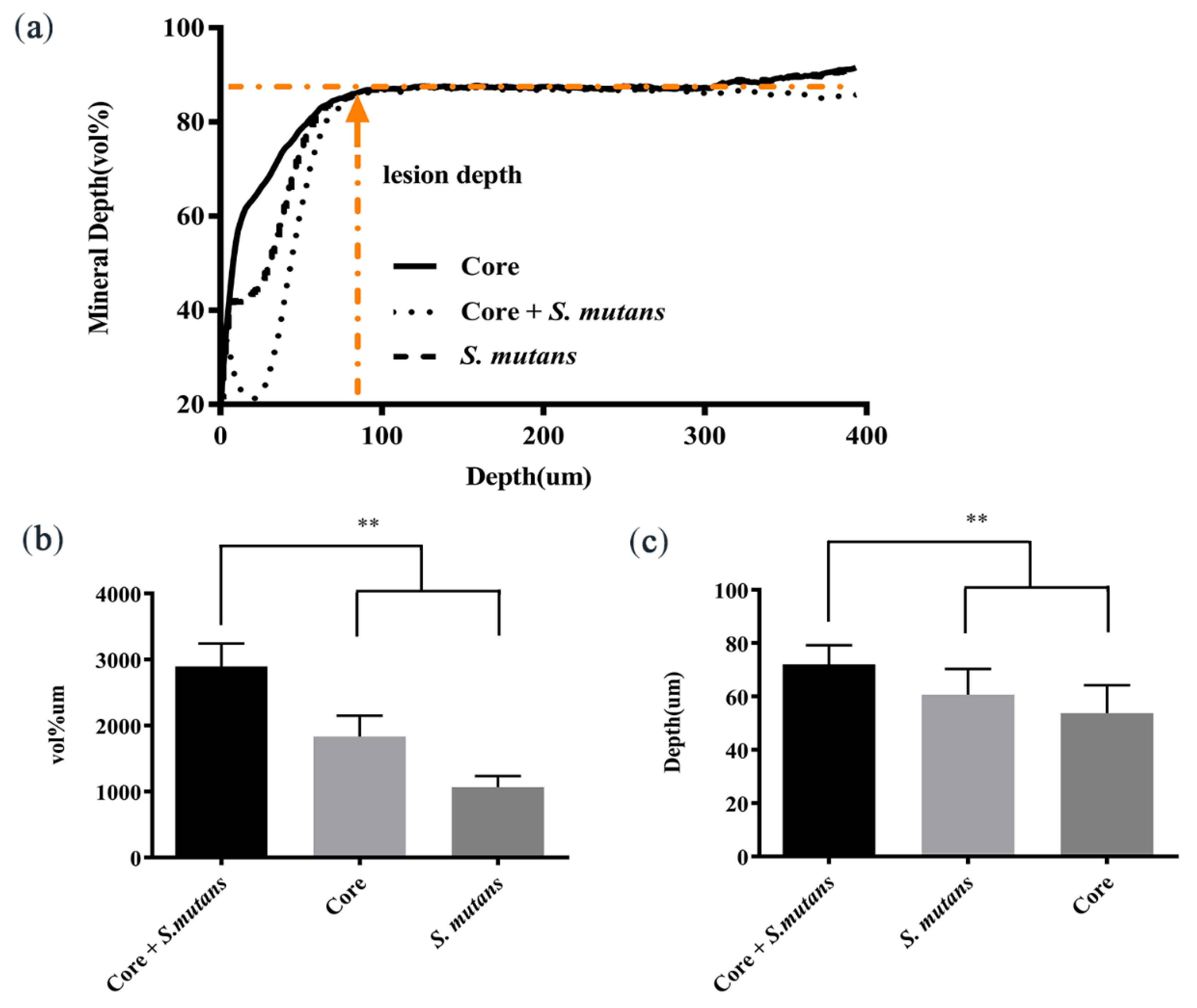
3.3. Core Microbiota Enhanced the Enamel Demineralization In Vitro

To evaluate the demineralization ability of different biofilms, we harvested the biofilm demineralized enamel blocks after 72 h of treatment and tested them using transverse microradiography (TMR), which is the gold standard for detecting demineralization. The Core + S. mutans biofilm had the highest demineralization degree, followed by the S. mutans group biofilms, and the Core group had the lowest demineralization degree (Figure 3a). The integrated mineral loss was 2895 (±345.25) Vol%µm for the Core + S. mutans group; 1831.82 (±315.50) Vol%µm for the S. mutans group and 1077.78 (±175.98) Vol%µm for the Core group (Figure 3b). The demineralized depth was 72.05 (±7.18) µm for the Core + S. mutans group; 60.62 (±9.70) µm for the S. mutans group, and 53.74 (±10.43) µm for the Core group (Figure 3c). All three groups of biofilms exhibited demineralization ability on enamel and the Core + S. mutans biofilm had both the deepest demineralization depth and the most integrated mineral loss (p < 0.01). These results showed that the core microbiota promoted the enamel demineralization of S. mutans biofilm in vitro.

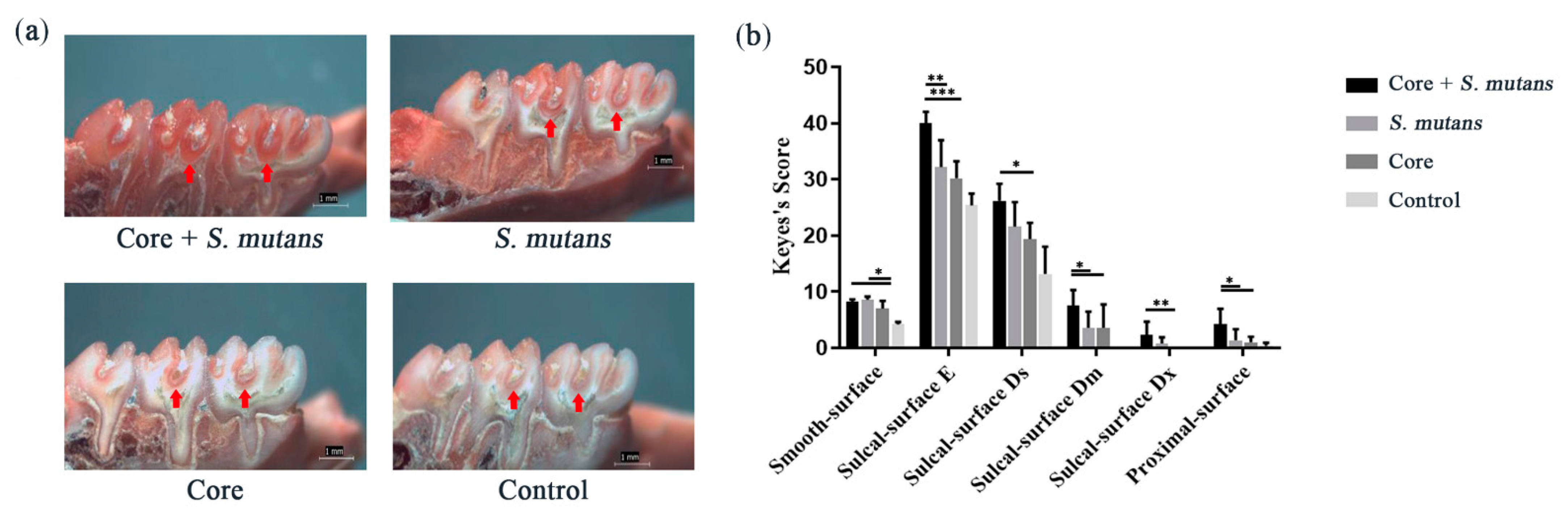
3.4. The Core Microbiota Increased Cariogenic Potential in a Rat Model

To explore whether the core microbiota would induce dental caries in vivo, we conducted the caries model on 20-day-old Wistar rats. For a better colonization of our selected bacteria, antibiotics were given by oral administration to eliminate the host microbiota. The bacteria were given for the first continuous seven days and the Keyes 2000 diet was fed to the rats for the whole experiment. These rats were sacrificed for Keyes scoring three weeks after bacterial challenge. As shown in Figure 4, significantly more carious lesions including enamel (E), moderate dentinal (Dm), and proximal-surface carious lesions were observed in the Core + S. mutans group compared with the S. mutans group.

4. Discussion

Dental caries including ECC, is a multifactorial disease [37,38]. Microbes are considered as one of the main pathogenic factors of ECC. A large amount of evidence proves that there might be a core microbiome of diseases in the oral cavity [39,40]. This also partially explains why some people are susceptible to caries while others are not. However, most of the literature have inferred the existence of core microbiome by sequencing and have found that tremendous species and genera were related to caries but failed to construct models to verify the casual relationship between dental caries and the core microbiota. A longitudinal study further narrowed the core microbiome and demonstrated that the core microbiota played an important role in ECC [13]. Referring to this literature, for the first time, we developed a core microbiota biofilm model containing four representative bacteria of ECC during its occurrence and development, which proved to promote the progress of dental caries both in vitro and in vivo.
Many studies have also shown that the four microorganisms we selected including V. parvula, F. nucleatum, P. denticola, and L. wadei are closely related to caries, especially ECC. In our study, we found that after 24 h, 48 h, and 72 h of culture, V. parvula accounted for more than half of the core microbial biofilm. Similarly, a large number of studies have proven that Veillonella content in ECC is higher than that in non-caries children, suggesting that it is closely related to ECC [13,41,42,43,44]. In another study, Fusobacterium was found to be significantly higher in ECC [45], in which F. nucleatum is closely associated with ECC, especially severe ECC [43,46]. Our study found that the amount of F. nucleatum in the biofilm at 72 h was about twice that at 24 h in the Core + S. mutans biofilm, which also seems to suggest that F. nucleatum is more related to severe ECC. Meanwhile, with the extension of the Core + S. mutans biofilm culture time, the content of P. denticola also increased. P. denticola was found to be one of the most prevalent species in the S-ECC, shown to be associated with dental caries. There is also speculation that the proportion of P. denticola would reduce after comprehensive restorative and preventive dental treatment [47,48]. Our results showed that when S. mutans was added, the content of L. wadei increased at the early stage of biofilm formation, which implied that S. mutans might promote the growth of L. wadei. L. wadei was found to be overrepresented in caries-active dental plaque compared to caries-free [49]. Moreover, a large number of Fusobacterium, Prevotella, and Leptotrichia were observed in a group of children with active caries [13,43,50]. In summary, these previous studies proved the rationality of our choice of four strains as core microbiota.
We found that the core microbiota had the effects of promoting the growth of S. mutans. There have been some controversies about the effect of V. parvula on S. mutans. Some previous studies have shown that V. parvula might contribute to acid production and the growth of S. mutans [43,51,52]. However, it has also been found that V. parvula had little effect on the S. mutans growth when co-cultured with S. mutans and Streptococcus gordonii compared with S. mutans alone [53]. There might be a certain relationship between V. parvula and S. mutans, but the impact of V. parvula in the Core microbiota on S. mutans remains to be studied. Similarly, F. nucleatum is also one of the first Gram-negative species to establish plaque biofilms [54]. It is one of the important “bridge” organisms in the naturally formed dental plaque [55], and has a central role in the ecology of dental plaque [56]. F. nucleatum has also been studied with the ability of affecting the growth and survival of S. mutans. However, the effect of core microbiota on S. mutans has not been previously studied. Our results showed that when the core microbiota presented, the growth of S. mutans increased by about four times, suggesting that the core microbiota promoted the S. mutans growth in the biofilms. In this study, we did not validate the function of these four species individually, but considered them as one factor. This is a limitation of our study, and we will further investigate the function of these individual species in our future studies.
The microbial composition of mature tooth biofilm is quite stable, but the pH of the biofilm could have small fluctuations [57]. A lower pH value is favored by demineralization, while a higher pH value is good for remineralization [9]. The fine-tuned balance of the oral ecosystem is disrupted, allowing disease-promoting bacteria to over grow and cause dental caries [8,37,58]. Interestingly, in our experiments, we found that the core microbiota itself produced limited acid, but could promote acid production of S. mutans. The integrated mineral loss of the enamel block increased by about 60% when the core microbiota existed. However, the environment in the oral cavity is very complicated, and saliva washing and food intake may have an impact on dental plaque. In vitro experiments cannot fully reproduce the complexity of in situ situation [59]. We therefore established an animal model to determine the role of the core microbiota with the presence of acidogenic and aciduric bacteria S. mutans and sugar. We found that rats in the Core + S. mutans group had the most severe dental caries.

5. Conclusions

In conclusion, we obtained core microbiota in ECC from a previous clinical research and validated its cariogenesis effects in vivo and in vitro. Subsequent clinical verification is needed in future investigations. The core microbiota containing four representative strains of ECC could promote the growth of S. mutans, acid production, demineralization, and the development of caries in rat (graphical abstract). This also brings new ideas and challenges to the prevention and treatment of ECC. However, how core microorganisms interact with S. mutans and promote the development of dental caries remain to be further studied.

Author Contributions

Conceptualization, J.C., L.K. and X.P.; Methodology, J.C., L.K., X.P. and Y.C.; Software, J.C., L.K. and X.P.; Validation, M.L. and J.L.; Formal analysis, J.C., L.K. and X.P.; Investigation, J.C., L.K. and X.P.; Resources, B.R.; Data curation, B.R., M.L. and J.L.; Writing—original draft preparation, J.C., L.K. and X.P.; Writing—review and editing, X.Z. and L.C.; Visualization, J.C., L.K. and X.P.; Supervision, X.Z. and L.C.; Project administration, X.Z. and L.C.; Funding acquisition, X.Z. and L.C. All authors have read and agreed to the published version of the manuscript.

Funding

This research was supported by the National Natural Science Foundation of China [81870759 (LC) 81430011(XZ)], the Youth Grant of the Science and Technology Department of Sichuan Province, China [2017JQ0028 (LC)], and Innovative Research Team Program of Sichuan Province (L.C). The authors declare no potential conflicts of interest with respect to the authorship and/or publication of this article.

Institutional Review Board Statement

The study was conducted according to the guidelines of the Declaration of Helsinki, and approved by the West China School of Stomatology Institute Review Board (WCSHIRB) ethics committee on 25 May 2017, and the record number was WCHSIRB-D-2017-115.

Informed Consent Statement

Not applicable.

Acknowledgments

Our thanks go to the State Key Laboratory of Oral Diseases, West China School of Stomatology of Sichuan University for their support throughout the research project.

Conflicts of Interest

The authors declare no conflict of interest.

References

  1. Hemadi, A.S.; Huang, R.; Zhou, Y.; Zou, J. Salivary proteins and microbiota as biomarkers for early childhood caries risk assessment. Int. J. Oral Sci. 2017, 9, e1. [Google Scholar] [CrossRef] [Green Version]
  2. Duangthip, D.; Chen, K.J.; Gao, S.S.; Lo, E.C.M.; Chu, C.H. Managing Early Childhood Caries with Atraumatic Restorative Treatment and Topical Silver and Fluoride Agents. Int. J. Environ. Res. Public Health 2017, 14, 1204. [Google Scholar] [CrossRef] [Green Version]
  3. Anil, S.; Anand, P.S. Early Childhood Caries: Prevalence, Risk Factors, and Prevention. Front. Pediatrics 2017, 5, 157. [Google Scholar] [CrossRef] [Green Version]
  4. Struzycka, I. The oral microbiome in dental caries. Pol. J. Microbiol. 2014, 63, 127–135. [Google Scholar] [CrossRef] [PubMed]
  5. Hajishengallis, E.; Parsaei, Y.; Klein, M.I.; Koo, H. Advances in the microbial etiology and pathogenesis of early childhood caries. Mol. Oral Microbiol. 2017, 32, 24–34. [Google Scholar] [CrossRef] [Green Version]
  6. Cappello, F.; Rappa, F.; Canepa, F.; Carini, F.; Mazzola, M.; Tomasello, G.; Bonaventura, G.; Giuliana, G.; Leone, A.; Saguto, D.; et al. Probiotics Can Cure Oral Aphthous-Like Ulcers in Inflammatory Bowel Disease Patients: A Review of the Literature and a Working Hypothesis. Int. J. Mol. Sci. 2019, 20, 5026. [Google Scholar] [CrossRef] [PubMed] [Green Version]
  7. Jiang, W.; Jiang, Y.; Li, C.; Liang, J. Investigation of supragingival plaque microbiota in different caries status of Chinese preschool children by denaturing gradient gel electrophoresis. Microb. Ecol. 2011, 61, 342–352. [Google Scholar] [CrossRef] [PubMed]
  8. Twetman, S. Prevention of dental caries as a non-communicable disease. Eur. J. Oral Sci. 2018, 126, 19–25. [Google Scholar] [CrossRef] [Green Version]
  9. Manji, F.; Dahlen, G.; Fejerskov, O. Caries and Periodontitis: Contesting the Conventional Wisdom on Their Aetiology. Caries Res. 2018, 52, 548–564. [Google Scholar] [CrossRef] [PubMed] [Green Version]
  10. Xiao, C.; Ran, S.; Huang, Z.; Liang, J. Bacterial Diversity and Community Structure of Supragingival Plaques in Adults with Dental Health or Caries Revealed by 16S Pyrosequencing. Front. Microbiol. 2016, 7, 1145. [Google Scholar] [CrossRef]
  11. Yang, F.; Ning, K.; Zeng, X.; Zhou, Q.; Su, X.; Yuan, X. Characterization of saliva microbiota’s functional feature based on metagenomic sequencing. SpringerPlus 2016, 5, 2098. [Google Scholar] [CrossRef] [PubMed] [Green Version]
  12. Turnbaugh, P.J.; Ley, R.E.; Hamady, M.; Fraser-Liggett, C.M.; Knight, R.; Gordon, J.I. The human microbiome project. Nature 2007, 449, 804–810. [Google Scholar] [CrossRef] [PubMed]
  13. Teng, F.; Yang, F.; Huang, S.; Bo, C.; Xu, Z.Z.; Amir, A.; Knight, R.; Ling, J.; Xu, J. Prediction of Early Childhood Caries via Spatial-Temporal Variations of Oral Microbiota. Cell Host Microbe 2015, 18, 296–306. [Google Scholar] [CrossRef] [PubMed] [Green Version]
  14. Qiu, W.; Zheng, X.; Wei, Y.; Zhou, X.; Zhang, K.; Wang, S.; Cheng, L.; Li, Y.; Ren, B.; Xu, X.; et al. d-Alanine metabolism is essential for growth and biofilm formation of Streptococcus mutans. Mol. Oral Microbiol. 2016, 31, 435–444. [Google Scholar] [CrossRef] [PubMed]
  15. Jang, J.Y.; Song, I.S.; Baek, K.J.; Choi, Y.; Ji, S. Immunologic characteristics of human gingival fibroblasts in response to oral bacteria. J. Periodontal Res. 2017, 52, 447–457. [Google Scholar] [CrossRef]
  16. Sarkar, J.; McHardy, I.H.; Simanian, E.J.; Shi, W.; Lux, R. Transcriptional responses of Treponema denticola to other oral bacterial species. PLoS ONE 2014, 9, e88361. [Google Scholar] [CrossRef] [Green Version]
  17. Poppleton, D.I.; Duchateau, M.; Hourdel, V.; Matondo, M.; Flechsler, J.; Klingl, A.; Beloin, C.; Gribaldo, S. Outer Membrane Proteome of Veillonella parvula: A Diderm Firmicute of the Human Microbiome. Front. Microbiol. 2017, 8, 1215. [Google Scholar] [CrossRef]
  18. Chen, J.; Li, T.; Zhou, X.; Cheng, L.; Huo, Y.; Zou, J.; Li, Y. Characterization of the clustered regularly interspaced short palindromic repeats sites in Streptococcus mutans isolated from early childhood caries patients. Arch. Oral Biol. 2017, 83, 174–180. [Google Scholar] [CrossRef]
  19. Tian, Y.; He, X.; Torralba, M.; Yooseph, S.; Nelson, K.E.; Lux, R.; McLean, J.S.; Yu, G.; Shi, W. Using DGGE profiling to develop a novel culture medium suitable for oral microbial communities. Mol. Oral Microbiol. 2010, 25, 357–367. [Google Scholar] [CrossRef] [Green Version]
  20. Zhang, K.; Wang, S.; Zhou, X.; Xu, H.H.K.; Weir, M.D.; Ge, Y.; Li, M.; Wang, S.; Li, Y.; Xu, X.; et al. Effect of Antibacterial Dental Adhesive on Multispecies Biofilms Formation. J. Dent. Res. 2015, 94, 622–629. [Google Scholar] [CrossRef] [Green Version]
  21. Wang, H.; Wang, S.; Cheng, L.; Jiang, Y.; Melo, M.A.S.; Weir, M.D.; Oates, T.W.; Zhou, X.; Xu, H.H.K. Novel dental composite with capability to suppress cariogenic species and promote non-cariogenic species in oral biofilms. Mater. Sci. Eng. C Mater. Biol. Appl. 2019, 94, 587–596. [Google Scholar] [CrossRef] [PubMed]
  22. Ge, Y.; Ren, B.; Zhou, X.; Xu, H.H.K.; Wang, S.; Li, M.; Weir, M.D.; Feng, M.; Cheng, L. Novel Dental Adhesive with Biofilm-Regulating and Remineralization Capabilities. Materials 2017, 10, 26. [Google Scholar] [CrossRef] [PubMed] [Green Version]
  23. Mashima, I.; Theodorea, C.F.; Thaweboon, B.; Thaweboon, S.; Nakazawa, F. Identification of Veillonella species in the tongue biofilm by using a novel one-step polymerase chain reaction method. PLoS ONE 2016, 11, e0157516. [Google Scholar] [CrossRef]
  24. Mima, K.; Nishihara, R.; Qian, Z.R.; Cao, Y.; Sukawa, Y.; Nowak, J.A.; Yang, J.; Dou, R.; Masugi, Y.; Song, M. Fusobacterium nucleatum in colorectal carcinoma tissue and patient prognosis. Gut 2016, 65, 1973–1980. [Google Scholar] [CrossRef] [PubMed] [Green Version]
  25. Cheng, L.; Zhang, K.; Weir, M.D.; Liu, H.B.; Zhou, X.D.; Xu, H.H.K. Effects of antibacterial primers with quaternary ammonium and nano-silver on Streptococcus mutans impregnated in human dentin blocks. Dent. Mater. 2013, 29, 462–472. [Google Scholar] [CrossRef] [Green Version]
  26. Xiao, J.; Klein, M.I.; Falsetta, M.L.; Lu, B.W.; Delahunty, C.M.; Yates, J.R.; Heydorn, A.; Koo, H. The Exopolysaccharide Matrix Modulates the Interaction between 3D Architecture and Virulence of a Mixed-Species Oral Biofilm. PLoS Pathog. 2012, 8, e1002623. [Google Scholar] [CrossRef] [Green Version]
  27. Heydorn, A.; Nielsen, A.T.; Hentzer, M.; Sternberg, C.; Givskov, M.; Ersbøll, B.K.; Molin, S. Quantification of biofilm structures by the novel computer program COMSTAT. Microbiology 2000, 146, 2395–2407. [Google Scholar] [CrossRef] [Green Version]
  28. Huang, X.; Cheng, L.; Exterkate, R.A.; Liu, M.; Zhou, X.; Li, J.; ten Cate, J.M. Effect of pH on Galla chinensis extract’s stability and anti-caries properties in vitro. Arch. Oral Biol. 2012, 57, 1093–1099. [Google Scholar] [CrossRef]
  29. Han, Q.; Li, B.; Zhou, X.; Ge, Y.; Wang, S.; Li, M.; Ren, B.; Wang, H.; Zhang, K.; Xu, H.H.K.; et al. Anti-Caries Effects of Dental Adhesives Containing Quaternary Ammonium Methacrylates with Different Chain Lengths. Materials 2017, 10, 643. [Google Scholar] [CrossRef] [Green Version]
  30. Beighton, D.; Gilbert, S.C.; Clark, D.; Mantzourani, M.; Al-Haboubi, M.; Ali, F.; Ransome, E.; Hodson, N.; Fenlon, M.; Zoitopoulos, L.; et al. Isolation and identification of bifidobacteriaceae from human saliva. Appl. Environ. Microbiol. 2008, 74, 6457–6460. [Google Scholar] [CrossRef] [Green Version]
  31. Schwendicke, F.; Korte, F.; Dorfer, C.E.; Kneist, S.; Fawzy El-Sayed, K.; Paris, S. Inhibition of Streptococcus mutans Growth and Biofilm Formation by Probiotics in vitro. Caries Res. 2017, 51, 87–95. [Google Scholar] [CrossRef]
  32. Caglar, E.; Sandalli, N.; Twetman, S.; Kavaloglu, S.; Ergeneli, S.; Selvi, S. Effect of yogurt with Bifidobacterium DN-173 010 on salivary mutans streptococci and lactobacilli in young adults. Acta Odontol. Scand. 2005, 63, 317–320. [Google Scholar] [CrossRef]
  33. Nam, H.J.; Kim, Y.M.; Kwon, Y.H.; Kim, I.R.; Park, B.S.; Son, W.S.; Lee, S.M.; Kim, Y.I. Enamel Surface Remineralization Effect by Fluorinated Graphite and Bioactive Glass-Containing Orthodontic Bonding Resin. Materials 2019, 12, 1308. [Google Scholar] [CrossRef] [PubMed] [Green Version]
  34. Kielbassa, A.M. In situ induced demineralization in irradiated and non-irradiated human dentin. Eur. J. Oral Sci. 2000, 108, 214–221. [Google Scholar] [CrossRef]
  35. Liu, S.Y.; Wu, T.M.; Zhou, X.D.; Zhang, B.; Huo, S.B.; Yang, Y.T.; Zhang, K.K.; Cheng, L.; Xu, X.; Li, M.Y. Nicotine is a risk factor for dental caries: An in vivo study. J. Dent. Sci. 2018, 13, 30–36. [Google Scholar] [CrossRef] [PubMed]
  36. Keyes, P.H. Dental caries in the molar teeth of rats. II. A method for diagnosing and scoring several types of lesions simultaneously. J. Dent. Res. 1958, 37, 1088–1099. [Google Scholar] [CrossRef] [PubMed]
  37. Philip, N.; Suneja, B.; Walsh, L.J. Ecological Approaches to Dental Caries Prevention: Paradigm Shift or Shibboleth? Caries Res. 2018, 52, 153–165. [Google Scholar] [CrossRef] [PubMed]
  38. Sanz, M.; Beighton, D.; Curtis, M.A.; Cury, J.A.; Dige, I.; Dommisch, H.; Ellwood, R.; Giacaman, R.A.; Herrera, D.; Herzberg, M.C.; et al. Role of microbial biofilms in the maintenance of oral health and in the development of dental caries and periodontal diseases. Consensus report of group 1 of the Joint EFP/ORCA workshop on the boundaries between caries and periodontal disease. J. Clin. Periodontol. 2017, 44 (Suppl. 18), S5–S11. [Google Scholar] [CrossRef]
  39. Jiang, W.; Zhang, J.; Chen, H. Pyrosequencing analysis of oral microbiota in children with severe early childhood dental caries. Curr. Microbiol. 2013, 67, 537–542. [Google Scholar] [CrossRef]
  40. Jiang, W.; Ling, Z.X.; Lin, X.L.; Chen, Y.D.; Zhang, J.; Yu, J.J.; Xiang, C.; Chen, H. Pyrosequencing Analysis of Oral Microbiota Shifting in Various Caries States in Childhood. Microb. Ecol. 2014, 67, 962–969. [Google Scholar] [CrossRef]
  41. Kanasi, E.; Dewhirst, F.E.; Chalmers, N.I.; Kent, R., Jr.; Moore, A.; Hughes, C.V.; Pradhan, N.; Loo, C.Y.; Tanner, A.C. Clonal analysis of the microbiota of severe early childhood caries. Caries Res. 2010, 44, 485–497. [Google Scholar] [CrossRef] [PubMed] [Green Version]
  42. Ling, Z.; Kong, J.; Jia, P.; Wei, C.; Wang, Y.; Pan, Z.; Huang, W.; Li, L.; Chen, H.; Xiang, C. Analysis of oral microbiota in children with dental caries by PCR-DGGE and barcoded pyrosequencing. Microb. Ecol. 2010, 60, 677–690. [Google Scholar] [CrossRef]
  43. Tanner, A.C.; Mathney, J.M.; Kent, R.L.; Chalmers, N.I.; Hughes, C.V.; Loo, C.Y.; Pradhan, N.; Kanasi, E.; Hwang, J.; Dahlan, M.A.; et al. Cultivable anaerobic microbiota of severe early childhood caries. J. Clin. Microbiol. 2011, 49, 1464–1474. [Google Scholar] [CrossRef] [Green Version]
  44. Agnello, M.; Marques, J.; Cen, L.; Mittermuller, B.; Huang, A.; Chaichanasakul Tran, N.; Shi, W.; He, X.; Schroth, R.J. Microbiome Associated with Severe Caries in Canadian First Nations Children. J. Dent. Res. 2017, 96, 1378–1385. [Google Scholar] [CrossRef] [PubMed]
  45. Corby, P.M.; Lyons-Weiler, J.; Bretz, W.A.; Hart, T.C.; Aas, J.A.; Boumenna, T.; Goss, J.; Corby, A.L.; Junior, H.M.; Weyant, R.J.; et al. Microbial risk indicators of early childhood caries. J. Clin. Microbiol. 2005, 43, 5753–5759. [Google Scholar] [CrossRef] [Green Version]
  46. Unsal, G.; Topcuoglu, N.; Ulukapi, I.; Kulekci, G.; Aktoren, O. Scardovia Wiggsiae and the Other Microorganisms in Severe Early Childhood Caries. J. Dent. Oral Care Med. 2017, 3, 2454–3276. [Google Scholar]
  47. Kamalendran, N. Oral Microbiota of Children Undergoing Comprehensive Treatment for Severe Early Childhood Caries. Ph.D. Thesis, University of Otago, Dunedin, New Zealand, 2015. [Google Scholar]
  48. Hurley, E.; Barrett, M.P.J.; Kinirons, M.; Whelton, H.; Ryan, C.A.; Stanton, C.; Harris, H.M.B.; O’Toole, P.W. Comparison of the salivary and dentinal microbiome of children with severe-early childhood caries to the salivary microbiome of caries-free children. BMC Oral Health 2019, 19, 13. [Google Scholar] [CrossRef] [Green Version]
  49. Peterson, S.N.; Snesrud, E.; Liu, J.; Ong, A.C.; Kilian, M.; Schork, N.J.; Bretz, W. The dental plaque microbiome in health and disease. PLoS ONE 2013, 8, e58487. [Google Scholar] [CrossRef] [PubMed] [Green Version]
  50. Luo, A.H.; Yang, D.Q.; Xin, B.C.; Paster, B.J.; Qin, J. Microbial profiles in saliva from children with and without caries in mixed dentition. Oral Dis. 2012, 18, 595–601. [Google Scholar] [CrossRef]
  51. Marsh, P.D. Microbial ecology of dental plaque and its significance in health and disease. Adv. Dent. Res. 1994, 8, 263–271. [Google Scholar] [CrossRef] [PubMed]
  52. Gross, E.L.; Beall, C.J.; Kutsch, S.R.; Firestone, N.D.; Leys, E.J.; Griffen, A.L. Beyond Streptococcus mutans: Dental caries onset linked to multiple species by 16S rRNA community analysis. PLoS ONE 2012, 7, e47722. [Google Scholar] [CrossRef] [PubMed]
  53. Liu, J.; Wu, C.; Huang, I.H.; Merritt, J.; Qi, F. Differential response of Streptococcus mutans towards friend and foe in mixed-species cultures. Microbiology 2011, 157, 2433–2444. [Google Scholar] [CrossRef] [PubMed] [Green Version]
  54. Könönen, E. Development of oral bacterial flora in young children. Ann. Med. 2000, 32, 107–112. [Google Scholar] [CrossRef] [PubMed]
  55. Kolenbrander, P.E.; Palmer, R.J., Jr.; Periasamy, S.; Jakubovics, N.S. Oral multispecies biofilm development and the key role of cell-cell distance. Nat. Rev. Microbiol. 2010, 8, 471–480. [Google Scholar] [CrossRef] [PubMed]
  56. Haake, S.K.; Yoder, S.C.; Attarian, G.; Podkaminer, K. Native plasmids of Fusobacterium nucleatum: Characterization and use in development of genetic systems. J. Bacteriol. 2000, 182, 1176–1180. [Google Scholar] [CrossRef] [PubMed] [Green Version]
  57. Fejerskov, O.; Scheie, A.A.; Manji, F. The Effect of Sucrose on Plaque Ph in the Primary and Permanent Dentition of Caries-Inactive and Caries-Active Kenyan Children. J. Dent. Res. 1992, 71, 25–31. [Google Scholar] [CrossRef]
  58. Kilian, M.; Chapple, I.L.; Hannig, M.; Marsh, P.D.; Meuric, V.; Pedersen, A.M.; Tonetti, M.S.; Wade, W.G.; Zaura, E. The oral microbiome—an update for oral healthcare professionals. Br. Dent. J. 2016, 221, 657–666. [Google Scholar] [CrossRef] [PubMed]
  59. Kantarci, A.; Hasturk, H.; Van Dyke, T.E. Animal models for periodontal regeneration and peri-implant responses. Periodontol. 2000 2015, 68, 66–82. [Google Scholar] [CrossRef] [Green Version]
Figure 1. Composition of the biofilm and biofilm pH monitoring. (a) Composition of the core biofilm at 24 h, 48 h, and 72 h. (b) Composition of the Core + S. mutans biofilm at 24 h, 48 h, and 72 h. (c) S. mutans counting of the Core + S. mutans biofilm and S. mutans biofilm at 24 h, 48 h, and 72 h (n = 3). (d) Ratio of S. mutans in Core + S. mutans/S. mutans. (e) Medium pH of the biofilms at 24 h, 48 h, and 72 h (n = 3). Data are expressed as means ± standard errors. * p < 0.05; ** p < 0.01.
Applsci 11 03638 g001
Figure 2. Biofilm structure by SEM and EPS distribution by CLSM. (a) Structure of the 72 h Core + S. mutans biofilm, S. mutans biofilm, and the core biofilm (n = 3). (b) Double-labeling of 72 h biofilms. Green, bacteria (SYTO 9); red, EPS (Alexa Fluor 647) (n = 3). (c) The distributions of EPS and bacteria at different heights of the Core + S. mutans biofilm. (d) The distributions of EPS and bacteria at different heights of the S. mutans biofilm. (e) The distributions of EPS and bacteria at different heights of the core biofilm. (f) The depth of the biofilm. (g) Quantification of EPS /bacteria biomass of the 72 h biofilms. ** p < 0.01.
Applsci 11 03638 g002
Figure 3. Enamel demineralization ability of the biofilms (n = 3). (a) Demineralization of the 72 h biofilm treated enamel slides. (b) Quantification of the integrated mineral loss. (c) Quantification of the demineralized depth. ** p < 0.01.
Applsci 11 03638 g003
Figure 4. In vivo cariogenesis ability of different biofilms. (a) Representative hemisectioned molars in each group under stereoscopic microscopy. (b) Caries score by Keyes’ method (n = 5). E = Enamel caries; Ds = slight dentinal caries; Dm = moderate dentinal caries; Dx = extensive dentinal caries. The severity decreased in the order of Core + S. mutans, S. mutans, Core and control groups. * p < 0.05; ** p < 0.01; *** p < 0.001.
Applsci 11 03638 g004
Table 1. Primers used in this study.
OrganismPCR PrimersSource
S. mutansForward:5′-TTGACGGTGTTCGTGTTGAT-3′
Reverse:5′-AAAGCGATAGGCGCAGTTTA-3′
This study
V. parvulaForward: 5′-GTAACAAAGGTGTCGTTTCTCG-3′
Reverse: 5′-CGTAACATCTTCCGAAACTTTC-3′
[23]
F. nucleatumForward:5′-CAACCATTACTTTAACTCTACCATGTTCA-3′
Reverse: 5′-GTTGACTTTACAGAAGGAGATTATGTAAAAATC-3′
[24]
P. enticolaForward:5′-GGGGATAAAGTGAGGGACGT-3′
Reverse:5′-GGCGCCTACATTTCACAACA-3′
This study
L. wadeiForward:5′-AAGCCTGCCCTGGAAACTAT-3′
Reverse:5′-CACTTCAGCGTCAGTTACCG-3′
This study
Publisher’s Note: MDPI stays neutral with regard to jurisdictional claims in published maps and institutional affiliations.

